Archives

Neonatal Hyperthyroidism

INTRODUCTION


Neonatal hyperthyroidism in most cases is transient and results from the transplacental passage of maternal stimulating TSH receptor antibodies (TRAb) known as neonatal Graves’ disease (GD).  Permanent non autoimmune neonatal hyperthyroidism is rare and is due to activating mutations of TSH receptor or due to somatic activating mutations in the stimulatory alpha subunit of the guanine nucleotide-binding protein (GNAS gene) in McCune-Albright syndrome. Exposure to topical iodine has also been reported as a rare cause of hyperthyroidism in newborns.


TRANSIENT NEONATAL HYPERTHYROIDISM


Neonatal Graves’ disease (GD) is usually a self-limited disease, but it can be life threatening and permanently damage the brain.  Neonatal GD is caused by transplacental passage of TSH receptor antibodies (TRAb) with stimulatory activity.


TRAb are Immunoglobulin of G class and freely cross the placenta. Different types of TRAb can be found: TRAb that bind to the TSH receptor and stimulates the production of thyroid hormones, (TSH receptor stimulating antibodies, TSI), TRAb that bind to the TSH receptor, do not stimulate the production of thyroid hormones and can block the binding of TSH (TSH receptor blocking antibodies TBI).


Hyperthyroidism develops only in babies born to mothers with the most potent stimulatory activity in serum. This corresponds to 1-2% of mothers with Graves’ disease, or 1 in 50,000 newborns, an incidence that is approximately four times higher than is that for transient neonatal hypothyroidism due to maternal TSH receptor blocking antibodies. Some mothers have mixtures of stimulating and blocking antibodies in their circulation, the relative proportion of which may change over time. Not surprisingly, the clinical picture in the fetus and neonate of these mothers is more complex and depends not only on the relative proportion of each activity in the maternal circulation at any one time but on the rate of their clearance from the neonatal circulation postpartum.


Occasionally, neonatal hyperthyroidism may even occur in infants born to hypothyroid mothers. A prospective study showed that 40% of patients treated for Graves’ disease with radioactive iodine had TRAb detectable after 5 years (13). In these situations, the maternal thyroid has been destroyed either by prior radioablation, surgery, or by coincident destructive autoimmune processes so that potent thyroid stimulating antibodies, present in the maternal circulation, are silent in contrast to the neonate whose thyroid gland is normal. Persistence of TRAb after thyroidectomy is higher in females with Graves’ ophthalmopathy or smokers. Fetal/neonatal thyrotoxicosis can occur also in newborn from hypothyroid mothers with chronic lymphocytic thyroiditis.


CLINICAL MANIFESTATIONS


TABLE 1. Situations That Should Prompt Consideration of Neonatal Hyperthyroidism

·       Unexplained tachycardia, goiter or stare

Unexplained petechiae, hyperbilirubinemia, or hepatosplenomegaly

·       History of persistently high TSH receptor antibody titer in mother during pregnancy

·       History of persistently high requirement for antithyroid medication in mother during pregnancy

·       History of thyroid ablation for hyperthyroidism in mother

·       History of previously affected sibling


Maternal TSH receptor antibody-mediated hyperthyroidism may present in utero. Fetal hyperthyroidism is suspected in the presence of fetal tachycardia (pulse greater than 160/min) especially if there is evidence of failure to thrive. Obstetric complications are common. Fetal goiter (fetal neck circumference >95%) can by monitored by ultrasound using nomograms for fetal thyroid growth. Fetal goiter can cause esophageal and/or tracheal obstructions and polyhydramnios. Fetal goiter can also be due to transplacental passage of antithyroid drugs that cause hypothyroidism in the fetus.


TABLE 2. Clinical Manifestations in the Fetus

Unexplained tachycardia,

Failure to thrive

Intrauterine growth retardation

Goiter

Advanced bone age

Prematurity

Craniosynostosis, microcephaly

Fetal death


In the neonate infant typically, the onset is during the first one two weeks of life but can occur by 45 days. This is due both to the clearance of maternally administered antithyroid drug (propylthiouracil- PTU, methimazole- MMI, or carbimazole) from the infant ’s circulation and to the increased conversion of T4 to the more metabolically active T3 after birth. Rarely, as noted earlier, the onset of neonatal hyperthyroidism may be delayed until later if higher affinity blocking antibodies are also present.

In the newborn infant, characteristic signs and symptoms include tachycardia, irritability, poor weight gain, and prominent eyes.  Goiter, when present, may be related to maternal antithyroid drug treatment as well as to the neonatal Graves’ disease itself.


Rarely, infants with neonatal Graves’ disease present with thrombocytopenia, jaundice, hepatosplenomegaly, and hypoprothrombinemia, a picture that may be confused with congenital infections such as toxoplasmosis, rubella, or cytomegalovirus. In addition, arrhythmias and cardiac failure may develop and may cause death, particularly if treatment is delayed or

inadequate. In addition to a significant mortality rate that approximates 20% in some older series, untreated fetal and neonatal hyperthyroidism is associated with deleterious long-term consequences, including premature closure of the cranial sutures (cranial synostosis), failure to

thrive, and developmental delay. The half-life of TSH receptor antibodies is 1 to 2 weeks. The duration of neonatal hyperthyroidism, a function of antibody potency and the rate of their metabolic clearance, is usually 2 to 3 months but may be longer.


TABLE 3. Clinical Manifestations in the Neonate

Irritability, hyperexcitability, sleep disorders

Tachycardia, hypertension, cardiac failure

Flushing, sweating

Respiratory distress, pulmonary hypertension

Goiter, stare

Feeding difficulties, increased appetite but no/poor weight gain

Diarrhea

Unexplained petechiae, hyperbilirubinemia, jaundice, or hepatosplenomegaly

Craniosynostosis, microcephaly,

Death


LABORATORY EVALUATION


The recent guidelines for management of hyperthyroidism and the updated guidelines for the management of thyroid disease during pregnancy released from the American Thyroid Association ATA both suggest determining TRAb levels in pregnant women with Graves’ disease at 18-22 weeks instead of 20-24 weeks of gestation because a severe case of fetal Graves’ disease has occurred at 18 weeks of pregnancy.


Because of the importance of early diagnosis and treatment, infants at risk for neonatal hyperthyroidism should undergo both clinical and biochemical assessment as soon as possible.

All neonates born from a woman with TRAb positivity in pregnancy should undergo determination of TRAb from cord blood at delivery. If TRAb is negative, the risk to neonatal hyperthyroidism is negligible (Sensitivity is around 100%). FT3, FT4 and TSH determination from cord blood did not predict neonatal hyperthyroidism. Increases in FT4 on day 3 to 5 seems to better indicate the onset of hyperthyroidism. Situations that should prompt consideration of neonatal hyperthyroidism are listed in Table 1. A high index of suspicion is necessary in babies of women who have had thyroid ablation because in them a high titer of TSH receptor antibodies would not be evident clinically. Similarly, women with persistently elevated TSH receptor antibodies and with a high requirement for antithyroid medication are at an increased risk of having an affected child.


The diagnosis of hyperthyroidism is confirmed by the demonstration of an increased concentration of circulating T4 (and free T4, and T3, if possible) accompanied by a suppressed TSH level in neonatal or fetal blood. Results should be compared with normal values during gestation. Fetal ultrasonography may be helpful in detecting the presence of a fetal goiter and in monitoring fetal growth. Demonstration in the baby or mother of a high titer of TSH receptor antibodies will confirm the etiology of the hyperthyroidism and, in babies whose thyroid function testing is normal initially, indicate the degree to which the baby is at risk.


 In general, babies likely to become hyperthyroid have the highest TSH receptor antibody titer but levels of maternal TRAb in the serum as low as 4.4 U/L has been associated with neonatal thyrotoxicosis.  If TSH receptor antibodies are not detectable, the baby is very unlikely to become hyperthyroid. In the latter case, it can be anticipated that the baby will be euthyroid, have transient hypothalamic-pituitary suppression, or have a transiently elevated TSH, depending on the relative contribution of maternal hyperthyroidism versus the effects of maternal antithyroid medication, respectively. Close follow up of all babies with abnormal thyroid function tests or detectable TSH receptor antibodies is mandatory.


THERAPY


In the fetus, treatment is accomplished by maternal administration of antithyroid medication. Until recently PTU was the preferred drug for pregnant women in North America, but current recommendations suggest the use of MMI rather than PTU after the first trimester because of concerns about potential PTU-induced hepatotoxicity. The goals of therapy are to utilize the minimal dosage necessary to normalize the fetal heart rate and render the mother euthyroid or slightly hyperthyroid. In the neonate MMI (0.25 to 1.0 mg/kg/day) has been used initially in 3 divided doses. If the hyperthyroidism is severe, a strong iodine solution (Lugol’s solution or SSKI, 1 drop every 8 hours) is added to block the release of thyroid hormone immediately. Often the effect of MMI is not as delayed in infants as it is in older children or adults, a consequence of decreased intrathyroidal thyroid hormone storage. Therapy with both antithyroid drug and iodine is adjusted subsequently, depending on the response. Propranolol (2 mg/kg/day in 2 or 3 divided doses) is added if sympathetic overstimulation is severe, particularly in the presence of pronounced tachycardia. If cardiac failure develops, treatment with digoxin should be initiated, and propranolol should be discontinued. Rarely, prednisone (2 mg/kg/day) is added for immediate inhibition of thyroid hormone secretion. Measurement of TSH receptor antibodies in treated babies may be helpful in predicting when antithyroid medication can be safely discontinued. Lactating mothers on antithyroid medication can continue nursing as long as the dosage of PTU or MMI does not exceed 400 mg or 40 mg, respectively. The milk/serum ratio of PTU is 1/10 that of MMI, a consequence of pH differences and increased protein binding, so one might anticipate less transmission to the infant, but concerns about potential PTU toxicity need to be considered. At higher dosages of antithyroid medication, close supervision of the infant is advisable. A review about management of neonates born to mothers with Graves’ disease has been recently published.


PERMANENT NEONATAL HYPERTHYROIDISM


Rarely, neonatal hyperthyroidism is permanent and is due to a germline mutation in the TSH receptor (TSH-R) resulting in its constitutive activation. A gain of function mutation of the TSH-R should be suspected if persistent neonatal hyperthyroidism occurs in the absence of detectable TSH-R antibodies in the maternal circulation. Prematurity, low birth weight, and advanced bone age are common. Most cases result from a mutation in exon 10 which encodes the transmembrane domain and intracytoplasmic tail of the TSH-R, a member of the G protein coupled receptor superfamily. Less frequently, a mutation encoding the extracellular domain has been described. An autosomal dominant inheritance has been noted in many of these infants; other cases have been sporadic, arising from a de novo mutation.


Early recognition is important because the thyroid function of affected infants is frequently difficult to manage medically and, when diagnosis and therapy is delayed, irreversible sequelae, such as cranial synostosis and developmental delay may result. For this reason, early, aggressive therapy with either thyroidectomy or even radioablation has been recommended.


Two clinical forms were described: the first one is the “familial non-autoimmune autosomal dominant hyperthyroidism” (FNAH). High variable age of manifestation from neonatal period to 60 years, with variability also within the same family is reported. Goiter is present in children, with nodules in older age. The second one is “Persistent sporadic congenital non autoimmune hyperthyroidism” (PSNAH) includes forms with sporadic (de novo) germline mutations in the TSH-R. PSNAH is characterized by fetal-neonatal onset or within 11 months and more severe hyperthyroidism requiring early aggressive therapy.


ThyroidfunctioninbabieswithagainoffunctionmutationoftheTSH receptormaybe difficult tomanagemedically and,whendiagnosis andtherapyisdelayed,irreversible sequelae,suchas cranial synostosis anddevelopmental delaymayresult.Thyroidablationmayberequired.Thyroidsurgeryis thepreferredapproachif anexperienced pediatric surgeonisavailable.Thetimingatwhichthyroidectomy canbeperformedwilldependoninstitutionalpreference.  Ifthis is notfeasible,thenradioablationmaybenecessary. Guidelines about this rare condition have recently been published.


MCCUNE ALBRIGHT SYNDROME


McCune Albright is a syndrome due to somatic activating mutations in Gsαgene and can rarely present with neonatal hyperthyroidism.


ACKNOWLEDGEMENTS


This chapter is, in part, based on the previous version written by Professor Rosalind Brown.


GUIDELINES


Ross DS, Burch HB, Cooper DS, Greenlee MC, Launberg P, Maia AL, Rivkees S, Samuels M, Sosa JA, Stan MN, Walter MA. 2016 American Thyroid Association Guidelines for Diagnosis and management of hyperthyroidism and other causes of thyrotoxicosis. Thyroid. 2016; 2:1343-1421.


Alexander EK, Pearce EN, Brent GA, Brown RS, Chen H, Dosiou C, Grobman WA, Launberg P, Lazarus JH, Mandel SJ, Peeters RP, Sullivan S. 2017 Guidelines of the American Thyroid Association for the diagnosis and management of thyroid disease during pregnancy and the postpartum. Thyroid: 27:315.


REFERENCES


Samuels SL, Namoc SM, Bauer AJ. Neonatal thyrotoxicosis Clin Perinatol 45:31-40 2018.


Barbesino G, Tomer Y. Clinical utility of TSH receptor antibodies. J Clin Endocrinol Metab.

2013; 98:2247-2255.


van Dijk MM, Smits IH, Fliers E, Bisschop PH. Maternal thyrotropin receptor antibody concentration and the risk of fetal and neonatal thyrotoxicosis: a systematic review. Thyroid 2018: 28:257-


Rivkees S, Pediatric Graves’ disease: management in the post -propylthiouracil Era. Int J Pediatr Endocrinol. 2014 10


Van der Kaay D, Wasserman JD, Palmert MR. Management of neonates born to mothers with Graves’ disease. Pediatrics. 2016;137:e20151878


Paschke R, Niedzela M, Vaidya B, Persani L, Rapaport B, Leclere J. The management of

familial and persistent sporadic non-autoimmune hyperthyroidism caused by thyroid stimulating

hormone receptor germline mutations. Eur Thyroid J. 2012; 1:142-147.


Segni M. Disorders of the Thyroid Gland in Infancy, Childhood and Adolescence. In: Feingold KR, Anawalt B, Boyce A, Chrousos G, Dungan K, Grossman A, Hershman JM, Kaltsas G, Koch C, Kopp P, Korbonits M, McLachlan R, Morley JE, New M, Perreault L, Purnell J, Rebar R, Singer F, Trence DL, Vinik A, Wilson DP, editors. Endotext [Internet]. South Dartmouth (MA): MDText.com, Inc.; 2017 Mar 18.


Kleinau G, Vassart G. TSH Receptor Mutations and Diseases. In: Feingold KR, Anawalt B, Boyce A, Chrousos G, Dungan K, Grossman A, Hershman JM, Kaltsas G, Koch C, Kopp P, Korbonits M, McLachlan R, Morley JE, New M, Perreault L, Purnell J, Rebar R, Singer F, Trence DL, Vinik A, Wilson DP, editors. Endotext [Internet]. South Dartmouth (MA): MDText.com, Inc.; 2017 Jul 24.


Congenital Adrenal Hyperplasia: Diagnosis and Emergency Treatment

CLINICAL RECOGNITION


Congenital adrenal hyperplasia (CAH) is a group of autosomal recessive disorders that arise from defective steroidogenesis. The production of cortisol in the zona fasciculata of the adrenal cortex occurs in five major enzyme-mediated steps. CAH results from deficiency in any one of these enzymes. Impaired cortisol synthesis leads to chronic elevations of ACTH via the negative feedback system, causing overstimulation of the adrenal cortex and resulting in hyperplasia and over-secretion of the precursors to the enzymatic defect. The forms of CAH are summarized in Table 1. Impaired enzyme function at each step of adrenal cortisol biosynthesis leads to a unique combination of elevated precursors and deficient products. The most common enzyme deficiency that accounts for more than 90% of all CAH cases is 21-hydroxylase deficiency (21OHD).


Table 1. Types of Congenital Adrenal Hyperplasia

Condition

Onset

Abnormality

Genitalia

Mineralocorticoid Effect

Gene

Chromosomal Location

Typical Features

Lipoid CAH
Congenital
StAR Protein

Female, with no sexual development
Salt wasting

StAR 

8p11.2
All steroid products low

Lipoid CAH

Congenital

P450scc

Female, with no sexual development
Salt wasting

CYP11A

15q23-24
All steroid products low

3β-HSD deficiency Congenital
3β-HSD

Females virilized, males hypovirilized
Salt wasting

HSD3B2

1p13.1
Elevated DHEA, 17-pregnenolone, low androstenedione, testosterone, elevated K, low Na, CO2

17α-OH deficiency Congenital

P450c17

Males hypovirilized, Hyperkalemic low-renin hypertension

CYP17

CYP17

10q24.3
Decreased androgens and estrogen, elevated DOC, corticosterone

Classic 21-OH deficiency, salt wasting
Congenital

P450c21

Females prenatally virilized, males unchanged
Salt wasting occurs in ¾ of 21OHD patients

CYP21 

6p21.3
Elevated 17-OHP, DHEA, and androstenedione, elevated K, low Na, CO2, low aldosterone, high plasma renin

Classic 21-OH deficiency, simple virilizing
Congenital P450c21

Females prenatally virilized, males unchanged
No salt wasting

CYP21 

6p21.3
Elevated 17-OHP, DHEA, and androstenedione, normal electrolytes

Non-classic 21-OH deficiency

Postnatal
P450c21

All with normal genitalia at birth, hyperandrogenism postnatally
No salt wasting

CYP21 

6p21.3
Elevated 17-OHP, DHEA, and
androstenedione on ACTH stimulation

11β-OH deficiency Congenital

P450c11B1

Females virilized, males unchanged
Low-renin hypertension

CYP11B1  

8q24.3
Elevated DOC, 11-deoxycortisol (S); androgens, low K, elevated Na, CO2

P450 Oxidoreductase deficiency (POR), Congenital

P450 oxidoreductase


Males undervirilized, females unchanged

Variable degree of mineralocorticoid deficiency

P450 Oxidoreductase gene (POR)

7q11.2

Combined and variable enzymatic defects of P450c21, P450c17 and P450aro

Wide range of phenotypes: normal to genital ambiguity +/- skeletal abnormalities (Antley Bixler type)


Classical CAH occurs in 1:13,000 to 1:15,000 live births. It is estimated that 75% of patients have the salt-wasting (SW) phenotype and the rest have simple-virilizing (SV) phenotype. Non-classical 21-OHD CAH (NC-CAH) is more common, and is one of the most common disorders in the Ashkenazi Jewish population with 1 in 27 Jews affected.  CAH owing to 11β-hydroxylase deficiency (11β-OHD) is the second most common cause of CAH, accounting for 5-8% of all cases. The other forms of CAH are considered rare diseases and the incidence is unknown in the general population.


PATHOPHYSIOLOGY

Adrenal steroidogenesis occurs in three major pathways: glucocorticoids, mineralocorticoids, and sex steroids as shown in Figure 1. Glucocorticoids (particularly cortisol), androgens, and estrogens are synthesized in the zona fasciculata and reticularis; and aldosterone in the zona glomerulosa. The HPA feedback system is mediated through the circulating level of plasma cortisol by negative feedback of cortisol on CRF and ACTH secretion. Therefore, a decrease in cortisol secretion leads to increased ACTH production, which in turn stimulates (1) excessive synthesis of adrenal products in those pathways unimpaired by the enzyme deficiency and (2) an increase of precursor molecules in pathways blocked by the enzyme deficiency.

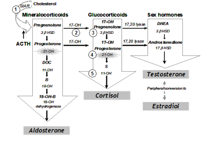
Figure 1. Pathways of Adrenal Steroidogenesis: Five enzymatic steps necessary for cortisol production are shown in numbers. 1= 20, 22 desmolase, 2= 17 hydroxylase (17-OH), 3=3ß-hydroxysteroid dehydrogenase (3ß HSD), 4=21 hydroxylase (21-OHD), 5=11ß hydroxylase (11-OH) In the first step of adrenal steroidogenesis, cholesterol enters mitochondria via a carrier protein called StAR. ACTH stimulates cholesterol cleavage, the rate limiting step of adrenal steroidogenesis.


The clinical symptoms of the five different forms of CAH result from the particular hormones that are deficient and those that are produced in excess as outlined in Table 1. In 21 OHD-CAH, there is an accumulation of 17-hydroxyprogesterone (17-OHP), a precursor to the 21-hydroxylation step, which is then shunted into the intact androgen pathway, where the 17,20-lyase enzyme converts the 17-OHP to D4-androstenedione, which is converted into androgens. Mineralocorticoid deficiency is a feature of SW-CAH, the most severe form of CAH. The enzyme defect in NC-CAH is only partial and salt wasting in this mild form of the disease does not occur. The analogy of all other enzyme deficiencies in terms of precursor retention and product deficiencies are shown in Table 1.


CLINICAL FEATURES

Genitalia

Females with Classical 21-OHD and 11β-hydroxylase deficiency CAH present at birth with virilization of their genitalia. Adrenocortical function begins around the 7th week of gestation; thus, a female fetus with classical CAH is exposed to adrenal androgens at the critical time of sexual differentiation (approximately 9 to 15 weeks gestational age). This leads to clitoral enlargement, fusion and scrotalization of the labial folds, and rostral migration of the urethral/vaginal perineal orifice, placing the phallus in the male position. Degrees of genital virilization are classified into five Prader stages (see Figure 2).

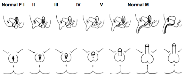
Figure 2. Different degrees of virilization according to the scale developed by Prader

Stage I: clitoromegaly without labial fusion

Stage II: clitoromegaly and posterior labial fusion

Stage III: greater degree of clitoromegaly, single perineal urogenital orifice, and almost complete labial fusion

Stage IV: increasingly phallic clitoris, urethra-like urogenital sinus at base of clitoris, and complete labial fusion

Stage V: penile clitoris, urethral meatus at tip of phallus, and scrotum-like labia (appear like males without palpable gonads)

Prader, A. Helv Paediatr Acta, 1954. 9:230-248.


Internal female genitalia, such as the uterus, fallopian tubes and ovaries, develop normally. Females with classical CAH maintain the internal genitalia potential for fertility.


Postnatal Effects, Growth and Puberty

Lack of appropriate postnatal treatment in boys and girls results in continued exposure to excessive androgens, causing progressive penile or clitoral enlargement, the development of premature pubic hair, axillary hair and acne. Advanced somatic and epiphyseal development occurs with exaggerated growth and is usually accompanied by premature epiphyseal maturation and closure, resulting in a final adult height that is typically significantly below that expected from parental heights. Excess glucocorticoid treatment can also lead to poor growth. The mean age at onset of puberty in both males and females is slightly younger than the general population. In those who are inadequately treated, central precocious puberty can occur. Following the onset of puberty, in a majority of successfully treated patients, the milestones of further development of secondary sex characteristics in general appear to be normal. In female adolescents and adults, signs of hyperandrogenism may include male-pattern alopecia (temporal balding), acne, hirsutism, menstrual irregularities, secondary PCOS and impaired fertility. Although the expected age of menarche may be delayed in females with classical CAH, when adequately treated many have regular menses after menarche.  In males, short stature and impaired fertility are observed.


Gender Role Behavior and Cognition

Prenatal androgen exposure in females affected with classical forms of CAH not only has a masculinizing effect on the development of the external genitalia, but also on childhood behavior. Both physical and behavioral masculinization are related to each other and to genotype, indicating that behavioral masculinization in childhood is a consequence of prenatal androgen exposure. The majority of genetic females with CAH retain the female gender identity even in the setting of prenatal androgen exposure and postnatal hyperandrogenism.


Fertility

Difficulty with fertility in females with CAH may be due to anovulation, secondary polycystic ovarian syndrome, irregular menses, non-suppressible serum progesterone levels, or an inadequate introitus. Fertility is reduced in SW-CAH with rare reports of pregnancy. Non-classical CAH is an important and frequently unrecognized form of infertility. Males with CAH, particularly if poorly treated, may have reduced sperm counts and low testosterone as a result of high androstenedione concentrations which suppress gonadotropins and testicular adrenal rest tumors. Testicular adrenal rest tumors (TART) are thought to arise from aberrant adrenal cells in the testes; TARTs are always benign and mostly bilateral. Microscopic examination shows that adrenal rest cells are present in the testicles of all male patients with CAH and often detected radiographically in those with longstanding poorly controlled disease. Regular testicular examination and periodic testicular ultrasonography are recommended for early detection of adrenal rest tumors of the testes. However, MRI studies have been increasingly used to diagnose TARTs.


Salt-Wasting 21-Hydroxylase Deficiency

When the loss of 21-hydroxylase function is severe, adrenal aldosterone secretion is not sufficient for sodium reabsorption by the distal renal tubules, and individuals suffer from salt wasting as well as cortisol deficiency and androgen excess. Infants with renal salt wasting have poor feeding, weight loss, failure to thrive, vomiting, dehydration, hypotension, hyponatremia, and hyperkalemic metabolic acidosis progressing to adrenal crisis (azotemia, vascular collapse, shock, and death). Adrenal crisis can occur as early as age one to four weeks. Affected males who are not detected in a newborn screening program are at high risk for a salt-wasting adrenal crisis because their normal male genitalia do not alert medical professionals to their condition. It is important to recognize that the extent of genital virilization may not differ among SV-CAH and SW-CAH.


Simple-Virilizing 21-Hydroxylase Deficiency

The salient features of classical SV-CAH are prenatal virilization and progressive postnatal masculinization with rapid somatic growth and advanced epiphyseal maturation leading to early epiphyseal closure and likely short stature. There is no evidence of mineralocorticoid deficiency in this disorder and serum electrolyte concentrations are normal. Diagnosis at birth of a female with SV-CAH is usually made immediately because of the apparent genital ambiguity. Since the external genitalia are not affected in newborn males, hyperpigmentation may be the only clue suggesting increased ACTH secretion and cortisol deficiency. Diagnosis at birth in males thus rests on prenatal or newborn screening.


Non-Classical 21-Hydroxylase Deficiency

Individuals with the non-classical (NC) form of 21-OHD have only mild to moderate enzyme deficiency and present postnatally, eventually developing signs of hyperandrogenism. Females with NC-CAH do not have virilized genitalia at birth. NC-CAH may present at any age after birth with a variety of hyperandrogenic symptoms. While serum cortisol concentration is typically low in patients with the classic form of the disease, it is usually normal in patients with NC 21-OHD. Similar to classical CAH, NC-CAH may cause premature development of pubic hair, acne, secondary PCOS, advanced bone age with accelerated linear growth velocity, and short stature. In adult males, early balding, acne, infertility or short stature may prompt the diagnosis of NC-CAH.


DIAGNOSIS

Diagnosis of CAH must be suspected in infants born with ambiguous genitalia. The physician is obliged to make the diagnosis as quickly as possible to initiate therapy. The diagnosis and rational decision of sex assignment must rely on the determination of genetic sex, the hormonal determination of the specific deficient enzyme, genotype, and an assessment of the patient's potential for future sexual activity and fertility. As indicated in Table 1, each form of CAH has its own unique hormonal profile, consisting of elevated levels of precursors and elevated or diminished levels of adrenal steroid products. Diagnosis of the 21-OHD CAH can also be confirmed biochemically by a hormonal evaluation. In a randomly timed blood sample, a very high concentration of 17-hydroxyprogesterone (17-OHP), the precursor of the defective enzyme, is diagnostic of classical 21-OHD. Such testing is the basis of the newborn-screening program developed to identify classically affected patients who are at risk for salt wasting crisis. False-positive results are, however, common with premature infants. Appropriate references based on weight and gestational age are therefore in place in many screening programs. False negative results may occur if samples are drawn late in the afternoon as adrenal hormones exhibit diurnal variation.  The gold standard for hormonal diagnosis is the corticotropin stimulation test (250 μg cosyntropin intravenously), measuring levels of 17-OHP and Δ4 androstenedione at baseline and 60 min. These values can then be plotted in the published nomogram (Figure 4) to ascertain disease severity. The corticotropin stimulation test should not be performed during the initial 24 hours of life as samples from this period are typically elevated in all infants and may yield false-positive results. Establishing a genetic diagnosis is not only important for the genotype-phenotype correlation, but also for genetic counseling for future pregnancies and for genetic counseling for the patient and his/her reproductive future.


For 21-OHD CAH, genetic analysis of the CYP21A2 gene may provide more clues to predict phenotypic severity. In about 50% of the causative genotypes, genotype-phenotype correlation can be found, although certain mutations can lead to variable phenotypes in different population groups especially in the simple virilizer group. Sequencing of the entire gene should be performed to detect rare mutations when genotype–phenotype non-concordance is observed in patients with CAH.


Newborn screening for CAH, which utilizes 17 hydroxyprogesterone levels, is a useful tool for early detection of CAH prior to the development of adrenal crisis in the affected neonate.   However, screening is associated with a high rate of false positive results as levels are affected by prematurity and birth weight. Molecular genetics, especially genotyping of the CYP21A2 gene should be considered as a second-tier screening test in the new born screening program.


Prenatal testing for CAH in utero has historically utilized invasive techniques like amniocentesis and chorionic villus sampling which cannot be done prior to 14 weeks of gestation. Prenatal dexamethasone treatment must begin prior to genital formation occurring at approximately 9 weeks, in order to avoid genital ambiguity in the affected female fetus. Massive parallel sequencing using hybridization probes on cell-free fetal DNA in maternal plasma indicated that the fetal CAH status was correctly deduced as early as 5 weeks 6 days of gestation. This is a noninvasive technique that accurately diagnoses CAH before the ninth week of gestation.


TREATMENT

Routine Treatment

br />

The goal of therapy in CAH is to both correct the deficiency in cortisol secretion and to suppress ACTH overproduction. Proper treatment with glucocorticoid reduces stimulation of the androgen pathway, thus preventing further virilization and allowing normal growth and development. The usual requirement of hydrocortisone (or its equivalent) for the treatment of classical 21-OHD form of CAH is about 10-15 mg/m2/day divided into 2 or 3 doses per day and for non-classical 21-OHD 5-8 mg/m2/day divided into 2 or 3 doses per day. Hydrocortisone is the glucocorticoid of choice in the pediatric age group. Prednisolone and dexamethasone are not used in growing children given growth suppressive effects. A small dose of dexamethasone at bedtime (0.25 to 0.5 mg) is usually adequate for androgen suppression in non-classical adult patients. Adequate biochemical control is assessed by measuring serum levels 17-OHP and androstenedione; serum testosterone can be used in females and prepubertal males (but not newborn males). We recommend that hormone levels are measured at a consistent time in relation to medication dosing, usually 1-2 hours after the morning corticosteroid. Titration of the dose should be aimed at maintaining 17-OHP concentrations below 1000 ng/dL and androstenedione concentrations below 200 ng/dl. Over-treatment should be avoided because it can lead to Cushing syndrome. Patients with salt wasting CAH have elevated plasma renin in response to the sodium-deficient state, and they require treatment with the salt-retaining 9α-fludrocortisone acetate. The average dose is 0.1 mg daily (0.05-0.2 mg daily). Infants should also be started on salt supplementation, as sodium chloride, at 1-2 g daily, divided into several feedings. Measurements of plasma renin and aldosterone are used to monitor the efficacy of mineralocorticoid therapy.   Advancement of bone age is monitored by bone age x-rays. Growth hormone therapy, in conjunction with a GnRH analogue, has been shown to be effective in improving final adult height. Patients may also experience peripheral precocious puberty, which requires treatment with gonadotropin-releasing hormone analogues. Aromatase inhibitors and growth hormone therapy should only be used in patients with a very short predicted final stature or in clinical trials. Use of aromatase inhibitors in CAH has been shown decrease bone maturation rates and some increase in adult height but the differences were not statistically significant.


Treatment During Illness and Emergency


Adrenal crisis can present as hypotension or shock and serum electrolyte abnormalities (hypoglycemia, hyponatremia, hyperkalemia, acidosis). During adrenal crisis, an immediate bolus of hydrocortisone 50-100 mg can be given intravenously or intramuscularly followed by hydrocortisone 100 mg/m2/day given as either continuous infusion or divided at least every 6 hours. Rehydration can be started with 20ml/kg isotonic saline with D5 as rapid bolus followed by repeat boluses or continuous infusion guided by level of dehydration. Hypoglycemia may require dextrose bolus and an initial bolus of 0.5-1 gram/kg of dextrose can be given intravenously at 2-3 ml per minute. If hyperkalemia is present, cardiac monitoring should be done to monitor for EKG changes. If changes are present, hyperkalemia should be treated using insulin with glucose infusion with or without other measures.


In non-life-threatening periods of illness or physiologic stress, the corticosteroid dose should be increased to 2 or 3 times the maintenance dose for the duration of that period, divided into 3 daily doses. Each family should be given injection kits of hydrocortisone, i.e. Solu-Cortef, for emergency use, and all family members should be trained in its intramuscular administration. The injectable dose of hydrocortisone in an emergency is 25 mg for infants, 50 mg for children under 40 kg, and 100 mg for children over 40 kg and for adults. In the event of a surgical procedure, 5-10 times the daily maintenance dose of hydrocortisone is needed, with 25-100 mg hydrocortisone IM/IV administered before and during a surgical procedure (as per infant, child, adult recommendations above), followed by high doses of hydrocortisone during the first 24-48 post-operative hours; the dose can then be tapered over the following days to the normal preoperative schedule. Stress doses of dexamethasone should not be given because of the delayed onset of action. It is not necessary for increased mineralocorticoid doses during these periods of stress. It is imperative for all patients who are receiving corticosteroid replacement therapy, such as patients with CAH, to wear a Medical Alert bracelet or medallion that will enable correct and appropriate therapy in case of emergencies. It is also crucial to re-educate parents at regular intervals on the life-threatening nature of this emergency.


GUIDELINES


Speiser PW, Arlt W, Auchus RJ, Baskin LS, Conway GS, Merke DP, Meyer-Bahlburg HFL, Miller WL, Murad MH, Oberfield SE, White PC. Congenital Adrenal Hyperplasia Due to Steroid 21-Hydroxylase Deficiency: An Endocrine Society Clinical Practice Guideline.

J Clin Endocrinol Metab. 2018 Nov 1;103(11):4043-4088


Rodriguez A, Ezquieta B, Labarta JI, Clemente M, Espino R, Rodriguez A, et al. Recommendations for the diagnosis and treatment of classic forms of 21-hydroxylase-deficient congenital adrenal hyperplasia. An Pediatr (Barc). 2017;87(2):116 e1- e10.


REFERENCES


New M, Yau M, Lekarev O, Lin-Su K, Parsa A, Pina C, Yuen T, Khattab A. Congenital Adrenal Hyperplasia. In: Feingold KR, Anawalt B, Boyce A, Chrousos G, Dungan K, Grossman A, Hershman JM, Kaltsas G, Koch C, Kopp P, Korbonits M, McLachlan R, Morley JE, New M, Perreault L, Purnell J, Rebar R, Singer F, Trence DL, Vinik A, Wilson DP, editors. Endotext [Internet]. South Dartmouth (MA): MDText.com, Inc.; 2000-2017 Mar 15


El-Maouche D, Arlt W, Merke DP. Congenital adrenal hyperplasia. Lancet. 2017 Nov 11;390(10108):2194-2210


Fluck CE, Miller WL. P450 oxidoreductase deficiency: a new form of congenital adrenal hyperplasia. Curr Opin Pediatr. 2006;18(4):435-41.


Yilmaz R, Sahin D, Aghayev A, Erol OB, Poyrazoglu S, Saka N, et al. Sonography and Magnetic Resonance Imaging Characteristics of Testicular Adrenal Rest Tumors. Pol J Radiol. 2017;82:583-8.


New MI, Abraham M, Gonzalez B, Dumic M, Razzaghy-Azar M, Chitayat D, et al. Genotype-phenotype correlation in 1,507 families with congenital adrenal hyperplasia owing to 21-hydroxylase deficiency. Proc Natl Acad Sci U S A. 2013;110(7):2611-6.


Balsamo A, Baldazzi L, Menabo S, Cicognani A. Impact of molecular genetics on congenital adrenal hyperplasia management. Sex Dev. 2010;4(4-5):233-48.


New MI, Tong YK, Yuen T, Jiang P, Pina C, Chan KC, et al. Noninvasive prenatal diagnosis of congenital adrenal hyperplasia using cell-free fetal DNA in maternal plasma. J Clin Endocrinol Metab. 2014;99(6):E1022-30.


Lin-Su K, Harbison MD, Lekarev O, Vogiatzi MG, New MI. Final adult height in children with congenital adrenal hyperplasia treated with growth hormone. J Clin Endocrinol Metab. 2011;96(6):1710-7.


Disorders of Sexual Differentiation in Newborns

INTRODUCTION      


In 2006, new nomenclature for conditions previously referred to as intersex was proposed in a consensus statement from the Lawson Wilkins Pediatric Endocrine Society and European Society of Pediatric Endocrinology in response to advanced identification of molecular genetic causes of sex. Disorders of sexual differentiation (DSD) are congenital conditions within which the development of chromosomal, gonadal and phenotypic sex is atypical. These disorders have a broad differential including variations in sex chromosomes, variations in genes involved in gonadal and genital development, disorders in steroidogenesis within the gonads and adrenals, maternal factors, and endocrine disruptors.   Classification of these disorders is based on sex chromosomes as such 46XX DSD, 46XY DSD, Ovotesticular DSD, and 46XX testicular DSD. 

Approximately 1-2% of live births are affected with atypical genitalia, including isolated hypospadias in males. The incidence of 46XX DSD is 1:15,000. The incidence of 46XY DSD is higher at 1:5000.

CLINICAL RECOGNITION

Given the potential association with glucocorticoid and mineralocorticoid deficiencies in CAH, the birth of a child with atypical genitalia constitutes a medical emergency requiring immediate evaluation. Further, the parents’ reaction to the birth of a child with atypical genitalia is one of shock and concern about which gender to assign, whether or not to decide for early genital surgery, and what to expect regarding the long-term outcome in terms of gender, sexual function, fertility, and general quality of life.  In order to provide appropriate counseling to the family, there is an urgency to determine the etiology.

PATHOPHYSIOLOGY

The phenotypic sex of a newborn is the result of external genital development that is under the influence of sex-determining genes as well as both endogenous and exogenous hormone exposures. 

The commitment of the bipotential primordial gonads to become testes or ovaries begins at 6 weeks and is fully achieved at 13-14 weeks.  Gonadal differentiation is controlled by a number of time and dosage-sensitive genes including the SRY gene on the Y chromosome, SOX9, and WNT4 genes.  The expression SRY and SOX9 and suppression of WNT4 expression is crucial to testicular differentiation.  The expression of WNT4 in the absence of SRY and SOX9 expression allows for ovarian differentiation. Leydig cells produce insulin like factor 3 (INSL3) which is responsible for transabdominal phase of testicular descent.

Fetal productions of androgens from the Leydig cells within the testes and from the adrenal glands begins at approximately 8-9 weeks. External genitalia develop concurrently around the 9th week of gestation under the influence of androgens, mainly dihydrotestosterone (DHT). Testosterone is the principal hormone produced by the testes and is required for the onset of virilization and promotion of Wolffian ducts.  Testosterone is converted to DHT by 5-alpha reductase.  DHT leads to the development of the prostate, scrotum and phallus. 

Anti-Mullerian Hormone (AMH) produced from Sertoli cells in the testes is required to support the development of Wolffian ducts including vas deferens, epididymis and seminal tubules in males. In females, Mullerian ductal structures including the uterus, fallopian tubes and cervix develop in the absence of AMH.

Disorders of Sexual Differentiation

46XX DSD

Patients with 46XX DSD are genotypic females with virilized characteristics. In 46XX DSD, the degree of genital virilization can be classified into five Prader stages. Stage 1, with the mildest degree of virilization, is characterized by clitoromegaly without labial fusion.  Stage 5, with the highest degree of virilization, is characterized by clitoromegaly with the urethral meatus at the tip, labial fusion, and scrotal-like appearance of the labia.  

46XX DSD can result from exogenous androgen exposure, endogenous adrenal androgen production or placental aromatase deficiency. Congenital adrenal hyperplasia (CAH) is the most common cause of 46XX DSD. The most common enzyme defects leading to CAH are 21-hydroxylase deficiency and 11-hydroxylase deficiency.  In a very rare form of CAH owing to p450 oxidoreductase deficiency, there is a mutation in the P450 oxidoreductase (POR) enzyme which causes partial deficiency of 21-hydroxylase and 17a-hydroxylase/17,20 lyase activities. Affected females can present with virilization of the external genitalia, glucocorticoid deficiency, and skeletal malformations such as craniosynostosis.


Maternal hyperandrogenism during gestation can cause virilization of the external genitalia in females when the placental aromatase is overwhelmed. The hyperandrogenism can be due to luteomas, androgen producing tumors and exogenous exposure. 

Maternal aromatase deficiency leads to decreased production of estrogen from androgen precursors. This leads to conversion of fetal DHEAS to androstenedione and testosterone by placental 3-beta hydroxysteroid dehydrogenase and virilization of female fetus. 

The majority of 46XX testicular DSD cases are caused by translocation between the X and Y chromosome, involving the SRY gene.

46XY DSD

Patients with 46XY DSD are genotypic males with under-virilization.  Micropenis is defined as a penile length less than 2.5 standard deviations below the mean penile length (<2.5 cm in a full-term newborn).  The severity of hypospadias is based on the distance of the urethral opening from its normal position at the tip of the phallus.  Lack of testicular palpation in the scrotum may signify cryptorchidism, vanishing testes, or gonadal dysgenesis.

46XY DSD can be caused by atypical testicular formation, low testosterone or dihydrotesterone production, or defects in the androgen receptor. In complete gonadal dysgenesis, there is no testicular development and patients present as phenotypic female with delayed puberty or amenorrhea.  Up to 20% of these cases occur due to deletion or mutation of the SRY gene.

Defects in androgen biosynthesis can lead to under-virilization in a 46XY patient. These defects can occur at various points along the production pathway of testosterone from cholesterol. Adrenal dysfunction is associated with defects in steroidogenic enzymes such as steroidogenic acute regulatory protein (StAR), p450 side chain cleavage enzyme, 3 beta HSD type 2, 17 alpha hydroxylase/17,20 lyase. Other defects of testosterone production can occur in the following enzymes: 7-dehydrocholesterol reductase causing Smith Lemli Opitz syndrome and 17 beta hydroxysteroid dehydrogenase. Affected males with 5-alpha-reductase deficiency have atypical genitalia (small phallus and perineal hypospadias).  With rises in testosterone at puberty, progressive virilization with phallic enlargement and testicular descent is seen.

Androgen insensitivity syndrome has been reported to be the main cause of 46XY DSD and is due to mutation in the androgen receptor.  In complete androgen insensitivity, the androgenic effects of testosterone and dihydrotestosterone are abolished and patients have unambiguously female appearing external genitalia. In partial androgen insensitivity, the androgenic effects are attenuated and patients can present on a spectrum of under-virilization.

Mutations in genes responsible for sex determination such as SRY, SOX9, and SF1 lead to 46XY complete gonadal dysgenesis.  Duplication of the DAX1 gene is associated with male to female sex reversal. 

Endocrine disruptors with anti-androgenic effects such as diethylstilbestrol or phthalates can also lead to atypical genitalia in males.

Table 1.  Laboratory Values to Differentiate Between Etiologies of Ambiguous Genitalia in Newborns with a 46, XY Chromosomal Complement.

Diagnosis

T

DHT

MIS

Androgen Insensitivity
Syndrome (AIS)

Normal/up

Normal/up

Normal

5α-Reductase Deficiency

Normal/up

Low

Normal

Testosterone Biosynthetic Defect orLeydig Cell Hypoplasia

Low

Low


Normal

Gonadal Dysgenesis

Low

Low

Low

T=testosterone, DHT=dihydrotestosterone and MIS=müllerian inhibiting substance.

OVOTESICULAR DSD

Ovotesticular DSD, one of the rarest forms of DSD, describes patients that were previously categorized as true hermaphrodites.  The gonads of patients with ovotesticular DSD contain both ovarian and testicular tissue.  Thus, the presentation of genital ambiguity can be variable. In ovotesticular DSD in which the gonads contain both ovarian and testicular tissue, the majority have an XX chromosomal constitution.  Complex mosaicism (XX/XY) are seen in approximately 10% of cases. Patients can present with a wide variety of genital ambiguity as well as a mixture of Wolffian and Mullerian structures.

DIAGNOSIS

Determination of chromosomal sex by karyotype with FISH analysis for SRY and pelvic ultrasound to evaluate for the presence of a uterus should be performed immediately. Currently, the only newborn screening test for steroid disorders is the measurement of 17-hydroxyprogesterone for 21-hydroxylase deficiency.   Further laboratory evaluation to accurately diagnose the specific underlying defect should be directed by a pediatric endocrinologist.  If CAH is suspected, measurement of adrenal hormones, ACTH stimulation testing, and molecular genetic testing can elucidate the form of CAH. Each form of CAH has its own unique hormonal profile, consisting of elevated levels of precursors and elevated or diminished levels of adrenal steroid products. HCG stimulation testing to assess testosterone and dihydrotestosterone response may be particularly helpful in 46XY DSD to assess testicular androgen production.  Molecular genetic evaluations should be guided by chromosomal and hormonal evaluations.

Chromosomal sex can be determined prenatally invasively by chorionic villus sampling and amniocentesis and noninvasively via free fetal DNA in the maternal blood. Thus, DSD may be suspected in utero if the phenotype on prenatal ultrasonogram is discordant with the chromosomal sex. 

THERAPY

When considering the gender of rearing, the prognosis for masculinization of brain and behavior, the anatomic and physiologic character of the reproductive tract with its potential for development and function in regard to both sexuality and fertility, and the social environment of the infant should be taken into account along with the genetic sex.  Both male and female gender assignment should be thoroughly considered.

Sex hormone replacement is needed to induce pubertal development.  Testosterone is used in the treatment of patients with testosterone deficiency (46XY DSD).  Different forms of testosterone (topical and intramuscular) are available and treatment will vary depending on what is best for the patient.  A short course of testosterone can be given during infancy to induce penile growth prior to surgical correction.  For 46XY DSD patients with functioning Sertoli cells, HCG can be used to stimulate testicular production.  Estrogen is used in the treatment of those reared female.  Estrogen is available as an oral tablet or transdermal patch.  Estrogen doses should be initiated at the lowest dose possible and slowly increased to a maximum of 0.625 mg/day of conjugated estrogen to allow for gradual breast development.  Progesterone supplementation with estrogen is recommended in patients with a uterus.


Glucocorticoids are needed to treat congenital adrenal hyperplasia. They suppress the pituitary glands oversecretion of adrenocorticotropic hormone and thus decrease the production of precursor hormones.  This also leads to a decrease in adrenal androgen production in forms of CAH associated with 46XX DSD.

The aim of surgical repair in patients with atypical genitalia reared in the female gender is generally to remove the redundant erectile tissue, preserve the sexually sensitive glans clitoris, and provide a normal vaginal orifice.  A medical indication for early surgery other than for sex assignment is recurrent urinary tract infections as a result of pooling of urine in the vagina or urogenital sinus. In the past, it was routine to recommend early corrective surgery for neonates born with ambiguous genitalia. However, in recent years, the implementation of early corrective surgery has become increasingly controversial due to lack of data on long-term functional outcome.  It is advised that all surgical decisions remain the prerogative of families in conjunction with experienced surgical consultants.

The process of assigning and accepting a gender of rearing for a child with ambiguous genitalia and of deciding the necessity of genital surgery is challenging. A team approach that combines the insights of the DSD-experienced pediatrician, endocrinologist, psychologist/psychiatrist, surgeon, and the child’s parents or guardian is essential. Although there is no consensus as to the appropriate age to disclose a condition, it is recommended to proceed gradually in line with the child’s cognitive and psychological development.

GUIDELINES

Audi L, Ahmed SF, Krone N, Cools M, McElreavey K, Holterhus PM, Greenfield A, Bashamboo A, Hiort O, Wudy SA, McGowan R, The EUCA: GENETICS IN ENDOCRINOLOGY: Approaches to molecular genetic diagnosis in the management of differences/disorders of sex development (DSD): position paper of EU COST Action BM 1303 'DSDnet'. Eur J Endocrinol 2018;179:R197-R206.

Lee PA, Houk CP, Ahmed SF, Hughes IA, International Consensus Conference on Intersex organized by the Lawson Wilkins Pediatric Endocrine S, the European Society for Paediatric E: Consensus statement on management of intersex disorders. International Consensus Conference on Intersex. Pediatrics 2006;118:e488-500.

Speiser PW, Arlt W, Auchus RJ, Baskin LS, Conway GS, Merke DP, Meyer-Bahlburg HFL, Miller WL, Murad MH, Oberfield SE, White PC: Congenital Adrenal Hyperplasia Due to Steroid 21-Hydroxylase Deficiency: An Endocrine Society Clinical Practice Guideline. J Clin Endocrinol Metab 2018;103:4043-4088.

REFERENCES

Lee PA, Nordenstrom A, Houk CP, Ahmed SF, Auchus R, Baratz A, Baratz Dalke K, Liao LM, Lin-Su K, Looijenga LH, 3rd, Mazur T, Meyer-Bahlburg HF, Mouriquand P, Quigley CA, Sandberg DE, Vilain E, Witchel S, Global DSDUC: Global Disorders of Sex Development Update since 2006: Perceptions, Approach and Care. Horm Res Paediatr 2016; 85:158-180.


Blackless M, Charuvastra A, Derryck A, Fausto-Sterling A, Lauzanne K, Lee E: How sexually dimorphic are we? Review and synthesis. Am J Hum Biol 2000; 12:151-166.

Krishnan S W, AB: Ambiguous Genitalia in Newborns; in New MI LO, Parsa A, Yuen T, O'Malley B, Hammer G (ed) Genetic Steroid Disorders. San Diego, CA, Elsevier, 2014, pp 87-97.

New M, Yau M, Lekarev O, Lin-Su K, Parsa A, Pina C, Yuen T, Khattab A. Congenital Adrenal Hyperplasia. In: Feingold KR, Anawalt B, Boyce A, Chrousos G, Dungan K, Grossman A, Hershman JM, Kaltsas G, Koch C, Kopp P, Korbonits M, McLachlan R, Morley JE, New M, Perreault L, Purnell J, Rebar R, Singer F, Trence DL, Vinik A, Wilson DP, editors. Endotext [Internet]. South Dartmouth (MA): MDText.com, Inc.; 2000- 2017 Mar 15.

Domenice S, Arnhold IJP, Costa EMF, Mendonca BB. 46,XY Disorders of Sexual Development. In: Feingold KR, Anawalt B, Boyce A, Chrousos G, Dungan K, Grossman A, Hershman JM, Kaltsas G, Koch C, Kopp P, Korbonits M, McLachlan R, Morley JE, New M, Perreault L, Purnell J, Rebar R, Singer F, Trence DL, Vinik A, Wilson DP, editors. Endotext [Internet]. South Dartmouth (MA): MDText.com, Inc.; 2000- 2017 May 3.


Fibrous Dysplasia

ABSTRACT


Fibrous dysplasia (FD) is an uncommon mosaic disorder falling along a broad clinical spectrum. It arises from post-zygotic mutations in GNAS, resulting in constitutive activation of the cAMP pathway-associated G-protein, Gsα, and proliferation of undifferentiated skeletal progenitor cells.  FD may occur in isolation, or in association with skin pigmentation and hyperfunctioning endocrinopathies, termed McCune-Albright syndrome (MAS). Disease may involve any part or combination of the skeleton, ranging from an isolated, asymptomatic monostotic lesion, to severe polyostotic disease resulting in fractures, deformity, functional impairment, and progressive scoliosis. FD may be diagnosed clinically in patients with polyostotic disease and/or extraskeletal features of MAS; however, biopsy is typically required to diagnose monostotic disease. Management is focused on treating endocrinopathies, preventing fractures, optimizing function, and treating pain. All patients should be evaluated and treated for extraskeletal features of MAS at the time of diagnosis. In particular control of growth hormone excess is important to prevent craniofacial FD expansion, and control of FGF23-mediated hypophosphatemia is important to prevent fracture, deformity, and bone pain. A mainstay of FD treatment is surgical, and practitioners should be aware that techniques and procedures used in other skeletal disorders, such as bone grafting and prophylactic optic nerve decompression, are frequently ineffective in FD. There are currently no medical therapies capable of altering the disease course in FD. Bisphosphonates may be effective in treating FD-related bone pain but are unlikely to impact bone quality or lesion expansion. There is a critical need to develop novel therapies capable of altering the disease activity of FD lesions.  Ongoing efforts include developing drugs to target the mutant Gsα, and devising strategies for targeting mutant skeletal progenitor cells. For complete coverage of all related areas of Endocrinology, please visit our on-line FREE web-text, WWW.ENDOTEXT.ORG.


INTRODUCTION


Fibrous dysplasia of bone (FD) (OMIM#1174800) is an uncommon skeletal disorder resulting in fractures, pain, and functional impairment. Disease is mosaic and may occur in a single bone (monostotic) or multiple bones (polyostotic). FD thus falls along a broad clinical spectrum, ranging from an incidental, asymptomatic lesion to severe disabling disease. Lesions may occur in isolation or in association with extraskeletal features, most commonly café-au-lait skin macules and hyperfunctioning endocrinopathies including precocious puberty, hyperthyroidism, growth hormone excess, hypercortisolism, and fibroblast growth factor-23 (FGF23)-mediated hypophosphatemia. The combination of FD and/or any of these extraskeletal features is termed McCune-Albright syndrome (MAS).  


PATHOGENESIS


FD/MAS results from post zygotic somatic activating (gain-of-function) mutations in GNAS, which encodes the cyclic AMP pathway-associated G-protein, Gsα. GNAS mutations in FD/MAS are known to occur at one of two amino acid residues: Arg201 (>95% of reported cases) [1] or Gln227 (<5%) [2]. These mutations disrupt the intrinsic GTPase activity of Gsα, causing constitutive activation of adenylyl cyclase and inappropriate cyclic AMP signaling.  Involvement of the skin, bone, and endocrine systems is consistent with a mutational event early in embryogenesis, occurring prior to derivation of the 3 germ layers. The phenotype in individuals with FD/MAS is thus the result of the distribution of tissues containing the GNAS mutation, and the role played by Gsα signaling in those tissues. This mosaicism is reflected in the clinical features of FD/MAS, including the distinctive appearance of café-au-lait macules which follow developmental lines of Blaschko (Fig 1A), the patchy bone disease (Fig 1B) with extreme variability between individuals, and the lack of any reported cases of vertical transmission [3].  

The pathogenesis of FD results from replacement of normal bone and bone marrow by fibro-osseous lesions. Histologically lesions are comprised of skeletal progenitor cells, which proliferate in the multipotent state without the expected differentiation into osteoblast, adipocyte, and hematopoietic-supporting cell lines. Skeletal progenitor cell proliferation and abnormal bone formation result in the characteristic histologic features of FD, including marrow fibrosis, abnormally-shaped trabeculae, and abnormal skeletal matrix formation [4] (Fig 2).  Also characteristic is skeletal undermineralization with prominent osteoid, due in part to increased production of FGF23 by FD cells [5, 6].

Figure 1. Clinical and radiographic features of fibrous dysplasia/McCune-Albright syndrome. A. Fibrous dysplasia involving the lower extremities has resulted in a windswept deformity and impaired ambulation. Note the café-au-lait macules (white arrowheads) on the posterior trunk with characteristic jagged borders and location respecting the midline of the body. B. Technetium-99 bone scan shows patchy tracer uptake in affected areas in the skull and limbs (black arrows). C. Radiograph of a patient with axial fibrous dysplasia resulting in severe thoracolumbar scoliosis.

Figure 2. Histopathologic features of fibrous dysplasia. Hematoxylin-eosin stained sections in low (A) and high power (B) show irregular, discontinuous trabeculae (b) within a fibrous stroma (ft), demonstrating the typical “discontinuous pattern. Goldner’s trichrome stained sections in low (C) and high power (D) reveal osteomalacic changes including excess osteoid (asterisks) and severe undermineralization of the dysplastic bone.


CLINICAL FEATURES


The clinical features in individuals with FD are variable depending upon the location and extent of skeletal lesions. The most commonly involved areas are the proximal femurs and skull base; however, any combination of affected bones is possible. FD in the appendicular skeleton often presents with fractures, limp, and/or pain. Recurrent fractures and FD expansion may lead to deformity with pain and loss of ambulation (Fig 1A). In the craniofacial area FD may present with facial asymmetry or a painless “lump” in the skull. Progressive expansion of craniofacial lesions may rarely lead to functional deficits including vision loss, malocclusion, and obstruction of the nasal and otic canals. Rarely, cranial base deformities may lead to serious neurologic complications [7]. Axial FD commonly leads to scoliosis, which in rare cases may be progressive and potentially fatal [8-10] (Fig 1C).


Radiographic features of FD also vary depending upon location. In the appendicular skeleton lesions appear radiolucent with cortical thinning and a characteristic “ground glass” appearance (Fig 3A). Craniofacial FD may appear sclerotic on radiographs, while computed tomography shows expansile lesions with a homogeneous “ground glass” appearance (Fig 3B). In older individuals craniofacial FD may appear more heterogeneous with focal cystic and sclerotic areas (Fig 3C).

Figure 3. Radiographic appearance of fibrous dysplasia (FD). A. Extensive FD involvement of the bilateral femurs in an 11-year-old girl demonstrates the characteristic radiographic findings, including the homogeneous “ground glass” appearance, diffuse cortical thinning, and coxa vara (“shepherd’s crook”) deformities (white arrows). B. Computed tomography images of craniofacial FD from a 10-year-old girl demonstrate the typical homogeneous, “ground glass” appearance. White arrows indicate the optic nerves, which are encased in FD. C. Computed tomography images from the same patient at age 19 years demonstrates typical age-related changes, including the development of mixed solid and radiolucent, cystic-appearing lesions. As is typical in FD, this patient has had persistently normal vision despite encasement of the optic nerves.  


The natural course of FD is to progress during childhood and adolescence. Patients have grossly normal skeletal development in utero, without obvious features of bone disease at birth.  Skeletal lesions appear during the first several years of life and expand during linear growth.  The majority of FD lesions are apparent by age 10 years, typically with no new clinically significant lesions occurring after age 15 years [11]. FD lesions typically become less active in adulthood, which is mirrored histologically by age-dependent apoptosis of GNAS mutation-bearing cells in older patients [12], and biochemically by a progressive decline in bone turnover markers over time [13].


Malignant transformation occurs rarely in FD, consistent with the slight increased oncogenic potential associated with GNAS mutations [14]. Malignancies appear to be more common in patients exposed to ionizing radiation, including those receiving pituitary radiation for treatment of growth hormone excess [15, 16]. Clinicians should be alerted to the possibility of malignant transformation in lesions that are rapidly expanding or show signs of cortical disruption on radiographs. Another potential complication of FD is aneurysmal bone cysts, which typically present with acute onset of expansion and localized pain. These lesions may progress rapidly, and frequently require urgent surgical treatment.


EVALUATION AND MANAGEMENT


FD is typically diagnosed based on clinical and radiologic evaluation.  All patients with FD should undergo a complete skeletal evaluation at the time of diagnosis to determine the extent of the disease. This is best accomplished with technetium-99 scintigraphy or 18F-NaF PET/CT imaging to identify the areas of involvement, followed by radiographs of affected areas to better visualize lesions anatomically.


The differential diagnosis in patients with FD/MAS is variable depending upon the clinical presentation. Patients with prominent skin findings may be misdiagnosed with neurofibromatosis type 1 (NF-1), which also presents with café-au-lait macules and skeletal abnormalities. The location and distribution of skin lesions in NF-1 includes multiple smooth-bordered macules, which are generally distinct from the jagged bordered lesions in MAS that typically respect the midline of the body. Skeletal findings in NF-1 are less common and include tibial pseudoarthroses and kyphoscoliosis. There are multiple types of fibro-osseous lesions with similar radiographic and histologic features to FD, including ossifying fibromas, osteofibrous dysplasia, and giant cell tumors of bone [17, 18]. These lesions are generally solitary and not associated with extraskeletal features. 


The utility of molecular testing for diagnostic purposes should be considered on a case-by-case basis. GNAS mutation detection is highly variable depending upon the level of mosaicism and the sensitivity of the sequencing technique [1, 19].  In patients with monostotic FD and no additional features of MAS, mutation testing may be helpful in distinguishing FD from other fibro-osseous skeletal lesions. In patients with polyostotic disease and/or typical extraskeletal features of MAS, mutation testing is not required to establish the diagnosis, and is unlikely to inform clinical management.  


Management in FD is focused on treating endocrinopathies, preventing fractures, optimizing function, and treating pain. There are currently no medical therapies which are capable of altering the disease course. Orthopedic surgery is an important component of management however, practitioners should be aware that techniques used in other skeletal disorders may be unsuccessful in FD. In particular bone grafting, curettage, and external fixation are commonly used techniques that were previously considered standard in FD, but are now known to be frequently ineffective [20]. Diagnosis and treatment of scoliosis is extremely important, as it may be rapidly progressive and lead to fatal respiratory compromise. Surgical fusion has been shown to be effective in treating FD patients with progressive scoliosis [13].  


Surgical management of craniofacial FD lesions should similarly be approached cautiously, as complete resection is typically not possible, and partially resected lesions frequently regrow post-operatively [21]. Optic canal involvement is common but only rarely leads to vision loss (Fig 3). Conservative management of optic canal FD has been shown to be superior in asymptomatic patients; prophylactic decompression without objective evidence of optic neuropathy may lead to vision loss and is contraindicated [22]. All patients with craniofacial FD should undergo baseline and yearly ophthalmologic, otolaryngologic, and audiologic evaluations. Patients with FD involving the skull base should also be undergo screening imaging for cranial base deformities around skull age, and should be evaluated regularly for associated neurologic symptoms [7].


Evaluation and treatment for extraskeletal features of MAS is an important component of clinical management in FD. Patients should undergo a staging workup at the time of diagnosis, including complete history and physical examination, review of growth curves, biochemical testing for endocrinopathies, and ultrasonography of the thyroid and testes [23]. 


Hyperthyroidism may have a deleterious effect on bone density, increasing the risk of skeletal deformities in patients with FD [7, 10]. Hyperthyroidism is treated with anti-thyroidal drugs and thyroidectomy [23]. Peripheral precocious puberty results in increased linear growth and premature epiphyseal maturation, which may compromise final adult height. Symptoms are generally well-controlled with the aromatase inhibitor letrozole in girls, which may be used in combination with a testosterone blocker in boys, as well as leuprolide in children with co-existent central precocious puberty [24-26]. Growth hormone excess has been shown to increase FD expansion, particularly in the craniofacial region, leading to an increased risk of macrocephaly and vision loss [27, 28]. This risk may be partially ameliorated with early diagnosis and treatment [29]. Therapeutic options include somatostatin analogues and pegvisomant, with total hypophysectomy generally reserved for refractory cases [30].


FGF23-mediated hypophosphatemia may have a significant impact on disease severity in FD.  Although urinary phosphate wasting is common in FD patients, frank hypophosphatemia occurs less frequently due to a compensatory increase in cleavage of FGF23 into its inactive fragments [31]. The degree of FGF23 overproduction appears to be related to overall disease burden, with frank hypophosphatemia occurring more frequently in individuals with greater amounts of FD. The course of FGF23-mediated hypophosphatemia in any individual may therefore be variable, with low phosphate levels “unmasked” during periods of rapid growth or disease progression. Hypophosphatemia may also potentially normalize as growth and disease activity wanes. In addition to the classic rachitic findings of growth deceleration and long bone deformities, FD patients with uncontrolled hypophosphatemia are also at higher risk for fractures [32], deformities [7, 10] and bone pain [33]. Treatment is similar to other disorders of FGF23 excess, and includes oral phosphorus and calcitriol. 


Bone pain is a common feature of FD, and pain management is an important component of clinical care. The pathophysiology of FD-related bone pain is not well-understood [34], and there does not appear to be a clear relationship between pain and overall FD burden [33]. The prevalence and severity of pain is generally greater in adults, however pain is frequently unrecognized and untreated in children [33]. In patients who present with pain, it is important to exclude acute injury, impending fracture, or other causes that may require orthopedic intervention. Patients should also be evaluated and treated for hypophosphatemia, in which pain is frequently the presenting symptom. In the absence of structural or metabolic causes, a reasonable management strategy for bone pain is to use a step-wise approach, starting with conservative measures such as non-steroidal anti-inflammatory medications, rest, and application of heat or cold packs. In pain not responsive to conservative measures, intravenous bisphosphonates such as zoledronic acid or pamidronate may be helpful. In general, these should be administered based on symptoms rather than a set schedule, using the lowest effective dose and interval. 


A number of reports have investigated the efficacy of bisphosphonates, medications which increase bone density by inhibiting bone-resorbing osteoclasts, as a potential treatment for FD.  Early uncontrolled case series reported subjective improvements in pain and variable effects on the radiographic appearance of FD lesions [35-37]. A placebo-controlled trial of the oral bisphosphonate alendronate showed no effects on pain or FD lesion appearance [38]. Retrospective analyses have demonstrated that bisphosphonates do not appear to prevent progression of FD lesions in children [13], and do not appear to prevent progression of spinal deformity in patients with scoliosis [10]. Concerningly, osteonecrosis of the jaw has been reported in patients with FD, with a prevalence of 5.4% in one series [39]. The role of bisphosphonates in FD management has not been fully elucidated; however, at present there is little evidence to support any effect of bisphosphonates on FD quality or lesion expansion, and the author recommends their use be limited to treatment of bone pain.   


FUTURE DIRECTIONS


Current therapeutic options for treatment of FD are inadequate, and there is a critical need to develop medical therapies capable of altering the disease course. Ongoing efforts include developing drugs to target activity of the mutant Gsα [40], and devising strategies for targeting mutant skeletal progenitor cells [41]. Recent evidence suggests a possible role for denosumab, a monoclonal inhibitor of receptor activator of nuclear kappa-B ligand, for treatment of FD. This medication has been approved for treatment of giant cell tumors [42], and showed preliminary positive effects on bone turnover and pain in case reports of FD [43-45]. Of concern are severe, life-threatening metabolic derangements associated with denosumab treatment in one FD patient [44]. Currently there is insufficient evidence to support safety or efficacy of denosumab treatment for FD, and its use should be limited to research protocols.


ACKOWLEDGEMENTS


This research was supported by the Intramural Research Program of the NIH, NIDCR.


REFERENCES


  1. Lumbroso, S., F. Paris, and C. Sultan, Activating Gsalpha mutations: analysis of 113 patients with signs of McCune-Albright syndrome--a European Collaborative Study. J Clin Endocrinol Metab, 2004. 89(5): p. 2107-13.
  2. Idowu, B.D., et al., A sensitive mutation-specific screening technique for GNAS1 mutations in cases of fibrous dysplasia: the first report of a codon 227 mutation in bone. Histopathology, 2007. 50(6): p. 691-704.
  3. Happle, R., The McCune-Albright syndrome: a lethal gene surviving by mosaicism. Clin Genet, 1986. 29(4): p. 321-4.
  4. Riminucci, M., et al., The histopathology of fibrous dysplasia of bone in patients with activating mutations of the Gs alpha gene: site-specific patterns and recurrent histological hallmarks. J Pathol, 1999. 187(2): p. 249-58.
  5. Riminucci, M., et al., Osteoclastogenesis in fibrous dysplasia of bone: in situ and in vitro analysis of IL-6 expression. Bone, 2003. 33(3): p. 434-42.
  6. Riminucci, M., et al., FGF-23 in fibrous dysplasia of bone and its relationship to renal phosphate wasting. J Clin Invest, 2003. 112(5): p. 683-92.
  7. Pan, K.S., et al., Chiari I Malformation and Basilar Invagination in Fibrous Dysplasia: Prevalence, Mechanisms, and Clinical Implications. J Bone Miner Res, 2018. 33(11): p. 1990-1998.
  8. Leet, A.I., et al., Fibrous dysplasia in the spine: prevalence of lesions and association with scoliosis. J Bone Joint Surg Am, 2004. 86-A(3): p. 531-7.
  9. Mancini, F., et al., Scoliosis and spine involvement in fibrous dysplasia of bone. Eur Spine J, 2009. 18(2): p. 196-202.
  10. Berglund, J.A., et al., Scoliosis in Fibrous Dysplasia/McCune-Albright Syndrome: Factors Associated with Curve Progression and Effects of Bisphosphonates. J Bone Miner Res, 2018.
  11. Hart, E.S., et al., Onset, progression, and plateau of skeletal lesions in fibrous dysplasia and the relationship to functional outcome. J Bone Miner Res, 2007. 22(9): p. 1468-74.
  12. Kuznetsov, S.A., et al., Age-dependent demise of GNAS-mutated skeletal stem cells and "normalization" of fibrous dysplasia of bone. J Bone Miner Res, 2008. 23(11): p. 1731-40.
  13. Florenzano, P., et al., Age-Related Changes and Effects of Bisphosphonates on Bone Turnover and Disease Progression in Fibrous Dysplasia of Bone. J Bone Miner Res, 2019.
  14. Ruggieri, P., et al., Malignancies in fibrous dysplasia. Cancer, 1994. 73(5): p. 1411-24.
  15. Hansen, M.R. and J.C. Moffat, Osteosarcoma of the Skull Base after Radiation Therapy in a Patient with McCune-Albright Syndrome: Case Report. Skull Base, 2003. 13(2): p. 79-83.
  16. Liu, F., et al., A case of McCune-Albright syndrome associated with pituitary GH adenoma: therapeutic process and autopsy. J Pediatr Endocrinol Metab, 2011. 24(5-6): p. 283-7.
  17. Kumar, K.A., et al., Management and Treatment Outcomes of Maxillofacial Fibro-osseous Lesions: A Retrospective Study. J Maxillofac Oral Surg, 2015. 14(3): p. 728-34.
  18. Steffner, R., Benign bone tumors. Cancer Treat Res, 2014. 162: p. 31-63.
  19. Narumi, S., et al., Quantitative and sensitive detection of GNAS mutations causing mccune-albright syndrome with next generation sequencing. PLoS One, 2013. 8(3): p. e60525.
  20. Stanton RP, I.E., Springfield D, Lindaman L, Wientroub S, Leet A., The surgical management of fibrous dysplasia of bone. Orphanet J Rare Dis, 2012. 24 (7): p. Suppl 1:S1.
  21. Boyce, A.M., et al., Surgical Management of Polyostotic Craniofacial Fibrous Dysplasia: Long-Term Outcomes and Predictors for Postoperative Regrowth. Plast Reconstr Surg, 2016. 137(6): p. 1833-9.
  22. Amit, M., et al., Surgery versus watchful waiting in patients with craniofacial fibrous dysplasia--a meta-analysis. PLoS One, 2011. 6(9): p. e25179.
  23. Boyce, A.M. and M.T. Collins, Fibrous Dysplasia/McCune-Albright Syndrome, in GeneReviews(R), R.A. Pagon, et al., Editors. 1993, University of Washington, Seattle: Seattle WA.
  24. Feuillan, P., et al., Letrozole treatment of precocious puberty in girls with the McCune-Albright syndrome: a pilot study. J Clin Endocrinol Metab, 2007. 92(6): p. 2100-6.
  25. Boyce, A.M., et al., Characterization and management of testicular pathology in McCune-Albright syndrome. J Clin Endocrinol Metab, 2012. 97(9): p. E1782-90.
  26. Estrada, A., et al., Long-term outcomes of letrozole treatment for precocious puberty in girls with McCune-Albright syndrome. Eur J Endocrinol, 2016. 175(5): p. 477-483.
  27. Roszko, K.L., M.T. Collins, and A.M. Boyce, Mosaic Effects of Growth Hormone on Fibrous Dysplasia of Bone. N Engl J Med, 2018. 379(20): p. 1964-1965.
  28. Akintoye, S.O., et al., Characterization of gsp-mediated growth hormone excess in the context of McCune-Albright syndrome. J Clin Endocrinol Metab, 2002. 87(11): p. 5104-12.
  29. Boyce, A.M., et al., Optic Neuropathy in McCune-Albright Syndrome: Effects of Early Diagnosis and Treatment of Growth Hormone Excess. J Clin Endocrinol Metab, 2012.
  30. Salenave, S., et al., Acromegaly and McCune-Albright syndrome. J Clin Endocrinol Metab, 2014: p. jc20133826.
  31. Bhattacharyya, N., et al., Mechanism of FGF23 processing in fibrous dysplasia. J Bone Miner Res, 2012. 27(5): p. 1132-41.
  32. Leet, A.I., et al., Fracture incidence in polyostotic fibrous dysplasia and the McCune-Albright syndrome. J Bone Miner Res, 2004. 19(4): p. 571-7.
  33. Kelly, M.H., B. Brillante, and M.T. Collins, Pain in fibrous dysplasia of bone: age-related changes and the anatomical distribution of skeletal lesions. Osteoporos Int, 2008. 19(1): p. 57-63.
  34. Chapurlat, R.D., et al., Pathophysiology and medical treatment of pain in fibrous dysplasia of bone. Orphanet J Rare Dis, 2012. 7 Suppl 1: p. S3.
  35. Liens, D., P.D. Delmas, and P.J. Meunier, Long-term effects of intravenous pamidronate in fibrous dysplasia of bone. Lancet, 1994. 343(8903): p. 953-4.
  36. Chapurlat, R.D., et al., Treatment of fibrous dysplasia of bone with intravenous pamidronate: long-term effectiveness and evaluation of predictors of response to treatment. Bone, 2004. 35(1): p. 235-42.
  37. Plotkin, H., et al., Effect of pamidronate treatment in children with polyostotic fibrous dysplasia of bone. J Clin Endocrinol Metab, 2003. 88(10): p. 4569-75.
  38. Boyce, A.M., et al., A randomized, double blind, placebo-controlled trial of alendronate treatment for fibrous dysplasia of bone. J Clin Endocrinol Metab, 2014: p. jc20141371.
  39. Metwally, T., et al., Fibrous Dysplasia and Medication-Related Osteonecrosis of the Jaw. J Oral Maxillofac Surg, 2016. 74(10): p. 1983-99.
  40. Bhattacharyya, N., et al., A high throughput screening assay system for the identification of small molecule inhibitors of gsp. PLoS One, 2014. 9(3): p. e90766.
  41. Piersanti, S., et al., Transfer, analysis, and reversion of the fibrous dysplasia cellular phenotype in human skeletal progenitors. J Bone Miner Res, 2010. 25(5): p. 1103-16.
  42. Singh, A.S., N.S. Chawla, and S.P. Chawla, Giant-cell tumor of bone: treatment options and role of denosumab. Biologics, 2015. 9: p. 69-74.
  43. Benhamou, J., D. Gensburger, and R. Chapurlat, Transient improvement of severe pain from fibrous dysplasia of bone with denosumab treatment. Joint Bone Spine, 2014. 81(6): p. 549-50.
  44. Boyce, A.M., et al., Denosumab treatment for fibrous dysplasia. J Bone Miner Res, 2012. 27(7): p. 1462-70.
  45. Ganda, K. and M.J. Seibel, Rapid biochemical response to denosumab in fibrous dysplasia of bone: report of two cases. Osteoporos Int, 2014. 25(2): p. 777-82.


Emergencies in Childhood Diabetes

INTRODUCTION


Diabetes Mellitus, after asthma, is the second most common chronic condition in children.  It can be diagnosed at any age and the incidence has been rising. Type 1 Diabetes (T1D) is by far the most common type of diabetes in children and its incidence has been increasing slowly but steadily. By definition T1D is an autoimmune condition caused by immune-mediated destruction of beta cells, with resultant development of pancreatic auto-antibodies (Type 1a).  However, 10-15% of patients with a clinical phenotype of T1D do not have detectable antibodies and are classified as having Type 1b.  Besides autoimmunity, beta cell loss can be caused by absence or loss of the pancreas, cystic fibrosis, and drug-induced diabetes.  In these cases, the management of diabetes is very similar to the management of autoimmune T1D.  Type 2 Diabetes (T2D) in children is characterized by progressive loss of beta cell function due to severe insulin resistance and often requires insulin therapy.  The incidence of T2D has also been increasing in children, commensurate with the rise of obesity. Therefore, the key to treating most children with diabetes is replacement of insulin and most of the emergencies in children with diabetes are associated with either inadequate or excess insulin administration.  


NEW ONSET DIABETIC KETOACIDOSIS (DKA)

Clinical Recognition


The onset of T1D can be insidious because the immune-mediated loss of b-cells is typically slow, occurring over several months to years before hyperglycemia is manifest.  The earliest signs of hyperglycemia begin with polyuria, followed shortly by polydipsia.  A typical scenario in children is that of a few weeks’ history of increasing fatigue, intermittent polyuria and polydipsia, a recent viral illness and/or travel followed by exacerbation of these symptoms.  If not recognized at this stage, hyperglycemia further impairs b-cell function and insulin secretion, which eventually leads to increasing lipolysis to provide free fatty acids as an alternative substrate for energy generation.  Oxidation of free fatty acids leads to accumulation of acetoacetic and b-hydroxybutyric acids (ketones).  When the level of ketones exceeds the child’s capacity to buffer the acidosis, the blood pH begins to decrease to below 7.3.  In addition, the osmotic diuresis caused by the hyperglycemia leads to dehydration and lactic acidosis which further contributes to the acidosis and to increasing insulin resistance, all leading to a vicious cycle and rapid worsening in the hemodynamic status and development of severe DKA.  The severity of DKA is divided into mild (pH 7.2-7.3, or bicarbonate <15 mmol/L), moderate (pH 7.1-7.2, or bicarbonate <10 mmol/L), and severe (pH <7.1, or bicarbonate <5 mmol/L).  The younger the child the faster the progression towards DKA, and the more severe the acidosis can be, with ensuing classic signs of dehydration, Kussmaul breathing, and potential obtundation and even coma.


Diagnosis


The diagnosis of new onset DKA relies on a high index of suspicion because most children presenting with new onset T1D have no family history of T1D.  In addition, infants and toddlers may not have the classical series of symptoms that usually precede presentation with DKA and may present with non-specific symptoms such as increased irritability, difficulty sleeping, or poor feeding.  Because the initial hyperglycemia can be exacerbated by a viral illness, these non-specific symptoms can be attributed to the viral illness and not to diabetes.  Therefore, a careful review of the history of the illness and the presenting symptoms is essential for making the right diagnosis.  Important clues include increased number of daily diapers or parental description of “heavy diapers especially in the morning”, the presence of diaper rash in infants or vaginal yeast infection in older girls, increased frequency of using the bathroom during school, nocturnal enuresis in a child who has already been trained, increased appetite yet continued weight loss, deterioration of performance in sports or school, and the presence of a sweet fruity smell from the child.

Once suspected, new onset T1D is easily confirmed by obtaining a basic metabolic panel which includes glucose and bicarb measurements.  A glucose level >200 mg/dl, with symptoms of hyperglycemia confirms the diagnosis of diabetes, while a lower than normal bicarb level suggests the presence of DKA, which can be confirmed by measuring urine or serum ketone levels. 


Treatment


Once diagnosed, new onset DKA in children is best managed if possible in a pediatric specialty center with expertise in treating children with DKA.  This is due to the fact that a) fluid replacement in children must be calculated with greater precision based on body weight and surface area, and be provided in a manner that minimizes the risk of rapid shifting in osmolality between intra- and extracellular spaces, which can occur more easily in children because of their relatively immature hemodynamic and cerebral autoregulatory processes.  Rapid shifting of fluids can lead to cerebral edema, the most serious complication of DKA especially in younger children presenting with new onset T1D; b) children, especially pre-pubertal children, are far more sensitive to insulin, requiring much smaller doses than what is usually needed in adults; and c) even within the pediatric population, the approach to managing DKA in infants and toddlers is quite different than in older patients. Adolescents in peak puberty present a different challenge because of their often-severe resistance to insulin.  For these and other reasons, we strongly recommend that children in DKA get transferred to a center with pediatric intensive care with the ability to provide close monitoring and management according to childhood specific treatment protocols.  If initial stabilization of the child is required before transfer, it should be done in consultation with a pediatric intensivist and/or endocrinologist if possible.

The first steps in the treatment of DKA are shown in Table 1.


Table 1. The First Steps in the Treatment of DKA

1) Determine current weight and height (or length) of the child, to calculate body surface area.

2) A careful physical examination looking for signs of infection, the presence of acanthosis nigricans, and assess the neurological status and degree of dehydration.

3) Initial blood sampling for laboratory measurements of electrolytes and glucose, BUN, creatinine, a blood gas, ketones, and a complete blood count.

4) Provide oxygen if needed.

5) Obtain samples for culturing if evidence of infection exists.  Antibiotics should be considered if the child is febrile.

6) Establish peripheral intravenous access.  Central line access is rarely needed


FLUID REPLACEMENT


All children presenting with DKA have some degree of dehydration, and must receive fluid replacement immediately.  When thinking of fluid replacement, a few important principles should be kept in mind:

a) Estimating total fluid deficit in a child with DKA can be difficult: High concentrations of glucose in the extracellular space pulls water out of the cells and the osmotic diuresis maintains a relatively large urine output even when severe fluid depletion has occurred. Therefore, intravascular volume and urinary volume are not good indicators of the degree of total fluid deficit.  A decreased urine excretion in the face of hyperglycemia is often a late sign of severe dehydration.  Therefore, it is a good practice to start with an assumption of 10% dehydration in patients with severe DKA and 5-7% dehydration with moderate DKA. 

b) Serum sodium is often falsely low due to dilution of the extracellular fluid (ECF) caused by high glucose concentration in this compartment.  Corrected, true sodium concentration can be calculated by adding 1.6 x current measured serum glucose – 100 to the measured serum sodium. Serum urea nitrogen and hematocrit levels are more reliable indicators of the status of the ECF and can be used for monitoring changes in the ECF after fluid replacement.
 
c) While fluid repletion is essential, caution must be taken as to the speed of fluid administration.  Rapid fluid infusion can result in rapid changes in the differential osmolality between the ECF and intracellular space, leading to a shift of water into the cells which is the main cause of cerebral edema.  Naturally, when the peripheral circulation is compromised, resuscitation should be provided as quickly as possible using normal saline (or Ringer’s lactate solution).  Once adequate circulation and tissue perfusion have been established, it is essential to slow down the rates of fluid replacement.  Additional boluses of fluids should be given over 1-2 hours whenever possible.  Subsequently, fluid administration for deficit replacement must be provided evenly over the next 48 hours, and should not include replacement of ongoing urinary losses.  At this stage, normal saline can be used for the initial 6 hours, followed by half normal saline, with added potassium.

d) Fluid administration will result in a significant decrease in glucose concentration.  Once serum glucose has decreased to 250-300 mg, a glucose-containing solution (5-12.5% dextrose) can be added to the saline solution as part of the fluid replacement.  This is aimed at slowing down the rate of decrease in serum osmolality and to provide sufficient substrate for administering insulin (see below).


INSULIN

A major component of the acidosis in DKA is caused by the accumulation of ketones which is the result of absent or insufficient insulin secretion or action.  While rehydration improves the hyperglycemia and acid-base status, it is insufficient to correct the pH back to normal.  Insulin administration is essential for restarting glucose metabolism and halting fatty acid oxidation and further ketone production.

Because children’s sensitivity to insulin varies markedly, starting with an intravenous insulin infusion provides the ability to titrate rapidly in response to changes in serum glucose, primarily to avoid hypoglycemia and relative rapid drops in serum glucose.  Most children can start at a rate of 0.05-0.1 unit/kg/hour.  Smaller children often require the lower end of this range; However, it is not surprising to occasionally see a toddler requiring up to 0.8 unit/kg/hour in the presence of a concomitant infection.

Insulin infusion rates can be titrated down if serum glucose concentrations are decreasing, but only if acidosis is correcting and the anion gap is closing.  If not, it is important to keep the insulin infusion rates higher and increase the glucose infusion rate to raise serum glucose above 200 mg/dL.  Failure to provide adequate insulin delays normalization of the acid base status.


POTASSIUM REPLACEMENT

An important consideration is potassium (K) replacement.  Most children presenting with new onset DKA have a relative depletion of intracellular potassium even if initial serum K is in the normal range.  Once hydration is provided and insulin is started, K shifts from the ECF into the intracellular space resulting in hypokalemia and potential arrythmias.  Thus, K should always be added into the fluids administered for the slow phase of rehydration.  On the contrary, it has been established that administration of bicarbonate should not be routine, and in fact may be harmful to the CNS, except in extreme cases with severe acidosis (pH <6.9) and severe hypokalemia.


LABORATORY MONITORING


Initial laboratory monitoring should include a CBC, a chemistry panel (Na, K, Cl, bicarb, BUN, Creatinine, glucose, Ca, Phosphate, Mg), and a venous blood gas.  The chemistry panel and blood gas should be done hourly initially, and can be reduced in frequency to every 2-4 hours after significant improvement in the clinical and the acid base status of the child.  After complete resolution of the acidosis, laboratory monitoring can be less frequent, and should focus on serum K and phosphate.  Assessment of serum glucose must continue at intervals of 3-4 hours and additional rapid acting insulin is provided when serum glucose values exceed 200 mg/dl.


Additional laboratory testing, which can be done after stabilization and resolution of the DKA, includes pancreatic auto-antibodies, thyroid function tests, serum total IgA level, and a tissue transglutaminase antibody measurement to screen for Celiac disease.  Pancreatic auto-antibodies against the following 5 antigens should be included: Islet cells, GAD 65, Zn-T8, IA-2 (or ICA 512), and insulin. TSH and Free T4 should be done only after complete resolution of the acute phase of the illness and any concomitant infection to avoid falsely abnormal values.


Follow-Up

After recovery from the DKA and normalization of pH and anion gap, the child can be transitioned from intravenous to subcutaneous insulin administration.  It is essential to recognize that subcutaneous insulin takes time to begin and reach peak action, which means that intravenous insulin should be continued for a few hours after the first injection of insulin.  Ideally, iv insulin should be continued until 1-2 hours after the first injection of SQ rapid acting insulin (Aspart or Lispro), or 4 hours after the first injection of long acting insulin (Glargine or Detemir).

Although it is obviously important to differentiate between children with new onset T1D and T2D because of the implications for long term therapy, the initial management of DKA in both populations should not be different.  With increasing rates of obesity in children, more youth are presenting with T2D at younger ages.  It is thought that children who develop T2D at a younger age do so because of more progressive beta cell failure than in adults.  Thus, the initial DKA at presentation with T2D is physiologically and biochemically similar to DKA in new onset T1D, with the main targets of therapy being fluid and insulin replacement.  Measuring pancreatic auto-antibodies and documentation of careful family history, will eventually aid in differentiating T1D from T2D, especially in children who are overweight or obese.  

RECURRENT DKA

When DKA occurs in a child with an established diagnosis of diabetes, it is almost always due to insulin omission (for a variety of reasons), inadequate administration of insulin, or relative insulin insufficiency due to inadequate adjustment of insulin dosing in the context of conditions that cause temporary insulin resistance, such as generalized or localized infections.

With wider use of insulin pumps, one of the most common causes of recurrent DKA, especially in young children, is unrecognized pump failure.  Patients treated with an insulin pump receive only rapid acting insulin as a continuous infusion with superimposed boluses for meals and for correcting high blood glucose, without the use of long acting insulin.  When pump failures occur, delivery of rapid acting insulin stops, and ketones can begin to accumulate within 2-4 hours.  If unrecognized and uncorrected within this time frame, mild-moderate DKA can result, with varying degrees of symptomatology, from minor nausea and stomach ache, to vomiting, headache, and acidosis.

Pump failures are often mechanical, ranging from a kinked or clogged catheter of the infusion set, to presence of air bubbles in the tubing (which can interrupt delivery of insulin for several hours, especially in small children), dislodging of the infusion set, or simply a spontaneous pump malfunction.  Other common causes include use of ineffective insulin that has been degraded or contaminated, injection of insulin (by a pump or syringes) into a severely hypertrophied area, which interferes with insulin absorption, errors in programming the pump, and running out of insulin or battery in the pump, especially at night.

Management of DKA in these cases is similar to new onset DKA, often associated with faster recovery and return to baseline once insulin administration is initiated.  It is important in such cases to work with the child and family to identify and remedy the cause of the DKA.  Whenever possible, the treating physician should communicate with the diabetes provider(s) to arrange for prompt follow up with emphasis on specific training to avoid recurrence in the future.

Another common cause of recurrent DKA in adolescents is intentional insulin omission.  Particular attention should be given to adolescents with history or evidence of eating disorders and recent weight loss, as they may be more sensitive to insulin than anticipated.  Psychological evaluations and therapy should be arranged upon discharge of these patients.


DKA WITHOUT HYPERGLYCEMIA IN T1D


These cases are actually more challenging than those presenting with hyperglycemia, and are more prone to mismanagement.  A typical scenario is that of a young child with T1D, who has not been eating well for at least one day due to a gastro-intestinal infection with or without vomiting and diarrhea.  In young children, a greater proportion of the daily insulin is given for carbohydrate consumption, up to 75% of total daily dose.  Therefore, a child who is not eating even for one day will miss significant amounts of insulin and begin the process of lipolysis very quickly and become ketonemic.  The lack of food consumption is associated with euglycemia. Fluid loss in euglycemic DKA is modest because of the absence of an osmotic diuresis. If fasting is prolonged with extended consumption of glycogen stores, hypoglycemia can develop, along with ketonemia and acidosis.

A careful history review with the parents will clarify these scenarios and direct the management of DKA towards earlier start of intravenous glucose infusion and insulin administration to block further production of ketones.  In these cases, even if the child is able to take some fluids orally, intestinal absorption is generally not optimal and should not be relied upon.  With adequate intravenous hydration with intravenous or SQ insulin, the DKA can resolve fairly quickly.

These scenarios are not limited to small children and can certainly occur in older children and even adolescents with poor oral intake due to a GI illness, prolonged fasting, or severe carbohydrate restriction.  The management principles are similar and rely on providing glucose which in turn allows for more insulin dosing.  The latter can be provided by intravenous infusion, SQ intermittent dosing, or even with an insulin pump if the child already has one, as long as there is adequate peripheral tissue perfusion to ensure appropriate absorption of insulin.

An emerging cause of euglycemic DKA is the use of a new class of drugs, the SGLT-2 inhibitors.  Although still rare in children, some older adolescents with T1D or T2D are prescribed SGLT-2 inhibitors, which primarily cause forced glucosuria, preventing a rise in blood glucose, even when insulin boluses for food are omitted.  Management of these cases is similar to what is described above in this section, and patients should be counseled against use of SGLT-2 inhibitors without further consultation with a pediatric endocrinology team.

One final note is in regard to patients who are using a continuous glucose monitor (CGM).  With significantly improved accuracy of CGM devices, more and more pediatric patients are using CGM, which can be used for treatment decisions.  However, in the presence of moderate to severe dehydration and impaired tissue perfusion, CGM readings may not be accurate and should not be relied upon in an emergency setting.


DKA IN PATIENTS WITH T2D


As stated above, generally speaking, when children develop T2D at such a young age, they are likely to have lost significant endogenous beta cell function.  Most children with T2D are either treated with metformin alone, insulin alone, or a combination.  Those who require insulin have little endogenous insulin and can develop DKA in the same way children with T1D do, and their DKA should be treated in the same way.  Children on metformin or metformin + insulin, may have some insulin secretion capacity, but do not have the ability to increase their insulin production at times of physiological stress which exacerbates their insulin resistance.  Therefore, if they do present with DKA, this is an indication of relative insulin insufficiency and they should also be treated similarly to children with T1D.  These scenarios include a special group labelled ketosis-prone T2D, who can be effectively treated with metformin alone, but have a tendency to develop DKA despite sufficient insulin production capacity at baseline conditions.

Because of their general insulin resistance, children with T2D presenting with recurrent DKA generally have concomitant hyperglycemia and rarely present with DKA and euglycemia except when treated with SGLT-2 inhibitors as described above.

A special group that is being recognized more often lately are children with monogenic diabetes, who can eventually progress towards partial insulin insufficiency and can present for the first time with either new onset or recurrent DKA.  In particular, maturity onset diabetes of the young (MODY) type 1 and 3 are the most common.  They usually have negative pancreatic auto-antibodies, their phenotype is not suggestive of insulin resistance, and there is a strong family history of non-type 1 diabetes in several generations.  Once again, when presenting in DKA their management follows the same principles.

HYPOGLYCEMIA


Hypoglycemia is the most common acute complication of diabetes in children and represents the greatest challenge in managing children with diabetes.  Surprisingly, the definition of hypoglycemia in children remains controversial and somewhat nebulous, and varies for different ages.


Clinical Recognition

 
While in the general population, a blood glucose (BG) level of <70 mg/dL is considered low (hypoglycemia) and results in clinical symptoms, children with diabetes spend significant time with BG levels above 200 mg/dL, thus shifting upward the brain threshold for exhibiting signs and symptoms of hypoglycemia.  Conversely, recent episodes of hypoglycemia can shift downward the threshold for exhibiting and recognizing hypoglycemia.  Recent consensus statements from the American Diabetes Association (ADA) and the International Society for Pediatric and Adolescent Diabetes (ISPAD) have defined hypoglycemia in a child with diabetes as any BG level that results in symptoms or signs of impaired cognitive function.  These hypoglycemic levels frequently change as the range of BG levels shift up or down over time, even within short periods of time.

In the context of diabetes, any level of hypoglycemia (resulting in symptoms) can be considered an emergency because of the potential for the BG level to drop further within minutes.  Therefore, any BG <70-80 mg/dL is generally considered a “hypoglycemia alert” and should be managed urgently.  This is particularly true in children with diabetes because of their higher sensitivity to both insulin and physical activity, resulting in more rapid drops in BG levels. 

The current consensus is that a true emergency is considered a BG <54 mg/dL (3 mMol/L), a level that indicates a clinically important hypoglycemia that is likely to be associated with defective counter-regulatory response and impaired cognitive function.  At these levels, a child is at a much higher risk for more severe cognitive impairment, with or without loss of consciousness or seizure, and requiring external assistance (severe hypoglycemia).


Pathophysiology


By far the most common cause of hypoglycemia in children with diabetes is excess insulin.  Most children are treated with intensive insulin therapy with a basal/bolus regimen, by multiple daily injections or via an insulin pump.  Boluses of rapid acting insulin are determined for each meal based on current BG value at the time before the meal and on the amount of carbohydrates (carbs) to be consumed in that meal.  This requires fairly accurate carb counting, followed by entry of the estimated carbs and the BG number into a dose calculator, which can be a simple sliding scale hard copy sheet, a smart phone app, or a smart insulin pump.  This process involves multiple steps, which naturally presents opportunities for estimation and transcription errors, leading sometimes to over-dosing of insulin.

A common scenario, especially in young children, is refusal to eat or to finish a meal after a bolus had been given, or vomiting shortly after a meal.  A history of repeated episodes of vomiting and/or repeated hypoglycemia after meals in an adolescent should alert the provider to the possibility of an eating disorder.

The second frequent cause of hypoglycemia in children is exercise.  Patients and caregivers are trained on adjusting insulin dosing to compensate for the enhanced sensitivity to insulin during and after any physical activity.  These include lowering doses of long acting insulin or rapid acting insulin, lowering a bolus amount for the meal before or after exercise, or consuming extra carbs which are not dosed for.  For patients on insulin pumps, a temporary basal rate reduction for several hours beginning before the activity is planned is often necessary.  Omission of such adjustments can lead to hypoglycemia which can occur during or up to few hours after the exercise.


Management

Most hypoglycemic episodes in children should be managed in a timely manner, at the patient location, such as home, school, or sports fields, to avoid extended brain deprivation of glucose and prevent acute and long-term sequelae.  Parents of children and persons with diabetes who are taking insulin or insulin secretagogues (sulfonylureas), should be trained to recognize the signs and symptoms of hypoglycemia and how to manage hypoglycemia.

Once a child exhibits signs of hypoglycemia, treatment should be initiated after measuring the BG regardless of level.  If the child is cooperative and able to take anything by mouth, simple and fast absorbing carbohydrates should be given.  Examples include glucose tablets, clear juice, soda, regular sugar, cake frosting, and a variety of candies.  In late adolescents, like in adults, 10-20 grams of carbs can be given to treat any hypoglycemia, and can be repeated 15 minutes later if needed.  However, in smaller children, it is imperative to recognize that smaller amounts of carbs should be used to prevent a rebound hyperglycemia.  As little as 2-4 grams are often sufficient in toddlers to raise the BG 30-60 mg/dL.

If a child is combative, unconscious, or seizing, oral treatment should not be attempted, as this carries the risk of aspiration or injury.  Instead, glucagon should be given intramuscularly as soon as possible.  For most children, a dose of 0.5 mg (half the amount provided in glucagon emergency kits) is effective in raising BG to >100 mg/dL, but up to 1.0 mg can be given to older adolescents.  Although rare, a dose of glucagon can be repeated within 30 minutes if the BG level does not rise above 100 mg/dL.

HYPERGLYCEMIC HYPEROSMOLAR STATES

The hyperglycemic hyperosmolar state (HHS) is very rare in children, but is worth reviewing because of the associated high mortality rate, which approximates 20%.  As the name indicates, HHS is characterized by severe hyperglycemia, hyperosmolarity, and dehydration in the absence of ketoacidosis.  Treatment of HHS is directed at replacing the usually severe fluid deficit and slowly correcting the hyperglycemia, osmolality, and electrolyte disturbances with low-dose insulin infusion similar to treating DKA.


Pathophysiology


HHS is typically initiated by severe hyperglycemia caused by both relative insulin deficiency and elevation of counter regulatory hormones, which cause a decrease in peripheral glucose utilization and increase glucose production, respectively.  The resulting severe hyperglycemia leads to polyuria and severe dehydration, with subsequent decrease in glomerular filtration and glucose clearance, which further exacerbates the hyperglycemia and hyperosmolality.  The absence of ketones in HHS is attributed to the relative but not absolute deficiency of insulin, and the higher insulin to glucagon ratio than that in DKA. 

As in adults, HHS occurs in children with T2D, but it can also be the first presentation of new onset T1D, caused by use of beverages with high carbohydrates, such as regular soda or juice, to alleviate the accompanying polydipsia.  Other common precipitating factors of HHS are infections, cystic fibrosis, and use of certain anti-psychotic medications.

Diagnosis


The progression of HHS is typically slower than DKA, resulting in delayed diagnosis and presentation with more severe hyperglycemia, dehydration, and altered mental status.  Diagnostic criteria for HHS include a BG >600 mg/dL (33.3 mmol/L), absence of appreciable acidosis (pH>7.30 and bicarb >15), anion gap <12, and an effective serum osmolality >320 mmol/kg.  Recent consensus guideline recommended calculating the effective serum osmolality as [2 (serum Na)] + [glucose in mmol/L].  This is based on the fact that altered mental status is usually manifest at serum Na >160 mmol/L, or calculated effective serum osmolality >320 mmol/L.


Treatment

The main objectives of treating HHS are very similar to treating DKA: restoration of circulatory volume and tissue perfusion; correction of hyperglycemia and electrolyte imbalance; and identification and treatment of the precipitating event(s).  Because HHS is rare in children, most published guidelines are based on experiences with adults.  Therefore, practical management of HHS in children follow the same guidelines as for DKA, including estimation of fluid deficit, careful replacement of the deficit after initial management of shock if present, keeping in mind that fluid administration results in significant decrease in BG concentration and drop in serum osmolality.  Once osmolality stops declining, insulin infusion can be started while fluid replacement continues, with the goal of maintaining BG at 250-300 mg/dL.


GUIDELINES


Wolfsdorf JI, Allgrove J, Craig ME, Edge J, Glaser N, Jain V, Lee WW, Mungai LN, Rosenbloom AL, Sperling MA, Hanas R; ISPAD Clinical Practice Consensus Guidelines 2014. Diabetic ketoacidosis and hyperglycemic hyperosmolar state. International Society for Pediatric and Adolescent Diabetes. Pediatr Diabetes. 2014 Sep;15 Suppl 20:154-79


Jones TW. On behalf of the ISPAD hypoglycemia guidelines writing group. Defining relevant hypoglycemia measures in children and adolescents with type 1 diabetes. Pediatr Diabetes. 2018; 19:354-355.


REFERENCES


Gosmanov AR, Gosmanova EO, Kitabchi AE. Hyperglycemic Crises: Diabetic Ketoacidosis (DKA), And Hyperglycemic Hyperosmolar State (HHS). In: Feingold KR, Anawalt B, Boyce A, Chrousos G, Dungan K, Grossman A, Hershman JM, Kaltsas G, Koch C, Kopp P, Korbonits M, McLachlan R, Morley JE, New M, Perreault L, Purnell J, Rebar R, Singer F, Trence DL, Vinik A, Wilson DP, editors. Endotext [Internet]. South Dartmouth (MA): MDText.com, Inc.; 2000- 2018 May 17


Spanakis EK, Cryer PE, Davis SN. Hypoglycemia During Therapy of Diabetes. In: Feingold KR, Anawalt B, Boyce A, Chrousos G, Dungan K, Grossman A, Hershman JM, Kaltsas G, Koch C, Kopp P, Korbonits M, McLachlan R, Morley JE, New M, Perreault L, Purnell J, Rebar R, Singer F, Trence DL, Vinik A, Wilson DP, editors. Endotext [Internet]. South Dartmouth (MA): MDText.com, Inc.; 2000- 2018 Jun 18.


Umpierrez G, Korytkowski M. Diabetic emergencies - ketoacidosis, hyperglycaemic hyperosmolar state and hypoglycaemia. Nat Rev Endocrinol. 2016 Apr;12(4):222-32.

Dayna E. McGill & Lynne L. Levitsky. Management of Hypoglycemia in Children and Adolescents with Type 1 Diabetes Mellitus. Current Diabetes Reports (2016) 16(9): 88.


Dhatariya KK, Vellanki P. Treatment of Diabetic Ketoacidosis (DKA)/Hyperglycemic Hyperosmolar State (HHS): Novel Advances in the Management of Hyperglycemic Crises (UK Versus USA). Current Diabetes Reports. 2017 May;17(5):33.


Yau M, Sperling M. Treatment of Diabetes mellitus in Children and Adolescents. In: Feingold KR, Anawalt B, Boyce A, Chrousos G, Dungan K, Grossman A, Hershman JM, Kaltsas G, Koch C, Kopp P, Korbonits M, McLachlan R, Morley JE, New M, Perreault L, Purnell J, Rebar R, Singer F, Trence DL, Vinik A, Wilson DP, editors. Endotext [Internet]. South Dartmouth (MA): MDText.com, Inc.; 2000- 2017 Sep 25


Bialo SR, Agrawal S, Boney CM, Quintos JB. Rare complications of pediatric diabetic ketoacidosis. World J Diabetes. 2015 Feb 15;6(1):167-74


Jefferies CA, Nakhla M, Derraik JG, Gunn AJ, Daneman D, Cutfield WS. Preventing Diabetic Ketoacidosis. Pediatr Clin North Am. 2015 Aug;62(4):857-71