Archives

Existing and Emerging Molecular Targets for The Pharmacotherapy of Obesity

ABSTRACT


Obesity is pandemic and a multidisciplinary approach is critical for its management. Anti-obesity treatment includes lifestyle modifications combined with anti-obesity medications. Anti-obesity drugs target either central nervous system pathways, which regulate sensations of satiety and fullness, or peripheral modulators of digestion, metabolism and lipogenesis. Combined anti-obesity agents is a novel, promising field, especially the co-administration of gut hormone analogues with centrally acting molecules. Consequently, it is hoped that in the near future, individualized pharmacological management of obesity could be meaningfully achieved by targeting different pathways governing energy homeostasis and weight regulation.  This chapter reviews potential molecular targets of the energy homeostasis system along with new anti-obesity drugs currently under investigation.

INTRODUCTION                                                                                                                      


The pathophysiology that leads to obesity is considered a novel field for research. Understanding human metabolism and the homeostatic mechanisms of weight regulation includes comprehension of the interaction between central nervous system and peripheral modulators of weight maintenance. Current anti-obesity molecular pharmacotherapy is based on single molecule anti-obesity drugs that act either via enhancement of satiety feeling, inhibition of hunger, or triggering of catabolism. However, on average, the weight-lowering effects of these medications are modest at best and side effects are common.


According to current clinical practice guidelines for pharmacological management of obesity published in 2015 by The Endocrine Society, if a patient’s weight is not responsive to lifestyle intervention, weight loss pharmacotherapy can be offered for a BMI ≥27kg/m2 when an obesity-related comorbidity is present, or when the BMI is ≥30kg/m2 (1). In fact, pharmacologic weight management should be considered in patients who meet these weight criteria and have any of a number of chronic conditions in which obesity is considered to play a major role, including type 2 diabetes mellitus (T2DM), cardiovascular disease, hypertension, dyslipidemia, obstructive sleep apnea, nonalcoholic fatty liver disease, certain cases of malignancies (i.e. endometrial, breast, colon) (2), osteoarthritis, depression (3), and infertility (4).


Currently, there are six anti-obesity medications that have received US Food and Drug Administration (FDA) approval: orlistat, phentermine, phentermine/topiramate extended release (ER), lorcaserin, naltrexone sustained release (SR)/bupropion SR, and liraglutide (the only injectable formulation). At the same time, the European Medicines Agency (EMA) has approved only three of these: orlistat, bupropion/naltrexone and liraglutide.


Considering the extent to which obesity impairs health alone or through expression of one or more of these comorbidities, the need for new molecular pharmaceutic agents is crucial. As detailed below, future weight-loss medications will be based on our knowledge of key regulatory sites of weight regulation and energy homeostasis so as to achieve greater efficacy while minimizing off-target side effects, characteristics that are necessary for approval by both American and European drug regulatory agencies.


TARGETS OF PHARMACOTHERAPY IN THE MANAGEMENT OF OBESITY


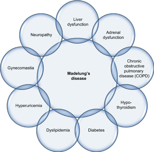
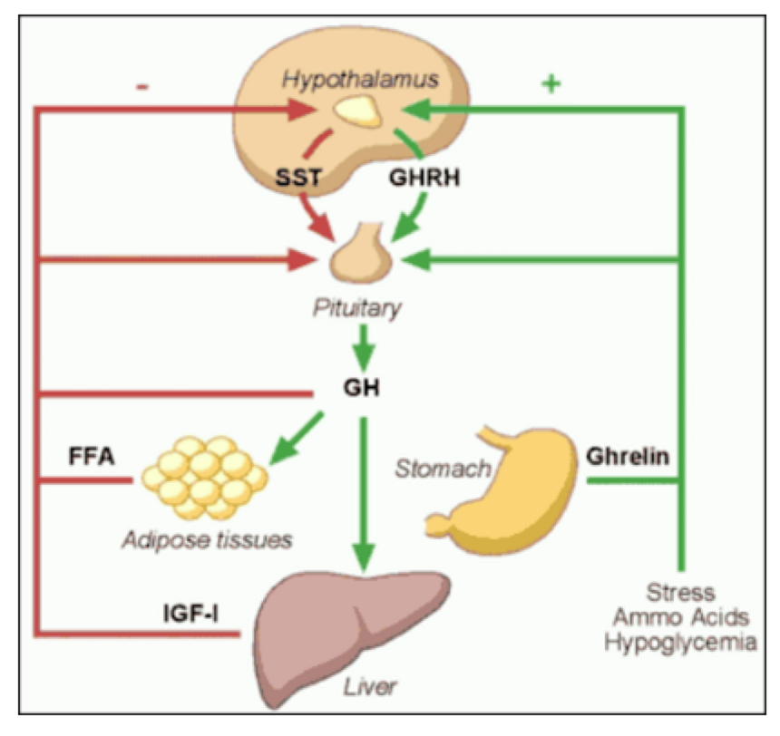
Novel insights provided by pathophysiology indicate the presence of a complex homeostatic system in which information about the energy reserve status and the meal quality and content is relayed from the periphery (gastrointestinal tract, pancreas, and adipose tissue) via specific orexigenic and anorexigenic peptides and hormones to the central nervous system (CNS). Peripheral peptide hormones are released postprandially and travel in the circulation to bind to their receptors in the homeostatic regulatory centers in the CNS, notably the arcuate nucleus (ARC) of the hypothalamus and the dorsal vagal complex (DVC) in the brainstem medulla. The ARC contains neurons expressing key orexigenic neurotransmitters, agouti-related peptide (AgRP) and neuropeptide Y (NPY), as well as anorexigenic neurotransmitters, proopiomelanocortin (POMC) and cocaine- and amphetamine-regulated transcript (CART). Food intake is thus modulated by complementary mechanisms so as to maintain energy and weight homeostasis. New drug therapies have begun to focus on combination therapy using medications that target more than one of these central pathways, thereby achieving more favorable weight loss outcomes. In addition, combining treatments may provide a better safety profile given that lower doses of each drug when used together may achieve better weight loss than higher doses of a single agent (see Figure 1 below).


Factors That Influence Appetite


The regulation of satiety and appetite depends on the interaction of three major factors: biological systems, modern macro-environmental exposures, and micro-environmental influences. Biological systems are shaped by genetic and epigenetic influences from early-life events that govern development of orexigenic and anorexigenic neuro-hormonal pathways involved in the pathophysiology of obesity. Modern macroenvironment (food production, consumption, availability, social structure, weather influencing physical activity, television and technology, cultural norms, endocrine disruptors) and microenvironment (nutrition, exercise, sleep, stressful lifestyle, circadian rhythm) play an important role in the conformational development of cognitive and emotional brain regions, thus predisposing to the obese phenotype.


Genetic Factors of Physical Activity


Specific genes predict to what extent adults remain active. This is evidenced in a study examining identical twins in which environmental factors shared by children at age 13 accounted for 78% to 84% of sport participation, whereas genetic differences provided no contribution at all. At the age of 17 to 18 the genetic influences represented 36% of the variance in the level of participation in sports, and by age 18 to 20, genetic factors were responsible for almost all (85%) of the differences in participation in sports.

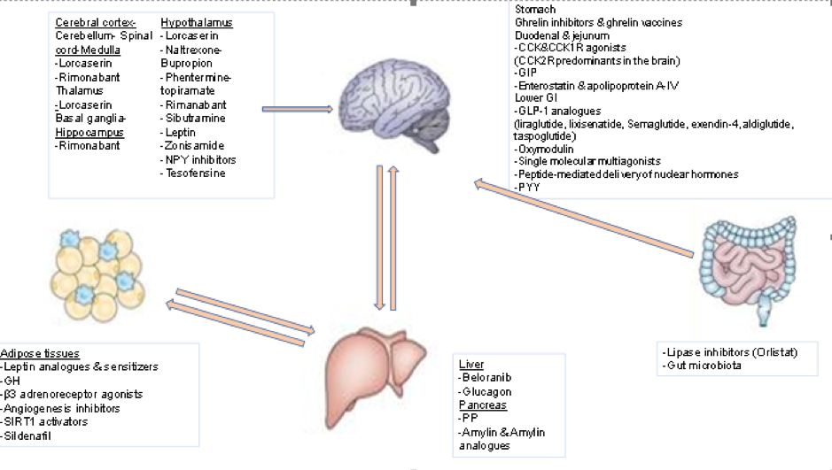
Figure 1. Sites of Action of the Most Important Anti-Obesity Drugs

CENTRALLY-ACTING ANTI-OBESITY DRUGS


Monoamine Neurotransmitter Modulators


With the exception of the glucagon-like peptide 1 (GLP-1) receptor agonist liraglutide, currently available weight loss medications act on the central nervous system to enhance dopamine, norepinephrine, and serotonin action to enhance satiety, diminish hunger, and consequently affect weight loss. Drug combinations have opened new horizons as they use multiple neural pathways, leading to better results with less adverse events. Recently, a review of fifty reports involving 43,443 subjects compared the efficacy of the central acting anti-obesity drugs lorcaserin (5HT2c receptor agonist), naltrexone-bupropion (opioid receptor antagonist combined with a norepinephrine releasing agent that stimulates POMC neuronal firing), phentermine-topiramate (a norepinephrine and dopamine modulator plus a carbonate anhydrase inhibitor), and liraglutide. It was found that the maximal mean weight loss relative to placebo was -3.06, -6.15, -7.45, and -5.5kg after 1 year with mean weight regain +0.48kg, +0.91kg, +1.27kg, +0.43kg the following year, respectively.  In these studies, the one-year drop-out rate was 40.9%, 49.1%, 34.9%, 24.3%, respectively (5).


Leptin, Leptin Analogues and Leptin Sensitizers


Leptin is a protein secreted primarily by white adipose tissue (WAT). It directly stimulates anorexigenic POMC neurons and inhibits adjacent orexigenic NPY neurons in the ARC of the hypothalamus, thus promoting satiety, increasing energy expenditure, and resulting in weight loss (6). Circulating levels of leptin increase with adiposity and decline following body weight reduction; the latter might be implicated in the total and resting energy expenditure reduction seen after weight loss. The discovery of leptin in 1994 was a seminal event in obesity research. It helped to establish that body weight should be viewed as a disorder with a strong biological basis rather than simply the result of poor lifestyle choices. Studies with congenitally leptin-deficient, severely obese subjects revealed that administration of physiological doses of leptin decreased food intake and body weight (7). Obese individuals, however, are leptin-resistant and have increased circulating leptin levels. Whether administration of leptin could overcome leptin resistance and exert an anti-obesity effect was tested in a placebo-controlled study with 47 obese men and women given varying doses of recombinant human leptin (0.03 mg/kg and 0.30 mg/kg, respectively) for 24 weeks and advised to eat 500 kcal less than body requirements each day. A dose-dependent decrease in body weight was shown, ranging from -1.3 kg in the placebo group to -1.4 kg in the 0.03 mg/kg leptin-treated group, and to -7.1 kg in the 0.30 mg/kg leptin-treated group (8). These results suggested that leptin resistance can be overcome with high doses of leptin but resulting in only modest weight loss similar to currently approved medications.  In addition, whether these effects can be sustained long-term is not known. Reports were similar from animal studies testing the effect of leptin sensitizers targeting the protein tyrosine phosphatase-1B (PTP1B)(9)(10) or the chemical chaperones that repair ER stress, including 4-phenyl butyric acid (PBA) and tauroursodeoxycholic acid (TUDCA) (11), each of which demonstrated reduced food intake and body weight. Like leptin treatment, sustainability of these anti-obesity effects is still not clear.


Weight loss is associated with reduction in energy expenditure, which makes long term weight loss maintenance difficult (12). Furthermore, 6 days of high fat diet in mice suffice to dramatically decrease the levels of phosphorylated signal transducer and activator of transcription 3 (p-STAT3) in the arcuate nucleus (13) while short term overfeeding of normal weight mice can lead to an increase of leptin resistance (14). Besides the inefficiency of leptin analogues as monotherapy, combinations of leptin with amylin (15), fibroblast growth factor 21(FGF21), exendin4, (16), or a GLP-1/glucagon co-agonist (17) were proposed. Only the combination with the GLP-1/glucagon co-agonist has shown improvement of leptin sensitivity (18). Apart from diet, stress of endoplasmic reticulum contributes to leptin resistance (19). Several plant-derived substances, such as celastrol (20) and withaferin (21) have been tested in diet-induced obese rodents for improvement of this pathway that leads to leptin resistance.


METRELEPTIN


Metreleptin (MYALEPT) is an injectable human recombinant leptin analogue approved in Japan for metabolic disorders including lipodystrophy and in USA as first-line treatment for non-HIV related forms of generalized lipodystrophy (leptin deficiency, congenital/acquired lipodystrophy) (22). A previous indication for hypothalamic amenorrhea has been withdrawn (23). (see Table 1)


Table 1. Metreleptin (MYALEPT)

FDA approved/Phase

Approved in Japan for lipodystrophy disorders and in USA for non-HIV lipodystrophy

Mechanism of action

Human recombinant leptin injectable analogue

Clinical Benefits

↓blood glucose, triglycerides, hepatic fatty steatosis

Adverse events

Headache, hypoglycemia, decreased weight, abdominal pain

-previous indication for hypothalamic amenorrhea discontinued


PRAMLINTIDE/METRELEPTIN


The combination of amylin-leptin (pramlintide-metreleptin) has been shown to be effective in the treatment of obesity. The anti-obesity properties of the combined treatment with pramlintide and metreleptin (pramlintide/metreleptin) were tested and showed a significant weight reduction of 12.7 ± 0.9% (11.5 ± 0.9 kg) without plateauing in obese patients during a 20-week trial period (24). The sponsors subsequently announced positive results from a 28-week proof-of-concept study with pramlintide and metreleptin combination treatment in overweight or obese subjects. The combination treatment reduced body weight on average by 12.7%, significantly more than treatment with pramlintide alone (8.4%), which is interpreted as 10 pounds more weight loss with the combined treatment. Remarkably, subjects receiving pramlintide/metreleptin continued to lose weight until the end of the study, compared to those treated with pramlintide alone, whose weight loss had stabilized towards the end of the study. The magnitude of weight loss was found to be dose-dependent and baseline BMI-dependent. Patients with a starting BMI less than 35 kg/m2 experienced the best weight loss efficacy with the combined treatment. A year later, the results of the 52-week blinded, placebo-controlled Phase II extension study of pramlintide/metreleptin were announced. The results indicated sustained and robust weight loss through the combined treatment; again, the most robust efficacy was seen in patients with a BMI less than 35 kg/m2 (25). Although the pramlintide/metreleptin combination seemed to be the next promising anti-obesity drug to be marketed, the sponsors discontinued its development in 2011, following commercial reassessment of the program (26).


Melanocortin-4 Receptor Agonists


The melanocortin system has a highly significant role in the hypothalamic regulation of body weight and energy expenditure. Leptin inhibits the release of the orexigenic neuropeptides orexin and melanocortin-concentrating hormone (MCH) in the lateral hypothalamic area (LHA) through the release of CART and melanocyte-stimulating hormone (α-MSH). The latter derives from the cleavage of POMC by prohormone convertase-1 and acts via melanocortin-3 and -4 receptors (MC3R, MC4R) activation. α-MSH emerged as a promising novel anti-obesity drug, and intranasal administration of the melanocortin sequence MSH/ACTH4-10 to normal-weight subjects was shown to acutely increase subcutaneous WAT lipolysis (27) and decrease body fat by 1.7 kg, when administered for six weeks (28). It eventually proved not to induce any significant reduction in body weight or body fat when compared with placebo in a 12-week study of 23 overweight men.


In preclinical studies, obese primates treated for eight weeks with the MC4R agonist RM-493 (Setmelanotide) lost an average of 13.5% of their body weight, with significant improvements in both insulin sensitivity and cardiovascular function. In June 2014, the results from the first human Phase II trial were released, testing the hypothesis that an MC4R agonist increases resting energy expenditure in obese subjects. A total of 12 obese but otherwise healthy individuals were randomized and completed both RM-493 and placebo periods in this double-blind, placebo-controlled, two-period crossover study. Analysis of the data indicates that short-term treatment with RM-493 increased resting energy expenditure significantly (by 6.4% vs placebo), thus suggesting RM-493 may be clinically effective for treating obesity. In 2015, administration of Setmelanotide to obese individuals for a limited time increased resting energy expenditure (REE) by 6.4% and shifted substrate oxidation to fat (29). Currently, Setmelanotide is being tested as a therapeutic option for rare genetic disorders of obesity such as POMC deficiency, heterozygous deficiency obesity, and POMC epigenetic disorders (30-32). (see Table 2)


Table 2. Setmelanotide (RM-493)

FDA approved/Phase

Phase II

Mechanism of action

MC4R-agonist

Weight loss vs placebo

13.5%

Clinical Benefits

↑insulin sensitivity, cardiovascular function, energy expenditure, ↓ body weight

-tested for POMC deficiency, heterogenous deficiency obesity, POMC epigenetic disorders

Adverse events

Headache, arthralgia, nausea, spontaneous penile erection, female genital sensitivity


Melanin-Concentrating Hormone (MCH) Antagonists


The melanocortin-concentrating hormone (MCH) is an important orexigenic neuropeptide in the LHA. Its release is stimulated by NPY and inhibited by leptin, exerting its orexigenic effects through the MCH1 receptor (MCHR1) (33). Like NPY, MCH exerts pleiotropic effects on locomotor activity, sensory processing, anxiety, aggression, and learning. Thus, despite the role of MCH in hunger stimulation, MCHR1 blockade as an anti-obesity target is questionable because such inhibition could elicit undesirable side effects. In animal models, MCH antagonists have consistently demonstrated efficacy in reducing food intake acutely and in inhibiting body weight gain when given chronically (34). Five compounds have reached testing in human subjects. Although they were reported as well-tolerated, none has proceeded to Phase II studies. A major issue with many lead compounds is increased cardiovascular risk due to drug-induced QTc prolongation (35). Among others, the MCHR1 antagonist AMG 076 entered Phase I safety and tolerability testing in 2004, but there have been no subsequent reports of its status since 2005. The MCHR1 antagonist GW-856464 also entered Phase I studies in 2004; however, in 2010 it was reported that low bioavailability precluded further development. The MCHR1 antagonist NGD-4715 was safe and well-tolerated in a Phase I clinical trial, but its development ceased in 2013. Similarly, despite the reported tolerability and indication of efficacy of the MCHR1 antagonist ALB-127158, its development was terminated before the initiation of Phase II studies. Finally, the longest (28-day) Phase I study with BMS-830216, a pharmacological antagonist of MCH signaling (36) produced no indications of weight loss or reduced food intake and the compound did not proceed to Phase II studies.


Subtype-Selective Serotonin-Receptor Agonists


Central serotonin participates in feeding behavior and energy balance modulation, reducing food intake in animals and human beings.  This finding was supported by reports of two selective serotonin reuptake inhibitors (SSRIs) developed to treat depression, fluoxetine and sertraline, being associated with non-sustained weight loss in obese subjects. Thus, agonists to appropriate serotonin receptors are potentially valuable drugs. The serotonin (5-HT) system directly modulates the hypothalamic POMC (anorexigenic) and NPY (orexigenic) networks, enhancing satiety and causing hypophagia. These effects are mediated by 5-HT2C and 5-HT1B receptors, located on hypothalamic POMC and NPY neurons, respectively. Through the 5-HT1B receptors, serotonin inhibits the NPY/Agrp neurons, thereby decreasing the GABAergic inhibitory input to POMC cells; while through the 5-HT2C receptors it directly activates the anorexigenic POMC neurons. Via these actions, serotonin increases α-MSH and decreases AgRP release into the hypothalamic melanocortin system, promoting satiety. Between 1973 and 2000 there was an explosion in the pharmaceutic industry regarding central acting anti-obesity drugs. Three non-selective serotonin-receptor agonists were approved by FDA: fenfluramine (1973-1997), the combination phentermine-fenfluramine (1992-1997), and dexfenfluramine (1996-1997). These were all 5-HT1b agonists characterized for their ability to inhibit food consumption, but also had effects on other serotonin receptors that lead to unacceptable side effects (cardiac valvular thickening) and were voluntarily withdrawn from the market.


In 1997, when fenfluramine and dexfenfluramine were discontinued by the manufacturer, sibutramine, a serotonin and norepinephrine reuptake inhibitor emerged. Sibutramine has only little clinical relevance as an antidepressant but enhances weight loss due to an increase in energy expenditure and inhibition of food intake (37). In addition to weight loss, sibutramine was found to improve fasting levels of insulin, triglycerides, and high-density lipoprotein cholesterol. Sibutramine was also associated with increase of blood pressure, cardiovascular events, and cardiac arrhythmias (38). For these reasons, FDA withdrew it in 2010.


LORCASERIN


As activation of the 5-HT1B receptor has been implicated in both primary pulmonary hypertension (39) and valvopathy (40), the 5-HT2C receptor subtype has been proposed as a target for therapeutic intervention to allow weight loss. Several potent and selective 5-HT2Creceptor agonists proved to be effective in suppressing food intake and inducing weight loss in rodents, including WAY-163909 (41), CP-809101 (42), and vabicaserin (43). However, only lorcaserin (APD356) moved into clinical testing. Lorcaserin (Belviq) is a selective 5-HT2c receptor agonist, which belongs in the third generation of 5-HT-based anti-obesity drugs (44). It activates hypothalamic POMC neurons to induce satiety and decrease food intake but does not affect energy expenditure. Through actions on midbrain dopaminergic tone, it has been shown to suppress binge- food behaviors. Its action in addictive disorders is currently under investigation (45). Based on the outcome of the BLOOM (46) and BLOSSOM trials (47), in 2012 the FDA approved lorcaserin as an addition to a reduced-calorie diet and exercise for eligible patients (48). The efficacy of lorcaserin appears similar to that of orlistat (mean difference in weight loss between active and placebo treated groups approximately 3 to 4 kg) and perhaps slightly less than that of phentermine-topiramate. The impact of lorcaserin in patients with T2DM and BMI: 27-45kg/m2 was examined in the BLOOM-DM trial which showed a reduction of body weight by approximately 5kg versus 1.6kg in the placebo group, as well as significant decreases in heart rate, HDL levels, and waist circumference. Valvopathy was shown not to occur in excess with treatment and lorcaserin was generally well tolerated, with a low incidence of side effects such as headache, dizziness, fatigue, nausea. After the results of BLOOM-DM trial, a potential combination of GLP-1RA and 5-HT2A/C is now under investigation (49).


In a multicenter, randomized, double-blind, placebo-controlled, parallel-group study involving12,000 overweight and obese patients with cardiovascular disease or multiple cardiovascular risk factors (CAMELLIA-TIMI 61), the effect of long-term treatment with lorcaserin on major cardiovascular events and conversion to T2DM over a 5-year period were examined. After one year of treatment, 5% weight loss was observed in 38.7% and 17.4% in the lorcaserin and the placebo groups, respectively. Regarding cardiac risk, the lorcaserin group was non-inferior to the placebo group with slightly better values in cardiac risk factors (blood pressure, heart rate, glycemic control, lipid profile). Adverse events were rare in both groups, apart from the incidence of serious hypoglycemia in the lorcaserin group in those with diabetes managed using insulin or sulfonylureas (50, 51). In addition, lorcaserin administration decreased the incidence of T2DM by 19% in patients with prediabetes and by 23% in patients without diabetes. In patients with T2DM, lorcaserin resulted in a reduction of 0.33% in HbA1c compared with placebo at 1 year from a mean baseline of 7.0%. (see Table 3, 4)


Table 3. Lorcaserin (Belviq)

FDA approved/Phase

2012

Mechanism of action

Selective Serotonin 2C agonist

Weight loss vs placebo

3-4kg

Clinical Benefits

↓food intake, heart rate, HDL levels, waist circumference, HbA1c

Adverse events

Headache, dizziness, fatigue, nausea, dry mouth, constipation, heart valvopathy

-In diabetics: hypoglycemia, headache, back pain, cough, fatigue, risk of serotonin syndrome/neuroleptic malignant syndrome, valvular heart disease


Table 4. Clinical Trials of Lorcaserin

Clinical trial

Patients

Dose

Treatment, placebo from baseline

% of patients losing ≥5% of baseline weight

Comment


BLOSSOM

1-year randomized, double-blind, placebo-controlled trial

(2011)

4008 patients (18-65 y.o., BMI- 30-45kg/m2 or 27-29.9kg/m2 with comorbidity) randomized in a 2:1:2 ratio

i.10mg x2 po


ii.10mg x1 po


iii.placebo

i.-5.8kg


ii.-4.7kg


iii.-2.9kg

i.47.2%


ii.40.2%


iii.25%

Exclusion criteria: recent cardiovascular events, diabetes mellitus, BP >150/95mmHg

BLOOM

2-year randomized, double-blind, placebo-controlled trial

(2010)


3182 adults (mean BMI-36.2kg/m2) randomized to lorcaserin twice daily or placebo group. After 52 weeks, the placebo group continued placebo and lorcaserin group selected placebo or lorcaserin for 52 weeks

i.10mg x2 po


ii. placebo

i.-5.8kg


ii.-2.2kg

i.47.5%


ii.20.3%

Weight loss was greater in the group which continued lorcaserin for the second year

BLOOM-DM

1-year randomized, double-blind, placebo-controlled trial

(2012)

604 patients (HbA1c: 7-10%, BMI-27-45kg/m2, treatment with metformin, sulfonylurea or both)

i.10mg x2 po


ii.10mg x1 po


iii.placebo

i.-4.7kg


ii.-5.0kg


iii.-1.6kg

i.37.5%


ii.44.7%


iii.16.1%

↓heart rate, HDL levels, waist circumference in lorcaserin treated groups

NO valvopathy was statistically significant

CAMELLIA-TIMI 61

3.3-year randomized, placebo-controlled trial

(2018)


12,000 patients overweight/obese-three subgroups

A. diabetes

B. prediabetes

C. normoglycemic

i.10mg x2/day


ii. placebo

At 1 year the mean treatment difference:

 A: -2.6kg

 B: -2.8kg

 C: -3.3kg


At 1 year compared with placebo:

A: 37.4%

B: 39.7%

C: 42.3%

↓ BMI, waist circumference, waist-to-hip ratio, HbA1c, reduced microvascular complications


Bupropion


Bupropion is a dopamine and norepinephrine-reuptake inhibitor that has been marketed as an anti-depressant and for smoking cessation. Previous animal studies have clearly shown a dose-dependent satiety effect of bupropion following intraperitoneal injection (52). The acute effects of dopamine and noradrenaline reuptake inhibition on energy homeostasis demonstrated their additive effects on short-term food intake (53). Bupropion increases dopamine activity and POMC neuronal activation, thereby reducing appetite and increasing energy expenditure (54). Whether the acute meal terminating effects of bupropion documented in animal studies could be translated into long-term weight loss efficacy in humans was addressed by three clinical trials with overweight and obese adults (55, 56, 57) using different treatment doses (100 to 400 mg/d) and duration (up to 24 weeks). They have all shown bupropion to have dose-dependent modest weight reducing efficacy, plus a safe profile. One study that assessed the anti-obesity efficacy of bupropion over two years reported maintenance of weight loss during the continuation phase, while another demonstrated its efficacy even in depressed patients. Although the weight loss effect of bupropion was superior in non-depressed patients compared to those suffering from depression, the fact that bupropion was well-tolerated and effective in this group of patients provides a potential valuable adjunctive therapy to elevate mood in depressed subjects in whom weight gain secondary to antidepressant therapy is an issue. Cardiovascular effects, such as a rise in blood pressure and tachycardia, were usually mild, while the risk of seizure, which was high with the original bupropion formulation, has been significantly reduced with the advent of bupropion-SR and bupropion-ER.


An interesting finding of the previous studies was that the rather modest weight loss effect of bupropion reached a plateau by 24 weeks of treatment. This could be explained by the molecular pathophysiology of the weight reducing effects of bupropion, which directly stimulates the hypothalamic POMC neurons that in turn release α-MSH and β-endorphin. α-MSH mediates the anorectic effect of POMC activation, whereas β-endorphin exerts negative feedback on POMC neurons via opioid receptors (58). The latter possibly points to one of the compensatory mechanisms that limits long-term efficacy of bupropion and other weight loss modalities.


Naltrexone


Naltrexone is an opioid receptor antagonist. By blocking opioid receptors on the POMC neurons, feedback inhibition is prevented further increasing POMC activity. Monotherapy with opioid antagonists to decrease short-term food intake has been tested (59). Naltrexone failed to produce consistent or clinically meaningful weight loss, even at large doses (300 mg/d) (60), implying that a single opioid mechanism is unlikely to explain all aspects of ingestive behavior.


Bupropion/Naltrexone Sustained Release (SR)


The combined bupropion/naltrexone (NB) therapy induced significantly greater weight loss on a diet and exercise program over 56 weeks compared to monotherapy and placebo (61). In 2014, the FDA approved this combination (Contrave, Mysimba) for body weight management in adults who are overweight and obese. This combined therapy of opioid antagonist and aminoketone antidepressant is titrated over four weeks to the maximum dose. NB has shown remarkable benefit in patients with binge-eating disorder (BED) and concomitant alcohol abuse, but this result needs further evaluation (62). Four major 56-week phase III randomized, double-blind, placebo-controlled trials have shown the therapeutic effect of ΝΒ SR (COR-I, COR-II, COR-BMOD, COR-DIABETES) in different dosage combinations (see Table 6). In COR-I, the weight loss ratio on NB 16/360mg, NB 32/360mg or placebo was -5.0%, -6.1%, -1.3% (P<0.00) respectively. In COR-II, the weight loss ratio on NB 32/360mg or placebo was -6.4%, -1.2% (P<0.001) (63). In COR-BMOD, NB SR 32/360mg plus intensive behavioral modification was compared with the behavioral modification alone as a therapeutic option. The weight loss ratio was -11.5% versus -7.3% (P<0.001), respectively (64). Recently, COR-Diabetes has included patients with T2DM with or without antidiabetic treatment. The NB SR 32/360mg treatment resulted in -5.1% weight loss versus -1.8% in the placebo group (P<0.001). NB treatment resulted in a HbA1c reduction, cardiovascular benefit, and lipid profile improvement (65). Due to FDA request for further investigation of the effect of NB on major cardiovascular events, the LIGHT trial was created. Unfortunately, this trial terminated early following recommendation by the academic leadership of the study because confidential interim data were publicly released by the sponsor (66). (See Table 5, 6)


Table 5. Bupropion/Naltrexone Sustained Release (Contrave, Mysimba+

FDA approved/Phase

2014

Mechanism of action

Aminoketone antidepressant/Opioid antagonist

Weight loss vs placebo

4.8kg

Clinical Benefits

↓ appetite

Adverse events

Nausea, constipation, headache, vomiting, dizziness, insomnia, dry mouth, suicidal ideation, increase blood pressure/heart rate, hepatotoxicity, angle-closure glaucoma Uncontrolled hypertension, seizures, anorexia nervosa/bulimia, chronic opioid use, coadministration with MAO inhibitors


Table 6: Clinical trials of Naltrexone/Bupropion SR

Clinical trial

Patients

Dose

Treatment, placebo from baseline

% of patients losing ≥5% of baseline weight

Comment


COR I

1-year randomized, double-blind, placebo-controlled trial

(2010)

1742 patients randomly categorized in a 1:1:1 ratio

i.16/360mg po


ii.32/360mg po


iii. placebo

i.-5.0%


ii.-6.1%


iii.-1.3%


i.39%


ii.48%


iii.16%


COR II

1-year randomized, double-blind, placebo-controlled trial

(2013)

1496 patients randomly categorized in a 2:1 ratio to NB 32/360mg or placebo; patients on NB with <5% weight loss in 28-44 week were reassigned to continue 32/360mg or increase daily dose to NB 48/360mg

i.32/360mg (or increased daily dose 48/360mg)


ii.placebo

i.-6.4%


ii.-1.2%


i.50.5%


ii.17.1%

Random reassignment to higher dose did not change weight loss results

COR-BMOD

1-year randomized, double-blind, placebo-controlled trial

(2011)

793 patients with obesity randomly categorized in a 1:3 ratio

i. BMOD+ NB (32/350mg)


ii. BMOD+ placebo

i.-11.5%


ii.-7.3%

i.66.4%


ii.42.5%

The efficacy of NB is obvious, and a lifestyle change can increase weight loss

COR-DIABETES

1-year randomized, double-blind, placebo-controlled trial

(2013)

505 patients overweight/obese and T2DM with/without oral anti-hypoglycemic agents randomly categorized in a 2:1 ratio

i.32/360mg


ii. placebo

i.-5.0%


ii.-1.8%

i.44.5%

ii.18.9%

↓HbA1c, certain improvements in CVD risk factors.

↑ nausea, constipation, vomiting


Zonisamide


Given the pathophysiology behind the anti-obesity efficacy of the selective serotonin-receptor agonists and the dopamine-reuptake inhibitors, an ideal drug would combine serotonergic and dopaminergic activity. This is exactly the case of Zonisamide, a marketed antiepileptic drug that exerts dose-dependent biphasic dopaminergic (67) and serotonergic (68) activity. Its weight loss efficacy was investigated by a double-blind, placebo-controlled trial which reported a 32-week mean weight loss of 9.2 kg (1.7 kg) (9.4% loss) for the Zonisamide group (dose administered up to 600 mg/d) compared with 1.5 kg (0.7 kg) (1.8% loss) for the placebo group (P<0.001); Zonisamide was generally well-tolerated with only a few adverse effects (69). The findings were similar when the long-term effectiveness and tolerability of Zonisamide for weight control was examined in psychiatric outpatients using various psychotropic medications; the mean BMI reduction achieved was 0.8±1.7 kg/m2 and ranged from -2.9 kg/m2 to 4.7 kg/m2 (p<0.001), while the drug was generally safe and well-tolerated (70). Zonisamide was also assessed in the treatment of binge-eating (BE) disorder where it proved to be effective in reducing binge-eating frequency, severity of illness, and weight; however, the reports regarding its tolerability were conflicting (71). (see Table 7).


Table 7. Zonisamide

Mechanism of action

Selective serotonin-receptor agonist and dopamine-reuptake inhibitor

Weight loss vs placebo

7.8kg

Clinical Benefits

Assess in the treatment of binge-eating disorder

Adverse events

Nausea, headache, insomnia


Zonisamide/Bupropion SR


Whether the anti-obesity efficacy of Zonisamide is increased when combined with bupropion (dopamine and norepinephrine -reuptake inhibitor) has been evaluated in a few Phase II clinical trials with different combined doses; the bupropion SR/Zonisamide SR combination is marketed under the trade name Empatic. In its 24-week, double-blind, placebo-controlled Phase IIb trial (72), patients completing 24 weeks of bupropion SR 360 mg/Zonisamide SR 360 mg therapy lost 9.9% of their baseline body weight, or 22 pounds, compared to 1.7% for placebo patients (p<0.001). Of patients who completed 24 weeks of therapy, 82.6% lost at least 5% of their baseline body weight and 47.7% lost at least 10% of their baseline body weight compared to 18.9% and 5.7% of placebo patients, respectively (p<0.001 for both). Patients experienced significant weight loss as early as by their first post-baseline visit at week four. Importantly, patients continued to lose weight until the end of the trial period with no evidence of a weight loss plateau. Early results showed that patients lost an average of 14% over 48 weeks. Improvements were observed in key markers of cardiometabolic risk such as waist circumference, triglycerides, fasting insulin, and blood pressure. The most commonly reported adverse events for all patients were headache, insomnia, and nausea. The most common adverse events leading to discontinuation were insomnia, headache, and urticaria (hives). There were no serious adverse events attributed by investigators to the study drug. There were no statistically or clinically meaningful differences between the drug and placebo on measures of cognitive function, depression, suicidality or anxiety. These reports revealed a significant weight-reduction effect for the combination Bupropion/Zonisamide. However, the safety concerns (73) will need to be addressed in the upcoming Phase III studies before firm conclusions about its safety profile can be drawn. (see Table 8)


Table 8. Zonisamide/Bupropion (Empatic)

FDA approved/Phase

Phase II completed

Mechanism of action

Selective serotonin-receptor agonist and dopamine-reuptake inhibitor/dopamine and norepinephrine reuptake inhibitor

Weight loss vs placebo

9.9% of their baseline weight

Clinical Benefits

↓cardiometabolic risk

Adverse events

Headache, insomnia, nausea, urticaria


Topiramate


Topiramate is another anticonvulsant agent associated with weight loss. It is a sulphamate-substituted fructose that is approved as an antiepileptic/antimigraine agent and has multiple effects on the CNS, including action on the orexigenic GABA systems causing appetite suppression (74). A 6-month dose-ranging study in obese human subjects addressing its anti-obesity efficacy at doses of 64, 96, 192, and 384 mg/day (in divided twice-daily dosing) concluded that all doses produced significantly greater weight loss compared to placebo, and that weight loss in the 192 mg/day group was similar to the 384 mg/day group (75). This is important as topiramate has been associated with several neuropsychiatric effects, especially when administered at high doses (of 192 mg/d or more). Another study investigating the weight loss efficacy and safety of topiramate doses of 96, 192, and 256 mg/day over a 1-year period in obese subjects using the immediate release form tablets (before the development of the controlled-release formulation). Clinically significant weight loss (7.0, 9.1, and 9.7% of their baseline body weight for the doses of 96, 192, and 256 mg/day, respectively), was reported compared to 1.7% body weight loss in the placebo group (P<0.001) plus improvements in blood pressure and glucose tolerance (76). Finally, several other studies investigated the therapeutic effect of topiramate in patients with BED and bulimia (77) that are both associated with obesity; the results were very promising regarding control of symptoms in both disorders. (see Table 9)


Table 9. Topiramate

FDA approved/Phase

Phase II completed

Mechanism of action

Sulphamate-substituted fructose acts on GABA system

Weight loss vs placebo

7.0%(96mg),9.1%(192mg), 9.7% (256mg/day)

Clinical Benefits

Assess in the treatment of binge-eating, bulimia

Adverse events

Headache, insomnia, nausea, urticaria


Phentermine


Phentermine is a sympathomimetic amine, which has anorexigenic action, that also releases insignificant quantities of dopamine. Thus, it is characterized by lower abuse potential (78). Its main mechanism of action involves catecholamine release in the hypothalamus resulting in enhanced satiety feeling and reduction of food intake (79). The most common side effects of phentermine as a sympathomimetic drug is heart rate increase, hypertension, dizziness, dry mouth, insomnia, irritability, and gastrointestinal disorders (80). Phentermine was the first FDA approved anti-obesity drug in 1959 for those aged >16 years old, but for only short-term use (maximum 3 months). The reason for the time limit is because the pharmaceutic industry had not updated labeling since 1959. In 1968, in a double-blind, placebo-controlled trial, 108 overweight or obese women were categorized into three groups that received continuously or intermittently (every 4 weeks) dosed phentermine or placebo, respectively. The weight loss was -12.2kg, -13.0kg or -4.8kg, respectively (81).


Currently, the off-label long-term use of phentermine is indicated only if there is clinical benefit, stable blood pressure and pulse rate in the absence of cardiovascular history or substance abuse disorders. In a recently published retrospective cohort study, 13,972 patients were observed for 6, 12 and 24 months after phentermine initiation. They were categorized in five groups based on the time of phentermine administration: short-term use, short-term intermittent use, medium-term continuous use, medium-term intermittent use, long-term continuous use. Weight-loss, changes in blood pressure, heart rate, and incidence of cardiovascular events (myocardial infarction, stroke, angina, coronary artery bypass grafting, carotid artery intervention, death) were examined. Weight loss was greater among off-label groups than referent group of short-term use with results depending on the duration of phentermine initiation. Specifically, at six months, short-term intermittent patients lost 1.8% further body weight relative to short-term single patients and medium-term intermittent patients lost 5.6% further body weight relative to short-term single patients. At twelve months, the medium-term intermittent use group lost further 5.6% body weight relatively to the short-term use group. At twenty-four months, long-term the continuous use group lost 7.4% additional body weight in comparison with the short-term use group. Concerning safety of phentermine, changes in heart rate and diastolic blood pressure were insignificant at six, twelve, and twenty-four months. Interestingly, although the referent group showed a slight increase in systolic blood pressure (+0.5-3.2 mmHg) at twenty-four months, all groups had slightly lower systolic blood pressure than the referent group at twelve- and twenty-four-months follow-up period. Lastly, the incidence of major cardiovascular events was low. So, it was shown that the off-label over three months therapy with phentermine was superior to short–term administration with greater weight-loss effect and cardiovascular safety. More studies with fewer limitations should follow in order to support these findings (82). In 2013, a clinical trial comparing phentermine as monotherapy or as part of a combined therapy, took place and resulted in a weight loss of 5.1% at 28 weeks follow-up period in favor of the combined phentermine/topiramate group.(see Table 10)


Table 10. Phentermine

FDA approved/Phase

1959

Mechanism of action

Norepinephrine release and minor dopamine release

Weight loss vs placebo

0.23kg/week

Clinical Benefits

Lower abuse potential

Adverse events

Stimulation, insomnia, dry mouth, constipation, primary pulmonary hypertension

Contraindicated in cardiovascular disease, coadministration with MAO inhibitors, hyperthyroidism, glaucoma, drug abuse


Phentermine/Topiramate ER


Because of dose-related side effects seen with topiramate treatment including suicidality, metabolic acidosis, acute myopia, and secondary angle closure glaucoma, a lower dose of topiramate was used (in a special controlled release formulation) in a novel anti-obesity drug called Qsymia. The main mechanism of action of Phentermine/Topiramate extended release(ER) is possibly the alteration of various neurotransmitters, including inhibition of voltage-dependent sodium channels, glutamate receptors, and carbonic anhydrase as well as potentiation of γ-aminobutyrate activity (83).Two large randomized, double-blind, placebo-controlled trials took place (EQUIP and CONQUER) followed by a 2-year extension trial (SEQUEL). In the EQUIP trial 1,267 patients with BMI>35kg/m2were allocated in two groups and received phentermine/topiramate ER 3.75/23mg and 15/92mg, respectively, once daily. With 59.9% of the patients discontinuing, this trial found no statistically significant difference between the two groups regarding weight reduction (84). In the CONQUER trial 2,487 patients were allocated in three groups and received phentermine/topiramate ER 7.5/46mg, phentermine/topiramate ER 15/92mg, and placebo, respectively. The results were in favor of the combined therapy while the greater dosage resulted in greater weight loss with mean weight loss -7.8kg, -9.8kg, and -1.2kg in the three respective groups (85). Patients who completed the CONQUER trial entered the SEQUEL trial for 52 weeks. The weight loss as percentage of the initial weight was -9.3%, -10.5% and -1.8% in the three respective groups. A statistically significant improvement of lipid profile, glycemic control, and waist circumference in the phentermine/topiramate ER groups was reported (86). Based on the positive results from three Phase III studies, in 2012 FDA approved topiramate/phentermine extended-release as an adjunct to a reduced-calorie diet and increased physical activity for chronic weight management in eligible adults. Meanwhile however, approval was denied by European regulatory authorities, who cited potential risk to the heart and blood vessels, psychiatric side effects, and cognitive side effects in explaining their decision (see Table 11, 12).


Table 11. Topiramate/Phentermine Extended Release (ER) (Qsymia)

FDA approved/Phase

2012

Mechanism of action

Norepinephrine release, GABA modulation, voltage-gated ion channel modulation, stop of AMPA/kainite excitatory glutamate receptors and carbonic anhydrase

Weight loss vs placebo

6,6kg

Clinical Benefits

↓ lipid profile, HbA1c, waist circumference

Adverse events

Paresthesia, dizziness, dysgeusia, insomnia, constipation, dry mouth, fetal toxicity, metabolic acidosis, cognitive impairment

Contraindicated in: Glaucoma, hyperthyroidism, coadministration with MAO inhibitors


Table 12. Clinical Trials of Phentermine/Topiramate ER

Clinical trial

Administration

N

Treatment, placebo from baseline

% of patients losing ≥5% of baseline weight

Comment


CONQUER

Double-blind, placebo-controlled trial over 1 year

(2011)

4-week titration+ 52 weeks of treatment:

15/92mg po

or 7.5/46mg po

2487 patients (BMI:27-45kg/m2 with 2+ risk factors

i.15/92mg


ii.7.5/46mg


iii. placebo


i.-9.8kg


ii.-7.8kg


iii.-1.2kg


i.70%


ii.62%


iii.21%

↑improvement in blood pressure, waist circumference, lipid levels, fasting glucose and insulin

SEQUEL

2-year study overall;1-year extension of CONQUER

(2012)

227 patients completed the original blinded treatment


i.15/92mg


ii.7.5/46mg


iii. placebo

i.-10.9kg


ii.-9.6kg


iii.-2.1kg

i.79.3%


ii.75.2%


iii.30%


Neuropeptide Y (NPY) Inhibitors


The ARC NPY neurons inhibit the anorexigenic POMC neurons (via NPY Y1 and Y5 receptors) and promote the release of the orexigenic neuropeptides orexin and MCH in the LHA, thus promoting food intake. Therefore, NPY blockade could be a promising target for body weight management. Animal experiments (in mice) have shown that pharmacologic blockade or genetic deletion of the Y1- and Y5-receptors reduces food intake and weight, with Y1-receptor signaling appearing to be the major mediator of the orexigenic effects of NPY. However, NPY is the most abundant central neuropeptide and regulates many functions beyond feeding; thus, targeting NPY neurons/Y receptors specifically for obesity is not easy and could result in unacceptable side effects. In addition, experimental medical blockade of NPY signaling with the Y5-receptor antagonist MK-0577 failed to cause any significant weight loss in a 1-year clinical trial (87). On the other hand, the oral, once-daily, centrally acting selective Y5-receptor antagonist velneperit, previously known as S-2367, induced a mean placebo-adjusted weight loss of 5.0% from initial weight (p <0.0001) over 54 weeks of therapy and was accompanied by improvement of lipid profile and waist circumference reduction (88).Nevertheless, velneperit did not proceed in markets due to disappointing results in phase IIb trials. However, the combined Y1/Y5-receptor antagonism may prove more effective, though we are not aware of any Y1/Y5-receptor antagonist in development to date. In contrast to Y1 and Y5, the Y2- and Y4-receptors are the targets of the satiety hormones PYY and pancreatic polypeptide (PP), respectively, and, as mentioned below, two drugs, a Y2/Y4-receptor agonist (obinepitide and a selective Y4-receptor agonist (TM30339)) are in Phase I/II clinical trials and are yielding results that appear quite promising as regards weight loss. A combined anti-obesity medication of velneperit/orlistat is under way (phase II clinical trial), also with promising results (89).


Dopamine antagonists


The mesolimbic dopamine system was proven to play a critical role in compulsive overeating or binge eating, which is one of the main reasons why people become overweight or obese. There is some evidence that blocking the action of dopamine in animals can reduce food intake, particularly of foods that are high in fat and sugar. GSK 598809 is a D3 antagonist that blocks dopamine. Preliminary data from human studies failed to show any significant effect on body weight control (90).


Tesofensine


Tesofensine (TE) is a presynaptic inhibitor of norepinephrine, dopamine, and serotonin. Like sibutramine, it suppresses appetite and may result in significant weight loss, as this was shown when given for the treatment of Parkinson’s disease, but also in a multi-dose, dose-ranging trial where 203 obese patients were randomly assigned to Tesofensine (0.25, 0.5, and 1.0 mg) or placebo once daily. Phase II testing of the drug has been completed. After 24 weeks, mean weight reduction was greater in the Tesofensine groups (-6.7, -11.3, -12.8 kg, for the three doses, respectively) compared with placebo (-2.2 kg). Additionally, an improvement in lipid profile and glycemic control was observed. A dose-dependent increase in blood pressure was observed along with a 7.4bpm increase in pulse rate in the 0.5mg/day group. Adverse events such as headache and mood alterations were also present in all groups especially in the 1mg/day group (91). In another trial, 32 males were allocated in two groups and received 2mg/day Tesofensine and placebo, respectively. The interesting point in this trial was that the patients were free to consume their usual amounts levels of food and exercise as usual. However, in the Tesofensine group they lost 1.8kg over 2 weeks because Tesofensine increased visual analog scale ratings of satiety and 24h fat oxidation in comparison with placebo. Even if an increase in total energy expenditure was not observed, an increase in sleeping energy expenditure was found. Altogether, Tesofensine induces weight loss by promoting the satiety feeling and slightly increasing metabolic rate (92). The effect of Tesofensine in appetite sensations was evaluated in another phase II trial, in which patients were allocated in 4 groups and received 0.25mg, 0.5mg, 1mg and placebo, respectively, for 24 weeks. For the first 12 weeks, a dose-dependent increase in the satiety feeling was noticed even though this feeling faded away as the trial was in progress (93). In 2010, a study on the abuse effect of Tesofensine, bupropion, atomoxetine, and placebo in comparison to d-amphetamine took place and concluded that the studied substances had no abusive action (94). Tesofensine has been shown to increase both blood pressure and pulse rate. In 2018, a phase III clinical trial was powered by the pharmaceutic industry producing Tesofensine. In this study 372 patients were allocated in three groups and received Tesofensine 0.25mg, 0.5mg and placebo. Furthermore, a combination of Tesofensine/metoprolol is recently being examined against hypothalamic injury-induced obesity and Prader-Willi syndrome (95). (see Table 13)


Table 13. Tesofensine

FDA approved/Phase

Phase III

Mechanism of action

Triple monoamine reuptake inhibitor of dopamine, norepinephrine, serotonin

Weight loss vs placebo

4.5-10.6%

Clinical Benefits

Pharmacological similar to sibutramine

↓ appetite, body weight, lipid profile, blood glucose

Adverse events

Headache, mood alterations, potentially increase heart rate, blood pressure, psychiatric disorders

.

Lisdexamfetamine dimesylate


Another sympathomimetic, Lisdexamfetamine dimesylate, at certain doses appears effective in decreasing binge-eating days in patients with BED compared with placebo, according to a study published online by JAMA Psychiatry (96). The study included 259 and 255 adults with BED in safety and intention-to-treat analyses, respectively. Patients received lisdexamfetamine 30, 50 or 70 mg/day or placebo. BE days per week decreased in the 50 mg and 70 mg groups but not in the 30 mg group compared with placebo. Confirmation of these findings in ongoing clinical trials may result in improved pharmacologic treatment for moderate to severe BED.


Cannabinoid-1 Receptor (CB1) Antagonists


Among other neurotransmitter systems, the cannabinoid system modulates the hypothalamic melanocortin and NPY feeding networks. It has been shown that administration of cannabinoid-1 receptor (CB1) agonists and antagonists induces hyperphagia and hypophagia, respectively. These observations led to development of rimonabant, a cannabinoid-1 receptor antagonist, for the treatment of obesity, which was shown quite effective in promoting weight loss; however, it increased the incidence of mood-related disorders (97). As a result, in 2009, rimonabant was withdrawn from the market and the development of other cannabinoid-1 receptor antagonists for the treatment of obesity has also been discontinued. Before withdrawal, rimonabant was shown to have advantages in glycemic control and cardiovascular events (98). In 2010, another CB1 antagonist (AM6545) was found to have less psychological side effects and to induce satiety feeling and weight loss in animal studies (99). (see Table 14)


Table 14. Cannabinoid Type-1 Receptor Antagonists (SR141716, AM251, AM6545)

Mechanism of action

Antagonism of cannabinoid type-1 receptors stimulates anorexigenic signaling

Clinical Benefits

↓ body weight, blood glucose, cardiovascular events

-AM6545: has limited CNS penetration

Adverse events

Mood alterations


Human Chorionic Gonadotropin (hCG)


Human chorionic gonadotropin (hCG) in the form of subcutaneous injection and oral or sublingual diet drops has been advertised as aiding in weight loss of one to two pounds daily, absence of hunger, and maintenance of muscle tone. Clinical trials, however, failed to support this claim (100). In fact, FDA recommended avoiding buying over-the-counter weight loss products which contain hCG. One might ask why the hCG diet has so many enthusiastically supporting it. The reason may be that this diet needs to be accompanied by severe calorie restriction, to only 500-800 calories per day. Anyone following such recommendations is bound to lose weight, if only short-term. Most crucially, though, since hCG has been reported to induce serious side effects, this drug should not be used for the treatment of obesity. In addition, very low-calorie diets have not been shown to be superior to conventional diets for long-term weight loss, plus they have risks, such as gallstone formation, irregular heartbeat, and an imbalance of electrolytes. Therefore, if weight loss is the goal, there are safer ways to make it happen.


Nesfatin-1


Nesfatin-1 is a satiety molecule, which was first described in rats and is derived from its precursor molecule nucleobindin2 (NUCB2) (101). It is expressed both centrally in hypothalamic food intake-regulatory nuclei, the nucleus paraventricular and the arcuate nucleus, and peripherally, in the stomach, pancreas, adipose tissue, and testis. In the gastric oxyntic mucosa, nesfatin-1 is co-expressed with the orexigenic peptide ghrelin in X/A-like cell in rats and humans. The anorexigenic action of nesfatin-1 is based on its ability to cross the blood-brain barrier. It is notable that NUCB2/nesfatin-1 not only decreases food intake, gastric emptying, and small intestine motility, but also reduces glucose and increases insulin levels (102). Intracerebroventricular (icv) injection of full length nesfatin-1 caused a significant reduction of food intake in rats and mice (103). These findings suggest that downstream signaling might be altered, a hypothesis to be further investigated. The fact that nesfatin-1 acts in a leptin-independent way, indicates that it might be a new molecular target in the pharmacotherapy of obesity. The identification of the yet unknown nesfatin-1 receptor will allow the development of nesfatin-1 agonists and antagonists. Whether peripheral nesfatin-1 is primarily involved in the regulation of food intake is questionable and should be further investigated.


GASTROINTESTINAL AND PANCREATIC PEPTIDES THAT REGULATE FOOD INTAKE


The gut-brain axis plays an important role in food consumption regulation. During food intake, information regarding meal quality and content and short-term alterations in nutrient status is relayed from the gastrointestinal (GI) tract and pancreas to the brain which in turn determines meal size. Apart from feeding, a few satiation signals optimize these processes by influencing gastrointestinal motility and secretion. Several peptides have been identified that mediate this GI system-brain communication including satiety signals such as gastrin releasing peptide (GRP), cholecystokinin (CCK), peptide YY (PYY), glucagon-like peptide-1 (GLP-1), pancreatic polypeptide, glucagon, and amylin, as well as the orexigenic peptide ghrelin. While the anorexigenic peptides are secreted during feeding, ghrelin is secreted before meals and acts to increase hunger and meal initiation. Some of the GI and pancreatic peptides implicated in the regulation of food intake act directly on regions of the brain involved in the regulation of food intake, including the ARC in the hypothalamus and the area postrema, while others act outside of the CNS.  For example, modulating the activity of neurons such as the vagus nerve, which projects to the nucleus of the solitary tract in the brain stem.


CCK and CCK1R Agonists


CCK is the first described intestinal satiation peptide (104). It is produced by the mucosal I cell (105) of the duodenum and jejunum, and the enteric nervous system, in response to luminal nutrients, especially lipids and proteins. Through endocrine and/or neural mechanisms, CCK regulates numerous GI functions, including satiation, by acting on two CCK-specific receptors: the CCK receptor 1 (CCK1R), expressed mainly in the GI system, and the CCK2R that predominates in the brain. The vagus nerve plays a critical role in CCK-induced satiation as it contains CCK1R, indicating the afferent pathway through which CCK relays satiation signals from the GI to the hindbrain region. Corroborating this hypothesis is the well-documented attenuation of CCK-induced satiation following abdominal subdiaphragmatic vagotomy (106). In addition, CCK inhibits gastric emptying, thereby augmenting gastric distention and mechanoreceptor stimulation, which in turn augments the anorectic effects of CCK (107). Despite the satiety effect of CCK, its potential as an anti-obesity target is questionable. Human studies with intravenously infused CCK carboxy-terminal octapeptide (CCK-8) have shown decreases in meal size and duration in a dose-dependent manner (108). However, the CCK satiating effects were very short-lived, usually not lasting more than 30 minutes, which raises issues as to its importance in long-term body weight regulation. In an animal study, chronic CCK administration with up to 20 peripheral injections per day, although reducing meal size, was associated with increased meal frequency, leaving body weight unaffected (109). Finally, the reports from trials testing CCK1R agonists as potential anti-obesity drugs were disappointing (110). It is currently suggested that there might be a role for CCK in body weight regulation not as a monotherapy but possibly as an adjunctive/synergistic therapy to long-term adiposity signals, such as leptin (111).


Glucagon-Like Peptide-1 Analogues


The dominant role of GI in satiation (112) is mediated not only by the gastric mechanoreceptors and upper intestinal neuropeptides such as CCK, but also by gut satiation peptides that are secreted from lower-intestine enteroendocrine cells in response to ingested food. They in turn diffuse through interstitial fluids to activate nearby nerve fibers and/or enter the bloodstream to function as hormones and augment the perception of GI fullness by acting in specific parts of the CNS. There is a well-defined duodenal-ileal communication (the ileal brake) via which the proximal intestine informs the distal intestine as to meal quality and content so that the latter modulates/restricts feeding duration, proximal GI motility, and gastric emptying, while it also regulates metabolic responses to nutrient intake. GLP-1 appears to engage such a mechanical and behavioral brake effect on eating and is produced primarily by L cells in the distal small intestine and colon. Along with glucagon and oxyntomodulin, GLP-1 is cleaved from proglucagon, which is expressed in the gut, pancreas, and brain. The GLP-1 equipotent bioactive forms GLP17–36 and GLP17-37 are rapidly inactivated in the circulation by dipeptidyl peptidase-4 (DPP4). Therefore, GLP-1 analogues that have a slightly different molecular structure, but a significantly longer duration of action compared to wild GLP-1 have been used for therapeutic interventions in patients with diabetes, in whom they significantly improved glycemic control, fasting plasma glucose, β-cell function, and probably β-cell regeneration. Currently, the GLP-1 analogues used in clinical practice for diabetes control are exenatide, lixisenatide, dulaglutide, liraglutide, and semaglutide. Beyond the improved glycemic control achieved, clinical studies have also demonstrated anorectic effects and significant weight loss via these agents (113, 114). Although the exact mechanisms by which GLP1 induces anorexia are not yet fully known, it is suggested that vagal and possibly direct central pathways are involved (115). The GLP-1 receptor R (GLP1R) is the principle mediator of the anorectic effects of GLP-1 (116) and is expressed by the gut, pancreas, brainstem, hypothalamus, and vagal-afferent nerves (117).


LIRAGLUTIDE


Its mechanism of action is both central and peripheral targeting satiety centers of the brain and regulating glucose metabolism. It is the only injectable medication for obesity and is titrated from 0.6mg to 3.0mg over 4 weeks. The most common side effects of liraglutide and generally of GLP1 analogues are gastrointestinal (nausea, diarrhea, constipation, vomiting, dyspepsia, abdominal pain) and rarely pancreatitis. The product has a boxed warning stating that thyroid C-cell tumors have been seen in rodents but the relevance of this in humans is uncertain. The drug should not be used in patients with a personal or family history of medullary thyroid carcinoma (MTC) or in patients with multiple endocrine neoplasia syndrome type 2. Three major trials, SCALE-Obesity, Prediabetes, SCALE-Diabetes, SCALE-Maintenance, have established the therapeutic benefit of liraglutide for weight loss. The SCALE-Obesity, Prediabetes evaluated liraglutide in patients who were overweight and obese but did not have diabetes. The study included 3,731 individuals who were assigned to treatment with liraglutide 3 mg or a placebo. Patients were also counseled on diet and exercise. At the end of the 56-week trial, the liraglutide group lost an average 8% (7.2kg) of their body weight compared to 2.6% (2.8kg) in the placebo group (118); net weight loss was 4.4kg. In the SCALE-Diabetes trial, 846 adults who were overweight or obese and had T2DM were allocated to receive either daily 3.0mg liraglutide or placebo, with mean weight loss -6.0% and -2.0%, respectively (119). In the SCALE-Maintenance trial, 422 adults who were overweight or obese and had lost >5% of initial body weight with a calorie restriction diet were allocated to receive either liraglutide or placebo, respectively, with mean weight loss -6.2% and -0.2%, respectively (120).Recently, Saxenda (liraglutide 3.0mg) has asked for a label update based on the results of the LEADER trial, which studied the effects of the lower dose version of liraglutide (1.8 mg) used to treat diabetes. According to this trial, which examined a population with T2DM and established cardiovascular disease,1.8mg liraglutide daily showed statistically significant reduction of cardiovascular death, of non-fatal myocardial infarction (heart attack), and of non-fatal stroke by 13% versus placebo, when added to standard care. (121) (see Table 15, 16).


Table 15. Liraglutide (Saxenda)

FDA approved/Phase

2014

Mechanism of action

Glucagon-like peptide-1 agonist

Weight loss vs placebo

4.4kg

Clinical Benefits

↓cardiovascular death, non-fatal myocardial infarction, non-fatal stroke

Adverse events

Nausea, hypoglycemia (serious if co-administrated with insulin), gastrointestinal disorders, fatigue, dizziness, abdominal pain, increased lipase, acute pancreatitis, acute gallbladder disease, increase heart rate, suicidal ideation, thyroid c-cell tumors seen in mice

Contraindicated in: History of medullary thyroid carcinoma or multiple endocrine neoplasia 2


Table 16. Clinical Trials of Liraglutide

Clinical trial

Patients

Dose

Treatment, placebo from baseline

% of patients losing ≥5% of baseline weight

Comment


SCALE-Obesity+Prediabetes

1-year randomized, double-blind, placebo-controlled trial

(2015)

3731 patients overweight/obese without DM (61.2% had prediabetes) randomly divided into two groups

i.3.0mg sc once daily


ii. placebo

i.-8.4kg


ii.-2.8kg

i.63.2%


ii.27.1%

Improvement of body weight, glycemic index, blood pressure, waist circumference

SCALE-Diabetes

1-year randomized, double-blind, placebo-controlled trial

(2015)

846 adults with T2DM overweight/obese

i.3.0mg sc once daily


ii. placebo

i.-6.0%


ii.-2.0%

i.54.2%


ii.21.4%

More GI disorders in the liraglutide group. No pancreatitis was reported

SCALE-Maintenance

1-year randomized, double-blind, placebo-controlled trial

(2013)

422 adults overweight/obese who had lost ≥5% of initial body weight during a calorie-restriction period were randomized

i.3.0mg sc once daily


ii. placebo

i.-6.2%


ii.-0.2%

i.81.4%


ii.48.9%

A combination of liraglutide, diet, exercise induced further weight loss and improvement in certain cardiovascular risk factors


SEMAGLUTIDE


Semaglutide is a novel long-acting GLP1 analogue indicated for T2DM and awaiting approval for obesity at higher doses. The efficacy of this anti-obesity drug has been proven by the SUSTAIN 1-6 trials. In these trials, patients who were overweight or obese, with and without T2DM, with or without antidiabetic medications, were allocated in groups which received semaglutide in two different dosages (0.5mg or 1.0mg) or placebo or another anti-diabetic therapy. The superiority of semaglutide 1.0mg against semaglutide 0.5mg or placebo or another anti-diabetic agent was obvious (122). In SUSTAIN 7, Semaglutide administered in subcutaneous injections once weekly was compared with Dulaglutide. Mean weight loss was greater in the group which received 1.0mg semaglutide (-4.9kg) vs the groups that received 0.5mg semaglutide (-3.6kg), 1.5mg Dulaglutide (-3kg), and0.75mg Dulaglutide (-2.3kg). Additionally, oral semaglutide is currently approved for the treatment of T2DM. In order to avoid malabsorption, semaglutide is administrated 30 minutes before breakfast. Apart from semaglutide, other oral GLP-1 agonists, such as TTP054/TTP-054 and ZYOG1, are under investigation (122). Two other trials, STEP, which studies the effects of semaglutide in patients with obesity, and SELECT, which investigates the cardiovascular effects of semaglutide in patients with obesity are currently underway (123). PIONEER, which examines the cardiovascular safety of oral administration of semaglutide in patients with T2DM, recently showed the non-inferiority of this medication to placebo (124). (see Table 17).


Table 17. Clinical Trials of Semaglutide

Clinical trial

Study Design

Dose

Treatment, placebo from baseline

% of patients losing ≥5% of baseline weight

SUSTAIN 1

Double-blinded

For 30 weeks

i.0.5mg sc once weekly

ii.1.0mg sc once weekly

iii. placebo

i. -3.7kg

ii. -4.5kg

iii. -1.0kg

i.37%

ii.45%

iii.7%

SUSTAIN 2

Double-blinded

Duration: 56 weeks

i.0.5mg sc once weekly

ii.1.0mg sc once weekly

iii. sitagliptin 100mg per po once daily

i.-4.3kg

ii.-6.1kg

iii.-1.9kg

i.46%

ii.62%

iii.18%

SUSTAIN 3

Open-label

Duration:56 weeks

i.1.0mg sc once weekly

ii. exenatide extended release 2.0mg

i.-5.6kg

ii.-1.9kg

i.52%

ii.17%

SUSTAIN 4

Open-label

Duration: 30 weeks

i.0.5mg sc once weekly

ii.1.0mg sc once weekly

iii. insulin glargine

i.-3.5kg

ii.-5.2kg

iii.+1.2kg

i.37%

ii.51%

iii.5%

SUSTAIN 5

Double-blinded

Duration:30 weeks

ii.0.5mg sc once weekly

ii.1.0mg sc once weekly

iii. placebo

i.-3.7kg

ii.-6.4kg

iii.-1.4kg

i.42%

ii.66%

iii.11%

SUSTAIN 7

Open-label

Duration: 40 weeks

i.0.5mg sc once weekly

ii.0.75mg dulaglutide sc once weekly

iii.1.0mg sc once weekly

iv.1.5mg dulaglutide sc once weekly

i.-4.6kg

ii.-2.3kg


iii.-6.5kg

iv.-3.0kg

i.44%

ii.23%


iii.63%

iv.30%

SUSTAIN 6

(CVD outcomes)

Double-blinded

Duration:104 weeks

i. 0.5mg sc once weekly

ii.1.0mg sc once weekly

iii. placebo 0.5mg

iv. placebo 1.0mg

i.-3.6kg

ii.-4.9kg

iii.-0.7kg

iv.-0.5kg

Non inferior

SUSTAIN 8

Phase 3b

Semaglutide vs canagliflozin


SUSTAIN 9

Semaglutide as an add-on to SGLT2 monotherapy or in combination with either metformin or sulfonylurea


OTHER LONG-ACTING GLP-1 ANALOGUES


Other long-acting GLP-1 analogues are currently being investigated for weight loss in addition to diabetes treatment. Once-daily 13-week treatment with 20 μg or 30 μg of lixisenatide reduced body weight significantly more compared to placebo (-3 kg for lixisenatide 20 μg; p<0.01, -3.47 kg for lixisenatide 30 μg; p<.01, -1.94 kg for placebo) (125). Current findings regarding CJC-1134-PC, which is a conjugate of exendin-4 and recombinant human albumin and represents a once-weekly glucagon-like peptide-1 receptor agonist, suggest that it provides similar reduction in body weight compared with exenatide twice-daily. It may have a more favorable adverse event profile which might improve patient compliance and probably total weight loss in the long-term (126). Finally, albiglutide and taspoglutide are two novel GLP-1 analogues currently being investigated. A recent review that examined the efficacy, safety, and perspective for the future of the once-weekly GLP-1 receptor agonists exenatide, taspoglutide, albiglutide, LY2189265 and CJC-1134-PC, and compared them to the currently available agonists, exenatide BID and liraglutide QD, concluded that the long-acting agonists are not superior compared to the currently used exenatide BID and liraglutide QD regarding weight loss (127).In a separate development, an orally administered PYY3-36 and GLP-1 combination has been formulated using a sodium N-[8-(2-hydroxybenzoyl) amino] caprylate (SNAC) carrier (127). Early studies revealed that the neuropeptides delivered orally in this way had a pharmacodynamic profile consistent with the reported pharmacology, were rapidly absorbed by the gastrointestinal tract, and reached concentrations several-fold higher than those seen naturally postprandially (128). Oral GLP-1 (2-mg tablet) alone and in combination with PYY3-36 (1-mg tablet) showed enhanced fullness at meal onset and induced a significant reduction in energy intake. Exenatide-CCK (129) and Liraglutide-Setmelanotide (130) have been also introduced as different combined anti-obesity therapies which act synergistically on POMC-deficient patients.


Single Molecule Multi-Agonists


The main therapeutic idea of this category is based on the concept that a single molecule could target multiple receptors (at least two; multi-agonist), thus allowing synergistic action of both pharmaceutical agents.


GLUCAGON-LIKE PEPTIDE 1/GLUCAGON


As mentioned before, GLP-1 analogues are effective anti-obesity medications and improve glucose intolerance. Glucagon has direct action on the liver by stimulating gluconeogenesis and glycogenolysis (131). It can even result in hyperglycemia and T2DM. Of note, patients with T2DM are characterized by impaired glucagon secretion. However, glucagon in CNS decreases food intake, increases energy expenditure via brown fat thermogenesis, decreases fat accumulation via lipolysis and lipid synthesis inhibition, improves cardiac performance, inhibits gastric motility, and stimulates autophagy. In 2009, the first human study announced that low-dose co-infusion of GLP-1 and glucagon could decrease food intake and increase energy expenditure (132). Therapy with a GLP-1/glucagon multi-agonist was created when amino acids 17, 18, 20, 21, 23 of glucagon were substituted in the glucagon molecule by the respective GLP-1 residues (133). The alanine at position 2 of the peptide was substituted with Aminoisobuturic acid (Aib) to protect the molecule from DDP-IV inactivation, and a lactam bridge was introduced between amino acids 16 and 20 to stabilize the secondary structure to ensure glucagon receptor potency. Once weekly administration of this pharmaceutical agent, for 4 weeks, in diet-induced obesity in mice, resulted in improvement of obesity, hepatic steatosis, glucose control, and lipid profile. Increase in energy expenditure was observed only with the multi-agonist therapy, but not with the glucagon monotherapy. Moreover, it was found that therapy with the multi-agonist improved leptin sensitivity in DIO mice (134). Different GLP-1/glucagon multi-agonists are currently under investigation (135). Interestingly, an oxyntomodulin multi-agonist was under investigation concurrently with the GLP1/glucagon multi-agonist.


OXYNTOMODULIN


Oxyntomodulin (OXM) is a 37-amino acid anorexigenic peptide hormone produced in the L-cells of the distal small intestine and colon, where it co-localizes with GLP-1 and PYY. Animal studies have shown weight reduction and improved glucose metabolism following chronic OXM injections beyond that explained by food intake restriction, suggesting an additional effect of OXM on energy expenditure. Just like GLP1, OXM is a product of proglucagon gene believed to modulate energy homeostasis at least in part via GLP1R, although its GLP1R binding affinity is about 100 times lower than that of GLP1 (136). Centrally however, GLP1 and OXM have different targets, as OXM activates neurons in the hypothalamus (137), whereas GLP1 acts in the hindbrain and other autonomic control areas (138). In human studies, acute anorectic effect of OXM was demonstrated by intravenously infused OXM (139). A reduction in food intake was also seen and retained during chronic administration in a 4-week trial with OXM injections three times a day 30 minutes before meals in a group of overweight and obese volunteers (n = 14). OXM reduced nutrient intake (35% ± 9%) resulting in significant weight loss compared to placebo (2.3 ± 0.4 kg vs 0.5 ± 0.5 kg, respectively). The findings of another study with twelve overweight or obese human volunteers who underwent a randomized, double-blinded, placebo-controlled study were similar; an ad libitum test meal was used to measure energy intake during intravenous infusions of either PYY3-36 or OXM or combined PYY3-36/OXM. Again, OXM significantly reduced energy intake compared to placebo, although the combined treatment had superior effects compared to PYY3-36 or OXM monotherapy. Human studies have also clearly demonstrated the direct effect of OXM on energy expenditure (140); this effect was later confirmed by indirect calorimetry (141). These modest but favorable results suggest significant promise for OXM-based therapies for obesity. In addition to the established action of OXM on appetite, another mechanism that potentially plays a role in energy intake and glucose metabolism is gastric emptying. Intravenous infusion of OXM reduced gastric emptying in humans (142). Whether reduction in gastric emptying is involved in the acute and long-term metabolic effects of OXM is not yet clear. Nevertheless, the immediate future will reveal OXM’s role in obesity management. However, as for other peptide hormones, their clinical application is limited by their short circulatory half-life, a major component of which is cleavage by DPP-IV. Therefore, structurally modified analogues with an altered OXM pharmacological profile have been produced with longer duration of action, good safety profile, and positive effects on body weight (and glucose metabolism) management in animal studies (143). These findings bring closer their usage in human clinical trials. Furthermore, the crystal structure of OXM has been determined, and this advance should facilitate the rational design of oxyntomodulin peptidomimetics to be tested as oral anti-obesity pharmaceuticals. Even so, despite the promising weight reduction efficacy of OXM, only a small number of development projects appears to be at an advanced stage. TKS1225 is an OXM analogue. The present status of this molecule is unknown. OXY-RPEG has been engineered via its proprietary reversible pegylation technology to increase its half-life and increased potency. In preclinical testing, OXY-RPEG was significantly superior to twice daily injections of OXM in the reduction of food intake and the degree and durability of weight loss. In 2009, an oxyntomodulin-based multi-agonist peptide with glucagon and GLP-1 agonistic actions were created. This multi-agonist had advanced action comparing to the one that Day et al had introduced at the same year (144). A 2-weeks trial in DIO mice showed weight loss and glucose control improvement. This beneficial action was obvious even in mice without GLP1 or glucagon receptor confirming the superiority of this analogue. Oxyntomodulin functions endogenously as a physiologic co-agonist, but regarding its small bioactivity, it is mainly characterized by its function as biosynthetic precursor to glucagon.


GLUCAGON-LIKE PEPTIDE 1/AMYLIN


In 2010, salmon calcitonin-exendin-4 combined therapy achieved reduction of food intake and weight in non-human primates (145). Of note, the human amylin receptor subtypes consist of calcitonin receptor and receptor activity-modifying proteins. This observation was the first step in the development of multi-agonist molecules targeting GLP-1 and Amylin (146). Two of these peptide hybrids (phybrids) had a C-terminally truncated Exenatide, which was covalently linked to the N-terminus of an amylin analogue (Davalintide) through either a repeat β-ala-β-ala dipeptide, or through triple-glycine linear repeat. Administration of phybrids resulted in greater weight loss in non-human primates than monotherapy, although similar to that achieved by a physical commixture of the single hormones. Another GLP1/Amylin phybrid was introduced, which used a full-length Exenatide sequence linked to Davalintide viaan intervening 40-kDa PEG. This phybrid reduced both blood glucose and body weight in a dose-dependent fashion.    


GLUCAGON-LIKE PEPTIDE 1/GLUCOSE-DEPENDENT INSULINOTROPIC POLYPEPTIDE


This single-molecule multi-agonist was quite controversial. Glucose-Dependent Insulinotropic Polypeptide (GIP), is a 42-amino acid peptide, produced by K-cells in the duodenum and jejunum and released into the general circulation upon stimulation by dietary lipids (147). The investigation following the discovery of this new peptide, showed that GIP is the first incretin hormone. It acts directly on the pancreas augmenting glucose-stimulated insulin secretion (148). It is worth mentioning that GIP has the ability to enhance both insulin secretion in hyperglycemia and glucagon release in hypoglycemia (149). A few years later, the role of GIP in obesity development became apparent. GIP acts on adipocytes enhancing adipogenesis, inhibition of lipolysis, stimulation of de novo lipogenesis (150) and on chylomicrons stimulating triglyceride release. It also affects adipocyte glucose and fatty acid uptake and adipocyte lipoprotein lipase enzyme activity (151). It is remarkable that although GIP was regarded as an obesogenic hormone, mice overexpressing GIP showed improved β-cell function and improved glycemic control and were resistant to DIO (152). Additionally, in studies with mice, it was shown that the chronic GIP agonist administration improves glucose metabolism without body weight changes (153). In 2013, two single-molecule multi-agonists GLP1/GIP were introduced, whose action was based on the insulinotropic action of both components (153). GIP agonist enhanced GLP1 action upon glucose metabolism and GLP1 could mitigate obesogenic effect of GIP via its anorectic effect. The biochemical structure of multi-peptide was similar to GLP1/glucagon multi-agonist i.e. a single peptide with potency at both receptors (GIP residues were introduced in the median and the C-terminal part of peptide; certain modifications that increased activity on the glucagon receptor were removed; the C-terminus of the peptide ended with the nine amino acid extension found in exendin-4 and an Aib was added at position 2 to protect against DPP-IV inactivation) (154). Several clinical trials in mice, rodent models, non-human primates and humans were performed, concluding that the GLP1/GIP multi-agonist therapy reduced food intake, and consequently body weight, improved glycemic control, lipid profile and lipolysis but without any improvement in energy expenditure.


GLUCAGON LIKE PEPTIDE 1/GLUCAGON/GLUCOSE-DEPENDENT INSULINOTROPIC POLYPEPTIDE


The creation of this single-molecule multi-agonist was based on the biochemical structure of GLP1/GIP and GLP1/glucagon multi-peptides. An Aib at position 2 both protected the molecule from DPP-IV inactivation and decreased its potency at the glucagon receptor; an amino acid lysine at position 10 was fatty-acylated via a γ-glutamic acid linker to palmitic acid; amino acids at positions 16,17, 20, 27, 28 replaced balanced glucagon bioactivity; a C-terminal exendin-4 extension sequence (CEX) succeeds agonism at all three receptors 10-fold greater than native hormones (155). The main mechanism of action is based on the combination of the anorectic effect of GLP-1, the lipolytic and thermogenic characteristics of glucagon, and the action of GIP on β-cell function and glycemic control. Contrary to GLP-1/GIP multi-agonist, which doesn’t affect energy expenditure, this triple agonist increases energy expenditure. (see Table 18)


Table 18. Single Molecule Multi-Agonists

Drug name

Clinical benefits

Adverse events

Glucagon-like peptide 1/glucagon

oxyntomodulin, MED10382, G530S (Glucagon analogue/Semaglutide), GC-co-agonist 1177

↓ food intake, obesity, hepatic steatosis, HbA1c, lipid profile

↑energy expenditure


Glucagon-like peptide1/amylin co-agonism

↓ blood glucose and body weight dose-dependently


Glucagon-like peptide 1/glucose-dependent insulinotropic polypeptide

↓ blood glucose, lipid profile, food intake, body weight,

↑ lipolysis

No improvement in energy expenditure

Glucagon-like peptide 1/glucagon/glucose-dependent insulinotropic polypeptide

↓body weight, HbA1c, hepatosteatosis, cholesterol, ↑energy expenditure, lipolysis


Peptide-Mediated Delivery of Nuclear Hormones


The use of nuclear hormones as an agent of GLP-1 and Glucagon is a novel promising therapy in the treatment of obesity. Nuclear hormones are characterized by high potency and pleiotropic action as well as unwanted adverse effects. The basic idea involves a linkage of a nuclear hormone to a peptide, usually through a linker that would allow metabolism of the nuclear hormone only within the targeted cell reducing the undesirable effects in other tissues. However, in the cell types that possess the specific peptide receptor, its activation should lead to internalization of the ligand-nuclear hormone receptor complex. In this case, the peptide receptor plays the role of a gateway into the cell. Upon internalization, biological processing of a suitably designed linker would release the nuclear hormone and allow activation of its intracellular receptor. Although a promising option, not all nuclear hormones can be used as peptide-mediated agents. They should have high tissue selectivity, ability to be internalized and compatibility to peptide wanted. Estrogens, tri-iodothyronine, and dexamethasone are the nuclear hormones that have been tested.


GLUCAGON-LIKE PEPTIDE 1-MEDIATED DELIVERY OF ESTROGEN


Glucagon-Like Peptide 1-mediated delivery of estrogen was first introduced in 2012. The use of estrogens was indicated by the fact that estrogen replacement therapy in postmenopausal women improved multiple cardio metabolic parameters (156). Furthermore, estrogens have anabolic, insulinotropic, and anorectic effects (157). The combination of estrogen and GLP-1 was found to improve body weight and glycemic control in rodent models with the metabolic syndrome (158). The weight-lowering effect was due to appetite suppression, while the GLP-1/E2 combination showed greater potency comparing to GLP-1 analog or E2 alone. Further clinical trials enhanced this finding showing an influence on feeding behavior. Additive contribution of GLP-1/E2 on pancreatic islet function, cytoarchitecture and protection from deleterious insults such as lipotoxicity was found in 2015 (159). Despite the powerful metabolic benefits associated with estrogen action, effects on the reproductive endocrine system and oncogenic potential have restricted their clinical use in postmenopausal women. Furthermore, many aspects of molecular pharmacology and mechanism of action remain unresolved. Specifically, neither the precise intracellular processing of the GLP-1/E2 conjugate, which results in active estrogen cargo release, nor the molecular identity that delivers estrogen activity, have been determined. It is possible that estrogens enhance brain penetration and alter the bio-distribution of the conjugate to more privileged sites for central nervous action.


GLUCAGON-MEDIATED DELIVERY OF TRI-IODOTHYRONINE


Glucagon and thyroid hormone can separately lower body weight and LDL cholesterol in humans. Thyroid hormones act both on liver, regulating hepatic lipid metabolism and hepatosteatosis, and in adipose cells, increasing energy expenditure and enhancing lipolysis (160, 161). On the other hand, they can cause cardiac hypertrophy, tachycardia, muscle catabolism, and bone deterioration. Glucagon receptors are highly concentrated not only in the liver, which is the preferred site for T3 action, but also in adipose tissues, kidney, and the cardiovascular system resulting in metabolic enhancement along with toxicity risk. Considering all of the above, a glucagon/T3 conjugate was created. A native T3 combined with a DPP4-protected C-terminally extended glucagon analog via a peptide spacer (162). Several control compounds were also generated to permit appropriate pharmacological comparisons. These additional peptides included a conjugate with selective chemical substitution in the peptide to suppress glucagon activity, a compound with a linker that proved metabolically stable and was incapable of intracellular T3 release, and a third control conjugate that carried a metabolically-inert thyroid hormone. Finan found that the conjugate glucagon/T3 corrected lipid metabolism in rodent models with dietary-induced metabolic syndrome. The above findings showed that the body-weight effect of the conjugate can partially be governed by actions in adipose depots because glucagon receptors exist in rodent adipocytes, less than in liver. Moreover, the glucagon/T3 conjugate effect is supported by the uncoupling protein 1-mediated thermogenesis, enhanced FGF21 secretion and biased by PGC-1 cofactor signaling. Interestingly, the combination of glucagon/T3 seems to decrease arterial plaque area in LDL receptor -/- mice and fibrosis in mice with advanced fatty liver disease.  Although the above data demonstrate the cardiovascular benefit of this conjugate, further chemical improvements should be made in order to be safe for chronic use in higher mammals and especially humans.


GLUCAGON-LIKE PEPTIDE 1-MEDIATED DELIVERY OF DEXAMETHASONE


It is widely known that dietary-induced obesity causes chronic peripheral and central inflammation (163). Glucocorticoids are widely known for their anti-inflammatory characteristics, but due to their ubiquitous action profile, their therapeutic use can lead to off-target effects. In 2017, DiMarchi and Tschop created a GLP1/dexamethasone conjugate which managed to improve body weight in DIO mice, in a superior way to GLP1 or dexamethasone alone.  This combination improved hypothalamic inflammation, astrocytosis, microgliosis, and insulin sensitivity. The targeted delivery of dexamethasone to GLP1R-positive cells prevented typical dexamethasone off–target effects on glucose metabolism, bone density, and the hypothalamus-pituitary-adrenal axis activity (164). (see Table 19)


Table 19. Peptide-Mediated Delivery of Nuclear Hormones

Drug name

Clinical benefits

Glucagon-like peptide 1/estrogen

↓ food intake, body weight, HbA1c

Glucagon/tri-iodothyronine

↓ lipid profile, arterial plaque and fibrosis in advanced fatty liver disease

Glucagon-like peptide 1/dexamethasone

↓ hypothalamic inflammation, astrocytosis, microgliosis, ↑insulin sensitivity


Peptide Y (PYY)


PYY is a 36-amino acid anorexigenic peptide with a hairpin-like U-shaped fold secreted from the entero-endocrine L-cells of the ileum and colon in response to feeding. PYY presents in two major forms, PYY1-36 and PYY3-36. More specifically, PYY is a member of the pancreatic polypeptide-fold (PP-fold) family which also includes NPY and PP and interacts with a family of receptors (mainly Y2R). It is produced postprandially, in response and proportionally to caloric load, by the distal-intestinal L cells along with oxyntomodulin (OXM) and GLP-1. Just like GLP-1 and OXM, PYY1-36 is rapidly proteolyzed by DPP4. However, unlike the other two neuropeptides, the cleaved product PYY3-36, is bioactive. Human studies have shown that PYY delays gastric emptying and promotes satiety (165), while short-term intravenous administration of PYY3-36 , at doses generating physiologic postprandial blood excursions, was shown to decrease calorie intake by approximately 30% in lean and obese subjects, without causing nausea, affecting food palatability, or altering fluid intake, nor was it followed by compensatory hyperphagia (166). Another study confirmed the above findings, reporting dose-dependent reductions of food intake (maximal inhibition, 35%; P<0.001 vs control) and calorie intake (32%; P<0.001) after intravenous infusions of several different concentrations of PYY3-36 (167). Sloth et al. first showed the significantly higher energy expenditure following PYY3-36 intravenous infusion compared with PYY1-36 or control. In a recent study, the effect of infused PYY3-36 on energy intake was compared to that of OXM or the combined PYY3-36/OXM treatment; the results demonstrated that energy intake was significantly less with the combined treatment compared to PYY3-36 or OXM monotherapy (168). Whether these findings pointed to a weight loss efficacy of PYY was evaluated in a 12-week trial of 133 obese patients who were randomly assigned to intranasal PYY3-36 (200 or 600 mcg three times a day before meals) or placebo, in conjunction with diet and exercise. At the 200 mcg dose, PYY3-36 failed to reduce body weight, while 60% of patients treated with the high PYY3-36 dose (600 mcg three times a day) dropped out due to nausea and vomiting, so that no meaningful inference could be drawn from the few patients who completed the study on 600 mcg. These findings contrast with those in rodents (169, 170) and nonhuman primates (171) where PYY3-36 preparations reduce body weight. One suggested explanation is that the PYY3-36 effect is critically modulated by the time of injection. As the main anorexigenic effect of PYY is by Y2R-mediated NPY inhibition, PYY is obviously more effective at times that the orexigenic NPY is increased. In accordance with this theory is the reported weight loss effect of PYY3-36 when injected in rodents in the fasting state or in the early dark cycle — times when NPY is naturally induced (172).


PYY3-36 is structurally similar to pancreatic polypeptide (PP); PYY3-36 acts mainly through Y2R, while PP acts through Y4R. Obinepitide (TM30338), a synthetic dual-analogue of PYY3-36 and PP that stimulates both Y2/Y4-receptors, has been developed. Pre-clinical studies have shown that obinepitide efficiently reduces weight in obese mice. Furthermore, initial studies in humans have shown that once-a-day subcutaneous administration of obinepitide in obese human subjects inhibited food intake, at a statistically significant level, up to at least nine hours after dosing (173). Various PYY analogues have been created including intravenous, oral or nasal formulations. Interestingly, the combined therapy of PYY3-36 and GLP-1 receptor agonist (exendin-4) was found to decrease food intake and body weight in an additive manner in animal models and humans. Specifically, this synergistic result was attributed to the enhancement of c-fos reactivity in special cerebral nuclei (174). (see Table 20)


Table 20. PYY

Mechanism of action

Anorexigenic peptide which decreases gastric motility, increases satiety, inhibits NPY receptors

Clinical Benefits

↓ appetite, decreases food intake, ghrelin levels

Adverse events

Short-time action


Ghrelin Vaccines and Ghrelin Inhibitors


Ghrelin is a 28-amino acid peptide produced primarily by the stomach and proximal small intestine (175). It is the only known circulating orexigenic hormone and signals both on vagal afferents and in the arcuate nucleus where it powerfully enhances NPY orexigenic signaling (176, 177). Its levels increase before meals and are suppressed by ingested nutrients, with carbohydrates being the most effective ones (compared to proteins and lipids). Ghrelin’s suppression results from neutrally transmitted (non-vagal) intestinal signals, augmented by insulin. An experimental ghrelin vaccine, CYT009-GhrQb, was discontinued in 2006 as it did not have the expected effects on weight loss. A novel one conjugated to the hapten, keyhole limpet hemocyanin (KLH), tested in rodent models, was shown to decrease feeding and induce weight loss (178). NOX-B11 is a ghrelin-neutralizing RNA spiegelmer that attaches to the active form of ghrelin and blocks its ability to bind to its receptor thus blocking the orexigenic activity of exogenously administrated ghrelin in rats (179). However, NOX-B11 did not affect basal food intake in nonfood-deprived rats, thus this treatment may only be efficacious when plasma ghrelin levels are high, such as before a meal or during times of food restriction (dieting).Since the discovery that the effects of ghrelin are primarily mediated by the GH secretagogue receptor (GHSR) 1a, there have been multiple potent, selective, and orally bioavailable ghrelin antagonists produced with good pharmacokinetic (PK) profiles that are currently in preclinical testing. An amide derivative 13d (Ca2+ flux IC50 = 188 nM, [brain]/[plasma] = 0.97 @ 8 h in rat), for example, showed a 10% decrease in 24-hour food intake in rats, and over 5% body weight reduction after 14-day oral treatment in diet-induced obese (DIO) mice (180).


Moreover, the discovery of ghrelin O-acyltransferase (GOAT) as the enzyme that catalyzes ghrelin octanoylation, revealed several therapeutic possibilities including the design of drugs that inhibit GOAT and block the attachment of the octanoyl group to the ghrelin third serine residue; such GOAT inhibitors could potentially prevent or treat obesity (181). Octanolyation of ghrelin by GOAT on its third amino acid (serine-3) is necessary for the hormone’s biological functions. Octanoylated ghrelin enhances hyperphagia and increases gastrointestinal motility. Furthermore, it reduces insulin secretion causing glucose dysfunction, enhances thermogenesis, adipogenesis and liver lipogenesis, limiting lipolysis at the same time (182). So, inhibiting GOAT could impede the production of acyl-ghrelin and increase desacyl-ghrelin, thus improving glucose homeostasis. In 2010, GO-CoA-Tat was created. A peptide-based bi-substrate analog which inhibited GOAT activity. The chronic treatment with GO-CoA-Tat, resulted in body weight stabilization in vehicle-treated mice fed MCT-rich HFD. Additionally, a decrease of fat mass was shown, but not of lean mass (183). Another study on Siberian hamsters also resulted in improvement in ingestive behavior. Remarkably, after 48h food deprivation, GO-CoA-Tat attenuated food foraging, food intake, and food hoarding post-refeeding relative to animals treated with saline. GO-CoA-Tat treated mice improved their blood glucose (184).


Another promising anti-obesity agent against ghrelin is a brain penetrant CAMKK2 inhibitor. Generally, CAMKK2 has been identified as the hypothalamic AMPK kinase that transduces Ca2+-mediated ghrelin signaling, inhibiting selectively hypothalamic AMPK and NPY’s downstream orexigenic effect. 4t, a 2,4-diaryl 7-azaindole, was created in order to inhibit AMPK phosphorylation in a hypothalamus-derived cell line. When this agent was tested in rodents, it managed to reduce ghrelin-induced food intake (185) (see Table 21).


Table 21. Ghrelin Vaccine (NOX-B11)

Mechanism of action

Ghrelin vaccine

Clinical Benefits

↓ food intake, hypothalamic orexigenic signals, ↑energy expenditure

Adverse events

No weight loss seen in human trials


Fat-Specific Satiation Peptides


ENTEROSTATIN AND APOLIPOPROTEIN A-IV


Enterostatin and apolipoprotein A-IV appear to be GI peptides that are specifically stimulated by fat ingestion and subsequently regulate intake and/or metabolism of lipids. Although peripheral and central enterostatin administration decreases dietary fat intake in animals (while enterostatin-receptor antagonists did the opposite) (186), its administration to humans has shown no effects on food intake, appetite, energy expenditure, or body weight (187). Similarly, apolipoprotein A-IV, which is synthesized and secreted exclusively by the small intestine (primarily by the jejunum, but also by the duodenum and ileum), acts as a satiety factor that is downregulated by leptin (188) and upregulated by insulin and PYY in both rodents and humans (189). Although exogenous administration of apolipoprotein A-IV was quite effective concerning meal size, food intake, and weight gain reduction in rats (190), data is lacking regarding apo A-IV therapeutic administration in humans and its effects on body weight.


Pancreatic Satiation Peptides


PANCREATIC POLYPEPTIDE (PP)


Pancreatic polypeptide (PP) is a 36-amino acid peptide that is structurally similar to PYY. It is primarily produced in the pancreas in response to ingestion of food and in proportion to caloric load (191). Animal studies have shown that peripheral administration of PP decreases feeding (through Y4R in the area postrema), whereas centrally administrated PP increases it (through Y5R deeper in the brain) (192). In humans, intravenous infusion of PP (10 pmol/kg/min) (supra-physiological levels of PP) in ten healthy volunteers (men and women of normal body weight) caused a sustained decrease in both appetite and cumulative 24-hour energy intake by 25.3 +/- 5.8% (193). The findings of another study studying the anorexigenic effect of a lower infusion rate of PP (5 pmol/kg/min) in lean fasted volunteers were similar, holding promise for potential use as an anti-obesity agent (194). Another trial studying whether combined treatment with PP/PYY3-36 is superior regarding weight loss compared to either agent alone concluded that PP and PYY3-36 do not inhibit feeding additively in humans (195). Again, this study was conducted on lean subjects. Conversely, as previously mentioned, a synthetic analogue (TM30338) of both PYY3-36 and PP, which acts as an agonist of both the Y2 and Y4 receptors, yielded very promising results as concerns early meal termination when administered once-a-day subcutaneously in obese human subjects. Similarly, initial reports of a selective Y4-receptor agonist (TM30339) currently under development were also quite promising inducing reduction of food intake and promoting weight loss.


AMYLIN AND AMYLIN ANALOGUES


Amylin is a 37-amino acid neuroendocrine peptide hormone co-secreted postprandially with insulin by pancreatic β-cells. Among other properties, amylin is characterized by centrally mediated glucoregulatory and anorexigenic actions (196). It inhibits gastric emptying and glucagon secretion as well as decreases meal size and calorie intake (fat specific) (197) in a dose-dependent manner. These are vagus-independent actions and are exerted via binding to specific amylin receptors in the hindbrain area postrema (198), which is in contrast with the peripheral neural mechanisms engaged by most other gut peptides involved in energy homeostasis system regulation. The anorectic efficacy of amylin along with its glucoregulatory actions were investigated in human studies with the usage of pramlintide, a subcutaneous injectable amylin analogue which differs from amylin by only three amino acids. Studies in patients with type 1 and type 2 diabetes have shown great improvement in glycemic control plus sustained reductions in food intake and meal size, as well as mild progressive weight loss, following acute and long-term adjunctive pramlintide treatment (120 μg) (199). The most common adverse event associated with pramlintide usage was transient, mild-to-moderate nausea. This weight loss is noteworthy because it occurred in subjects with type 2 diabetes, on concomitant insulin therapy, and in the face of a significant A1C reduction, factors that all favor weight gain. Similar to the GLP-1 analogues discussed previously, pramlintide is currently approved for the treatment of type 1 and type 2 diabetes.


Whether pramlintide could constitute a potent anti-obesity agent was investigated in well-designed trials addressing this issue. In such a study (16-week randomized, double-blind, placebo-controlled), 204 individuals with obesity but not diabetes were treated with self-administered subcutaneous injections of pramlintide (nonforced dose escalation ≤ 240 μg) or placebo three times a day, 15 minutes before meals without concomitant lifestyle intervention (200). Pramlintide was generally well-tolerated and approximately 90% of the pramlintide-treated subjects were able to escalate to the highest dose of 240 μg three times a day. In contrast to the placebo-treated subjects who experienced minimal changes in body weight over the 16-week treatment period, the pramlintide-treated subjects attained significant weight loss from baseline as early as week 2, which was progressive up to week 16, with no evidence of a plateau. At week 16, the placebo-corrected reduction in body weight after pramlintide treatment was statistically significant compared with placebo (3.7 ± 0.5%, P < 0.001; 3.6 ± 0.6 kg, P < 0.001). Furthermore, the reduction in weight in pramlintide-treated subjects was accompanied by a significant reduction in waist circumference compared with placebo-treated subjects after 16 weeks of treatment (evaluable 4.3 ± 0.6 vs. 0.7 ± 0.9 cm, P < 0.01). At the end of the 16-week trial, 31% of the subjects treated with pramlintide achieved ≥ 5% weight loss compared to just 2% of the placebo group (P < 0.001). Interestingly, 8 weeks after treatment cessation, the pramlintide-treated subjects had on average regained one third of the overall weight loss observed by week 16. These findings constitute a proof of concept that pramlintide may have therapeutic use as an anti-obesity agent. Remarkably, at this higher dose (240 μg three times a day), the mean reduction in body weight with pramlintide treatment over 16 weeks was approximately twice that previously observed over a similar time-frame in insulin-treated subjects with type 2 diabetes who were treated with lower pramlintide doses (120 μg). This could suggest that higher doses of pramlintide might be necessary to achieve significant weight loss, although it is not yet clear whether concurrent insulin treatment was the main cause of that difference.


AMYLIN/PRAMLINTIDE COMBINATIONS


Previous animal studies have shown that amylin treatment significantly enhanced hypothalamic anorexigenic leptin signaling, while the combination treatment with amylin and leptin led to marked, synergistic reductions in food intake (up to 45%) and fat-specific weight loss (up to 15%). Recently, the weight-lowering effect of combined amylin/leptin agonism in human obesity was evaluated using the analogues pramlintide/metreleptin, respectively. As previously discussed, (see leptin), three trials addressing the weight loss efficacy of the combined treatment over 20, 28, and 52 weeks, respectively) reported sustained and robust weight loss by the combined treatment. Development was discontinued following commercial reassessment of the program. A Phase II study of davalintide, a second-generation analogue of amylin, for the treatment of obesity has also completed. In this study however, the weight loss efficacy and tolerability profile of davalintide was not superior to pramlintide, and was inferior to the pramlintide/metreleptin combination, thus resulting in deciding to halt further development of davalintide.


The anti-obesity effect of the combined treatment amylin/PYY3-36 was evaluated in an animal study, given that they both may have the potential for short-term signals of meal termination with anorexigenic and weight-reducing effects (201, 202). Statistical analyses revealed that food intake suppression with the combined treatment was synergistic, whereas body weight reduction was additive; this combination has not yet been studied in humans.  Additional preclinical studies looking at the safety and efficacy of the combined treatment with pramlintide/phentermine and pramlintide/sibutramine was evaluated in a randomized placebo-controlled study with 244 obese or overweight nondiabetic subjects (203). The results suggested that the weight loss achieved at week 24 with either combination treatment was greater than with pramlintide alone or placebo (P < 0.001; 11.1 +/- 1.1% with pramlintide + sibutramine, 11.3 +/- 0.9% with pramlintide + phentermine, -3.7 +/- 0.7% with pramlintide; -2.2 +/- 0.7% with placebo; mean +/- s.e.), without any major adverse events.


As mentioned above, the human amylin receptor subtypes consist of calcitonin receptor and receptor activity-modifying proteins. Because of their mechanism of action, amylin mimetics coupled with calcitonin receptor agonists, are known as dual action amylin and calcitonin receptor agonists (DACRA). DACRA KBP-088 showed greater efficacy relative to davalintide regarding in vitro receptor pharmacology and in vivo efficacy of food intake and body weight (204). DACRA KBP-088 and KBP-042 improved body weight, glycemic control and adipose hypertrophy in high-fat diet-fed rats (205). A long acting amylin analogue is also in phase I clinical trial as a once daily anti-obesity treatment (206). (Table 22)


Table 22. Amylin/Pramlintide Combinations

Drug name

FDA approved/Phase

Mechanism of action

Clinical Benefits

Adverse events

pramlintide

Approved for DM1, DM2

Amylin analogue

-in DM1, DM2: ↓ blood glucose, food intake, body weight, waist circumference

Nausea

Davalintide (AC2307)

Phase II

Amylin analogue

↓food intake, body weight, HbA1c

hypoglycemia

DACRA KBP-088, KBP-042


Dual amylin and calcitonin receptor agonist

↓body weight, glycemic control, adipose hypertrophy


PERIPHERAL MODULATORS OF THE EFFICIENCY OF DIGESTION, METABOLISM, AND LIPOGENESIS


Lipase Inhibitors


Apart from early termination of food intake augmented by the centrally acting appetite suppressants, another potential therapeutic anti-obesity approach is the induction of a negative energy balance through the inhibition of nutrient, particularly fat, absorption. Lipase inhibitors inhibit gastric and pancreatic lipases in the lumen of the gastrointestinal tract that decrease systemic absorption of dietary fat. Orlistat is currently the only marketed anti-obesity drug of this category licensed for the treatment of obesity (including weight loss and weight maintenance). Additionally, it has been proven to improve glucose metabolism and nonalcoholic fatty liver disease. The most common adverse events are gastrointestinal system and include oily spotting, flatus with discharge, diarrhea, fecal urgency, and vitamin malabsorption (207).


The only other pancreatic and gastrointestinal lipase inhibitor currently in clinical development is Cetilistat (ATL-962). A short-term (12-week) randomized, placebo-controlled study of weight reduction addressing the efficacy, safety, and tolerability of Cetilistat in obese patients reported that Cetilistat produced a clinically and statistically significant weight loss in obese patients to similar extents at all doses examined compared to placebo (60 mg t.i.d. 3.3 kg, P<0.03; 120 mg t.i.d. 3.5 kg, P=0.02; 240 mg t.i.d. 4.1 kg, P<0.001), plus it significantly improved other obesity-related parameters including waist circumference, serum cholesterol and low-density lipoprotein cholesterol levels. Cetilistat treatment was also well-tolerated and the common orlistat-induced GI adverse events, such as flatus with discharge and oily spotting, occurred in only 1.8-2.8% of subjects in the Cetilistat-treated group (208). The combined results from three Phase I clinical studies designed to investigate the efficacy, pharmacodynamics, and tolerability of a range of Cetilistat doses [50 mg t.i.d. (n = 7), 60 mg t.i.d. (n = 9), 100 mg t.i.d. (n = 7), 120 mg t.i.d. (n = 9), 150 mg t.i.d. (n = 16), 240 mg t.i.d. (n = 9) and 300 mg t.i.d. (n = 9)] compared with placebo or orlistat [120 mg t.i.d. (n = 9)] in healthy volunteers were published (209). They reported that Cetilistat is equipotent with orlistat regarding fecal fat excretion; it however achieves a much better tolerance profile, as the number of episodes of steatorrhea per subject in the orlistat group (4.11) was 2.5-fold greater than that in the Cetilistat-treated group. The different tolerance profile between the two lipase inhibitors, seems to be related to the physical form of the fat in the intestine (rather than the amount of fat) resulting from each medication. Thus, Cetilistat acts more like a detergent, whereas orlistat may promote the coalescence of micelles, leading to oil-drops and increased gastrointestinal adverse events. Finally, a 12-week trial compared the efficacy and safety of Cetilistat (40, 80 or 120 mg three times daily) and orlistat (120 mg t.i.d.) relative to placebo in obese patients with type 2 diabetes on metformin (210). In this study similar reductions in body weight were observed in patients receiving Cetilistat (80 or 120 mg t.i.d.) or orlistat; these reductions were significant compared to placebo (3.85 kg, P = 0.01; 4.32 kg, P = 0.0002; 3.78 kg, P = 0.008). Furthermore, treatment with Cetilistat (80 or 120 mg t.i.d.) or with orlistat significantly improved glycemic control relative to placebo; again, Cetilistat was well-tolerated and showed fewer discontinuations due to adverse events than in the placebo and orlistat groups. Based on the above findings, this novel lipase inhibitor is currently at the furthest stage in the clinical development of new drugs of this class (see Table 23).


Table 23. Lipase Inhibitors

Drug name

FDA approved

Mechanism of action

Weight loss vs placebo

Clinical Benefits

Adverse events

Orlistat (Xenical)

1999

Lipase inhibitor

2.6%

↓ HbA1c, nonalcoholic fatty liver disease

Gastrointestinal side effects, vitamin malabsorption

Contraindicated in:Chronic malabsorption syndrome, cholestasis

Cetilistat (ATL-962)


Pancreatic and gastric lipase inhibitor


↓body weight, lipid profile, waist circumference

Gastrointestinal (less than orlistat)


Growth Hormone (GH) and GH Lipolytic Domain Synthetic Analogues


Besides its growth effects, GH also possesses significant metabolic properties, including lipolysis induction. On the other hand, GH dynamics change with increasing adiposity and GH circulating levels and response to stimuli are repressed in obesity (211, 212). Taken together, it could be hypothesized that GH administration is an effective therapeutic option for weight loss and fat mass reduction in obese individuals. However, the majority of the 16 clinical trials of GH administration in obesity indicated little or no beneficial effects of GH treatment on body weight (213). There is a report from an Australia-based biotechnology company of the development of a modified fragment of amino acids 177-191 of GH (hGH177-191) (AOD-9604) that mimics the lipolytic effects of GH without producing growth effects. AOD-9604 however failed to induce significant weight loss in a 24-week trial of 536 subjects and its development as an anti-obesity agent was terminated (214). In 2018, it was announced that GH not only promotes lipolysis, but also enhances the creation of beige adipose tissue through activation of STAT5 and induction of ADRB3. Consequently, it promotes the adrenergic action of WAT.


β3-Adrenoreceptor Agonists


The β3-adrenergic receptor is expressed in adipocytes; its activation by cognate β-agonists cause lipolysis and increase thermogenesis. Thyroid hormones increase thermogenesis via the thyroid hormone receptor β subtype; however, to date, every attempt to develop selective thyroid hormone receptor agonists which are effective in adipose tissue without systemic side-effects has failed. In 2000, a selective human β3-agonist, L-796568, was developed (215). Although its acute (4-hour period) administration in overweight human subjects was associated with significant increase in energy expenditure (by ~8%) (216), a 28-day clinical trial investigating the efficacy of chronic use of L-796568 in overweight and obese non-diabetic men receiving the drug (350 mg/d) failed to display any significant changes in body composition or 24-hour energy expenditure (217). The ineffectiveness of β3-adrenreceptor activation to induce significant and sustained lipolysis in humans may be explained by the fact that human WAT expresses minimal levels of β3-adrenoreceptors; similarly, their expression is also low within human brown adipose tissue.


11β-Hydroxysteroid Dehydrogenase Type 1 Inhibitors


Previous studies have shown enhanced conversion of inactive cortisone to active cortisol through the expression of 11β-hydroxysteroid dehydrogenase type 1 (11βHSD1) in cultured omental adipose stromal cells (218); the autocrine action of cortisol may be crucial in the pathogenesis of central obesity and features of the metabolic syndrome, such as insulin resistance. The reports relating to effectiveness of carbenoxolone (nonselective 11β-HSD inhibitor) in reducing central obesity are conflicting (219). Currently, several pharmaceutical companies are developing selective 11β-HSD1 inhibitors that are effective in adipose tissue and may be more effective in improving insulin sensitivity and reducing body weight. Preliminary data from animal studies evaluating the weight-loss benefit of T-BVT, a new 11β-HSD1 pharmacological inhibitor with specificity for WAT, are very promising regarding its anti-obesity effectiveness and amelioration of multiple metabolic syndrome parameters (220). CNX-010-49, is another selective tissue-acting 11β-HSD1 inhibitor under investigation. Animal studies showed that this inhibitor acts on glucocorticoids and isoproterenol resulting in lipolysis in mature 3T3-L1 adipocytes. It not only enhances muscle glucose oxidation and mitochondrial biogenesis, but also reduces proteolysis and gluconeogenesis in primary mouse hepatocytes. As a result, it improves glucose control, lipid metabolism, and inhibits body weight gain without affecting feed consumption. A potential cardiovascular benefit was found because of the action of CNX-010-49 on plasminogen activator inhibitor-1 (PAI-1), interleukin-6 (IL-6), and fetuin-A (221). (see Table 24)


Table 24. 11β-Hydroxysteroid Dehydrogenase Type 1 Inhibitors

Drug name

Mechanism of action

Clinical Benefits

T-BVT

Selective to white adipose tissue 11β-HSD1


CNX-010-49

Selective to white adipose tissue 11β-HSD1

↑lipolysis,

↓ HbA1c, lipid metabolism, inhibits body weight gain without affecting feed consumption


Angiogenesis Inhibitors


Increasing adiposity is associated with expansion of the adipose capillary bed. Several vascular growth factors are produced by enlarged adipocytes, for example, vascular endothelial growth factor (VEGF), hepatocyte growth factor (HGF), and angiogenin, which may in turn facilitate the expansion of adipose tissue. Thus, anti-angiogenesis may eventually participate in the treatment of obesity. This hypothesis is strengthened by studies where the experimental administration of anti-angiogenic agents in mice from different obesity models resulted in significant weight reduction and adipose tissue loss (222). Remarkably, there were benefits on food intake, metabolic rate, and preferred energy substrate. These findings appeared to modulate fat tissue by altering vasculature. Although there are many foods and beverages containing naturally occurring inhibitors of angiogenesis (e.g. green tea, oranges, strawberries, lemons, red wine, ginseng, garlic, tomato, olive oil, etc.), no convincing clinical trials have been conducted investigating their anti-obesity effect so far. Currently, a Phase II trial using the anti-angiogenic/anti-MMP drug ALS-L1023 for the treatment of obesity is underway (223). Similarly, endostatin was found to have both anti-adipogenic and anti-angiogenic action protecting mice against dietary-induced obesity (224).


Sirtuin 1 (SIRT1) Activators


Sirtuin 1 (SIRT1) is a member of the Sirtuin family of proteins that comprises seven members in mammals (SirT1-T7). Sirtuin proteins have gained considerable attention due to their importance as physiological targets for treating diseases associated with aging. They contribute to cellular regulation interacting with metabolic pathways and may serve as entry points for drugs. SIRT1 has gained popularity as it has been linked with the French Paradox and the calorie restriction-mediated longevity and delayed incidence of several diseases associated with aging, such as cancer, atherosclerosis, and diabetes. The calorie restriction-induced modulations have been demonstrated in organisms ranging from yeast to mammals. White adipose tissue seems to be a primary factor in the longevity brought about through calorie restriction, as mice engineered to have reduced levels of WAT live longer (224). Corroborating this, it was found that food withdrawal is followed by SIRT1 binding and repression of genes controlled by the fat regulator PPAR-γ (peroxisome proliferator-activated receptor-γ), including genes mediating fat storage. This, in turn, activates fat mobilization and lipolysis and reduces WAT mass (225). In addition to PPAR-γ, SIRT1 also interacts with PGC-1α, inducing the expression of mitochondrial genes involved in oxidative metabolism and fatty acid oxidation, while it also enhances leptin sensitivity by repressing PTP1B. The weight restricting effects of SIRT1 were further supported by experiments with resveratrol (RSV), a potent allosteric SIRT1 activator, which was shown to protect mice from diet-induced obesity (226). Furthermore, mice treated with SRT1720, a potent, selective synthetic activator of SIRT1, were resistant to diet-induced obesity due to enhanced oxidative metabolism in skeletal muscle, liver, and brown adipose tissue, indicating the positive metabolic consequences of specific SIRT1 activation (227). Currently, several pharmaceutical companies are investigating specific SIRT1 activators in Phase I and Phase II trials for the treatment of type II diabetes and obesity (228) to define their utility in the treatment of obesity and metabolic diseases.


Cyclic-GMP Signaling in Anti-Obesity Pharmacotherapy


Cyclic nucleotides, including 3-5-cyclic guanosine monophosphate (cGMP) and 3-5-cyclic adenosine monophosphate (cAMP), are second messengers important in many biological processes. Knowledge of the role of cAMP in the regulation of energy homeostasis has been extended, thanks to its intimate relationship with AMPK (AMP-activated protein kinase) signaling; intracellular cAMP activates the AMPK signaling pathway. AMPK regulates energy balance at both cellular and whole-body levels (229). Activation of AMPK facilitates fatty acid oxidation and mitochondria biogenesis, which promotes energy expenditure (230). Interestingly, activation of AMPK in the hypothalamus promotes food intake behavior (231). e.g. physiologic processes in the same direction and induces weight loss by mutual reinforcement. Moreover, off-the-shelf approaches might be possible, given the existence of an established market for medications targeting cGMP pathways, with FDA- and EMA-approved drugs such as sildenafil and linaclotide. Sildenafil acts on adipocytes, possibly through cGMP-dependent protein kinase I and mechanistic/mammalian target of rapamycin (mTOR) signaling pathways, browning subcutaneous white fat, thus increasing energy expenditure (232).


Beloranib


Beloranib is an analogue of the natural chemical compound fumagillin and is a methionine aminopeptidase 2 (MetAP2) inhibitor acting to reduce production of new fatty acid molecules by the liver and converting stored fats into useful energy (233). It was first tested in 31 obese women, who were divided into four groups (0.1mg, 0.3mg, 0.9mg, or placebo twice weekly). A dose-dependent weight loss was shown after four weeks of 0.9mg Beloranib administration with mean 3.8kg loss vs 0.6kg in the placebo group. It also improved lipid metabolism and lowered C-reactive protein and adiponectin. A phase II double-blinded, randomized clinical trial examined the efficacy and safety of Beloranib administration (234).147 obese patients were divided into four groups: 0.6, 1.2, 2.4 mg subcutaneous injection or placebo. After twelve weeks of administration, a dose-dependent weight loss of -5.5, -6.9, -10kg, respectively, was reported, vs -0.4kg in the placebo group. The main adverse events were sleep disturbance and gastrointestinal abnormalities. Beloranib may also cause robust weight loss and hypophagia in rats with hypothalamic and genetic obesity (235). In 2015, however, a phase III clinical trial for Prader-Willi was stopped after a second patient death (236). (see Table 25)


Table 25. Beloranib

FDA approved/Phase

Phase III aborted in 2015 after second patient death in Prader-Willi trial

Mechanism of action

Fumagillin analogue with methionine aminopeptidase 2 inhibition that reduced fatty acid synthesis in the liver and converted stored fat into useful energy; originally designed as an angiogenesis inhibitor

Clinical Benefits

↑ weight loss, hypophagia,

↓ lipid metabolism, CRP, adiponectin, cardiovascular factors

Adverse events

Sleep disturbance, gastrointestinal abnormalities


Fibroblast Growth Factor (FGF21)


Fibroblast growth factor (FGF) 21, expressed primarily in the liver, but also found in adipose tissue, skeletal muscle, and pancreas, is a member of the FGF family and acts as a metabolic regulator of body weight, glucose metabolism, and lipid metabolism (237). In WAT, FGF21 induces glucose uptake and adiponectin secretion with browning of white adipose tissue. In brown adipose tissue, it stimulates glucose uptake and thermogenesis, thus increasing energy expenditure. In the liver, it blocks GH signaling, regulates fatty acid oxidation both in the fasted state and in mice consuming high-fat, low-carbohydrate ketogenic diet and it maintains lipid homeostasis (238). FGF21 is characterized by anti-inflammatory, anti-oxidative stress properties with its circulating concentration increasing during periods of muscle activity or critical stress (239). Although, it is an attractive anti-obesity and anti-diabetes target, FGF21 levels are increased in obese ob/ob and db/db mice and correlate positively with BMI in humans. Exogenous administration of FGF21 in DIO in mice show virtually no beneficial effects on glucose tolerance and lipid metabolism, suggesting that the obesity state is FGF21-resistant (240).


ALTERNATIVE AND COMPLEMENTARY TYPES OF TREATMENT OF OBESITY


Gut Microbiota


Recently, a major shift in research has occurred towards the investigation of gut microbiota effects on energy expenditure and metabolism. Gut microbiota are responsible for a significant amount of the interaction between the host and the nutritional environment. Soluble fiber such as galacto-oligosaccharides and fructo-oligosaccharides (FOS), are fermented by the gut microbiota into short-chain fatty acids (SCFAs) acetate, propionate and butyrate (241). This mechanism provides to host 10% of its daily energy requirement (242). These SCFAs are an energy source for colonic epithelium, liver, and peripheral tissues (243). By fermenting nondigestible dietary fibers, host metabolism is enhanced. In mice with DIO, SCFAs improved glucose metabolism, insulin resistance, and obesity. In other animal studies, butyrate-producing bacteria (F. prausnitzii) induced secretion of glucagon-like peptide 1 (GLP1) from colonic L cells through the fatty acid receptor FFAR2(244). Furthermore, butyrate and propionate activate intestinal gluconeogenesis. Butyrate, through a cAMP-dependent mechanism, promotes the gene expression involved in intestinal gluconeogenesis. Propionate, itself a substrate for intestinal gluconeogenesis, activates its expression viaa gut-brain neural circuit involving the fatty acid receptor FFAR3 (245).


Given the key role played by microbiota in host nutrient processing and metabolism, it is not surprising that data points to a strong relation between gut microbiota and obesity and diabetes in humans. A reduced gut microbial diversity and altered microbiota composition is observed in obese individuals. There is also a low rate of gut microbial richness and specific bacterial groups are enriched or decreased in obese patients in comparison with lean people (246). Moreover, chronic diseases, such as obesity, diabetes, and HIV are associated with chronic low-grade inflammation. Gut microbiota regulates this inflammation through several mechanisms. Lipopolysaccharides (LPSs) from the outer membrane of Gram-negative bacteria may translocate through the intestinal border and cause subsequent systemic inflammation (247). Indeed, the intestinal barrier of obese patients is more permeable compared with that of lean individuals. Bile acids are characterized by a strong relation with gut microbiota affecting host’s body-weight homeostasis. Bile acids are microbially altered metabolites that are first endogenously produced by the liver and further metabolized by the gut microbiota (248). FXR signaling is an important pathway connecting gut microbiota and bile acids.


Based on the above knowledge, several interventions involving manipulation of the microbiome have been proposed as anti-obesity treatment. A diet which contains soluble fiber, prebiotics and/or probiotics could enhance the growth of beneficial gut microbiota and boost host metabolism. Lately, there has been interest in berberine administration in T1D, T2D, gestational diabetes, and prediabetes. The early reports of interventions using probiotics appear successful (249). Fecal microbiota transplant (FMT), the transfer of fecal suspension from a healthy (lean) donor into the gastrointestinal tract of an individual with disease (obesity) in order to restore a healthy gut is a potentially novel option to treat obesity.  However, there is not enough data about the safety of this method, that is why it is only FDA approved for recurrent Clostridium difficile infection.


Anti-Obesity Vaccines (Ghrelin, Somatostatin, Ad36)


The idea of a vaccination against obesity is also intriguing. The main action of these vaccines would be based on suppressing appetite-stimulating hormones or blocking food absorption. Three vaccines have been tested so far:


  1. An anti-ghrelin vaccine was found not only to reduce appetite by decreasing hypothalamic orexigenic signals but also to increase energy expenditure in rodent and pigs (250). Despite the promising results in rodents, clinical trials in humans showed no weight loss despite the development of ghrelin autoantibodies after four injections of anti-ghrelin vaccine (251). Another study, however, showed that IgG anti-ghrelin autoantibodies could protect ghrelin from degradation, suggesting that an autoimmune response may be involved in the orexigenic effects of ghrelin (252).


  1. An anti-somatostatin vaccine. Somatostatin is a peptide hormone which is produced, mainly, in the hypothalamus as well as other tissues, such as the gastrointestinal system. Somatostatin has the ability to suppress GH and insulin-like growth factor 1 (IGF-1) secretion. Reduced GH is associated with obesity and increased adiposity. So, the somatostatin vaccine could increase the secretion of GH and IGF-1(253). However, clinical trials in mice failed to reduce food intake, though a 10% improvement of body weight was observed (254).


  1. A live adenovirus 36 (Ad36) vaccine. Adenovirus 36 increases the risk of obesity in humans, characterized by increased inflammation and adiposity (255). Mice were injected with live Ad36 vaccine and compared to the control group (unvaccinated) after 14 weeks. The control group had 17% greater body weight and 20% more epididymal fats versus the vaccinated group, which also had decreased inflammatory cytokines and macrophages in fat tissue (256). (see Table 26)


Table 26. Anti-Obesity Vaccines

Drug name

Mechanism of action

Weight loss vs placebo

Clinical Benefits

Anti-obesity vaccine: somatostatin vaccine

Increases the secretion of GH, IGF-1

10%


Adenovirus 36

Live adenovirus36


Decreases body weight, epididymal fat in mice, inflammatory cytokines and macrophages


Nanomedicine


The introduction of nanomedicine in the field of obesity treatment is highly novel (257). Nanoparticles can achieve targeted drug delivery along with minimized side effects. The poor water-solubility of anti-obesity drugs can be overcome via nano-encapsulation. More specifically, nanoemulsion of orlistat has been tried in order to overcome its high lipophilicity, to improve its dissolution and to avoid the pancreatic lipase inhibition caused by this pharmaceutical agent in vivo (258). Additionally, a conjugated polymer-nanocarrier was created in order to reduce the side effects of orlistat (259). In 2014, the ability of mesoporous silica particles to reduce body weight was investigated (260). They found that the silica particles embedded in food could sequestrate lipase in their small pores through a lipase-specific interaction, leading to decreased absorption of fat.


Appetite suppression is an alternative method to decrease food intake and impact energy homeostasis (261). As mentioned above, however, anti-ghrelin vaccine was formed using virus-like particles for obesity treatment. The passive delivery of anti-ghrelin antibodies did not lead to long-term inhibition of food intake. So, to solve this problem, investigators immunoconjugated ghrelin with virus proteins to create a vaccine that was able to trigger an immune response leading to generation of specific anti-ghrelin antibodies. This anti-ghrelin vaccine played an important role in maintaining energy homeostasis in a DIO murine model.


In other examples, nanomedicine has enhanced the action of antiangiogenic agents in the treatment of obesity. Detailed above, antiangiogenic therapy inhibits the progression of adipocyte hyperplasia and reduces weight gain. A targeting nanoparticle was created in order to enhance the accumulation of the antiangiogenic drug in WATs by delivering it to vascular endothelial cells. Unlike WAT, brown adipose tissue (BAT) is full of mitochondria and a robust vascular structure helps to induce thermogenesis, increasing energy expenditure, and decreasing body weight. Thus, two nanoparticle platforms delivering browning agents to adipose tissue vasculature were formed (262). PPARγ nuclear receptor agonists (including rosiglitazone) have been shown to be characterized by anti-inflammatory properties against obesity and atherosclerosis. However, they are associated with severe side effects that limit their therapeutic use (263). In another, a mitochondria-targeted nanoparticle delivers the proposed anti-obesity compound PLGA-bPEG-triphenylphosphonium (TPP) polymer (264). The PEG shell extends the circulation time of nanoparticles, and TPP could facilitate the internalization into the matrix space of mitochondria to achieve targeted drug delivery.


Instead of targeted delivery, a localized and sustained release of a browning agent is a promising alternative for facilitation of WAT browning. Two nanoparticles, one injectable (265) and one in a painless microneedle array patch (266) were introduced. In vivo studies revealed successful delivery of the model drug into the human adipose tissue followed by ~15% decrease of weight gain after a four-week treatment.


CONCLUSION


The field of anti-obesity molecular pharmacotherapy is expanding. The homeostasis of body weight and metabolism are tightly linked to the central nervous system. The latter is characterized by centers that send orexigenic and anorexigenic signals regulating starvation and satiety, reducing and increasing energy expenditure, respectively. Pharmaceutical multi-agents in single compounds containing active portions of two or more drugs may allow for simultaneous effects on several synergistic pathways affecting appetite control and energy expenditure. Such medications could achieve increased weight loss with fewer side effects. Furthermore, the possibility of improved formulations (e.g., injectable forms of anti-obesity drugs and or once weekly verses daily administration) serve to enhance compliance. Considering that obesity is a multifactorial disease, it needs multimodal treatment. In an era where a variety of different therapeutic options is the norm for the management of chronic diseases such as type 2 diabetes and hypertension, the hope is that this process will led to better personalized anti-obesity treatments, focusing on the special characteristics, needs, and comorbidities of each patient and the effectiveness and safety of the recommended therapy. Thus, before starting any therapy, it will be important to record the detailed medical profile of the patient. Hereditary or acquired diseases, lifestyle parameters, and psychiatric history have to be taken into account when anti-obesity treatment is tailored for each patient. Further on the therapeutic horizon and still in much need of research are the place for altering gut microbiota balance and development of anti-obesity vaccines, novel peptide-mediated delivery of nuclear hormones, single molecular multi-agonists, and nanotechnologies that improve drug delivery and hold promise in the future of molecular pharmacotherapy of obesity.


REFERENCES


  1. Apovian CM, Aronne LJ, Bessesen DH, McDonnell ME, Murad MH, Pagotto U, et al. Pharmacological Management of Obesity: An Endocrine Society Clinical Practice Guideline. The Journal of Clinical Endocrinology & Metabolism. 2015;100(2):342-62.
  2. Kopelman P. Health risks associated with overweight and obesity. Obesity Reviews. 2007;8(s1):13-7.
  3. de Wit L, Luppino F, van Straten A, Penninx B, Zitman F, and Cuijpers P. Depression and obesity: A meta-analysis of community-based studies. Psychiatry Research. 2010;178(2):230-5.
  4. Pories WJ, Swanson MS, MacDonald KG, Long SB, Morris PG, Brown BM, et al. Who would have thought it? An operation proves to be the most effective therapy for adult-onset diabetes mellitus. Ann Surg. 1995;222(3):339-52.
  5. Dong Z, Xu L, Liu H, Lv Y, Zheng Q, and Li L. Comparative efficacy of five long-term weight loss drugs: quantitative information for medication guidelines. Obes Rev. 2017;18(12):1377-85.
  6. Ahima RS, and Flier JS. Leptin. Annu Rev Physiol. 2000;62:413-37.
  7. Montague CT, Farooqi IS, Whitehead JP, Soos MA, Rau H, Wareham NJ, et al. Congenital leptin deficiency is associated with severe early-onset obesity in humans. Nature. 1997;387(6636):903-8.
  8. Heymsfield SB, Greenberg AS, Fujioka K, Dixon RM, Kushner R, Hunt T, et al. Recombinant leptin for weight loss in obese and lean adults: a randomized, controlled, dose-escalation trial. Jama. 1999;282(16):1568-75
  9. Elchebly M, Payette P, Michaliszyn E, Cromlish W, Collins S, Loy AL, et al. Increased insulin sensitivity and obesity resistance in mice lacking the protein tyrosine phosphatase-1B gene. Science. 1999;283(5407):1544-8
  10. Picardi PK, Calegari VC, Prada PO, Moraes JC, Araujo E, Marcondes MC, et al. Reduction of hypothalamic protein tyrosine phosphatase improves insulin and leptin resistance in diet-induced obese rats. Endocrinology. 2008;149(8):3870-80.
  11. Ozcan L, Ergin AS, Lu A, Chung J, Sarkar S, Nie D, et al. Endoplasmic reticulum stress plays a central role in development of leptin resistance. Cell Metab. 2009;9(1):35-51.
  12. Vasselli JR. The Role of Dietary Components in Leptin Resistance. Advances in Nutrition. 2012;3(5):736-8.
  13. Munzberg H, Flier JS, and Bjorbaek C. Region-specific leptin resistance within the hypothalamus of diet-induced obese mice. Endocrinology. 2004;145(11):4880-9.
  14. Wang J, Obici S, Morgan K, Barzilai N, Feng Z, and Rossetti L. Overfeeding rapidly induces leptin and insulin resistance. Diabetes. 2001;50(12):2786-91.
  15. Roth JD, Roland BL, Cole RL, Trevaskis JL, Weyer C, Koda JE, et al. Leptin responsiveness restored by amylin agonism in diet-induced obesity: evidence from nonclinical and clinical studies. Proc Natl Acad Sci U S A. 2008;105(20):7257-62.
  16. Müller TD, Sullivan LM, Habegger K, Yi C-X, Kabra D, Grant E, et al. Restoration of leptin responsiveness in diet-induced obese mice using an optimized leptin analog in combination with exendin-4 or FGF21. Journal of Peptide Science. 2012;18(6):383-93.
  17. Clemmensen C, Chabenne J, Finan B, Sullivan L, Fischer K, Kuchler D, et al. GLP-1/glucagon coagonism restores leptin responsiveness in obese mice chronically maintained on an obesogenic diet. Diabetes. 2014;63(4):1422-7.
  18. Trevaskis JL, Wittmer C, Athanacio J, Griffin PS, Parkes DG, and Roth JD. Amylin/leptin synergy is absent in extreme obesity and not restored by calorie restriction-induced weight loss in rats. Obes Sci Pract. 2016;2(4):385-91.
  19. Ozcan L, Ergin AS, Lu A, Chung J, Sarkar S, Nie D, et al. Endoplasmic reticulum stress plays a central role in development of leptin resistance. Cell Metab. 2009;9(1):35-51.
  20. Liu J, Lee J, Salazar Hernandez MA, Mazitschek R, and Ozcan U. Treatment of obesity with celastrol. Cell. 2015;161(5):999-1011.
  21. Lee J, Liu J, Feng X, Salazar Hernandez MA, Mucka P, Ibi D, et al. Withaferin A is a leptin sensitizer with strong antidiabetic properties in mice. Nat Med. 2016;22(9):1023-32.
  22. Meehan CA, Cochran E, Kassai A, Brown RJ, and Gorden P. Metreleptin for injection to treat the complications of leptin deficiency in patients with congenital or acquired generalized lipodystrophy. Expert Rev Clin Pharmacol. 2016;9(1):59-68.
  23. Gordon CM, Ackerman KE, Berga SL, Kaplan JR, Mastorakos G, Misra M, et al. Functional Hypothalamic Amenorrhea: An Endocrine Society Clinical Practice Guideline. The Journal of Clinical Endocrinology & Metabolism. 2017;102(5):1413-39.
  24. Roth JD, Roland BL, Cole RL, Trevaskis JL, Weyer C, Koda JE, et al. Leptin r     responsiveness restored by amylin agonism in diet-induced obesity: evidence from nonclinical and clinical studies. Proc Natl Acad Sci U S A. 2008;105(20):7257-62.
  25. Aronne LJ, Halseth AE, Burns CM, Miller S, and Shen LZ. Enhanced weight loss following coadministration of pramlintide with sibutramine or phentermine in a multicenter trial. Obesity (Silver Spring). 2010;18(9):1739-46.
  26. Amylin and Takeda Discontinue Development of Pramlintide/Metreleptin Combination Treatment for Obesity Following Commercial Reassessment of the Program. 2011.
  27. Wellhoner P, Horster R, Jacobs F, Sayk F, Lehnert H, and Dodt C. Intranasal application of the melanocortin 4 receptor agonist MSH/ACTH(4-10) in humans causes lipolysis in white adipose tissue. Int J Obes (Lond). 2012;36(5):703-8.
  28. Fehm HL, Smolnik R, Kern W, McGregor GP, Bickel U, and Born J. The melanocortin melanocyte-stimulating hormone/adrenocorticotropin(4-10) decreases body fat in humans. J Clin Endocrinol Metab. 2001;86(3):1144-8.
  29. Chen KY, Muniyappa R, Abel BS, Mullins KP, Staker P, Brychta RJ, et al. RM-493, a melanocortin-4 receptor (MC4R) agonist, increases resting energy expenditure in obese individuals. J Clin Endocrinol Metab. 2015;100(4):1639-45.
  30. Low MJ. Neuroendocrinology: New hormone treatment for obesity caused by POMC-deficiency. Nat Rev Endocrinol. 2016;12(11):627-8.
  31. Kühnen P, Clément K, Wiegand S, Blankenstein O, Gottesdiener K, Martini LL, et al. Proopiomelanocortin Deficiency Treated with a Melanocortin-4 Receptor Agonist. New England Journal of Medicine. 2016;375(3):240-6.
  32. An investigational, melanocortin-4 receptor (MC4R) agonist in clinical development for the treatment of rare genetic disorders of obesity. https://www.rhythmtx.com/our-pipeline/.
  33. Pissios P, Bradley RL, and Maratos-Flier E. Expanding the scales: The multiple roles of MCH in regulating energy balance and other biological functions. Endocr Rev. 2006;27(6):606-20.
  34. Ito M, Ishihara A, Gomori A, Egashira S, Matsushita H, Mashiko S, et al. Melanin-concentrating hormone 1-receptor antagonist suppresses body weight gain correlated with high receptor occupancy levels in diet-induced obesity mice. Eur J Pharmacol. 2009;624(1-3):77-83.
  35. Mendez-Andino JL, and Wos JA. MCH-R1 antagonists: what is keeping most research programs away from the clinic? Drug Discov Today. 2007;12(21-22):972-9.
  36. trials.gov C. Safety, Pharmacokinetics and Pharmacodynamics Study to Evaluate BMS-830216 in Obese Subjects. https://clinicaltrials.gov/ct2/show/NCT00909766?term=BMS-830216&draw=2&rank=1.
  37. Heal DJ, Aspley S, Prow MR, Jackson HC, Martin KF, and Cheetham SC. Sibutramine: a novel anti-obesity drug. A review of the pharmacological evidence to differentiate it from d-amphetamine and d-fenfluramine. Int J Obes Relat Metab Disord. 1998;22 Suppl 1:S18-28; discussion S9.
  38. Wooltorton E. Obesity drug sibutramine (Meridia): hypertension and cardiac arrhythmias. CMAJ. 2002;166(10):1307-8.
  39. Launay JM, Herve P, Peoc'h K, Tournois C, Callebert J, Nebigil CG, et al. Function of the serotonin 5-hydroxytryptamine 2B receptor in pulmonary hypertension. Nat Med. 2002;8(10):1129-35.
  40. Fitzgerald LW, Burn TC, Brown BS, Patterson JP, Corjay MH, Valentine PA, et al. Possible role of valvular serotonin 5-HT(2B) receptors in the cardiopathy associated with fenfluramine. Mol Pharmacol. 2000;57(1):75-81.
  41. Dunlop J, Sabb AL, Mazandarani H, Zhang J, Kalgaonker S, Shukhina E, et al. WAY-163909 [(7bR, 10aR)-1,2,3,4,8,9,10,10a-octahydro-7bH-cyclopenta-[b][1,4]diazepino[6,7,1hi]indol e], a novel 5-hydroxytryptamine 2C receptor-selective agonist with anorectic activity. J Pharmacol Exp Ther. 2005;313(2):862-9.
  42. Siuciak JA, Chapin DS, McCarthy SA, Guanowsky V, Brown J, Chiang P, et al. CP-809,101, a selective 5-HT2C agonist, shows activity in animal models of antipsychotic activity. Neuropharmacology. 2007;52(2):279-90.
  43. John Dunlop SW, James E. Barrett, Joseph Coupet, Boyd Harrison, Hossein, Mazandarani SN, Menelas N. Pangalos, Siva Ramamoorthy, Lee, Schechter DS, Gary Stack, Jean Zhang, Guoming Zhang and Sharon, and Rosenzweig-Lipson. Characterization of Vabicaserin (SCA-136), a Selective 5-HT2C Receptor Agonist ASPET Journals. 2011,March 14.
  44. Burke LK, and Heisler LK. 5-Hydroxytryptamine Medications for the Treatment of Obesity. Journal of Neuroendocrinology. 2015;27(6):389-98.
  45. Higgins GA, and Fletcher PJ. Therapeutic Potential of 5-HT2C Receptor Agonists for Addictive Disorders. ACS Chem Neurosci. 2015;6(7):1071-88.
  46. Smith SR, Weissman NJ, Anderson CM, Sanchez M, Chuang E, Stubbe S, et al. Multicenter, placebo-controlled trial of lorcaserin for weight management. N Engl J Med. 2010;363(3):245-56.
  47. Fidler MC, Sanchez M, Raether B, Weissman NJ, Smith SR, Shanahan WR, et al. A one-year randomized trial of lorcaserin for weight loss in obese and overweight adults: the BLOSSOM trial. J Clin Endocrinol Metab. 2011;96(10):3067-77.
  48. FDA approves Belviq to treat some overweight or obese adults. Home Healthc Nurse. 2012;30(8):443-4.
  49. Anderberg RH, Richard JE, Eerola K, Lopez-Ferreras L, Banke E, Hansson C, et al. Glucagon-Like Peptide 1 and Its Analogs Act in the Dorsal Raphe and Modulate Central Serotonin to Reduce Appetite and Body Weight. Diabetes. 2017;66(4):1062-73.
  50. Bohula EA, Wiviott SD, McGuire DK, Inzucchi SE, Kuder J, Im K, et al. Cardiovascular Safety of Lorcaserin in Overweight or Obese Patients. New England Journal of Medicine. 2018;379(12):1107-17.
  51. Bohula EA, Scirica BM, Inzucchi SE, McGuire DK, Keech AC, Smith SR, et al. Effect of lorcaserin on prevention and remission of type 2 diabetes in overweight and obese patients (CAMELLIA-TIMI 61): a randomised, placebo-controlled trial. Lancet. 2018;392(10161):2269-79.
  52. Zarrindast MR, and Hosseini-Nia T. Anorectic and behavioural effects of bupropion. Gen Pharmacol. 1988;19(2):201-4.
  53. Billes SK, and Cowley MA. Inhibition of dopamine and norepinephrine reuptake produces additive effects on energy balance in lean and obese mice. Neuropsychopharmacology. 2007;32(4):822-34.
  54. Greenway FL, Whitehouse MJ, Guttadauria M, Anderson JW, Atkinson RL, Fujioka K, et al. Rational design of a combination medication for the treatment of obesity. Obesity (Silver Spring). 2009;17(1):30-9.
  55. Anderson JW, Greenway FL, Fujioka K, Gadde KM, McKenney J, and O'Neil PM. Bupropion SR enhances weight loss: a 48-week double-blind, placebo- controlled trial. Obes Res. 2002;10(7):633-41.
  56. Jain AK, Kaplan RA, Gadde KM, Wadden TA, Allison DB, Brewer ER, et al. Bupropion SR vs. placebo for weight loss in obese patients with depressive symptoms. Obes Res. 2002;10(10):1049-56.
  57. Gadde KM, Parker CB, Maner LG, Wagner HR, 2nd, Logue EJ, Drezner MK, et al. Bupropion for weight loss: aninvestigation of efficacy and tolerability in overweight and obese women. Obes Res. 2001;9(9):544-51.
  58. Cowley MA, Smart JL, Rubinstein M, Cerdan MG, Diano S, Horvath TL, et al. Leptin activates anorexigenic POMC neurons through a neural network in the arcuate nucleus. Nature. 2001;411(6836):480-4.
  59. Yeomans MR, and Gray RW. Opioid peptides and the control of human ingestive behaviour. Neurosci Biobehav Rev. 2002;26(6):713-28.
  60. Mitchell JE, Morley JE, Levine AS, Hatsukami D, Gannon M, and Pfohl D. High-dose naltrexone therapy and dietary counseling for obesity. Biol Psychiatry. 1987;22(1):35-42.
  61. Greenway FL, Fujioka K, Plodkowski RA, Mudaliar S, Guttadauria M, Erickson J, et al. Effect of naltrexone plusbupropion on weight loss in overweight and obese adults (COR-I): a multicentre, randomised, double-blind, placebo-controlled, phase 3 trial. Lancet. 2010;376(9741):595-605.
  62. Calderone A, Calabro PF, Lippi C, Jaccheri R, Vitti J, and Santini F. Psychopathological Behaviour and Cognition in Morbid Obesity. Recent Pat Endocr Metab Immune Drug Discov. 2017;10(2):112-8.
  63. Apovian CM, Aronne L, Rubino D, Still C, Wyatt H, Burns C, et al. A randomized, phase 3 trial of naltrexone SR/bupropion SR on weight and obesity-related risk factors (COR-II). Obesity (Silver Spring). 2013;21(5):935-43.
  64. Wadden TA, Foreyt JP, Foster GD, Hill JO, Klein S, O'Neil PM, et al. Weight loss with naltrexone SR/bupropion SR combination therapy as an adjunct to behavior modification: the COR-BMOD trial. Obesity (Silver Spring). 2011;19(1):110-20.
  65. Hollander P, Gupta AK, Plodkowski R, Greenway F, Bays H, Burns C, et al. Effects of naltrexone sustained-release/bupropion sustained-release combination therapy on body weight and glycemic parameters in overweight and obese patients with type 2 diabetes. Diabetes Care. 2013;36(12):4022-9.
  66. Nissen SE, Wolski KE, Prcela L, Wadden T, Buse JB, Bakris G, et al. Effect of Naltrexone-Bupropion on Major Adverse Cardiovascular Events in Overweight and Obese Patients with Cardiovascular Risk Factors: A Randomized Clinical Trial. Jama. 2016;315(10):990-1004.
  67. Okada M, Kaneko S, Hirano T, Mizuno K, Kondo T, Otani K, et al. Effects of zonisamide on dopaminergic system. Epilepsy Res. 1995;22(3):193-205.
  68. Okada M, Hirano T, Kawata Y, Murakami T, Wada K, Mizuno K, et al. Biphasic effects of zonisamide on serotonergic system in rat hippocampus. Epilepsy Res. 1999;34(2-3):187-97.
  69. Gadde KM, Franciscy DM, Wagner HR, 2nd, and Krishnan KR. Zonisamide for weight loss in obese adults: a randomized controlled trial. Jama. 2003;289(14):1820-5.
  70. Lim J, Ko YH, Joe SH, Han C, Lee MS, and Yang J. Zonisamide produces weight loss in psychotropic drug-treated psychiatric outpatients. Prog Neuropsychopharmacol Biol Psychiatry. 2011;35(8):1918-21.
  71. McElroy SL, Kotwal R, Hudson JI, Nelson EB, and Keck PE. Zonisamide in the treatment of binge-eating disorder: an open-label, prospective trial. J Clin Psychiatry. 2004;65(1):50-6.
  72. Gadde KM, Yonish GM, Foust MS, and Wagner HR. Combination therapy of zonisamide and bupropion forweight reduction in obese women: a preliminary, randomized, open-label study. J Clin Psychiatry.2007;68(8):1226-9.
  73. Ohtahara S, and Yamatogi Y. Safety of zonisamide therapy: prospective follow-up survey. Seizure. 2004;13:S50-S5.
  74. Rosenfeld WE. Topiramate: a review of preclinical, pharmacokinetic, and clinical data. Clin Ther. 1997;19(6):1294-308.
  75. Bray GA, Hollander P, Klein S, Kushner R, Levy B, Fitchet M, et al. A 6-month randomized, placebo-controlled, dose-ranging trial of topiramate for weight loss in obesity. Obes Res. 2003;11(6):722-33.
  76. Wilding J, Van Gaal L, Rissanen A, Vercruysse F, and Fitchet M. A randomized double-blind placebo-controlledstudy of the long-term efficacy and safety of topiramate in the treatment of obese subjects. Int J Obes Relat Metab Disord. 2004;28(11):1399-410.
  77. McElroy SL, Arnold LM, Shapira NA, Keck PE, Jr., Rosenthal NR, Karim MR, et al. Topiramate in the treatmentof binge eating disorder associated with obesity: a randomized, placebo-controlled trial. Am J Psychiatry. 2003;160(2):255-61.
  78. Hendricks EJ, Srisurapanont M, Schmidt SL, Haggard M, Souter S, Mitchell CL, et al. Addiction potential of phentermine prescribed during long-term treatment of obesity. Int J Obes (Lond). 2014;38(2):292-8.
  79. Samanin R, and Garattini S. Neurochemical mechanism of action of anorectic drugs. Pharmacol Toxicol. 1993;73(2):63-8
  80. Yanovski SZ, and Yanovski JA. Long-term drug treatment for obesity: a systematic and clinical review. Jama. 2014;311(1):74-86.
  81. Munro JF, MacCuish AC, Wilson EM, and Duncan LJ. Comparison of continuous and intermittent anorectic therapy in obesity. Br Med J. 1968;1(5588):352-4.
  82. Lewis KH, Fischer H, Ard J, Barton L, Bessesen DH, Daley MF, et al. Safety and Effectiveness of Longer-Term Phentermine Use: Clinical Outcomes from an Electronic Health Record Cohort. Obesity (Silver Spring). 2019;27(4):591-602.
  83. Antel J, and Hebebrand J. Weight-reducing side effects of the antiepileptic agents topiramate and zonisamide. Handb Exp Pharmacol. 2012(209):433-66.
  84. Allison DB, Gadde KM, Garvey WT, Peterson CA, Schwiers ML, Najarian T, et al. Controlled-release phentermine/topiramate in severely obese adults: a randomized controlled trial (EQUIP). Obesity (Silver Spring). 2012;20(2):330-42.
  85. Gadde KM, Allison DB, Ryan DH, Peterson CA, Troupin B, Schwiers ML, et al. Effects of low-dose, controlled-release, phentermine plus topiramate combination on weight and associated comorbidities in overweight and obese adults (CONQUER): a randomised, placebo-controlled, phase 3 trial. Lancet. 2011;377(9774):1341-52.
  86. Garvey WT, Ryan DH, Look M, Gadde KM, Allison DB, Peterson CA, et al. Two-year sustained weight loss and metabolic benefits with controlled-release phentermine/topiramate in obese and overweight adults (SEQUEL): a randomized, placebo-controlled, phase 3 extension study. Am J Clin Nutr. 2012;95(2):297-308.
  87. Erondu N, Gantz I, Musser B, Suryawanshi S, Mallick M, Addy C, et al. Neuropeptide Y5 receptor antagonism does not induce clinically meaningful weight loss in overweight and obese adults. Cell Metab. 2006;4(4):275-82.
  88. Sargent BJ, and Moore NA. New central targets for the treatment of obesity. Br J Clin Pharmacol. 2009;68(6):852-60.
  89. Double-Blind, Multi-Center, Randomized Study to Assess the Efficacy and Safety of Velneperit (S-2367) and Orlistat Administered Individually or Combined with a Reduced Calorie Diet (RCD) in Obese Subjects. https://clinicaltrials.gov/ct2/show/NCT01126970.
  90. Nathan PJ, O'Neill BV, Mogg K, Bradley BP, Beaver J, Bani M, et al. The effects of the dopamine D(3) receptor antagonist GSK598809 on attentional bias to palatable food cues in overweight and obese subjects. Int J Neuropsychopharmacol. 2012;15(2):149-61.
  91. Astrup A, Madsbad S, Breum L, Jensen TJ, Kroustrup JP, and Larsen TM. Effect of tesofensine on bodyweight loss, body composition, and quality of life in obese patients: a randomised, double-blind, placebo-controlled trial. Lancet. 2008;372(9653):1906-13.
  92. Sjodin A, Gasteyger C, Nielsen AL, Raben A, Mikkelsen JD, Jensen JK, et al. The effect of the triple monoamine reuptake inhibitor tesofensine on energy metabolism and appetite in overweight and moderately obese men. Int J Obes (Lond). 2010;34(11):1634-43.
  93. Gilbert JA, Gasteyger C, Raben A, Meier DH, Astrup A, and Sjodin A. The effect of tesofensine on appetite sensations. Obesity (Silver Spring). 2012;20(3):553-61.
  94. Schoedel KA, Meier D, Chakraborty B, Manniche PM, and Sellers EM. Subjective and objective effects of the novel triple reuptake inhibitor tesofensine in recreational stimulant users. Clin Pharmacol Ther. 2010;88(1):69-78.
  95. Effect of Tesofensine on Weight Reduction in Patients with Obesity. . Updated April 22, 2013.
  96. McElroy SL, Mitchell JE, Wilfley D, Gasior M, Ferreira-Cornwell MC, McKay M, et al. LisdexamfetamineDimesylate Effects on Binge Eating Behaviour and Obsessive-Compulsive and Impulsive Features in Adults with Binge Eating Disorder. Eur Eat Disord Rev. 2016;24(3):223-31.
  97. Greenway FL, and Bray GA. Human chorionic gonadotropin (HCG) in the treatment of obesity: a critical assessment of the Simeons method. West J Med. 1977;127(6):461-3.
  98. Despres JP, Golay A, and Sjostrom L. Effects of rimonabant on metabolic risk factors in overweight patients with dyslipidemia. N Engl J Med. 2005;353(20):2121-34.
  99. Randall PA, Vemuri VK, Segovia KN, Torres EF, Hosmer S, Nunes EJ, et al. The novel cannabinoid CB1 antagonist AM6545 suppresses food intake and food-reinforced behavior. Pharmacol Biochem Behav. 2010;97(1):179-84.
  100. Bosch B, Venter I, Stewart RI, and Bertram SR. Human chorionic gonadotrophin and weight loss. A double-blind, placebo-controlled trial. S Afr Med J. 1990;77(4):185-9.
  101. Oh IS, Shimizu H, Satoh T, Okada S, Adachi S, Inoue K, et al. Identification of nesfatin-1 as a satiety molecule in the hypothalamus. Nature. 2006;443(7112):709-12.
  102. Gonzalez R, Reingold BK, Gao X, Gaidhu MP, Tsushima RG, and Unniappan S. Nesfatin-1 exerts a direct, glucose-dependent insulinotropic action on mouse islet beta- and MIN6 cells. J Endocrinol. 2011;208(3):R9-r16.
  103. Prinz P, Teuffel P, Lembke V, Kobelt P, Goebel-Stengel M, Hofmann T, et al. Nesfatin-130-59 Injected Intracerebroventricularly Differentially Affects Food Intake Microstructure in Rats Under Normal Weight and Diet-Induced Obese Conditions. Front Neurosci. 2015;9:422.
  104. Gibbs J, Young RC, and Smith GP. Cholecystokinin elicits Satiety in Rats with Open Gastric Fistulas. Nature. 1973;245(5424):323-5.
  105. Moran TH, and Kinzig KP. Gastrointestinal satiety signals II. Cholecystokinin. Am J Physiol Gastrointest Liver Physiol. 2004;286(2):G183-8.
  106. Moran TH, Baldessarini AR, Salorio CF, Lowery T, and Schwartz GJ. Vagal afferent and efferent contributions to the inhibition of food intake by cholecystokinin. Am J Physiol. 1997;272(4 Pt 2):R1245-51.
  107. Kissileff HR, Carretta JC, Geliebter A, and Pi-Sunyer FX. Cholecystokinin and stomach distension combine to reduce food intake in humans. Am J Physiol Regul Integr Comp Physiol. 2003;285(5):R992-8.
  108. Muurahainen N, Kissileff HR, Derogatis AJ, and Pi-Sunyer FX. Effects of cholecystokinin-octapeptide (CCK-8) on food intake and gastric emptying in man. Physiol Behav. 1988;44(4-5):645-9.
  109. West DB, Fey D, and Woods SC. Cholecystokinin persistently suppresses meal size but not food intake in free-feeding rats. Am J Physiol. 1984;246(5 Pt 2):R776-87.
  110. Castillo EJ, Delgado-Aros S, Camilleri M, Burton D, Stephens D, O'Connor-Semmes R, et al. Effect of oral CCK-1 agonist GI181771X on fasting and postprandial gastric functions in healthy volunteers. Am J Physiol Gastrointest Liver Physiol. 2004;287(2):G363-9.
  111. Morton GJ, Cummings DE, Baskin DG, Barsh GS, and Schwartz MW. Central nervous system control of food intake and body weight. Nature. 2006;443(7109):289-95.
  112. Woods SC, Lutz TA, Geary N, and Langhans W. Pancreatic signals controlling food intake; insulin, glucagon and amylin. Philos Trans R Soc Lond B Biol Sci. 2006;361(1471):1219-35.
  113. Verdich C, Flint A, Gutzwiller JP, Naslund E, Beglinger C, Hellstrom PM, et al. A meta-analysis of the effect of glucagon-like peptide-1 (7-36) amide on ad libitum energy intake in humans. J Clin Endocrinol Metab. 2001;86(9):4382-9.
  114. Buse JM, Leigh; Stonehouse, Anthony; Guan, Xuesong; Malone, James; Okerson, Ted; Maggs, David; Kim, Dennis. Exenatide Maintained Glycemic Control with Associated Weight Reduction Over Three Years in Patients with Type 2 Diabetes. Diabetes. June 2007; Vol. 56, pA73.
  115. Abbott CR, Monteiro M, Small CJ, Sajedi A, Smith KL, Parkinson JR, et al. The inhibitory effects of peripheral administration of peptide YY(3-36) and glucagon-like peptide-1 on food intake are attenuated by ablation of the vagal-brainstem-hypothalamic pathway. Brain Res. 2005;1044(1):127-31.
  116. Baggio LL, Huang Q, Brown TJ, and Drucker DJ. Oxyntomodulin and glucagon-like peptide-1 differentially regulate murine food intake and energy expenditure. Gastroenterology. 2004;127(2):546-58.
  117. Drucker DJ. The biology of incretin hormones. Cell Metab. 2006;3(3):153-65.
  118. Pi-Sunyer X, Astrup A, Fujioka K, Greenway F, Halpern A, Krempf M, et al. A Randomized, Controlled Trial of 3.0 mg of Liraglutide in Weight Management. New England Journal of Medicine. 2015;373(1):11-22.
  119. Davies MJ, Bergenstal R, Bode B, Kushner RF, Lewin A, Skjoth TV, et al. Efficacy of Liraglutide for Weight Loss Among Patients with Type 2 Diabetes: The SCALE Diabetes Randomized Clinical Trial. Jama. 2015;314(7):687-99.
  120. Wadden TA, Hollander P, Klein S, Niswender K, Woo V, Hale PM, et al. Weight maintenance and additional weight loss with liraglutide after low-calorie-diet-induced weight loss: the SCALE Maintenance randomized study. Int J Obes (Lond). 2013;37(11):1443-51.
  121. Aroda VR, Ahmann A, Cariou B, Chow F, Davies MJ, Jodar E, et al. Comparative efficacy, safety, and cardiovascular outcomes with once-weekly subcutaneous semaglutide in the treatment of type 2 diabetes: Insights from the SUSTAIN 1-7 trials. Diabetes Metab. 2019;45(5):409-18.
  122. Husain M, Birkenfeld AL, Donsmark M, Dungan K, Eliaschewitz FG, Franco DR, et al. Oral Semaglutide and Cardiovascular Outcomes in Patients with Type 2 Diabetes. N Engl J Med. 2019;381(9):841-51.
  123. Fosgerau K, and Hoffmann T. Peptide therapeutics: current status and future directions. Drug Discov Today. 2015;20(1):122-8.
  124. Semaglutide Effects on Heart Disease and Stroke in Patients with Overweight or Obesity (SELECT). https://clinicaltrials.gov/ct2/show/NCT03574597. Updated 2019.
  125. Barnett AH. Lixisenatide: evidence for its potential use in the treatment of type 2 diabetes. Core Evid. 2011;6:67-79.
  126. Pinelli NR, and Hurren KM. Efficacy and safety of long-acting glucagon-like peptide-1 receptor agonistscompared with exenatide twice daily and sitagliptin in type 2 diabetes mellitus: a systematic review and meta-analysis. Ann Pharmacother. 2011;45(7-8):850-60.
  127. Madsbad S, Kielgast U, Asmar M, Deacon CF, Torekov SS, and Holst JJ. An overview of once-weekly glucagon-like peptide-1 receptor agonists-available efficacy and safety data and perspectives for the future. Diabetes Obes Metab. 2011;13(5):394-407
  128. Steinert RE, Poller B, Castelli MC, Drewe J, and Beglinger C. Oral administration of glucagon-like peptide 1 orpeptide YY 3-36 affects food intake in healthy male subjects. Am J Clin Nutr. 2010;92(4):810-7.
  129. Trevaskis JL, Sun C, Athanacio J, D'Souza L, Samant M, Tatarkiewicz K, et al. Synergistic metabolic benefits of an exenatide analogue and cholecystokinin in diet-induced obese and leptin-deficient rodents. Diabetes Obes Metab. 2015;17(1):61-73.
  130. Clemmensen C, Finan B, Fischer K, Tom RZ, Legutko B, Sehrer L, et al. Dual melanocortin-4 receptor and GLP-1 receptor agonism amplifies metabolic benefits in diet-induced obese mice. EMBO Mol Med. 2015;7(3):288-98.
  131. Muller TD, Finan B, Clemmensen C, DiMarchi RD, and Tschop MH. The New Biology and Pharmacology of Glucagon. Physiol Rev. 2017;97(2):721-66.
  132. Cegla J, Troke RC, Jones B, Tharakan G, Kenkre J, McCullough KA, et al. Coinfusion of low-dose GLP-1 and glucagon in man results in a reduction in food intake. Diabetes. 2014;63(11):3711-20.
  133. Day JW, Ottaway N, Patterson JT, Gelfanov V, Smiley D, Gidda J, et al. A new glucagon and GLP-1 co-agonist eliminate obesity in rodents. Nat Chem Biol. 2009;5(10):749-57.
  134. Clemmensen C, Chabenne J, Finan B, Sullivan L, Fischer K, Kuchler D, et al. GLP-1/glucagon coagonism restores leptin responsiveness in obese mice chronically maintained on an obesogenic diet. Diabetes. 2014;63(4):1422-7.
  135. Brandt SJ, Gotz A, Tschop MH, and Muller TD. Gut hormone polyagonists for the treatment of type 2 diabetes. Peptides. 2018;100:190-201.
  136. 136. Dakin CL, Gunn I, Small CJ, Edwards CM, Hay DL, Smith DM, et al. Oxyntomodulin inhibits food intake in the rat. Endocrinology. 2001;142(10):4244-50.
  137. Dakin CL, Small CJ, Batterham RL, Neary NM, Cohen MA, Patterson M, et al. Peripheral oxyntomodulin reduces food intake and body weight gain in rats. Endocrinology. 2004;145(6):2687-95.
  138. Yamamoto H, Lee CE, Marcus JN, Williams TD, Overton JM, Lopez ME, et al. Glucagon-like peptide-1 receptor stimulation increases blood pressure and heart rate and activates autonomic regulatory neurons. J Clin Invest. 2002;110(1):43-52.
  139. Cohen MA, Ellis SM, Le Roux CW, Batterham RL, Park A, Patterson M, et al. Oxyntomodulin suppresses appetite and reduces food intake in humans. J Clin Endocrinol Metab. 2003;88(10):4696-701.
  140. Wynne K, Park AJ, Small CJ, Meeran K, Ghatei MA, Frost GS, et al. Oxyntomodulin increases energy expenditure in addition to decreasing energy intake in overweight and obese humans: a randomised controlled trial. Int J Obes (Lond). 2006;30(12):1729-36.
  141. Baggio LL, Huang Q, Brown TJ, and Drucker DJ. Oxyntomodulin and glucagon-like peptide-1 differentially regulate murine food intake and energy expenditure. Gastroenterology. 2004;127(2):546-58.
  142. Schjoldager B, Mortensen PE, Myhre J, Christiansen J, and Holst JJ. Oxyntomodulin from distal gut. Role in regulation of gastric and pancreatic functions. Dig Dis Sci. 1989;34(9):1411-9.
  143. Santoprete A, Capito E, Carrington PE, Pocai A, Finotto M, Langella A, et al. DPP-IV-resistant, long-actingoxyntomodulin derivatives. J Pept Sci. 2011;17(4):270-80.
  144. Pocai A, Carrington PE, Adams JR, Wright M, Eiermann G, Zhu L, et al. Glucagon-like peptide 1/glucagon receptor dual agonism reverses obesity in mice. Diabetes. 2009;58(10):2258-66.
  145. Bello NT, Kemm MH, Ofeldt EM, and Moran TH. Dose combinations of exendin-4 and salmon calcitonin produce additive and synergistic reductions in food intake in nonhuman primates. Am J Physiol Regul Integr Comp Physiol. 2010;299(3):R945-52.
  146. Sun C, Trevaskis JL, Jodka CM, Neravetla S, Griffin P, Xu K, et al. Bifunctional PEGylated exenatide-amylinomimetic hybrids to treat metabolic disorders: an example of long-acting dual hormonal therapeutics. J Med Chem. 2013;56(22):9328-41.
  147. Takeda J, Seino Y, Tanaka K, Fukumoto H, Kayano T, Takahashi H, et al. Sequence of an intestinal cDNA encoding human gastric inhibitory polypeptide precursor. Proc Natl Acad Sci U S A. 1987;84(20):7005-8
  148. Dupre J, Ross SA, Watson D, and Brown JC. Stimulation of insulin secretion by gastric inhibitory polypeptide in man. J Clin Endocrinol Metab. 1973;37(5):826-8.
  149. Pederson RA, and Brown JC. Interaction of gastric inhibitory polypeptide, glucose, and arginine on insulin and glucagon secretion from the perfused rat pancreas. Endocrinology. 1978;103(2):610-5.
  150. Oben J, Morgan L, Fletcher J, and Marks V. Effect of the entero-pancreatic hormones, gastric inhibitory polypeptide and glucagon-like polypeptide-1(7-36) amide, on fatty acid synthesis in explants of rat adipose tissue. J Endocrinol. 1991;130(2):267-72.
  151. Eckel RH, Fujimoto WY, and Brunzell JD. Gastric inhibitory polypeptide enhanced lipoprotein lipase activity in cultured preadipocytes. Diabetes. 1979;28(12):1141-2.
  152. Kim SJ, Nian C, Karunakaran S, Clee SM, Isales CM, and McIntosh CH. GIP-overexpressing mice demonstrate reduced diet-induced obesity and steatosis, and improved glucose homeostasis. PLoS One. 2012;7(7):e40156.
  153. Martin CM, Irwin N, Flatt PR, and Gault VA. A novel acylated form of (d-Ala(2))GIP with improved antidiabetic potential, lacking effect on body fat stores. Biochim Biophys Acta. 2013;1830(6):3407-13.
  154. Finan B, Ma T, Ottaway N, Muller TD, Habegger KM, Heppner KM, et al. Unimolecular dual incretins maximize metabolic benefits in rodents, monkeys, and humans. Sci Transl Med. 2013;5(209):209ra151.
  155. Finan B, Yang B, Ottaway N, Smiley DL, Ma T, Clemmensen C, et al. A rationally designed monomeric peptide triagonist corrects obesity and diabetes in rodents. Nat Med. 2015;21(1):27-36.
  156. Mauvais-Jarvis F, Manson JE, Stevenson JC, and Fonseca VA. Menopausal Hormone Therapy and Type 2 Diabetes Prevention: Evidence, Mechanisms, and Clinical Implications. Endocr Rev. 2017;38(3):173-88.
  157. Gao Q, Mezei G, Nie Y, Rao Y, Choi CS, Bechmann I, et al. Anorectic estrogen mimics leptin's effect on the rewiring of melanocortin cells and Stat3 signaling in obese animals. Nat Med. 2007;13(1):89-94.
  158. Finan B, Yang B, Ottaway N, Stemmer K, Muller TD, Yi CX, et al. Targeted estrogen delivery reverses the metabolic syndrome. Nat Med. 2012;18(12):1847-56.
  159. Schwenk RW, Baumeier C, Finan B, Kluth O, Brauer C, Joost HG, et al. GLP-1-oestrogen attenuates hyperphagia and protects from beta cell failure in diabetes-prone New Zealand obese (NZO) mice. Diabetologia. 2015;58(3):604-14.
  160. Angelin B, and Rudling M. Lipid lowering with thyroid hormone and thyromimetics. Curr Opin Lipidol. 2010;21(6):499-506.
  161. Lopez M, Varela L, Vazquez MJ, Rodriguez-Cuenca S, Gonzalez CR, Velagapudi VR, et al. Hypothalamic AMPK and fatty acid metabolism mediate thyroid regulation of energy balance. Nat Med. 2010;16(9):1001-8.
  162. Finan B, Clemmensen C, Zhu Z, Stemmer K, Gauthier K, Muller L, et al. Chemical Hybridization of Glucagon and Thyroid Hormone Optimizes Therapeutic Impact for Metabolic Disease. Cell. 2016;167(3):843-57.e14.
  163. Hotamisligil GS. Inflammation and metabolic disorders. Nature. 2006;444(7121):860-7.
  164. Quarta C, Clemmensen C, Zhu Z, Yang B, Joseph SS, Lutter D, et al. Molecular Integration of Incretin and Glucocorticoid Action Reverses Immunometabolic Dysfunction and Obesity. Cell Metab. 2017;26(4):620-32.e6.
  165. Pironi L, Stanghellini V, Miglioli M, Corinaldesi R, De Giorgio R, Ruggeri E, et al. Fat-induced ileal brake in humans: a dose-dependent phenomenon correlated to the plasma levels of peptide YY. Gastroenterology. 1993;105(3):733-9.
  166. Batterham RL, Cohen MA, Ellis SM, Le Roux CW, Withers DJ, Frost GS, et al. Inhibition of food intake in obese subjects by peptide YY3-36. N Engl J Med. 2003;349(10):941-8.
  167. Degen L, Oesch S, Casanova M, Graf S, Ketterer S, Drewe J, et al. Effect of peptide YY3-36 on food intake in humans. Gastroenterology. 2005;129(5):1430-6.
  168. Field BC, Wren AM, Peters V, Baynes KC, Martin NM, Patterson M, et al. PYY3-36 and oxyntomodulin can beadditive in their effect on food intake in overweight and obese humans. Diabetes. 2010;59(7):1635-9.
  169. Talsania T, Anini Y, Siu S, Drucker DJ, and Brubaker PL. Peripheral exendin-4 and peptide YY(3-36) synergistically reduce food intake through different mechanisms in mice. Endocrinology. 2005;146(9):3748-56.
  170. Scott V, Kimura N, Stark JA, and Luckman SM. Intravenous peptide YY3-36 and Y2 receptor antagonism in the rat: effects on feeding behaviour. J Neuroendocrinol. 2005;17(7):452-7.
  171. Moran TH, Smedh U, Kinzig KP, Scott KA, Knipp S, and Ladenheim EE. Peptide YY(3-36) inhibits gastric emptying and produces acute reductions in food intake in rhesus monkeys. Am J Physiol Regul Integr Comp Physiol. 2005;288(2):R384-8.
  172. Batterham RL, Cowley MA, Small CJ, Herzog H, Cohen MA, Dakin CL, et al. Gut hormone PYY(3-36) physiologically inhibits food intake. Nature. 2002;418(6898):650-4.
  173. 7TM Pharma Initiates Phase II Clinical Study with the Drug Candidate Obinepitide for the Treatment of Obesity. https://www.biospace.com/article/releases/7tm-pharma-initiates-phase-ii-clinical-study-with-the-drug-candidate-obinepitide-for-the-treatment-of-obesity-/.
  174. Kjaergaard M, Salinas CBG, Rehfeld JF, Secher A, Raun K, and Wulff BS. PYY(3-36) and exendin-4 reduce food intake and activate neuronal circuits in a synergistic manner in mice. Neuropeptides. 2019;73:89-95.
  175. Cummings DE, Foster-Schubert KE, and Overduin J. Ghrelin and energy balance: focus on current controversies. Curr Drug Targets. 2005;6(2):153-69.
  176. Date Y, Murakami N, Toshinai K, Matsukura S, Niijima A, Matsuo H, et al. The role of the gastric afferent vagalnerve in ghrelin-induced feeding and growth hormone secretion in rats. Gastroenterology. 2002;123(4):1120-8.
  177. Wren AM, Seal LJ, Cohen MA, Brynes AE, Frost GS, Murphy KG, et al. Ghrelin enhances appetite and increases food intake in humans. J Clin Endocrinol Metab. 2001;86(12):5992.
  178. Zorrilla EP, Iwasaki S, Moss JA, Chang J, Otsuji J, Inoue K, et al. Vaccination against weight gain. Proc Natl Acad Sci U S A. 2006;103(35):13226-31.
  179. Moran TH, and Dailey MJ. Minireview: Gut peptides: targets for antiobesity drug development? Endocrinology. 2009;150(6):2526-30.
  180. Xin Z, Serby MD, Zhao H, Kosogof C, Szczepankiewicz BG, Liu M, et al. Discovery and pharmacologicalevaluation of growth hormone secretagogue receptor antagonists. J Med Chem. 2006;49(15):4459-69.
  181. Yang J, Zhao TJ, Goldstein JL, and Brown MS. Inhibition of ghrelin O-acyltransferase (GOAT) by octanoylated pentapeptides. Proc Natl Acad Sci U S A. 2008;105(31):10750-5.
  182. Li Z, Mulholland M, and Zhang W. Ghrelin O-acyltransferase (GOAT) and energy metabolism. Science China Life Sciences. 2016;59(3):281-91.
  183. Barnett BP, Hwang Y, Taylor MS, Kirchner H, Pfluger PT, Bernard V, et al. Glucose and weight control in mice with a designed ghrelin O-acyltransferase inhibitor. Science. 2010;330(6011):1689-92.
  184. Teubner BJ, Garretson JT, Hwang Y, Cole PA, and Bartness TJ. Inhibition of ghrelin O-acyltransferase attenuates food deprivation-induced increases in ingestive behavior. Horm Behav. 2013;63(4):667-73.
  185. Price DJ, Drewry DH, Schaller LT, Thompson BD, Reid PR, Maloney PR, et al. An orally available, brain-penetrant CAMKK2 inhibitor reduces food intake in rodent model. Bioorg Med Chem Lett. 2018;28(10):1958-63.
  186. Okada S, York DA, Bray GA, Mei J, and Erlanson-Albertsson C. Differential inhibition of fat intake in two strains of rat by the peptide enterostatin. Am J Physiol. 1992;262(6 Pt 2):R1111-6.
  187. Kovacs EM, Lejeune MP, and Westerterp-Plantenga MS. The effects of enterostatin intake on food intake and energy expenditure. Br J Nutr. 2003;90(1):207-14.
  188. Doi T, Liu M, Seeley RJ, Woods SC, and Tso P. Effect of leptin on intestinal apolipoprotein AIV in response to lipid feeding. Am J Physiol Regul Integr Comp Physiol. 2001;281(3):R753-9.
  189. Attia N, Touzani A, Lahrichi M, Balafrej A, Kabbaj O, and Girard-Globa A. Response of apolipoprotein AIV and lipoproteins to glycaemic control in young people with insulin-dependent diabetes mellitus. Diabet Med. 1997;14(3):242-7.
  190. Fujimoto K, Machidori H, Iwakiri R, Yamamoto K, Fujisaki J, Sakata T, et al. Effect of intravenous administration of apolipoprotein A-IV on patterns of feeding, drinking and ambulatory activity of rats. Brain Res. 1993;608(2):233-7.
  191. Katsuura G, Asakawa A, and Inui A. Roles of pancreatic polypeptide in regulation of food intake. Peptides. 2002;23(2):323-9.
  192. Asakawa A, Inui A, Ueno N, Fujimiya M, Fujino MA, and Kasuga M. Mouse pancreatic polypeptide modulatesfood intake, while not influencing anxiety in mice. Peptides. 1999;20(12):1445-8.
  193. Batterham RL, Le Roux CW, Cohen MA, Park AJ, Ellis SM, Patterson M, et al. Pancreatic polypeptide reduces appetite and food intake in humans. J Clin Endocrinol Metab. 2003;88(8):3989-92.
  194. Jesudason DR, Monteiro MP, McGowan BM, Neary NM, Park AJ, Philippou E, et al. Low-dose pancreaticpolypeptide inhibits food intake in man. Br J Nutr. 2007;97(3):426-9.
  195. Neary NM, McGowan BM, Monteiro MP, Jesudason DR, Ghatei MA, and Bloom SR. No evidence of an additive inhibitory feeding effect following PP and PYY 3-36 administration. Int J Obes (Lond). 2008;32(9):1438-40.
  196. Lutz TA, Althaus J, Rossi R, and Scharrer E. Anorectic effect of amylin is not transmitted by capsaicin-sensitive nerve fibers. Am J Physiol. 1998;274(6):R1777-82.
  197. Roth JD, Hughes H, Kendall E, Baron AD, and Anderson CM. Antiobesity effects of the beta-cell hormone amylin in diet-induced obese rats: effects on food intake, body weight, composition, energy expenditure, and gene expression. Endocrinology. 2006;147(12):5855-64.
  198. Rushing PA, Hagan MM, Seeley RJ, Lutz TA, and Woods SC. Amylin: a novel action in the brain to reduce body weight. Endocrinology. 2000;141(2):850-3.
  199. Chapman I, Parker B, Doran S, Feinle-Bisset C, Wishart J, Strobel S, et al. Effect of pramlintide on satiety and food intake in obese subjects and subjects with type 2 diabetes. Diabetologia. 2005;48(5):838-48.
  200. Aronne L, Fujioka K, Aroda V, Chen K, Halseth A, Kesty NC, et al. Progressive reduction in body weight after treatment with the amylin analog pramlintide in obese subjects: a phase 2, randomized, placebo-controlled, dose-escalation study. J Clin Endocrinol Metab. 2007;92(8):2977-83.
  201. Roth JD, Roland BL, Cole RL, Trevaskis JL, Weyer C, Koda JE, et al. Leptin responsiveness restored by amylinagonism in diet-induced obesity: evidence from nonclinical and clinical studies. Proc Natl Acad Sci U S A. 2008;105(20):7257-62.
  202. Trevaskis JL, Coffey T, Cole R, Lei C, Wittmer C, Walsh B, et al. Amylin-mediated restoration of leptinresponsiveness in diet-induced obesity: magnitude and mechanisms. Endocrinology. 2008;149(11):5679-87.
  203. Aronne LJ, Halseth AE, Burns CM, Miller S, and Shen LZ. Enhanced weight loss following coadministration ofpramlintide with sibutramine or phentermine in a multicenter trial. Obesity (Silver Spring). 2010;18(9):1739-46.
  204. Gydesen S, Andreassen KV, Hjuler ST, Christensen JM, Karsdal MA, and Henriksen K. KBP-088, a novel DACRA with prolonged receptor activation, is superior to davalintide in terms of efficacy on body weight. Am J Physiol Endocrinol Metab. 2016;310(10):E821-7.
  205. Hjuler ST, Andreassen KV, Gydesen S, Karsdal MA, and Henriksen K. KBP-042 improves bodyweight and glucose homeostasis with indices of increased insulin sensitivity irrespective of route of administration. Eur J Pharmacol. 2015;762:229-38.
  206. Long-acting amylin analog (obesity / diabetes). https://www.zealandpharma.com/longacting-amylin-analog
  207. Drent ML, Larsson I, William-Olsson T, Quaade F, Czubayko F, von Bergmann K, et al. Orlistat (Ro 18-0647), a lipase inhibitor, in the treatment of human obesity: a multiple dose study. Int J Obes Relat Metab Disord. 1995;19(4):221-6
  208. Kopelman P, Bryson A, Hickling R, Rissanen A, Rossner S, Toubro S, et al. Cetilistat (ATL-962), a novel lipase inhibitor: a 12-week randomized, placebo-controlled study of weight reduction in obese patients. Int J Obes (Lond). 2007;31(3):494-9.
  209. Bryson A, de la Motte S, and Dunk C. Reduction of dietary fat absorption by the novel gastrointestinal lipase inhibitor cetilistat in healthy volunteers. Br J Clin Pharmacol. 2009;67(3):309-15.
  210. Kopelman P, Groot Gde H, Rissanen A, Rossner S, Toubro S, Palmer R, et al. Weight loss, HbA1c reduction, and tolerability of cetilistat in a randomized, placebo-controlled phase 2 trial in obese diabetics: comparison with orlistat (Xenical). Obesity (Silver Spring). 2010;18(1):108-15.
  211. Nelson CN, List EO, Ieremia M, Constantin L, Chhabra Y, Kopchick JJ, et al. Growth hormone activated STAT5is required for induction of beige fat in vivo. Growth Horm IGF Res. 2018;42-43:40-51.
  212. Scacchi M, Pincelli AI, and Cavagnini F. Growth hormone in obesity. Int J Obes Relat Metab Disord. 1999;23(3):260-71.
  213. Shadid S, and Jensen MD. Effects of growth hormone administration in human obesity. Obes Res. 2003;11(2):170-5.
  214. Valentino MA, Lin JE, and Waldman SA. Central and peripheral molecular targets for antiobesity pharmacotherapy. Clin Pharmacol Ther. 2010;87(6):652-62.
  215. Mathvink RJ, Tolman JS, Chitty D, Candelore MR, Cascieri MA, Colwell LF, Jr., et al. Discovery of a potent, orally bioavailable beta(3) adrenergic receptor agonist, (R)-N-[4-[2-[[2-hydroxy-2-(3-pyridinyl)ethyl]amino]ethyl]phenyl]-4-[4 -[4-(trifluoromethyl)phenyl]thiazol-2-yl]benzenesulfonamide. J Med Chem. 2000;43(21):3832-6.
  216. van Baak MA, Hul GB, Toubro S, Astrup A, Gottesdiener KM, DeSmet M, et al. Acute effect of L-796568, a novel beta 3-adrenergic receptor agonist, on energy expenditure in obese men. Clin Pharmacol Ther. 2002;71(4):272-9.
  217. Larsen TM, Toubro S, van Baak MA, Gottesdiener KM, Larson P, Saris WH, et al. Effect of a 28-d treatment with L-796568, a novel beta(3)-adrenergic receptor agonists, on energy expenditure and body composition in obese men. Am J Clin Nutr. 2002;76(4):780-8.
  218. Bujalska IJ, Kumar S, and Stewart PM. Does central obesity reflect "Cushing's disease of the omentum"? Lancet. 1997;349(9060):1210-3.
  219. Sandeep TC, Andrew R, Homer NZ, Andrews RC, Smith K, and Walker BR. Increased in vivo regeneration of cortisol in adipose tissue in human obesity and effects of the 11beta-hydroxysteroid dehydrogenase type 1 inhibitor carbenoxolone. Diabetes. 2005;54(3):872-9.
  220. Liu J, Wang L, Zhang A, Di W, Zhang X, Wu L, et al. Adipose tissue-targeted 11beta-hydroxysteroid dehydrogenase type 1 inhibitor protects against diet-induced obesity. Endocr J. 2011;58(3):199-209.
  221. Anil TM, Dandu A, Harsha K, Singh J, Shree N, Kumar VS, et al. A novel 11beta-hydroxysteroid dehydrogenase type1 inhibitor CNX-010-49 improves hyperglycemia, lipid profile and reduces body weight in diet induced obese C57B6/J mice with a potential to provide cardio protective benefits. BMC Pharmacol Toxicol. 2014;15:43.
  222. Kim YM, An JJ, Jin YJ, Rhee Y, Cha BS, Lee HC, et al. Assessment of the anti-obesity effects of the TNP-470 analog, CKD-732. J Mol Endocrinol. 2007;38(4):455-65.
  223. 223. The Company is developing an anti-abdominal obesity botanical drug, ALS-L1023, which is in phase III clinical trial in Korea. The company has already commercialized anti-abdominal obesity dietary supplement (Ob-X). https://www.pharmalicensing.com/detail.php?uid=64551.
  224. Bluher M, Kahn BB, and Kahn CR. Extended longevity in mice lacking the insulin receptor in adipose tissue. Science. 2003;299(5606):572-4.
  225. Picard F, Kurtev M, Chung N, Topark-Ngarm A, Senawong T, Machado De Oliveira R, et al. Sirt1 promotes fat mobilization in white adipocytes by repressing PPAR-gamma. Nature. 2004;429(6993):771-6.
  226. Lagouge M, Argmann C, Gerhart-Hines Z, Meziane H, Lerin C, Daussin F, et al. Resveratrol improves mitochondrial function and protects against metabolic disease by activating SIRT1 and PGC-1alpha. Cell. 2006;127(6):1109-22.
  227. Feige JN, Lagouge M, Canto C, Strehle A, Houten SM, Milne JC, et al. Specific SIRT1 activation mimics low energy levels and protects against diet-induced metabolic disorders by enhancing fat oxidation. Cell Metab. 2008;8(5):347-58.
  228. Huynh FK, Hershberger KA, and Hirschey MD. Targeting sirtuins for the treatment of diabetes. Diabetes Manag (Lond). 2013;3(3):245-57.
  229. Hardie DG, and Frenguelli BG. A neural protection racket: AMPK and the GABA(B) receptor. Neuron. 2007;53(2):159-62.
  230. Narkar VA, Downes M, Yu RT, Embler E, Wang YX, Banayo E, et al. AMPK and PPARdelta agonists are exercise mimetics. Cell. 2008;134(3):405-15.
  231. Minokoshi Y, Alquier T, Furukawa N, Kim YB, Lee A, Xue B, et al. AMP-kinase regulates food intake by responding to hormonal and nutrient signals in the hypothalamus. Nature. 2004;428(6982):569-74.
  232. Li S, Li Y, Xiang L, Dong J, Liu M, and Xiang G. Sildenafil induces browning of subcutaneous white adipose tissue in overweight adults. Metabolism. 2018;78:106-17.
  233. Chun E, Han CK, Yoon JH, Sim TB, Kim YK, and Lee KY. Novel inhibitors targeted to methionine aminopeptidase 2 (MetAP2) strongly inhibit the growth of cancers in xenografted nude model. Int J Cancer. 2005;114(1):124-30.
  234. Kim DD, Krishnarajah J, Lillioja S, de Looze F, Marjason J, Proietto J, et al. Efficacy and safety of beloranib for weight loss in obese adults: a randomized controlled trial. Diabetes Obes Metab. 2015;17(6):566-72.
  235. Elfers CT, and Roth CL. Robust Reductions of Excess Weight and Hyperphagia by Beloranib in Rat Models of Genetic and Hypothalamic Obesity. Endocrinology. 2017;158(1):41-55.
  236. McCandless SE, Yanovski JA, Miller J, Fu C, Bird LM, Salehi P, et al. Effects of MetAP2 inhibition on hyperphagia and body weight in Prader-Willi syndrome: A randomized, double-blind, placebo-controlled trial. Diabetes, obesity & metabolism. 2017;19(12):1751-61.
  237. Giralt M, Gavalda-Navarro A, and Villarroya F. Fibroblast growth factor-21, energy balance and obesity. Mol Cell Endocrinol. 2015;418 Pt 1:66-73.
  238. Fisher FM, and Maratos-Flier E. Understanding the Physiology of FGF21. Annu Rev Physiol. 2016;78:223-41.
  239. Gomez-Samano MA, Grajales-Gomez M, Zuarth-Vazquez JM, Navarro-Flores MF, Martinez-Saavedra M, Juarez-Leon OA, et al. Fibroblast growth factor 21 and its novel association with oxidative stress. Redox Biol. 2017;11:335-41.
  240. Fisher FM, Chui PC, Antonellis PJ, Bina HA, Kharitonenkov A, Flier JS, et al. Obesity is a fibroblast growth factor 21 (FGF21)-resistant state. Diabetes. 2010;59(11):2781-9.
  241. Flint HJ, Scott KP, Louis P, and Duncan SH. The role of the gut microbiota in nutrition and health. Nat Rev Gastroenterol Hepatol. 2012;9(10):577-89.
  242. Rosenbaum M, Knight R, and Leibel RL. The gut microbiota in human energy homeostasis and obesity. Trends Endocrinol Metab. 2015;26(9):493-501.
  243. Rooks MG, and Garrett WS. Gut microbiota, metabolites and host immunity. Nat Rev Immunol. 2016;16(6):341-52.
  244. Christiansen CB, Gabe MBN, Svendsen B, Dragsted LO, Rosenkilde MM, and Holst JJ. The impact of short-chain fatty acids on GLP-1 and PYY secretion from the isolated perfused rat colon. Am J Physiol Gastrointest Liver Physiol. 2018;315(1):G53-g65.
  245. De Vadder F, Kovatcheva-Datchary P, Goncalves D, Vinera J, Zitoun C, Duchampt A, et al. Microbiota-generated metabolites promote metabolic benefits via gut-brain neural circuits. Cell. 2014;156(1-2):84-96.
  246. Cotillard A, Kennedy SP, Kong LC, Prifti E, Pons N, Le Chatelier E, et al. Dietary intervention impact on gut microbial gene richness. Nature. 2013;500(7464):585-8.
  247. Cani PD, Possemiers S, Van de Wiele T, Guiot Y, Everard A, Rottier O, et al. Changes in gut microbiota control inflammation in obese mice through a mechanism involving GLP-2-driven improvement of gut permeability. Gut. 2009;58(8):1091-103.
  248. de Aguiar Vallim TQ, Tarling EJ, and Edwards PA. Pleiotropic roles of bile acids in metabolism. Cell Metab. 2013;17(5):657-69.
  249. Stefanaki C, Peppa M, Mastorakos G, and Chrousos GP. Examining the gut bacteriome, virome, and mycobiome in glucose metabolism disorders: Are we on the right track? Metabolism. 2017;73:52-66.
  250. Altabas V, and Zjacic-Rotkvic V. Anti-ghrelin antibodies in appetite suppression: recent advances in obesity pharmacotherapy. Immunotargets Ther. 2015;4:123-30.
  251. Colon-Gonzalez F, Kim GW, Lin JE, Valentino MA, and Waldman SA. Obesity pharmacotherapy: what is next? Mol Aspects Med. 2013;34(1):71-83.
  252. Takagi K, Legrand R, Asakawa A, Amitani H, Francois M, Tennoune N, et al. Anti-ghrelin immunoglobulins modulate ghrelin stability and its orexigenic effect in obese mice and humans. Nat Commun. 2013;4:2685.
  253. Monteiro MP. Obesity vaccines. Hum Vaccin Immunother. 2014;10(4):887-95.
  254. Haffer KN. Effects of novel vaccines on weight loss in diet-induced-obese (DIO) mice.J Anim Sci Biotechnol. 2012;3(1):21.
  255. Yamada T, Hara K, and Kadowaki T. Association of adenovirus 36 infection with obesity and metabolic markers in humans: a meta-analysis of observational studies. PLoS One. 2012;7(7):e42031.
  256. Na HN, and Nam JH. Proof-of-concept for a virus-induced obesity vaccine; vaccination against the obesity agent adenovirus 36. Int J Obes (Lond). 2014;38(11):1470-4.
  257. Zhang Y, Yu J, Qiang L, and Gu Z. Nanomedicine for obesity treatment. Sci China Life Sci. 2018;61(4):373-9.
  258. Sangwai M, Sardar S, and Vavia P. Nanoemulsified orlistat-embedded multi-unit pellet system (MUPS) with improved dissolution and pancreatic lipase inhibition. Pharm Dev Technol. 2014;19(1):31-41.
  259. Chen YL, Zhu S, Zhang L, Feng PJ, Yao XK, Qian CG, et al. Smart conjugated polymer nanocarrier for healthy weight loss by negative feedback regulation of lipase activity. Nanoscale. 2016;8(6):3368-75.
  260. Kupferschmidt N, Csikasz RI, Ballell L, Bengtsson T, and Garcia-Bennett AE. Large pore mesoporous silica induced weight loss in obese mice. Nanomedicine (Lond). 2014;9(9):1353-62.
  261. Mun EC, Blackburn GL, and Matthews JB. Current status of medical and surgical therapy for obesity. Gastroenterology. 2001;120(3):669-81.
  262. Xue Y, Xu X, Zhang XQ, Farokhzad OC, and Langer R. Preventing diet-induced obesity in mice by adipose tissue transformation and angiogenesis using targeted nanoparticles. Proc Natl Acad Sci U S A. 2016;113(20):5552-7.
  263. Di Mascolo D, C JL, Aryal S, Ramirez MR, Wang J, Candeloro P, et al. Rosiglitazone-loaded nanospheres for modulating macrophage-specific inflammation in obesity. J Control Release. 2013;170(3):460-8.
  264. Marrache S, and Dhar S. Engineering of blended nanoparticle platform for delivery of mitochondria-acting therapeutics. Proceedings of the National Academy of Sciences of the United States of America. 2012;109(40):16288-93.
  265. Jiang C, Cano-Vega MA, Yue F, Kuang L, Narayanan N, Uzunalli G, et al. Dibenzazepine-Loaded Nanoparticles Induce Local Browning of White Adipose Tissue to Counteract Obesity. Mol Ther. 2017;25(7):1718-29.
  266. Yu J, Zhang Y, Ye Y, DiSanto R, Sun W, Ranson D, et al. Microneedle-array patches loaded with hypoxia-sensitive vesicles provide fast glucose-responsive insulin delivery. Proc Natl Acad Sci U S A. 2015;112(27):8260-5.

Subcutaneous Adipose Tissue Diseases: Dercum Disease, Lipedema, Familial Multiple Lipomatosis and Madelung Disease

ABSTRACT


Subcutaneous adipose tissue diseases involving adipose tissue and its fascia, also known as adipofascial disorders, represent variations in the spectrum of obesity. The adipofascia diseases discussed in this chapter can be localized or generalized and include a common disorder primarily affecting women, lipedema, and four rare diseases, familial multiple lipomatosis, angiolipomatosis, Dercum disease, and multiple symmetric lipomatosis. The fat in adipofascial disorders is difficult to lose by standard weight loss approaches, including lifestyle (diet, exercise), pharmacologic therapy, and even bariatric surgery, due in part to tissue fibrosis. In the management of obesity, healthcare providers should be aware of this difficulty and be able to provide appropriate counseling and care of these conditions. Endocrinologists and primary care providers alike will encounter these conditions and should consider their occurrence during workup for bariatric surgery or hypothyroidism (lipedema) and in those that manifest, or are referred for, dyslipidemia or diabetes (Dercum disease). People with angiolipomas should be worked up for Cowden’s disease where a mutation in the gene PTEN increases their risk for thyroid and breast cancer. This chapter provides details on the pathophysiology, prevalence, genetics and treatments for these adipofascial disorders along with recommendations for the care of people with these diseases. 


INTRODUCTION


People with subcutaneous adipose tissue (SAT) diseases have fat within this compartment that grows abnormally in amount or structure, often causing pain and other discomfort.  Subcutaneous adipose tissue is loose connective tissue, or adipofascia, which is the most common type of connective tissue in vertebrates. The focus of this chapter is on abnormal SAT that has within it changes in blood vessels, lymphatic vessels, immune cells, mesenchymal stem cells, fascia, interstitial matrix organ, or other components that make up loose connective tissue. 


The SAT diseases discussed here include lipedema, which commonly occurs in women, and four rare adipose tissue diseases (RAD): familial multiple lipomatosis, angiolipomatosis, Dercum disease, and Madelung disease (1). Adipose tissue in SAT diseases is resistant to loss by usual measures including extreme dietary changes (both hypocaloric and in macronutrient content) and exercise. Because of this, it is often referred to persistent fat tissue. People with diabetes and/or obesity may have a mixture of normal and persistent fat, making the understanding of SAT diseases by clinicians important in the care of these patients. Persistent fat may also be found in conditions where adipose tissue proliferation occurs, such as during infection, in autoimmune diseases, in those with hypermobile joint disorders, or with exposure to environmental toxins. Information on subcutaneous adipose tissue diseases not discussed here can be found in recent reviews and other Endotext chapters, including those covering lipodystrophies (2-4), cellulite (5), obesity (6,7), and other fat depots such visceral fat (abdominal, perirenal, pericardial), and perivascular fat (8).


Along with the gut (9), subcutaneous adipose tissue is thought to be one of the largest endocrine organs in the body (10). Subcutaneous adipose tissue houses immune cells including monocytes/macrophages, mast cells, and lymphocytes, which produce some of the hormones secreted by fat tissue (11,12). Greater amounts of fat and immune cells result in an inflammatory process that can lead to insulin resistance and slow intrinsic pumping of lymphatic vessels, which, in turn, may prolong inflammation in this tissue (13).   


Patients, most often women with swelling, have slow blood flow in, and lymph flow out of, depots of increased fat on the abdomen (14,15) or the gynoid area (hips, thighs and buttocks) (16). Poor blood and lymph flow through fat tissue results in accumulation of fluid, cell waste material, proteins, cells and other metabolic products in the extracellular matrix (ECM) around adipocytes and other components of adipofascia, resulting in a hypoxic environment, especially in adipocytes furthest from their nutrient and oxygen sources. These adipocytes then send signals that recruit more immune cells, resulting in a state of sustained inflammation and tissue degradation. Connective tissue then replaces degraded tissue in a process called fibroplasia or fibrosis. When tissue ischemia occurs or ECM accumulation outpaces its degradation, fibrosis may become unchecked and lead to widespread pathological remodeling of the ECM culminating in permanent scar tissue that completely inhibits flow through the adipose tissue (17).


Obesity is a main cause of densification of fascia and fibrosis development in loose connective tissue (18). The result is a fibrotic mesh around adipocytes and fat lobules which has been well described (19). This sick fat, or adisopathy, increases the risk of metabolic disease (20). In addition, more fibrotic adipose tissue is less responsive to mobilization and reduction attempts through diet, exercise, use of weight loss medications, or bariatric surgery (19,21,22). All the SAT disorders in this chapter have a component of fibrosis in the tissue that prevents loss by usual measures. An important goal is to determine why the loose connective tissue in SAT diseases becomes fibrotic and adisopathic to prevent its occurrence and progression and treat when identified.


LIPEDEMA


Lipedema is a common SAT disease that was first described in 1940 at the Mayo clinic by Drs. Allen and Hines (23). A second seminal paper in 1951 provided a description of lipedema that is still commonly used for clinical diagnosis. Lipedema occurs almost exclusively in women but has been reported rarely in men (24-26).


Lipedema fat is located just under the skin on the limbs including upper arms, hips, buttocks, thighs, lower legs, generally sparing the trunk and feet. It feels nodular when palpated, may be painful to touch, and often has prominent superficial veins. Lipedema tissue can be found under the umbilicus and in some woman, a deeper nodular adipofascia is found in the lateral abdomen. This inner nodularity may reflect changes in the ECM that may be a precursor to lipedema fat if abdominal obesity develops. Disease defining key questions and physical examination characteristics can help to establish the diagnosis of lipedema (Figure 1).

Figure 1. Characteristics of lipedema that aid in establishment of diagnosis are listed, and many can be seen in the accompanying photo. This patient was diagnosed with Stage 3, type III and IV lipedema without lymphedema (see below). A quick and easy set of questions and exam findings are included to help in diagnosis of women with lipedema.

Classification of Lipedema


Lipedema is classified by stages and types (27). In Stage 1 (Figure 2) the skin surface is normal over an enlarged hypodermis that already has palpable pea-sized nodules in the fat. These pea-sized nodules represent enlargement of and fibrosis in the ECM and in the connective tissue surrounding the fat lobule. Stage 2 skin is uneven with indentations (like cellulite) representing thickening and contraction of underlying connective tissue fibers over increased fat with small to larger hypodermal masses. Lobular extrusions of skin, fat and fascia tissue in Stage 3 represent significant loss of elasticity in the adipofascia drastically inhibiting mobility; blood flow in and lymph flow out of the lobules is reduced resulting in inflammation followed by fibrosis; it is in this stage that fibrosis in the tissue becomes prominent and clearly palpable; fibrosis may or may not affect the skin and the skin may develop thinning and a looser connection to the underlying hypodermis (Figure 1). Modifications of diagnostic criteria for lipedema have been suggested (28).


Lymphedema can occur at any stage, but is more often found in women with Stage 3 lipedema when it is often called lipo-lymphedema or Stage 4 (26). Rather than use this combined term, however, it is preferable to identify the lipemia stage and state whether lymphedema is also present or not. Lymphedema can be identified in women with lipedema by visible swelling of the hands or feet, pitting edema, asymmetry between the size of one limb versus the other, and/or a positive Stemmer sign. A positive Stemmer sign occurs when edema in the limb makes it difficult to pinch skin on the great toe, top of the foot, finger, or dorsal hand. A negative Stemmer sign occurs when only skin is pinched. Other differences between lipedema and lymphedema include symmetry (lipedema tissue occurs symmetrically versus lymphedema, which is usually unilateral), sparing of the hands and feet in lipedema, and report by patients of pain in lipedematous but not lymphedematous tissue.

Figure 2. Three stages of legs of women with lipedema with subcategories of types. In Stage 1, the skin is smooth, and the legs can appear normal but there is pain, easy bruising and a nodular feel to the fat tissue. In Stage 2, the lipedema fat exhibits a mattress-like pattern indicating fibrosis under the skin that tethers on the skin that can be found on the upper legs (Type II) or extend down to the ankles (Type III). In lipedema Stage 3, there are folds of tissue and the lipedema fat usually extends down to the ankles. For description of the types of lipedema, see Figure 3.

The type of lipedema is defined by its anatomical location (29). Women with Type I lipedema have affected fat on the pelvis, buttocks and hips (saddle bag phenomenon). Women with Type II have affected fat from the buttocks to knees with formation of a tender deposits of fat around the inner side of and below the knee. Women with Type III lipedema have affected fat from the buttocks to ankles where a prominent “cuff,” or ledge, of fat tissue develops. Women with Type IV lipedema fat have affected arms and women with Type V lipedema, a rare type, have only affected lower legs. The most common phenotype of women with lipedema are combinations of II and IV or III and IV (Figure 3).

Figure 3. Types of lipedema fat. Lipedema fat may be located from the umbilicus down to the bottom of the hips (Type I), down to the medial knees usually including a pad of fat on the inner knee and below the knee (Type II), and down to the ankle (Type III) where a “cuff” of fat develops but spares the dorsal foot. Rarely only the lower legs are affected (Type V). Lipedema affecting the arms alone is rare (Type IV) and, instead, usually is found in combination with Type II or III lipedema. The arms can be variably affected with nodular lipedema fat around the cubit nodes, over the brachioradialis, down the medial arm to the wrist in line with the thumb or 5th digit, the entire lower arm, or the entire arm.

Prevalence of Lipedema


From one specialty lymphedema/obesity clinic in Germany, the prevalence of lipedema in women was estimated to be 11%. Estimates from similar clinics reported prevalence rates of lipedema ranging from 15 to 18.8% (30,31). The prevalence of lipedema in children in the United States in a vascular clinic was reported to be 6.5% (32). Examination of 62 women outside of clinics in Germany found a prevalence rate for all types of lipedema of 39% (33).  Using the lowest prevalence estimate in adults of 11%, over 16 million women in the US may be affected with lipedema.  


Genetics of Lipedema


The gene or genes underlying lipedema are not known, but another affected immediate family member has been reported in up to 60% of women (34-37). This is compatible with an autosomal dominant inheritance with incomplete penetrance (38) in which an affected parent has a 50% chance of passing lipedema to their child. Supportive of a genetic component, lipedema has been reported in children (32) and as early as infancy by some women. The phenotypic expression of lipedema may vary amongst affected females, especially if there is also obesity in the family. Males that carry the putative gene do not generally exhibit the phenotype, even the fathers of affected daughters. 


A mutation in the POU1F1/PIT-1 gene has been shown to cause multiple anterior pituitary deficiencies including thyroxine and growth hormone (GH) deficiency. A PIT-1 mutation was associated with the phenotypic presentation of lipedema in a short mother but not her short son who also carried the mutation, and not in her normal height daughter who did not carry the mutation (39). In mice with GH receptor (GHR) antagonism or lacking GH function, subcutaneous adipose tissue is increased more than other fat depots similar to lipedema in humans (40). No other cases of women with lipedema and a mutation in PIT-1 have been reported to date and women with lipedema are not known to be more likely to have short stature. Classic features of lipedema can be found in people with Williams syndrome caused by a chromosomal 7q11.23 microdeletion of ~1.6 million base pairs, which includes the elastin gene ELN (41). Loss of elasticity results in the reduction of the ability of tissue to contract back after being stretched. Changes in elasticity may therefore allow more fat to grow.  These reports suggest there may be more than one genetic mutation causing expression of the lipedema phenotype. Additional genes that may be important in the development of lipedema have been reviewed (42).


Pathophysiology of Lipedema


The cause of lipedema remains unknown. Given the predominance of occurrence in women, it is tempting to consider sex steroids, especially estrogen, as major triggers or contributors of this condition. That knee laxity in women peaks when estrogen levels decline between ovulation and post-ovulation is indictive of estrogen’s effect on connective tissue (43). Other observations that support this effect include reports that lipedema is often first noticed at the time of puberty in young girls and occasionally following pregnancy, when multiple hormone levels are high, including estrogen. Although data in men is sparse, those reported to have lipedema tend to have low testosterone or liver disease, both of which are associated with a relative increase in estrogen levels and therefore a higher estrogen to testosterone ratio (21). While higher levels of testosterone in women with polycystic ovarian syndrome are not protective against lipedema, the adipose tissue in women with this condition may be predisposed to lipedema due to abdominal obesity and inflammation associated with pre-diabetes or diabetes. A causative role for estrogen in the expression of lipedema remains speculative until well-controlled studies are conducted that quantify sex hormone levels, sex hormone receptors, tissue effects, metabolism or sex hormone driven pathways in men and women with lipedema. 


PROPOSED THEORIES TO EXPLAIN LIPEDEMA


Theory 1: Abnormal Blood Microvasculature


We and others (36,44,45) have advanced the theory that increased compliance from structural changes in connective tissue results in the ability to hold on to fluids, proteins and other constituents within the ECM and is causally important in the development of lipedema. As in lymphedema, changes in lipedema tissue may occur globally but are likely to also be found regionally in the same limb (46). Over 50% of women with lipedema are thought to have some kind of joint hypermobility consistent with a connective tissue disorder (25). Most women with lipedema and hypermobility fall into the Ehlers Danlos spectrum of disorders, the gene for which is not known (47,48). This hypothesis is consistent with loss of elasticity in tissue resulting in classic features of lipedema in Williams syndrome. Structures in loose connective tissue that contain elastin include blood vessels, lymph nodes, and connective tissue fascia that helps loose connective tissue hold its shape. Initial lymphatic vessels do not have elastic fibers, but elastic fibers support lymphatic vessels to open and close in response to pressure changes in the tissue; loss of elasticity could reduce the ability of lymphatic vessels to open with increased pressure in the ECM. Capillaries do not have elastic fibers but the loose connective tissue surrounding them does; as loose connective tissue enlarges due to growth of adipocytes, capillaries are at risk for dilation and distortion. Dilated and/or distorted capillaries may release their contents into tissues at a higher rate and/or amount in lipedema which initially, lymphatic vessels promptly pump out. Over time, due to compliance in fascia surrounding cells, malfunction of lymphatic vessels, and increased infiltrate leaving altered capillaries fill the ECM, with protein dense and salt-rich (49) deposits that accumulate in the interstitial space clogging flow through the tissue resulting in hypoxia. Hypoxia results in the secretion of hypoxia inducible factor (HIF)-1 by local adipocytes, which stimulates hypoxia response elements on a number of genes including the vascular endothelial growth factor (VEGF) gene and inducing proliferation of stem cells in the adipose tissue (50). Levels of VEGF have been shown to be elevated in women with lipedema (n=9) compared to women without lipedema (n=5) (51), supporting an underlying role for hypoxia in the pathogenesis of lipedema. 


Lymph fluid induces adipogenesis when added to adipocytes (52). Therefore, accumulation of pre-lymph fluid in the ECM may be a stimulating factor in adipogenesis. Mesenchymal stem cells isolated from lipedema stromal vascular fraction (SVF), a  heterogeneous collection of cells surrounding adipocytes within adipose tissue, contained the connective tissue cell marker CD90+ and endothelial/pericytic marker CD146+ (53). With ~50% of cells in the SVF expressing +CD146+, perivascular cells (pericytes) play a physiological role in vascular development and homeostasis (52). The presence of such high numbers of pericytes is consistent with a chronic capillary injury in lipedema leading to increased need for repair and increased protection of vessels.


As overworked lymphatic vessels eventually lose function, microaneurysms form in the lymphatic vessels in lipedemadous tissue, becoming high risk for breakage and leakage analogous to what happens in lymphedema (54,55). Adipokines, especially large adipokines such as leptin and monocyte chemoattractant protein (MCP)-1, become secreted primarily into the lymphatic system rather than blood capillaries (56). It would then follow that adipokine flow out of lipedema loose connective tissue would be diminished and systemic levels lower (e.g., leptin levels), leading to larger quantities of adipose tissue necessary to achieve weight homeostasis. 


Other potential contributors to expression of lipedema include release of lipids from leaky lymphatic vessels in the gut and tissue level (57), which could mediate induction of adipogenesis and have an important role in the development of the loose connective tissue in lipedema (58). Finally, inflammation in response to hypoxia or injuries could facilitate the development of fibrosis within loose connective tissue, not only reducing flow through the tissue further, but also impeding fat loss during weight reduction (19,21,22).


Theory 2: Abnormal Lymphatic Vasculature


Another theory posits that fluid accumulation in the ECM results from a primary defect in lymphatic vessels. Such accumulation enhances permeability issues in surrounding blood microvessels (59). In support, of this hypothesis, one study of women with lipedema and obesity noted a mismatch in the number of lymphatic vessels and the increased numbers of blood vessels in affected tissue (60). Instead there was an increase in the size (area) and area/perimeter ratio of the lymphatic vessels. Increased angiogenesis but fewer numbers and dilated lymphatic vessels has also been reported in a diet-induced obesity model in mice (61).  In another supportive study of lipedemadous tissue free of lymphedema, an expansion in the size of lymphatic vessels but no significant changes in transport in of lymphatic fluid was reported (62). However, against this hypothesis of a primary defect in lymphatic tissue as the proximal cause of lipedema is that lymphatic vessel function as determined by lymphangioscintigraphy appears normal in many women with early stages of lipedema and only later can reductions in lymphatic flow rate or function be detected in many women with late stage lipedema (54,63,64).  


Markers of Obesity, Cardiometabolic Health, and Aortic Disease in Women with Lipedema


Hypertrophic adipocytes, a marker of an inflammatory environment at risk for insulin resistance and other metabolic dysfunction, are reported in loose connective tissue in lipedema from women regardless of whether they were obese or not (58,65). Adipogenesis has also been identified in lipedema loose connective tissue (58) as has hypertrophy and hyperplasia of adipocytes in people who developed obesity after lifestyle changes. Unhealthy hypertrophic adipocytes undergo necrosis and become surrounded by macrophages that phagocytize the dead adipocytes forming crown like structures on histological exams of tissue. Crown-like structures have also been found in the loose connective tissue of women with lipedema (53,58). 


Adipose tissue stem cells collected from subcutaneous adipose tissue from people with obesity have reduced adipogenic potential and proliferative ability (66). The same reduction in adipogenic potential including a reduced capacity to produce leptin by cells in culture was found for adipose tissue removed by tumescent liposuction from women with lipedema compared to women without lipedema (67). These data support the possibility that even in early stages of lipedema when BMI is in the non-obese range, lipedema fat tissue shares characteristics of adipose tissue taken from people with obesity. Thus, even though femoral adipose tissue is known to be cardioprotective (68), this association weakens in later stages of lipedema. The later the stage, the greater the obesity and metabolic risk, including lower high density lipoprotein (HDL) cholesterol levels, higher diastolic and systolic blood pressures, higher reported history of hypertension, and higher percentages of pre-diabetes (69).  As such women with advanced lipedema should be closely monitored for these conditions as part of their ongoing care.


Women with lipedema are thought to have a connective tissue disease along the spectrum of hypermobile Ehlers Danlos. Transthoracic 2D echocardiography (2DE) and Doppler imaging revealed that women with Stage 2 lipedema in their early 40s with BMI ~30 kg/m2 had impaired left ventricular apical rotation and left ventricular twist compared to people with lymphedema and those without either disease (70). Another paper by the same group used 2DE and Doppler imaging demonstrated enlarged ascending aortic systolic and diastolic diameters resulting in aortic stiffness in women with lipedema compared to controls (44). Individuals with Williams syndrome with loss of elasticity and features of lipedema also have aortic stiffness (71). These cardiac changes may reflect an underlying connective tissue disorder in women with lipedema and the possible need for cardiovascular screening even if lipid levels and other markers of metabolic syndrome are normal.


Imaging of Lipedema

There are currently no imaging exams that can be used to definitively differentiate lipedema fat from non-lipedemadous adipose tissue.  However, some imaging studies may be useful.  Nuclear medicine lymphangioscintigraphy (NM LAS) may be helpful in differentiating the presence of lymphedema in patients with lipedema. Flow of Technetium-99m-sulfur colloid injected dermally and taken up by the lymphatic vessels starting at the toe or finger webbing can be normal in lipedema (72), or the lymphatics can be tortuous especially below the knee (73). Other authors found slower lymphatic flow and a marked asymmetry of the lymphatic system in women with lipedema as compared with women without lipedema (74,75).          


Dual energy X-ray absorptiometry scans (DEXA) can be used for assessing whole body composition including regional fat mass and lean body mass in addition to bone mineral density; some scanners also estimate visceral fat mass. One study suggested that DEXA can be used to strengthen the confirmation of a diagnosis of lipedema in women, differentiating them from women without lipedema by a cutoff value of 0.46 for fat mass in the legs (kg) adjusted for BMI (76). Even though many women with lipedema also have obesity, the authors assert this cutoff value allows for a separation of lipedema of the legs from women without lipedema regardless of obesity.


Ultrasound of lipedema tissue compared to control tissue or tissue from women with lymphedema demonstrates thinner skin in agreement with previous data (77), and increased thickness and hypoechogenicity of the subcutaneous fat throughout the lower limb suggesting a diffuse increase in aqueous material (78). The hypoechogenicity was most significant in the distal extremity (medial calf) and may provide support to a clinical diagnosis when found.  Another group found no difference between ultrasonographic features of women with and without lipedema including compressibility and echogenicity (79). The control women were reported to have obesity or lipohypertrophy, an enlargement of the legs that is phenotypically similar to lipedema but painless. The definition of lipohypertrophy is unclear in the literature where authors have stated that symptoms of lipohypertrophy resolve with elevation suggesting a fluid component associated with the fat tissue (80). In personal communication with the authors, they state the relief of symptoms is due to lowering of pressure on the venous system suggesting that venous disease is important in the diagnosis of lipohypertrophy. Other authors state that lipohypertrophy is a precursor to lipedema which may explain why the latter ultrasound data showed no differences. Clearly, better means of distinguishing lipedema from those with larger legs but no lipedema is needed.


Finally, widening of lymphatic vessels up to 2 mm has been found by magnetic resonance imaging (MRI) of the legs of women with lipedema; women with lipedema and lymphedema had lymphatic vessel enlargement >3 mm (81). If this dilation of lymphatic vessels is consistent with lymphostatic decompensation (failure of lymphatic vessel function) in lipedema as the authors suggest, then salt should be found in the skin of women with lipedema as lymphatic vessels regulate Na+, Cl– and water in the skin, where reduced lymphatic vessel numbers are paralleled by increased blood pressure (82). Indeed Crescenzi et al. found increased salt in the skin and loose connective tissue of women with lipedema compared to women without lipedema, even in earlier Stage 1 lipedema where women tend to not have obesity (49).


Conditions Associated with Lipedema


OBESITY


Women with lipedema are often are often thought of as having common obesity whether or not they meet BMI criteria for this condition. The two striking differences between women with lipedema and women with obesity are that women with lipedema often have tenderness of the affected tissue and/or easy bruising of the skin overlying the lipedema fat, which is not found in women with common obesity (Table 1). However, some women with lipedema do not have pain in their tissues.  It is unclear if women with lipedema with pain have the same disease as women without pain. Of note, women with lipedema may have no pain in their lipedema fat tissue when they are well-controlled under treatment regimens, however, they should still be considered to have lipedema. For example, women with lipedema who eat low inflammatory foods, avoiding processed starch and sugar, who exercise most days of the week and wear compression garments on their legs can have minimal to no pain. Therefore, a good history is important to identify a history of pain in the tissue that would be indicate the presence of lipedema. As described above, helpful measures to differentiate women with lipedema from women with common obesity include the disproportionate distribution of their adipose tissue between the trunk and legs, any family history of lipedema, as well as a historical inability to lose much fat from the lipedema-affected areas. On the other hand, when women with obesity and lipedema lose more substantial weight through medical or surgical interventions, they can lose some fat from the areas with lipedema, which can then leave them with rolls of excess skin along in the areas of remaining lipedema fat tissue (Figure 4). 


Table 1. Clinical Similarities and Differences Between Lipedema and Obesity

Sign/Symptom

Lipedema

Obesity

Sex affected

Females

Females and Males

Onset

Puberty

Any age

Increased fat

Common

Common

Gynoid disproportion

Common

Possible

Influenced by lifestyle

No

Yes

Tenderness of the tissue

Common

Absent

Easy bruising

Common

Absent

Pitting edema

Uncommon

Uncommon

Stemmer sign

Negative

Negative

Able to lose fat from the legs/hips

Minimal

Common


Obesity (especially abdominal/visceral obesity) and/or polycystic ovary syndrome can worsen lipedema severity. This is thought to be mediated by increases in adipokines, tumor necrosis factor alpha (TNFa) and leptin that often accompany these conditions, which are associated with venous disease (83). Venous dysfunction that can lead to leakage of fluid back into tissue due to reflux may also be an important contributor to worsening of both lipedema and lymphedema, when present (84,85). 

Figure 4. Fat due to obesity and fat due to lipedema can be intermixed on the legs. With weight loss, the obesity fat can be lost resulting in excess skin and lipedema fat tissue remaining on the legs.

LYMPHEDEMA


Women with lipedema are at risk for developing lymphedema, which may happens in lipedema Stage 3 > Stage 2 > Stage 1 (26). The presence of lymphatic disease or lymphedema increases the risk of cellulitis and wounds, which can be difficult to manage and disfiguring. Women with heavy limbs and swelling should be considered for manual lymphatic drainage and deeper tissue therapies such as instrument assisted soft tissue therapies (e.g., Astym therapy, Graston technique) or manual therapies (e.g., myofascial therapies or other deep tissue therapies (86)), followed by compression plus reduction of obese adipofascia to reduce the risk of developing lymphedema.


PSYCHOSOCIAL


Psychosocial issues are prominent in women with lipedema including appearance-related distress and depression (87), which can result in eating disorders (88). This is not surprising in the United States where very thin women or photos of women that have been photoshopped to accentuate the appearance of leanness are posted on the internet and on television. Imagine how a woman in today’s society might feel who developed lipedema at puberty and is told to diet and exercise by friends, family and healthcare providers to lose weight, something that she has done for years to no avail. One author polled women in Germany and found a high rate of suicide attempts in women with lipedema (89). Lower mobility associated with lipedema and obesity were also found to affect quality of life in women living with lipedema (87) and may contribute to social isolation. Prevention and management of obesity in women with lipedema becomes paramount to maintain their quality of life. High anxiety is associated with hypermobile joint disorders (90), and because hypermobile joint disorders are associated with lipedema (25), anxiety should be assessed and treated to help women living with lipedema.


DERCUM DISEASE (PAINFUL LIPOMAS)


Lipedema can be present in the same individual who also has Dercum disease (see below), and in this instance, would be considered a mixed disorder. Authors have tried to differentiate women with lipedema from those with Dercum disease by examining populations and finding that people with Dercum disease tend to have other pain disorders including higher pain scores, fibromyalgia, abdominal pain, and migraines, and more often have lipomas, cognitive dysfunction and shortness of breath; whereas women with lipedema have more often fibrotic tissue, easy bruising, hypermobile joints, venous disease and edema of the feet (25).


Numerous other conditions apart from those described above have been associated with lipedema (Table 2). Of note, hypothyroidism is found in 27% of women with lipedema (25,26).  In one case, a woman with lipedema was described as having lymphedema and multiple symmetric lipomatosis (91).


Table 2. Co-Morbidities and Complications Associated with Lipedema (60, 92-94)

Musculoskeletal

Soft Tissue

Vascular

Other

Gait disturbance

Obesity; fat deposits

Lymphedema/Idiopathic Edema

Pain

Change in posture (e.g., lordosis)

Loss of skin elasticity

Dilated Capillaries Microangiopathy

Psychological distress/anxiety

Genu valgum and arthritis of the knees

Thinning of the skin

Bruising

Shortness of breath

Ankle pronation

Lipomas

Varicose veins

Venous insufficiency

Cellulitis

Hypermobile joints (Hypermobile Ehlers Danlos?)

Cellulite; fibrosis

Cherry angiomas

Slow metabolic rate


Clinical Care of Women with Lipedema


Depending on whether the astute clinician makes the diagnosis of lipedema during the course of taking a history and physical, affected patients may more typically seek care for an associated co-morbidity. For example, women with lipedemia who have thyroid disease and/or obesity may regularly be referred by their primary care provider to an Endocrinologist. Endocrinologists should be clued in to a possible diagnosis of lipedema in women who present with difficulty losing weight from their hips, buttocks and legs, and who are convinced they have a thyroid issue, but thyroid labs are normal. In desperation, patients with lipedema may ask for a “complete set of thyroid labs” including thyroid stimulating hormone (TSH), free T4, free T3, reverse T3 and thyroid peroxidase (TPO) antibodies to ensure that there is no thyroid issue, which may cause tension during a clinical visit when a provider chooses not to order all these laboratories. Other times, women with joint hypermobility are often cared for by rheumatologists and orthopedic surgeons, and those with lipedema and venous disease are often followed by vascular surgeons, physical therapists and lymphedema specialists.


DIAGNOSIS


Once the possibility of lipedema is considered, a good medical history will include an assessment of the food eaten, patterns of exercise, and a timeline of development of lipedema signs and symptoms with special attention to hormonal transitions in women including puberty, pregnancy, or menopause. Additionally, helpful findings on history include pain and easy bruising in affected areas and a family history of similar traits in other female members. The upper arms and legs should then be examined for physical manifestations of lipedema as described in more detail below. In the author’s experience, diagnosing lipedema in a woman who presents thinking they have another condition, such as thyroid disease but with normal thyroid function tests, and providing education and treatment recommendations can be transformative for the patient’s life and greatly enhance the patient-physician relationship. 


The physical exam to diagnose lipedema can be performed quickly if a woman can be seen in her underwear after donning a gown.  Visual inspection to establish disproportionality between the upper and lower body fat should be done initially and include a measure of the waist and hip ratio, which is also helpful in diagnosing central obesity.  Following this, examination (both visually and by palpation of fat tissues) should be performed with special attention to characteristics described in Table 3.


Table 3. Examination of Subcutaneous Fat for Lipedema, With or Without Obesity

No Obesity

Head

Normal

Neck

Normal

Arms

Normal (nodular fat tissue may be found around cubital nodes)

Wrist

Normal

Hands

Normal; Stemmer sign negative (no edema)

Abdomen

Normal (nodules may be found deep laterally or under the umbilicus)

Buttocks

Increased loose connective tissue; may be nodular and heavy

Hips

Increased loose connective tissue; may be nodular and heavy

Thighs

Increased loose connective tissue; may be nodular and heavy

Medial knee

Nodular or enlarged fat pad; usually tender

Under knee

Fat pad; may be nodular

Shin

May be covered in fat making the shin hard to palpate

Lateral malleolus

May have fat pad underneath

Ankle

Cuff may be very small but present in Type III lipedema

Feet

Normal; stemmer sign negative

Skin

Bruising; livedo reticularis; may see peau d’orange with long-standing disease

With Obesity

Head

May have hair loss and increased fat

Neck

May have filling of the supraclavicular fossae

Arms

Nodular fat tissue on upper and/or lower arms and around cubital nodes; hanging fat on upper arm that may be heavy

Wrist

A cuff of fat may be present; bend the hand back to easily see the cuff

Hands

Fat may be found at the base of the thumb, between the MCP joints or over the hand

Abdomen

Increased deposit of fat above and/or below umbilicus

Buttocks

Increased loose connective tissue; may be nodular and usually heavy

Hips

Increased loose connective tissue; may be nodular and heavy

Thighs

Increased loose connective tissue; may be nodular and usually heavy

Medial knee

Nodular fat pad; usually tender

Under knee

Fat pad; may be nodular

Shin

Usually covered in fat making the shin hard to palpate

Lateral malleolus

May have fat pad underneath

Ankle

Cuff present in Type III lipedema

Feet

May have increased fat; Stemmer sign negative (no edema) when no lymphedema

Skin

Bruising; livedo reticularis; may see peau d’orange with long-standing disease

There is a wide variation in the phenotype of lipedema (see Figure 2 Types) therefore lack of one or more physical exam finding does not negative the presence of lipedema.


At present, lipedema does not have an International Classification of Disease (ICD)-10 code but an ICD-11 code of EF02.2 has been proposed.  In the meantime, other ICD-10 codes useful when caring for patients with lipedema are listed in Table 4.

.

Table 4. ICD-10 Codes for Clinical Visits for Patients with Lipedema

Sign/Symptom

ICD-10 Code

Lymphedema/Swelling (may be non-pitting)

I89.0

Edema unspecified

R60.9

Lipomatosis not elsewhere classified

E88.2

Chronic pain

G89.4

Venous insufficiency

I87.2

Varicose veins

I83.10

Overweight

E66.3

Other Obesity

E66.8

Obesity (ICD-10 code varies by BMI)

Z68


Treatments for Lipedema


FOOD PLANS


Many women with lipedema bring along family members that can attest to their healthy or minimal eating and beneficial exercise patterns as they tend not to be initially believed by healthcare providers. There is very little data on the use of diets to reduce lipedema fat.  Although poorly studied, it is generally accepted that lipedema fat is resistant to weight loss mediated through lifestyle, which compounds patients’ frustrations when weight loss expectations are not met. In the absence of specific recommendations, dietary counseling can focus on establishing healthy eating patterns for overall health improvement and weight management.


Food plans are important in helping manage obesity that accompanies lipedema with a minimal goal of stabilizing weight and a maximal goal of losing obesity weight. The most successful food plans are those with low processed carbohydrates including added sugars that reduce insulin levels and inflammation and, therefore, reduce adipogenesis (95); fasting between meals (no snacking) has been suggested (96). One group used a 1200 calorie diet along with complete decongestive therapy to reduce volume in the legs of women with lipedema (97), but evidence for long-term weight loss maintenance by this approach is lacking.


EXERCISE


Exercise is important for women living with lipedema as the muscle action helps pump blood and lymph fluid through the limbs. However, women with lipedema have ~67% of the normative value for quadriceps muscle strength compared to women without this condition matched for age and BMI (98). One theory is that fibrosis from the fat tissue extends into and reduces muscle function. The Dutch guidelines for lipedema recommend graded exercise programs aimed at strength training and conditioning for women with unhealthy lifestyles or physical limitations, although they recognize that the body parts affected by lipedema tend to increase in tissue volume despite activity (80). These authors also state that exercise and heat can increase swelling and pain in the lipedematous areas. Anecdotally, women with lipedema appear to benefit greatly from water exercises, which may be in part due to the compressive effect of water on the body that helps mobilize fluid and soften fibrotic tissues, as well as from water jets that may also help reduce fluid in the adipofascia. Some women with lipedema have a concern about showing their bodies in public due to the commonality of public body shaming (99). Cropped pant, swim tights and other swimwear coverings have enabled more women with lipedema to feel comfortable during public swimming.  Garments with compression are generally recommended for women with lipedema to wear during land-based exercises especially, when using Nordic poles that improve the adipofasica of the arms and legs.


COMPRESSION GARMENTS


Compression garments are usually worn on the legs with a high waist (to treat fat on the abdomen) and on the arms as needed. Compression can be lower in millimeters of mercury (mm Hg) for lipedema than for lymphedema. For example, 15-25 mm Hg or Class I 20-30 mm Hg, compared to Class II 30-40 mm Hg or Class III 40-50 mm Hg. The type of knit for a lower pressure garment can be circular knit, which means it is seamless and is knitted on a round cylinder. Circular knit garments have more stretch and are best suited for women with lipedema that have less lymphedema or swelling. They can also stretch to fit any shape and size. Flat knit garments have less stretch and therefore provide better edema control. Flat knit is recommended especially for women with lipedema who have an ankle cuff or unusual shape requiring a custom fit and usually have a seam. A durable medical equipment (DME) order can be provided to patients to take to a medical supply store if they are able to get insurance coverage for compression garments. Therapists treating patients with lipedema can provide guidance on compression wear.


VENOUS DISEASE


Venous insufficiency has been documented in 25% of women with lipedema (92,100). When pitting edema is present, venous insufficiency should be investigated in women with lipedema by a venous duplex ultrasound of the legs. These studies are performed in a vascular lab and should specify to look at the greater and lesser veins of the legs to evaluate for venous insufficiency and not just thrombus. Care should be taken to treat venous disease conservatively first as there is no data showing correction of venous insufficiency by surgical means will improve lipedema. Anecdotally in reports from women with or without lipedema, lymphedema can occur after surgical treatment of venous insufficiency of the greater saphenous vein (101).


BARIATRIC SURGERY


Women with lipedema without some upper body obesity may respond poorly to bariatric surgery with regard to weight loss (102) and often feel like failures or are mistakenly told (directly or indirectly) by their providers that it was their fault, with devastating psychological impact.  Indeed, women that lose minimal weight from their lower abdomen, hips, and legs after bariatric surgery should be examined for the presence of lipedema. Even with less-than-expected weight loss, patients with lipedema should still be considered candidates for bariatric surgery as several procedures (e.g., laparoscopic gastric bypass and sleeve gastrectomy) have shown weight-independent benefits on glucometabolic outcomes, especially prediabetes and diabetes, and cardiovascular risk. When women do lose weight and it includes a portion of their lipedema-affected regions, it often results in accentuation of the “saddle bag” look (See Figure 4) and may worsen their body image anxieties.  Optimally, women with lipedema should be identified prior to bariatric surgery, counseled on their condition and how it might influence their overall weight-loss response, and be offered complete decongestive therapy and compression garments to reduce the risk of developing lymphedema after bariatric surgery and to improve weight loss success.  In addition, pre-surgery is a good time to initiate consultation with a plastic surgeon regarding options of removal of excess skin removal once weight stability is established post-operatively (usually between 1 and 3 years).


LIPOSUCTION


Women with lipedema typically have several medically necessary reasons for undergoing liposuction to remove lipedema fat, including:

  • Loss of mobility
  • Reduced quality of life
  • Joint damage or altered gait
  • Chronic pain
  • Failure to improve signs and symptoms associated with lipedema despite conservative therapy


Complete decongestive therapy including manual lymphatic drainage, compression garments, a healthy eating plan, and as much activity as allowed or possible are important before liposuction to improve outcomes. Due to the increased vascularity of the lipedematous tissue and blood loss with liposuction, post-procedure anemia is not uncommon. Therefore, labs prior to surgery should include a complete blood count (CBC) with platelet level as well as coagulation labs to include activated prothrombin time (aPTT), prothrombin time (PT), thrombin time (TT), andfibrinogen.  People with normal coagulation labs and easy bruising can have hereditary and acquired platelet defects, hereditary disorders of vascular and perivascular tissues including Ehlers Danlos Syndrome, and other disorders of blood clotting.  Any woman with lipedema and a personal or family history of bleeding or clotting should work with a healthcare provider to determine if additional testing is needed before liposuction surgery (103).


Removal of lipedema fat by liposuction that spares lymphatic vessels (wet, not dry, technique) has been performed primarily in Europe, especially in Germany, since the 1990s (104-107).  The fat is saturated with Klein solution which includes saline or lactated Ringers solution, an anesthetic such as lidocaine or prilocaine, epinephrine, sodium bicarbonate buffer(108), usually without steroid (109).  This tumescent technique provides turgor to the tissue allowing blunt microcannula to slide through the fat tissue avoiding creation of shearing forces and tissue damage. When power assisted, tiny, rapid vibrations of the microcannula break up fat which is then suctioned out of the tissue. Water jet assisted liposuction (WAL) uses jets of saline and Klein solution to release fat for suction with minimal damage to cells and vessels(106) without the waiting period required to tumesce the tissue. Laser assisted tumescent liposuction is another technique which some reserve for fibrotic areas such as the posterior thighs.


Most affected women undergo liposuction in stages, involving removal of an area of lipedema fat from the lower body and arms followed by a period of recovery and healing before returning to remove an adjacent region. The average number of surgeries for a women with Stage II lipedema ranged between two and three (110), but some had more than five (37). Patients are either awake during the liposuction procedure with or without conscious sedation (104,111,112), or general anesthesia, the former allowing for rapid recovery (111). Some medications used in general anesthesia reduce the pumping activity of lymphatic vessels (113-116). Prior to undergoing liposuction by a qualified surgeon, therefore, a patient should have a thorough understanding of the surgeon’s technique, whether the surgeon uses general anesthesia along with the type of analgesia, the number of surgeries performed by the surgeon and outcomes and their complication rate. After liposuction, the surgically treated areas may be quite tender and uncomfortable for days to weeks.


Most studies on liposuction are from surgeons performing the procedure, are not randomized or controlled, and do not include external oversight of data collection. Nevertheless, current data are compelling for benefit. Twenty-five women with lipedema had significant improvements in pain, tension in the legs, excessive warmth, muscle cramps, leg heaviness, tired legs, swelling, itching, general involvement of the skin, difficulty walking, quality of life, and appearance of the legs six months post-liposuction surgery (104). A larger study of 85 women from the same clinic demonstrated significant improvements six months after surgery for all complaints with the greatest improvement in quality of life (110). In a longer study from a different clinic, 21 women over an average of 3.7 years after their first liposuction procedure and 2.9 years after the second liposuction showed improvement in the parameters of body disproportion, swelling, edema and quality of life, except for bruising which improved in all but two of the women (105).  A retrospective study of women with Stage I or II lipedema from the same clinic, four, and eight years after liposuction, showed sustained improvements during follow-up for parameters including pain, sensitivity to pressure, edema, bruising, restriction of movement, cosmetic impairment, reduction of overall quality of life and overall impairment (117). The most interesting data was the reduced need for combined decongestive therapy four years after liposuction, which decreased further after eight years (37). 


Any surgery, including liposuction, requires that efficacy of the procedure and the medical necessity be demonstrated to the insurance company.  What are currently needed are well conducted randomized, controlled trials of sufficient numbers of patients with lipedema to determine which patients do and do not benefit from liposuction. In the meantime, documenting patient baseline characteristics and outcomes by surgeons in the United States will be important to understand the benefits of liposuction for lipedema in the US population compared to reports from other countries (e.g., Germany). It is notable that surgeons agree that quality of life is strongly and consistently improved by liposuction (104,110,117,118). 


COMPLETE DECONGESTIVE THERAPY


Complete decongestive therapy (CDT) is commonly recommended for the treatment of lymphedema and includes skin care, education on home exercise programs, manual lymphatic drainage (MLD) therapy, wrapping as needed to reduce fluid build-up, and skin care recommendations performed by physical and occupational therapists and licensed massage therapists that have undergone additional training. Many women with lipedema benefit from CDT with reduced pain, limb volume and capillary fragility (119-121). Near-infrared fluorescence lymphatic imaging (NIRFLI) has added additional techniques to MLD including the “Fill and Flush” method (122). Complete decongestive therapy also improves lymph flow in brain lymphatic vessels (123). Deeper tissue therapies to reduce fibrosis in the lipedema tissues may also be beneficial for patients with lipedema. 


PNEUMATIC COMPRESSION DEVICES


Studies have shown the benefit of advanced pneumatic compression devices (PCDs) in the treatment of lymphedema. There are also studies on the benefits of PCDs in the treatment of lipedema (119,124). Important for the distorted and dilated capillaries in lipedema (36,88), PCDs decrease capillary fragility (120), improving vessel quality. Along with manual therapy to improve flow of fluid through lipedema tissue, PCDs are also recommended in conjunction with liposuction surgery for lipedema (110). If a woman with lipedema responds well to manual therapy, or she tries a PCD and has a reduction in tissue volume, she should be offered PCD therapy to continue treatment at home when insurance will no longer cover CDT or when distance or commitments prevent regular professional visits. The PCD should ideally be an E0652 device with a segmented, multi-ported pump that allows for individual pressure calibration at each port. This allows the patient to alter pressure in areas of severe pain or for different shaped tissue. Pump garments should wrap around and treat the abdomen and pelvis when the legs are pumped, and the chest when the arms are pumped.  If basic compression pumps are prescribed (E0650; E0651), compression garments to protect the abdomen, pelvis, chest and/or head should be worn during pumping. Without these compression garments, fluid is pushed up the leg into the abdominal and pelvic area where it accumulates due to lymphatic dysfunction. As this fluid sits in the tissue with all its nutrients and protein, evidence suggests it may stimulate further adipogenesis (125). With an E0652 pump, the abdomen is treated along with the leg and the chest is treated along with the arm preventing pooling of lymph fluid. PCDs can be easily ordered by writing a prescription for durable medical equipment with multiple suppliers.


DEEP TISSUE THERAPY


Women with lipedema treated with deep tissue manual therapy have reduced pain, fat tissue on the legs, tissue volume, tissue fibrosis and leaky or fibrotic vessels (86,126). This deep tissue therapy is in the spectrum of meridian massage shown to reduce body weight (127) and is thought to  improve lymphatic flow through lipedema fat tissue. Massage also reduced fat in preterm infants (128). Instrument-assisted soft tissue (IAST) therapy has cc in lipedema fat tissue with noted reduction in palpable fibrosis after treatment. Instrument-assisted soft tissue  techniques include Astym therapy, which increased fibroblast activation and number, production of fibronectin, movement, and decreased pain in patients with fibrosis (129) and Graston technique which reduces pain and improves movement (130), and are performed by physical therapists who can be located on websites for these techniques. Traditional Chinese gua sha tools or bian stones have been used to improve pain and function (131) as has cupping (132).  Pressure required to occlude lymphatic function in the upper limb was found to be 86 mm Hg, suggesting that deeper treatment into the tissue is safe, for example at pressures ranging from 15 to 25 mm Hg used to reduce scars (133), and will not damage lymphatic vessels (122).


PSYCHOLOGICAL SUPPORT


Women with lipedema have often spent years looking for answers and help for their condition.  Healthcare providers often hold strong negative attitudes and stereotypes about people with obesity, which may reduce the quality of care they provide to women with lipedema (134). Poor quality of life associated with mobility and appearance-related stress associated with lipedema can result in depression (87). For many patients, they experience a huge sense of relief when they finally get a diagnosis of lipedema after trying a myriad of diets and exercise programs, even bariatric surgery to lose the lipedema fat. In addition to treatment recommendations in this chapter, there are a number of things a healthcare provider can do to help improve the lives of people living with lipedema: 1) Reduce focus on body weight in lipedema and provide education on improving metabolism, reducing inflammation and improving quality of the lipedema fat tissue (reducing fibrosis); 2) Use motivational interviewing focusing on strides made to improve markers of health including healthy eating, activity, metabolic lab markers, and social interactions; 3) Ensure that the clinic environment is welcoming with tables and chairs that allow women with larger lower bodies to be comfortable; 4) Ensure that patients with lipedema have identity safety in clinic situations and encourage healthy social interactions at home and in on-line social groups that also provide safe affiliations known to improve satisfaction of life for women with lipedema (135); and 5) Ensure that the continuum of care includes adequate referral resources for counselling, physical therapy and message, and when indicated pain management specialists. Including providers that understand lipedema and the physical and the psychological burden this diagnosis carries for patients is especially important (134).


MEDICATIONS AND SUPPLEMENTS


There are no medications and supplements specifically for lipedema.  Instead, recommendations regarding use of medications and supplements for the treatment of lipedema should focus on reducing tissue inflammation, fibrosis, swelling, pain, and pharmacologic weight loss management for those who are overweight or have obesity.  Supplements used for lipedema are, in part, based on literature for lymphedema and venous disease, both complications of lipedema.  Some medications exacerbate symptoms in lipedema and should be avoided (Table 5). 


Sympathomimetic Amines


Sympathomimetic amines (SA) such as phentermine and amphetamine are approved by the food and drug administration (FDA) for the treatment of obesity. Sympathomimetic amines bind to adrenergic receptors (AR) located on adipocytes to induce lipolysis, reducing the storage of fat.  Adrenergic receptors are also located on blood vessels and lymphatic vessels. Activating AR on blood vessels induces vasoconstriction. Activating AR on lymphatic vessels improves the efficiency of lymphatic pumping by increasing the force of contraction (136); medications or supplements that improve lymphatic pumping are lymphagogues. Amphetamine and dextroamphetamine alone or in combination are also FDA-approved for the treatment of attention deficit disorder (ADD), attention deficit hyperactivity disorder (ADHD), and narcolepsy.  The use of SA for treatment of lipedema may be beneficial in reducing fat and improving lymphatic pumping. A retrospective questionnaire study found that low dose sympathomimetic amines improved quality of life, reduced weight, clothing size, pain and leg heaviness in women with lipedema (137). Contraindications of sympathomimetic amines include advanced arteriosclerosis, symptomatic cardiovascular disease, moderate to severe hypertension, hyperthyroidism, known hypersensitivity or idiosyncrasy to the sympathomimetic amines, and glaucoma.


Diosmin


Diosmin is a bioflavonoid found in the rind of citrus fruit and is traditionally prescribed for the treatment of inflammation associated with chronic venous insufficiency. Diosmin was shown to reduce oxidative stress markers in people with chronic venous insufficiency (138). Diosmin also functions as a lymphagogue, and in combination with its anti-inflammatory activity, reduces edema (138). Women with lipedema who have a feeling of heaviness in their legs, obvious edema, chronic venous insufficiency or Stage II and III lipedema report feeling less pain and improved swelling on diosmin based on the author’s experience. Diosmin can be found over the counter or ordered by prescription as a medical food.  Placing lemons, limes or other citrus in water to soak before drinking is a way to intake diosmin throughout the day.


Metformin


There are no current medications that can be used to reduce fibrosis already present in lipedema fat tissue, for which liposuction and deep tissue therapy are better modalities.  Metformin and resveratrol have been shown to reduce the development of hypoxia-inducible factor (HIF)-1 inflammation and fibrosis in mice fed a high fat diet (139). Metformin also prevented fibrosis and restored glucose uptake in fat after insulin stimulation, although it did not prevent side effects of doxorubicin that included tissue loss and inflammatory response (140).  Metformin should be considered early in women with obesity and lipedema Stages II and III where fibrosis in the fat tissue is prominent, as well as in women who have signs of metabolic syndrome (69).


Selenium


Selenium is a mineral found in the soil and in high concentration in Brazil nuts (Bertholletia excelsa). Selenium has been demonstrated to have anti-inflammatory effects on multiple levels of the inflammatory cascade (141-144).  Edema was significantly decreased after selenium intake in two placebo-controlled trials for people with lymphedema (145,146) and improved complete decongestive therapy while reducing the incidence of erysipelas infections in patients with chronic lymphedema (145). Each Brazil nut contains approximately 200 mcg of selenium with a no observed adverse effects for dietary intake of selenium up to 800 mcg daily (147).  Care must be taken to follow blood selenium levels as selenium deficiency and excess can both adversely affect glucose and lipid metabolism and potentiate the risk of development of type 2 diabetes in several animal studies, with less clear associations in human studies (148). One case report of a woman with lipedema showed reduced leg volume with a combination of selenium and Butcher’s broom (149).


Table 5: Medications and Supplements to Avoid When Treating People with Lipedema

Medication

Used for

Reason to avoid

Thiazolidinediones

Diabetes

Increases subcutaneous fat tissue; fluid retention

Calcium channel blockers

Hypertension

Fluid retention

Oral Corticosteroids*

Reduce inflammation

Weaken tissue; fluid retention; rebound inflammation

NSAIDs

Pain

Fluid retention

Sex hormones

Hormone replacement

Fluid retention; implicated to effect development of lipedema

Beta blockers

Cardiac health

Fluid retention

Clonidine

Hypertension

Fluid retention

Gabapentin

Pain

Fluid retention

Furosemide**

Edema

Concentrates protein in the interstitial organ eventually halting fluid flux

*Nasal or inhaled corticosteroids have less effect; oral corticosteroids should be used when medically necessary

**Aldactone and hydrochlorothiazide have less adverse effects in women with lipedema

NSAIDs: Non-steroidal anti-inflammatory drugs


Concluding Remarks on Lipedema


Lipedema is a common disease mostly in women resulting in an enlargement of the adipofascia on the limbs due to excess fibrosis in the tissue that typically defies expectations for loss by lifestyle, weight-loss medications, and bariatric/metabolic surgical interventions. The presence of fibrosis, especially in the interstitial spaces where it may serve to restrict blood and lymph out flow, is thought to contribute to the resistance of this tissue to weight loss. Women with lipedema should be recognized prior to weight loss efforts so that expectations can be discussed, and manual therapies and other treatments can be considered to improve outcomes.  Medications and supplements can be tried, but liposuction should be considered for women with lipedema who fail conservative measures and following weight loss with medications and/or bariatric surgery. There is a wide variety of presentations of lipedema in women due to co-morbidities and other genetic and environmental influences. Therefore, every affected woman should be considered on a spectrum and treatments personalized.


FAMILIAL MULTIPLE LIPOMATOSIS


Familial multiple lipomatosis (FML) is a rare adipose disorder (RAD) of multiple lipomas in subcutaneous fat (OMIM 151900).  Some members in an FML family may have only a few lipomas whereas others may have hundreds to thousands; it is not understood why there is unequal penetrance in families. Lipomas usually are not painful or tender to the touch except while growing; they may also cause a slight feeling of itching or burning when forming. Some lipomas can be tender if they develop in areas of pressure such as on the back of the legs, the lower back (pressure from a chair), or the lateral wrist due to repetitive stress such as comes from using a computer mouse (150,151). Another example of trauma-induced lipomatous growth includes movement of the xiphoid process (152).  


According to older FML literature (153), "pain may suddenly develop in one of the lipomas (called lipoma dolorosa), and will gradually extend to involve more and more of the discrete lipomas.” The authors state that lipoma dolorosa syndrome in families with FML is not the same as Dercum disease (see below) (154). This is confusing as individuals with painful lipomas in an FML family have been described as having Dercum disease. While painful lipomas in a person with FML may also be on the spectrum of Dercum disease, a more precise name is FML with painful lipomas, especially when a family history of FML is known.


It is interesting that by observation in some families with FML, the men will develop lipomas and the women often develop obesity in line with lipedema. This suggests an overlap between the development of one fat disorder (FML) and another (lipedema) and should prompt more detailed questions regarding other potentially affected family members.


Prevalence of FML


Familial multiple lipomatosis is considered to be a rare disease with an estimated prevalence of 1/50,000 (155).


Genetics of FML


Familial multiple lipomatosis is usually inherited in an autosomal dominant manner with males and females equally affected. The gene High Mobility Group AT-Hook 2 (HMGA2; 12q15) has been implicated in FML but is not thought to be causative. A mutation in partner and localizer of breast cancer (BRCA2, DNA-repair associated gene), called PALB2, was described in a family with FML (156). PALB2 is an intranuclear protein that anchors BRCA2 to nuclear structures.  PALB2 mutations are associated with a 2-fold increased risk of breast cancer, a Fanconi anemia subset, pancreatic cancer and ovarian cancer (156).


Conditions Associated with FML


In case reports, FML has been associated other rare or unusual disorders (Table 6).  Because multiple lipomas are often linked with mutations in tumor suppressor genes, FML can be considered clinically to be a marker for the presence of an underlying tumor suppression gene mutation and affected patients and their families should be appropriately screened.


For example, in MEN-1, lipomas have  been reported in association with a recessive mutation in a tumor suppressor gene (157).  In a family with retinoblastoma and multiple lipomas, the lipomas were present in people with a gene mutation in the RB1 gene who did not develop retinoblastomas (158). Multiple lipomas in Cowden's disease can be due to a germline inactivation of PTEN/MMAC1 that renders a person susceptible to thyroid and breast malignancies (159). Other genes including other tumor suppressor genes have been implicated in the growth of lipomas (160). For example, a mutation was found in the tumor suppression gene PALB2 in a family with multiple lipomas suggestive of a diagnosis of FML (156).  And finally, lipomatosis like that of FML has also been reported in two cases after chemotherapy (161,162), a treatment known to be associated with an increased risk of cancer development.  Because of these associations, people with multiple lipomas should be considered at increased risk for cancers and a referral to a geneticist considered.


Table 6. Disorders Found in Association with Multiple Lipomas

Atypical mole syndrome (163)

Gastroduodenal lipomatosis (no gastroduodenal lipomatosis in proband’s mother) (164)

Celiac disease (165)

Cowden’s disease (159)

Gastrointestinal stromal tumor (166)

Interhemispheric brain lipoma with corpus callosum hypoplasia and the malformation of cortical development in a young woman with refractory epilepsy (167)

Neurofibromatosis (NF1) (168)

Multiple endocrine neoplasia (MEN)-1 (169)

Retinoblastoma (Rb1) (158)

Legius Syndrome (SPRED 1); autosomal dominant; multiple café-au-lait macules and skin fold freckling, ± macrocephaly, a Noonan-like appearance, learning difficulties and/or attention deficit in children and lipomas in adults (170)


Pathophysiology of FML


The pathophysiology of lipoma growth in FML is not known. Single lipomas of subcutaneous fat tissue are the most common benign tumor growths in humans and may be induced by genetic changes, trauma, inflammation, or other causes.  As detailed above, multiple lipomas tend to be linked with tumor suppression genes. People with FML are known to be insulin sensitive, therefore an insulin-resistant metabolic cause of FML is unlikely (171). Additionally, the presence of the lipomas themselves do not confer insulin resistance.


Imaging of Lipomas in FML


Lipomas in FML are identified by palpation as connected to skin, surrounded by fat or connected to other structures such as muscle or solid fascial structures. Localized pain can assist in finding smaller lipomas. Silky or tight clothing can also assist in palpation. Sonographic evaluation is the best most inexpensive means to identify lipomas other than palpation, but the average sensitivity for three Radiologists when retrospectively reviewing sonographic exams of lipomas was only 48%, and average accuracy was 59% (172). Magnetic resonance imaging without contrast can be used to find lipomas (173), but small lipomas, lipomas without a capsule, and lipomas with minimal fibrosis or surrounding edema remain difficult to identify.  Radiologists were able to render the correct diagnosis for lipoma versus liposarcoma in 69% of cases (174). Computed tomography (CT) scans have been used to differentiate lipomas from liposarcomas (175) but should be used after sonography and MRI to avoid excess radiation exposure.


Evaluation of the Patient With FML


The initial workup for people with FML includes a family history of lipomas and cancer, and any associated conditions such as nevi or neuropathy. The exam incudes assessment for multiple lipomas usually located on the trunk, lower back, arms, and thighs; rarely on the upper back or calves. Skin should be examined for nevi and cherry angiomas, the latter seen commonly with multiple lipomatosis (176). Due to the associated cancer risk, the exam includes examination of the thyroid and breasts for nodules. Reflexes should be checked along with monofilament and/or vibration assessments for peripheral neuropathy. Cancer screening as appropriate for sex and age should be advised, and appropriate labs ordered (Table 7).  Although there is no definitive association of FML with dyslipidemia, statin therapy may be helpful in lowering lipoma size (177) and so a lipid panel is also appropriate.


Table 7. The FML Workup

Family history

Lipomas; cancer; nevi; celiac; neuropathy

Medical history

Lipomas; cancer; nevi; celiac; neuropathy

Physical Exam

Lipomas

Trunk, arms, low back, flanks, abdomen, thigh.

Attached to skin, muscle, other.

Skin

Nevi; cherry angiomas

Thyroid

Nodules

Nervous system

Reflexes; skin sensory exam (monofilament)

Laboratory Studies

Thyroid

TSH

Blood fats

Lipid panel; other per family history

Fibrin clot (found in angiolipoma)

D-dimer

Food/gluten intolerance

Celiac panel


Treatment of FML


The current management of FML includes screening for associated conditions such as cancer (Table 7) and consideration of a referral to genetics for tumor suppressor gene workups as needed. A healthy diet and an exercise plan to avoid or reduce obesity is important as obesity in families with FML can be associated with pain (154) and, anecdotally, triathletes notice a reduction in lipoma size during high intensity training. A statin has been shown to reduce a lipoma in a case report (177). Painful lipomas or those that interfere with activities of daily living can be excised as needed but these procedures can cause numerous scars (Figure 5).  Massive amounts of lipomas can occur on the arms, hips/flanks, buttocks and thighs. Therefore, this condition can be psychologically devastating and people with severe FML do not consider it benign.


Liposuction is an option to excision of lipomas in people with FML as it provides good results in terms of skin appearance, and there is reported lack of recurrence or growth or development of other lipomas in the same area for at least 12 months (178). Injections of collagenase have been shown to shrink or destroy lipomas with minimal pain and good cosmetic result in a published abstract (179). Similar data were found for the detergent, deoxycholic acid (180), but anecdotally care should be taken not to inject too much detergent that can remain in the tissue requiring excision to remove. Additional treatments such as cryotherapy have been suggested and reviewed (181). More data is needed for the efficacy of injections and other therapies for the lipomas in FML as they are preferable to more invasive and scarring surgical techniques.

Figure 5. Multiple scars after excision of lipomas in FML.

Concluding Remarks on FML


Familial multiple lipomatosis is a rare disease of multiple lipomas often associated with mutations in a tumor suppressor gene. Therefore, people identified with FML should be assessed for cancer. Liposuction should be considered to remove symptomatic lipomas and is preferable to surgical excision as multiple excisions leave many scars.


ANGIOLIPOMATOSIS


Angiolipomatosis also known as angiolipoma microthromboticum (OMIM 206550) is a rare disease of multiple angiolipomas and connective tissue that occurs commonly in men and usually begins after puberty; one case of a child with an angiolipoma in a family in which the father also had angiolipomatosis has been reported (182). Angiolipomas have been described as vascular malformations or vascular lipomas where blood vessels occupy between 10-90% of the angiolipoma. In families with familial angioliopomatosis, lipomas and angiolipomas can exist in the same person. Subcutaneous angiolipomas usually occur on the trunk and limbs, rarely on the head, hands, or feet (183). The angiolipomas can be the size of a rice grain, pea, a marble or much larger and are tender to the touch and can be associated with intense pain.  Angiolipomas may or may not be visible and may be palpable or non-palpable depending on their location and size. Numerous case reports describe epidural or extradural spinal angiolipomas, and rare cases report colonic (184), bronchial (185), joint (186), and testicular angiolipomas (187). Angiolipomas are known to be painful, although not always, and should be distinguished from other painful neoplasms (188). One case of angiolipomatosis was reported to occur after treatment with corticosteroids (189).


The loose connective tissue of angiolipomas contains adipose cells, fibrotic tissue, vessels with fibrin clots, and mast cells as salient features (Figure 6). Due to the large number of vessels, angiolipomas are bluish in color through the skin. Interestingly, the vessels in angiolipomas can grow from the dermis into the territory of the epidermis making the vessels palpable as small raised areas on the skin (Figure 7). A plethora of capillary “cherry” angiomas where a capillary grows and dilates through the epidermis may be found on the skin in areas of angiolipomas (Figure 8).

Figure 6. Angiolipoma with mast cell with enlarged multiple vessel lumens and degraded tissue. The black arrow points to a classic fried egg appearance of a mast cell stained with Alcian Blue in angiolipoma tissue. Red arrows point to small fat cell remnants likely non-functional as evidenced by the absence of nuclei. Blood vessels are numerous and large for location. The green arrow demonstrates the remnant of a capillary. Connective tissue is evident especially in the area surrounding the mast cell as bluish fibers. Magnification 100X.

Figure 7. Multiple cherry angiomas present on the legs and arms of a woman with angiolipomatosis.

Figure 8. Histological features of angiolipomas. A. Small area of hypervascularity in an angiolipoma (40X). B. Blood vessels in an angiolipoma grow up and through the epidermis and are palpable on the skin (40X). C. Empty and presumed dead and non-functional vessel on the left containing an eosinophil next to two functional blood vessel lumens containing red blood cells (100X). Microthrombi can be seen as pale areas especially between the right side of the dead vessel and the lumen of the active vessel. Dead vessels may result in hypoxia and ischemia causing pain. D. Non-functioning blood vessel to the right and smaller fat cells surrounded by an enlarged interstitial organ (40X).

Prevalence of Angiolipomatosis

The prevalence of angiolipomatosis is unknown but it is considered to be a rare disease (190,191). 


Genetics of Angiolipomatosis


Angiolipomatosis most often occurs sporadically, but a family history can be identified in a minority of cases as autosomal dominant (192) or autosomal recessive (193,194). There are no known genes identified to date for angiolipomatosis.

Pathophysiology of Angiolipomatosis

Angiolipomas likely arise from fascia and therefore may also be painful because fascia is highly innervated, and when inflamed, is a likely source of pain (195). Inflamed fascia has robust angiogenesis (196) and may be important in the initial development of angiolipomas as resident mesenchymal cells in fascia can develop into adipocytes (197). It is thought that microthrombi in angiolipomas leads to necrosis of blood vessels, adipocytes and other components of adipofascia. Other hypotheses regarding pain include nerve damage from limited blood flow and tethering by fibrotic tissue. 


Subcutaneous angiolipomas are assumed to be congenital in origin where pubertal hormones may induce differentiation of adult adipose-derived stromal adipogenic precursors that reside in adipofascia; these precursors develop into adipocytes in intimate association with blood vessels (197). Vascular proliferation is thought to occur after repeated trauma to the fascia resulting in the development of an angiolipoma. However, there is a question of whether angiolipomas can become autonomous as a cancer. Two of three cases of angiolipomas in one publication suggest a neoplastic nature for these tumors due to deletion of parts of chromosome 13, a region containing the retinoblastoma gene, a tumor suppressor gene (198). The neoplastic nature of angiolipomas should be considered in individuals with significant numbers of angiolipomas and anti-neoplastic treatments considered when other conservative therapies fail.

Imaging of Angiolipomatosis

Identification of angiolipomas in tissue by Ga-PSMA PET/CT (199), magnetic resonance imaging (200), and ultrasound (201) allows surgeons to identify superficial and deeper angiolipomas targeted for removal. 


Treatment of Angiolipomatosis


SURGICAL


The only definitive treatment of angiolipomas to date is individual resection by excision or liposuction (202). Angiolipomas are typically removed if they are painful or restrict movement. A surgical emergency may occur to prevent hemorrhage of angiolipomas which can compress the spinal cord (203,204). Karyotypes of DNA from the angiolipomas should be assessed to determine the neoplastic nature of the angiolipomas so as to prepare the patient for the potential of multiple resections throughout life (198).


A concern with resection of lipomas is that inflammation is often a sequela of the removal process. As fascia plays an important role in the pathophysiology of angiolipomas, generation of inflammation in the fascia by surgical techniques has been anecdotally noted to incite a pain crisis. Removal of angiolipomas must therefore be considered carefully and manual or IAST therapies for the fascia should be considered after any surgery to speed recovery and reduce pain.


PAIN MANAGEMENT


The necrosis of tissue in angiolipomas and inflammation of fascia with all of its nerve endings can cause severe pain in individuals with angiolipomatosis.  In a case report from Germany, the systemic administration of acetylsalicylic acid, diclofenac, ketotifen, ranitidine, tramadol, or tilidine combined with naloxone did not provide adequate pain relief. In contrast, the antidepressant doxepin, which also has antihistaminergic effects to control the release of mast cell mediators, demonstrated good therapeutic efficiency for the pain from angiolipomas (205).  Depending on the mast cell burden in angiolipomas and their systemic effects including flushing, itching, nausea, diarrhea, angioedema, pain and a cadre of other signs and symptoms (206,207), individuals with angiolipomatosis may be considered to have mast cell disease or mast cell activation disease. Treatments to reduce the burden of mast cells in angiolipomas such as histamine 1 and/or histamine 2 receptor blockers, montelukast, non-steroidal anti-inflammatory drugs, antihistaminergic bioflavonoids such as quercetin or pycnogenol, amphetamines, and possibly stronger immunosuppressants that have been used for mastocytosis such as sunitinib (208) or mast cell activation syndrome such as tofacitinib (209) or imatinib (210) may provide benefit for pain and growth of angiolipomas. Non-neoplastic therapy for mast cell activation disease should be considered prior to the use of antineoplastic agents, which have been described extensively (211). Patients with angiolipomatosis can have a poor quality of life due to extreme pain and fatigue and consider suicide.  In these individuals, use of anti-neoplastic agents should be considered early. 


Many individuals with angiolipomatosis require opioid pain management and should be under the care of pain management specialists. Unfortunately, opioids can activate mast cells requiring concurrent treatment of mast cell signs and symptoms (212). Opioids should not be withheld during a pain crisis, and in fact may need to be escalated before weaning back down after the pain crisis has resolved.


Concluding Remarks on Angiolipomatosis


People with angiolipomas have severe pain that can be out of proportion to the outward appearance of the individual. Treatment with mast cell stabilizers, pain medications, and surgical treatments of angiolipomas are all important in management.  More research is needed for this rare disease to enable individuals with angiolipomas to live a full and active life.


DERCUM DISEASE


Dercum disease (DD; OMIM 103200) is a term used to describe extremely painful adipofascial tissue that is resistant to loss by diet and exercise and poorly responsive to analgesics. Other names include adiposis dolorosa (a term that is also used to describe women with lipedema) and Morbus Dercum. While Dercum disease is defined as painful fatty masses accompanied by other signs and symptoms of a chronic healing cycle disorder (25) (213), there remains a lot of confusion in the literature as to what exactly Dercum disease is. One review article stated that people with Dercum disease have obesity and chronic pain (214), which can easily be confused with people who have obesity and chronic pain for a variety of reasons including fibromyalgia. The old classification of Dercum disease and a new classification remain inadequate to differentiate the overlapping disorders that are bundled together as Dercum disease (65,215,216) because they describe only the phenotype and not the history (Table 8).


Table 8. Comparison of Outdated Classifications of Dercum Disease.

Older Classification

Previous Recent Classification

Type I: Diffuse Type. Widespread occurrence of painful lipomas in a diffuse manner

Diffuse Type. Diffusely painful adipose tissue that may present as painful folds of fats containing fat nodules that feel like pearls located around lymph node beds. Mostly resembles lipedema but extends or start in the trunk which differentiates it from lipedema

Type II: Generalized Nodular

Nodular type. Intense pain in and around grape-like clustered lipomas of variable size most commonly on the arms, legs, lower back or thorax; can include angiolipomas. Most resembles familial multiple lipomatosis

Type III. Localized Nodular

Nodular type. Intense pain in and around grape-like clustered lipomas of variable size in defined areas; can include angiolipomas

Type IV: Juxta-articular

NA

NA

Mixed Type: Combination of diffuse and nodular


A better classification of painful adipofascia considers the history of the disease (Table 9). For example, women with lipedema who become obese and/or develop lymphedema can metabolically become toxic or ill leading to the growth of painful masses in fat tissue. The etiology of these masses is likely due to the presence of inflammation known to slow lymphatic pumping leaving more pre-lymph fluid in the ECM, inducing adipogenesis, as lymph (even pre-lymph) makes fat grow. Women with lipedema, obesity, and painful fatty masses have dominated some studies on Dercum disease leading the authors to describe women with Dercum disease as having obesity and chronic pain (214). The masses that develop in women with lipedema and metabolic syndrome are similar to those that develop on the abdomens of people who have obesity and do not have lipedema. These tender masses resolve with weight loss and have been called Ander’s disease or adiposis tuberosa simplex (217). A good history taken from a woman with lipedema and the label Dercum disease, may reveal the development of lipedema earlier in life and additional weight gain later in life with development of tender masses, allowing treatment to be focused on lipedema and obesity rather than on Dercum disease.


Table 9. Conditions with Painful Adipofascia which Have Been Labeled as Dercum Disease

Types

Comments

Obesity-associated

Non-painful lipomas resolve with weight loss (Ander’s disease)

Lipedema with obesity and/or lymphedema

Painful lipomas resolve with weight loss; lipedema fat tissue remains

Familial multiple lipomatosis (FML) with obesity

Lipomas may get smaller and pain reduce with weight loss

Angiolipomas with or without obesity

Weight loss does not affect angiolipomas but is important to reduce inflammation

Localized due to trauma (218)

Multiple lipomas in an area of trauma and not just a single lipoma; likely due to injury of the fascia as with angiolipomas

Toxic/infectious; present around lymph node beds or diffuse with or without obesity

Likely due to a healing cycle disorder, infection, methylation deficiency, high oxalate or other toxin overload.


Signs and symptoms of Dercum disease include chronic pain, fatigue, brain fog, insomnia, cardiac arrhythmia most often tachycardia (palpitations), gastrointestinal distress often similar to irritable bowel syndrome, muscle weakness, tremor or jerking of muscles (myoclonus), joint pains, insulin resistance and diabetes, hypothyroidism, and other autoimmune disorders (219).  The signs and symptoms of Dercum disease have been suggested to be in the spectrum of fibromyalgia (220).


Dr. Dercum’s first patient was a woman with obesity with fat similar to a woman with Stage 3 lipedema (221). Dr. Dercum and his medical resident described rapid changes in fat tissue shape and size in real time suggestive of edema or fluid shifts. Therefore, involvement of the lymphatic system is likely in Dercum disease. Irregular and thickened lymphatic vessels have been described in Dercum disease suggesting that an altered lymphatic system can contribute to changes in the adipofascial tissue that found by palpation (222). Once people with Dercum disease are more accurately described by phenotype, there will be a better chance of finding a gene or biomarkers.


Prevalence of Dercum Disease


Many women with lipedema have been miscategorized as having Dercum disease when actually they have lipedema and metabolic syndrome, making the prevalence estimate of 1/1000 for Dercum disease in Sweden too high (223). There are no other prevalence studies of Dercum disease although the angiolipomatosis type is considered rare and the obesity- or lipedema-associated types are likely common, but these individuals are better described with what they have, angiolipomas, obesity, or lipedema respectively, with metabolic disease rather than lumping them all together as Dercum disease simply due to the presence of pain in the tissue.


Genetics of Dercum Disease


There is(are) currently no known gene(s) for Dercum disease.  Continuing to include people with angiolipomas, FML, and lipedema under the same moniker of Dercum disease will make it very difficult to discover genes important for these diseases when examining populations. The study of genes for specific families may be more helpful to find gene mutations that can then be assessed in individuals with painful adipofascia.


One family with familial multiple lipomatosis was found to have members that developed pain in the lipomas consistent with Dercum disease (adiposis dolorosa). While many of the family members with FML and pain also had obesity, not all were. Therefore the authors concluded that “adiposis dolorosa may in fact be an expression of familial multiple lipomas” (154). It remains unclear, however, why some individuals would develop pain and some not in a family with FML. Fascia can become inflamed for a variety of reasons including surgery, trauma, infection, toxin or drug exposure, and development of obesity. Lipomas in people with FML are often connected by a tail of connective tissue to solid fascial structures in the body, and fascia is a source of preadipocytes (Figure 9).  It may be that lipomas in FML are a marker of fascial disease and that pain in and around the lipomas depends on the amount and extent of inflammation present.  Other genes may modify susceptibility to prolonged inflammation including those yet to be identified in fibromyalgia (224).

Figure 9. Lipomas with fascial component. A. Lipoma with obvious tail of connective tissue. Removal of the fascia is important along with the lipoma to reduce additional growth in the area of removal. B. Long piece of connective tissue weaving amongst multiple lipomas during resection.

Pathophysiology of Dercum Disease


The pathophysiology of Dercum disease needs to be determined by type, something that very few papers have done so accurately. In mostly women with lipedema type Dercum disease, substance P was lower in the spinal fluid compared to controls (225), confirming a strong pain component is present when women with lipedema develop obesity and metabolic syndrome. In another study, interleukin-6 levels were elevated in the fat from women with Dercum disease compared to women without lipedema supporting Dercum disease as an inflammatory disorder (226). Weight stabilization and when possible, weight loss in patients with obesity should be a focus for women who have developed metabolic disease, in addition to caring for their lipedema.


The juxta-articular type of Dercum disease where nodules in the adipofascial tissue are present around joints had been associated with rheumatoid arthritis (227). Lymph nodes are present around many joints including the elbow (cubit nodes), knees (popliteal nodes), and hips (femoral nodes), and in these locations adipofacial nodules have been found. Cases of juxta-articular Dercum disease suggest that inflammation in the adipofascia around joints may reduce lymphatic pumping in these areas resulting in a backup of fluid in the interstitial body leading to densification of fascia and eventually fibrosis around lobules of fat making them palpable as nodules. These nodules are tender due to inflammation of the fascia and nerves. As an example, a woman with rheumatoid arthritis was treated with tocilizumab, a humanized monoclonal antibody of class IgG1, targeting interleukin-6 receptors, and developed painful fatty masses of her knees documented by MRI (228). A similar pathophysiology would be likely for the trauma-induced Dercum disease. 


Familial multiple lipomatosis and angiolipomas have been previously discussed including why pain develops in angiolipomas. It is unclear why a person with FML would suddenly develop pain in and around lipomas qualifying for a diagnosis of Dercum disease (FML type with pain).  The presence of inflammation occurring in the body of a person with FML such as from obesity, trauma, hypermobile joint spectrum disorders, arthritis and any other inflammatory condition that includes the fascia in the inflammatory process likely accounts for the development of pain in FML. Resolving the inflammation in the fascia may reduce the pain and return the diagnosis back to FML alone.


One theory on the origin of Dercum Disease is based on the work of Robert Naviaux in which a failure of healing of inflammation occurs (213). According to Naviaux, a normal healing cycle includes normal wakefulness, restorative sleep, fitness and healthy aging. The cell “danger response” is an evolutionarily conserved cellular metabolic response activated when a cell encounters a threat that could injure or kill it, examples of which can be microbial, chemical, physical, or psychological in nature. Chronic disease occurs when cells fail to heal or contain inflammation, and a toxic repeating loop of incomplete recovery and re-injury occurs. Chronic pain disorders are included by Naviaux as a healing disorder and the author feels many people with Dercum disease fall into this category.


Imaging of Dercum Disease


The most inexpensive means to document lipomas in the adipofascia of people with Dercum disease is by ultrasound. Ultrasound findings include a hyperechogeneity (higher density) to the lipoma suggesting fibrotic tissue and no increased Doppler signal (minimal blood flow) (229).


Magnetic resonance imaging of the tissue of people with Dercum disease has found lymphedema in a woman and multiple lipomas in a man  (230). Nodular type lipomas have also been visualized by MRI in the tissue of people with Dercum disease (229). The lipomas were multiple, oblong, fatty lesions in the superficial subcutaneous adipose tissue, mostly < 2 cm in long axis diameter. A nodular ("blush-like") fluid signal was also found without the presence of contrast. According to Richard Semelka, MD, gadolinium contrast should not be used in people with Dercum disease unless absolutely necessary to avoid any risk for development of gadolinium deposition disease (231,232). MRI images demonstrate variability in the tissue of people with Dercum disease, from lymphedema to distinct lipomas, and exemplify the different phenotypes under the moniker of Dercum disease. To date there are no confirmed connections between multiple lipomas as in FML or trauma-induced Dercum disease and development of lymphedema.


Conditions Associated with Dercum Disease


Dercum disease has been associated with many conditions such as disrupted sleep cycle, headaches, cognitive difficulties, tachycardia, shortness of breath, and gastrointestinal symptoms (219). Many of these symptoms are consistent with mast cell activation disease (MCAD).  Therefore, MCAD is considered an associated condition in Dercum disease. Diabetes is common in Dercum disease (25,219) and cardiovascular disease should be evaluated for and treated in any person with Dercum disease especially if blood markers of inflammation are high, such as C-reactive protein. A woman with Dercum disease had a dysfunctional arteriolar venous reflex in her arm suggesting a blood vascular or nerve problem in Dercum disease (233).  Another case of a woman with the FML type of painful lipomas was described who had dizziness followed by left sided sensory-motor deficit suggestive of a vascular origin (234).  Anecdotally, some of the author’s patients with Dercum disease also have postural orthostatic tachycardia syndrome (POTS). Fibromyalgia is often an accompanying diagnosis in people with Dercum disease as are other pain syndromes such as migraines (25).


Many people with Dercum disease become concerned that the painful lipomas can spread throughout the body as with cancer.  Once case of a lipoma on the uterus of a woman with Dercum disease is known to the author, and two women with Dercum disease had invasive calcaneal lipomas that were resected.  Other lipomas in people with Dercum disease have been identified in the gastrointestinal system but these need to be verified. Lipomatous hypertrophy of the interatrial septum was found in one person with Dercum disease (235), but this type of fat can also be associated with obesity (236). Altered lymphatics were found in a few cases of Dercum disease (222). One family had Dercum disease with dysarthria, visual pursuit defect and progressive dystonia (237).


General swelling, a sensation of a “heaviness” in tissue, increased pain in one limb, or aching limbs all suggest that a lymphatic dysfunction may be present, which can be evaluated using lymphangioscintigraphy, which is performed in most nuclear medicine departments, or near infrared lymphatic imaging using indocyanine green, which is not yet to the point of being readily clinically available. Finding altered lymphatic function can change clinical management by steering practitioners towards prescribing manual lymphatic drainage therapy and compression garments to contain and support lymphatic flow.


Treatment of Dercum Disease


To maintain a healthy weight or lose excess adipofascial tissue, people with Dercum disease should be encouraged to eat a healthy diet such as Mediterranean, DASH, low processed sugar, plant-based low inflammatory foods, or foods that are low in histamine if mast cell activation disease is present or suspected; they should also undertake a graded exercise program.


PAIN MANAGEMENT OF DERCUM DISEASE


Signs and symptoms common in people with Dercum disease, including pain. should be treated symptomatically (Table 10). Opioids are often used for pain treatment for Dercum disease, but doses can escalate over time and care should be taken to try and find additional alternative treatments.


Table 10. Medications Used to Treat Pain and Other Symptoms in People with Dercum Disease

Medication

Comments

Deoxycholic acid (238)

Injection of deoxycholic acid reduced pain and size of lipomas in a man with FML type of Dercum disease

Doxepin (205)

Has antihistaminergic activity therefore useful for pain, depression and mast cell activation disease

Intravenous lidocaine (239)

Ketamine is often used in addition to or in place of lidocaine if not effective

Topical lidocaine (240)

Often combined with other medications in topical form such as EMLA (241)

Metformin (242)

Useful for metabolic disease and when inflammatory markers are high

Mexiletine and amitriptyline (243)

Mexilitine has been described as oral lidocaine and offers an alternative to opioids

Low dose naltrexone (244)

Effective for fibromyalgia pain, a condition often present in Dercum disease

Pregabalin (245)

Gabapentin has also been shown to reduce pain (246) but may increase edema

Sympathomimetic amines (137)

Phentermine, dextroamphetamine, amphetamine; sympathomimetic amines resolved lipomas and liver fat in two cases of Dercum disease.


NON-MEDICATION TREATMENT OF DERCUM DISEASE


People with Dercum disease should be offered manual or IAST therapies or pool/water therapy to reduce pain, improve mobility and impede progression of the disease. Manual lymphatic drainage combined with pregabalin improved weight and pain in a woman with Dercum disease  (245). It has also been reported that fascia improved, pain reduced, and fat was lost after women with lipedema and Dercum disease received deep tissue therapy (86,126).


Liposuction has been used as treatment for Dercum disease (247), reducing pain by one point on a visual analogue scale (248) and improving insulin sensitivity (249). Surgeons removing lipomas by liposuction must have experience in the removal of fibrotic tissue and manual or IAST therapies should be performed before and after any surgery to keep inflammation levels at a minimum.


Transcutaneous frequency rhythmic electrical modulation system (FREMS) reduced pain and the size of lipomas in one case of Dercum disease (250). Cycling hypobaric air around ten people with Dercum disease improved pain and mental quality of life after five days of therapy (251). Cycling air around the body by sequential pneumatic compression pump therapy is also useful for people with Dercum disease due to the presence of lymphatic dysfunction (222,229).


Concluding Remarks on Dercum Disease


People with Dercum disease have painful lipomas and other signs and symptoms of a healing disorder. The different types of Dercum disease need to be delineated before any gene or biomarker can be found. The pain and associated signs and symptoms of Dercum disease should be treated to improve quality of life. People with Dercum disease may be at high risk for cardiovascular disease and cardiovascular risk factors should be closely monitored and treated when appropriate.


MULTIPLE SYMMETRIC LIPOMATOSIS


Multiple symmetric lipomatosis (MSL; OMIM#151800) also known as Madelung disease, Launois-Bensaudesyndrome, cephalothoracic lipodystrophy, and benign symmetric lipomatosis is a rare disease first described by Brodie in 1846.This disorder is clearly not benign. Madelung reported data on 33 cases, but the classical description of the disease is attributed to Launois and Bensaude who published a detailed account of 65 cases in 1898. The literature on MSL was initially dominated by research on men with alcoholism; however, people who do not consume alcohol (252), women, and children are also affected (253).


There are different types of MSL described initially by two different groups and reclassified in 2018 based on a German cohort of 45 patients (Table 11 and Figure 10) (91). 


Table 11. Types of Multiple Symmetric Lipomatosis (Locations of Abnormal Fat Tissue)

Types

Old Classification (254)

Old Classification (255)

New German Classification (91)

I

Neck, shoulders, supraclavicular triangle, and proximal upper limbs

Neck, upper back, shoulder girdle,

and upper arms

Ia: Neck

Ib: Neck, shoulder girdle, upper arms

Ic: Neck, shoulder girdle, upper arms, chest, abdomen, upper and lower back

II

Abdomen and thighs

Shoulder girdle, deltoid region, upper arms, and thorax

Hips, bottom, and upper legs

III

Thigh or female type similar to lipedema

Gynecoid type: Thighs

and medial side of the knees

General distribution skipping head, forearms, and lower legs

IV

NA

Abdominal type: Abdomen

NA

Women tend to have Type II MSL and the authors state that it is difficult to differentiate women with lipedema from women with MSL type II. Criteria the authors used to distinguish the two are “the hips and bottom are affected [in Type II MSL] which are not affected in patients suffering from lipedema.”  Lipedema, however, does affect the hips and buttocks in Types I-III lipedema (Figure 3). Finding a gene or biomarker for lipedema and MSL will be ultimately be helpful in distinguishing these adipofascial disorders.

Figure 10. Two different presentations of MSL. The man has MSL with a Charcot Marie Tooth presentation with increased fat on the upper body even after multiple resections, and the woman on the right has increased fat on the arms and upper back consistent with the old classification of MSL Type II.

In rare cases, MSL SAT can invade the muscles of the tongue (256,257), vocal cords (258), and periorbital area (259). Tracheal or esophageal compression can occur resulting in superior vena cava syndrome (260).


Prevalence of MSL


Multiple symmetric lipomatosis is considered rare occurring 1:25,000 in a primarily male Italian population (261) and 1:25,000 in a German population where females outnumbered men 2.5:1 (91).


Genetics of MSL


Multiple deletions of mitochondrial DNA, and the myoclonus epilepsy and ragged red fibers (MERRF) tRNA(Lys) A>G(8344) mutation have been found in some cases of MSL (262,263) but not in others (264,265). Chalk et al. found no mitochondrial pathology or mutations in four siblings with MSL with a pattern favoring autosomal recessive (265). Another study examined individuals with mitochondrial mutations and found that MSL was a rare sign of mitochondrial disease with a strong association between multiple lipomas and lysine tRNA mutations (266). If triglyceride cannot be mobilized from fat, then along with adipogenesis (via microRNAs), fat would be expected to increase. Indeed, a mutation in the LIPE gene coding for hormone sensitive lipase was found to be mutated in a family with MSL and lipodystrophy (267).


Mutations in the MFN2 gene coding for mitofusin 2 have been found to cause MSL with Charcot Marie Tooth Disease (268). Mitofusin 2 helps to regulate the morphology of mitochondria by controlling the fusion process. Individuals with mutations in MFN2 have increased fat on the upper part of the body and a lipodystrophy or lack of fat on other aspects of the body. These data support pathophysiology of MSL hypothesis 1 above for the development of MSL but also support hypothesis 2 in that alcohol may cause widespread damage to mitochondria.


Pathophysiology of MSL


HYPOTHESIS 1: BROWN ADIPOSE TISSUE


The dorsocervical fat pad (buffalo hump) is thought to be a location of brown adipose tissue found both in MSL (264,269,270) and HIV-associated lipodystrophy (271-274) suggesting that the abnormal fat tissue in both of these conditions arises from brown adipocytes (275).  Uncoupling protein (UCP)-1 has been shown to be activated in HIV-associated lipodystrophy and in agreement, calcyphosine-like (CAPSL), important in early adipogenesis, was down-regulated and uncoupling protein (UCP)-1 upregulated in eleven individuals, one with familial MSL and ten with sporadic MSL disease (276). Stromal vascular cells grown out of MSL fat tissue resulted in multiloculated adipocytes consistent with brown adipocytes (277,278). These data suggest that altered pre-adipocyte mesenchymal stem cells, adipogenesis and energy metabolism are important in development of MSL fat. In support, microRNAs miR-125a-3p and miR-483-5p are significantly increased in the fat of patients with MSL. These microRNAs promote adipogenesis through regulating the RhoA/ROCK1/ERK1/2 pathway (279). Finally, stem cells from MSL tissue showed significantly higher proliferative activity (280) suggesting a defect in regulation of adipogenesis. That brown fat was not found by 18 F-fluorodeoxyglucose (18 F-FDG) uptake using PET/CT in areas of MSL tissue (281) does not rule out the brown fat hypothesis as there may be browning of MSL fat rather than as strict replication of brown adipocytes.


HYPOTHESIS 2: INFLAMMATION, ALCOHOL AND THE LYMPHATIC SYSTEM


Further research is needed to determine the exact pathophysiology involved in the development of MSL fat tissue, but the fact that alcohol is damaging to many tissues in the body suggests that inflammation may play a role. Interleukin-6 levels were elevated in MSL tissue compared to unaffected tissue (280), and ethanol intake increases CYP2E1 activity in adipose tissue, leading to apoptosis of adipocytes through activation of the pro-apoptotic Bcl-2 family protein Bid, resulting in activation of complement via C1q, and adipose tissue inflammation (282).


The liver produces over 50% of lymphatic fluid that enters the thoracic lymphatic ducts in the great veins in the neck (283).  When the liver is fatty or cirrhotic, the liver produces even more lymphatic fluid (284). That many men with men with MSL develop fat around the neck in the location of the thoracic ducts or abdomen where the digestive tract transports approximately 2/3 of lymphatic fluid, becomes intriguing and may suggest involvement of the lymphatic system.  Women have more developed vasculature, including lymphatics, in subcutaneous adipose tissue and therefore, if lymphatic vessels are important in the pathophysiology of MSL, they could be expected to have a different phenotype than men.  Rats provided acute alcohol intoxication were found to have mesenteric lymphatic hyperpermeability (thoracic duct was not examined), a peri-lymphatic adipose tissue inflammatory response, and an altered systemic adipokine profile (285). When lymphatic vessels leak, fat grows (52). Alcohol and other mediators of lymphatic vessel leakage may therefore play a role in MSL.


Disorders Associated with MSL


Associated disorders include liver disease, dyslipidemia, metabolic syndrome, hypertriglyceridemia, hypothyroidism, diabetes mellitus, and peripheral and autonomic neuropathy (Figure 11).

Figure 11. Disorders often associated with MSL (Madelung disease). Copyright © 2018 Szewc et al. (286). This work is published and licensed by Dove Medical Press Limited. Full terms of this license are available at https://www.dovepress.com/terms.php and incorporate the Creative Commons Attribution – Non Commercial (unported, v3.0) License (http://creativecommons.org/licenses/by-nc/3.0/).

Morbidity and mortality in MSL is thought to be high with sudden non-coronary death accounting for a large percentage of deaths in one series of primarily men (254). The neuropathology of MSL is a distal axonal demyelination different from that associated with alcohol intake and impairment of autonomic function has been suggested as a possible cause of sudden death; this impairment seems to prevalently involve the autonomic nervous system and not related to a high alcohol intake.


Treatment of MSL


Anyone with MSL should be encouraged to stop intake of alcohol. The only definitive treatment of MSL is liposuction or excision of the MSL tissue. The advantages of lipectomy is more complete removal of MSL tissue and better control of iatrogenic damage to nearby structures. Liposuction, however, achieves good cosmetic results and is simpler and less invasive than lipectomy (287). Multiple symmetric lipomatosis tissue tends to recur after liposuction and even excision. Therefore, other treatments are needed to slow down the progression of this disease to improve quality of life. Some believe that combining excision with liposuction can reduce recurrence (288).


Mesotherapy is a procedure that involves injections of multiple substances such as pharmaceuticals and/or vitamins into subcutaneous fat to reduce the fat tissue or cellulite. Such substances include phosphatidylcholine, multivitamins, pentoxifylline, aminophylline, hyaluronic acid, yohimbine, collagenase and others. Mesotherapy has been used to treat MSL but the injections can cause fibrosis which can make excision or liposuction difficult (289).


Concluding Remarks on MSL


Multiple symmetric lipomatosis is a rare adipofasial disorder associated with alcohol use, but not always. The pathophysiology is unknown but may involve early adipogenesis, mitochondrial dysfunction, and brown adipose tissue formation. Women with MSL may have lipedema and vice versa, therefore a gene or biomarker is needed to identify people with different types of MSL. Surgical treatment remains the only therapy for MSL.


OVERALL CONCLUSIONS ON ADIPOFASCIAL DISEASES


Aipofascial diseases occur when there is an increase in adipofascial tissue on the body that becomes fibrotic and is resistant to loss by lifestyle change. Until such time that better understanding of the pathophysiology of these disorder hints at other treatment modalities, these disorders often require removal by surgical means. Many of the diseases overlap, making identification difficult and will remain so until additional genes or biomarkers are clinically available. 


A comparison table of the five adipofascial disorders presented in this chapter can be helpful (Table 12).


Table 12.  Comparison of Adipofascial Diseases

Characteristic

Lipedema

DD

MSL

FML

Angiolipomas

Fat Location

Limbs

Global

Upper body

Trunk, arms, thighs

Global

Diet-resistant fat

Yes

Yes

Yes

Yes

Yes

Lipomas

+

+++

+++

+++

+++

Time SAT change

Puberty

Adult

Adult

Child, adult

Young adult

Painful SAT

Yes

Yes

Not usually

Not usually

Yes

Sex

Female

Female

Male

Male, female

Male, female

Lymphatic dysfunction

Yes

Yes

Yes

Possible

Unknown

Prevalence

Common

Rare

Rare

Rare

Rare

Associated conditions

Lymphedema

Autoimmune; diabetes

Neuropathy

Moles; neuropathy

Unknown

Inheritance Pattern

Autosomal dominant; incomplete penetrance

Autosomal dominant; sex-specific influence

Autosomal dominant or recessive

Autosomal dominant

Autosomal dominant; spontaneous

Gene

None

None

LIPE (267)

MFN2 (268)

tRNALys(266)

PALB2(156)

None

Biomarkers

None

None

miRNA (279)

None

None

Abbreviations: miRNA: microRNA; PALB2; Partner and localizer of BRCA2


REFERENCES


  1. Herbst KL. Rare adipose disorders (RADs) masquerading as obesity. Acta Pharmacol Sin 2012; 33:155-172. doi: 110.1038/aps.2011.1153.
  2. Luong Q, Huang J, Lee KY. Deciphering White Adipose Tissue Heterogeneity. Biology (Basel) 2019; 8(2).biology8020023. doi: 8020010.8023390/biology8020023.
  3. Hussain I, Patni N, Garg A. Lipodystrophies, dyslipidaemias and atherosclerotic cardiovascular disease. Pathology 2019; 51:202-212. doi: 210.1016/j.pathol.2018.1011.1004. Epub 2018 Dec 1027.
  4. Akinci B, Sahinoz M, Oral E. Lipodystrophy Syndromes: Presentation and Treatment. In: Feingold KR AB, Boyce A, Chrousos G, Dungan K, Grossman A, Hershman JM, Kaltsas G, Koch C, Kopp P, Korbonits M, McLachlan R, Morley JE, New M, Perreault L, Purnell J, Rebar R, Singer F, Trence DL, Vinik A, Wilson DP, ed. Endotext [Internet]. South Dartmouth (MA): MDText.com, Inc.; 2000-2018.
  5. Hogan S, Velez MW, Kaminer MS. Updates on the understanding and treatment of cellulite. Semin Cutan Med Surg 2018; 37:242-246. doi: 210.12788/j.sder.12018.12056.
  6. van der Valk ES, van den Akker ELT, Savas M, Kleinendorst L, Visser JA, Van Haelst MM, Sharma AM, van Rossum EFC. A comprehensive diagnostic approach to detect underlying causes of obesity in adults. Obes Rev 2019; 20:795-804. doi: 710.1111/obr.12836. Epub 12019 Mar 12831.
  7. Kyrou I, Randeva HS, Tsigos C, Kaltsas G, Weickert MO. Clinical Problems Caused by Obesity. In: Feingold KR AB, Boyce A, Chrousos G, Dungan K, Grossman A, Hershman JM, Kaltsas G, Koch C, Kopp P, Korbonits M, McLachlan R, Morley JE, New M, Perreault L, Purnell J, Rebar R, Singer F, Trence DL, Vinik A, Wilson DP, ed. Endotext [Internet]. South Dartmouth (MA): MDText.com, Inc.; 2000-2018.
  8. Gruzdeva O, Borodkina D, Uchasova E, Dyleva Y, Barbarash O. Localization of fat depots and cardiovascular risk. Lipids Health Dis 2018; 17:218. doi: 210.1186/s12944-12018-10856-12948.
  9. Ahlman H, Nilsson. The gut as the largest endocrine organ in the body. Ann Oncol 2001; 12:S63-68.
  10. Coelho M, Oliveira T, Fernandes R. Biochemistry of adipose tissue: an endocrine organ. Archives of medical science : AMS 2013; 9:191-200
  11. Chung KJ, Nati M, Chavakis T, Chatzigeorgiou A. Innate immune cells in the adipose tissue. Rev Endocr Metab Disord 2018; 19:283-292. doi: 210.1007/s11154-11018-19451-11156.
  12. Del Corno M, Conti L, Gessani S. Innate Lymphocytes in Adipose Tissue Homeostasis and Their Alterations in Obesity and Colorectal Cancer. Front Immunol 2018; 9:2556.:10.3389/fimmu.2018.02556. eCollection 02018.
  13. Chen Y, Rehal S, Roizes S, Zhu HL, Cole WC, von der Weid PY. The pro-inflammatory cytokine TNF-alpha inhibits lymphatic pumping via activation of the NF-kappaB-iNOS signaling pathway. Microcirculation 2017; 24(3).10.1111/micc.12364.
  14. Arngrim N, Simonsen L, Holst JJ, Bulow J. Reduced adipose tissue lymphatic drainage of macromolecules in obese subjects: a possible link between obesity and local tissue inflammation? Int J Obes (Lond) 2013; 37:748-750. doi: 710.1038/ijo.2012.1098. Epub 2012 Jul 1033.
  15. Blaak EE, van Baak MA, Kemerink GJ, Pakbiers MT, Heidendal GA, Saris WH. Beta-adrenergic stimulation and abdominal subcutaneous fat blood flow in lean, obese, and reduced-obese subjects. Metabolism 1995; 44:183-187.
  16. L'Hermitte F, Behar A, Paries J, Cohen-Boulakia F, Attali JR, Valensi P. Impairment of lymphatic function in women with gynoid adiposity and swelling syndrome. Metabolism 2003; 52:805-809.
  17. Kumar V, Abbas AK, Fausto N. Tissue renewal and repair: regeneration, healing, and fibrosis. In: Abbas VKAK, Fausto N, eds. Pathologic Basis of Disease. Philadelphia, PA, USA: Elsevier Saunders; 2005.
  18. Usunier B, Benderitter M, Tamarat R, Chapel A. Management of fibrosis: the mesenchymal stromal cells breakthrough. Stem Cells Int 2014; 2014:340257.:10.1155/2014/340257. Epub 342014 Jul 340214.
  19. Sun K, Tordjman J, Clement K, Scherer PE. Fibrosis and adipose tissue dysfunction. Cell Metab 2013; 18:470-477. doi: 410.1016/j.cmet.2013.1006.1016. Epub 2013 Aug 1015.
  20. Crewe C, An YA, Scherer PE. The ominous triad of adipose tissue dysfunction: inflammation, fibrosis, and impaired angiogenesis. J Clin Invest 2017; 127:74-82. doi: 10.1172/JCI88883. Epub 82017 Jan 88883.
  21. Chabot K, Gauthier MS, Garneau PY, Rabasa-Lhoret R. Evolution of subcutaneous adipose tissue fibrosis after bariatric surgery. Diabetes Metab 2017; 43:125-133. doi: 110.1016/j.diabet.2016.1010.1004. Epub 2016 Nov 1011.
  22. Bel Lassen P, Charlotte F, Liu Y, Bedossa P, Le Naour G, Tordjman J, Poitou C, Bouillot JL, Genser L, Zucker JD, Sokolovska N, Aron-Wisnewsky J, Clement K. The FAT Score, a Fibrosis Score of Adipose Tissue: Predicting Weight-Loss Outcome After Gastric Bypass. J Clin Endocrinol Metab 2017; 102:2443-2453. doi: 2410.1210/jc.2017-00138.
  23. Allen EV, Hines EAJ. Lipedema of the legs: A syndrome characterised by fat legs and orthostatic edema. Proc Staff Meet Mayo Clin 1940; 15:184-187
  24. Fife CE, Maus EA, Carter MJ. Lipedema: a frequently misdiagnosed and misunderstood fatty deposition syndrome. Adv 2010; 23:81-92; quiz 93-84.
  25. Beltran K, Herbst KL. Differentiating lipedema and Dercum's disease. Int J Obes (Lond) 2017; 41:240-245.
  26. Herbst K, Mirkovskaya L, Bharhagava A, Chava Y, Te CH. Lipedema Fat and Signs and Symptoms of Illness, Increase with Advancing Stage. Archives of Medicine 2015; 7:1-8
  27. Meier-Vollrath I, Schmeller W. Lipoedema — current status, new perspectives. . Journal der Deutschen Dermatologischen Gesellschaft 2004; 2:181-186
  28. Wold LE, Hines EA, Jr., Allen EV. Lipedema of the legs; a syndrome characterized by fat legs and edema. Ann Intern Med 1951; 34:1243-1250
  29. Coppel T, Cunneen J, Fetzer S, Gordon K, Hardy D, Jones J, McCarroll A, O'Neill C, Smith S, White C, Williams A. Best Practice Guidelines: The management of lipoedema. Wounds UK 2017;13(1). http://www.wounds-uk.com/best-practice-statements/best-practice-guidelines-the-management-of-lipoedema.
  30. Herpertz U. Krankheitsspektrum des Lipödems an einer Lymphologischen Fachklinik - Erscheinungsformen, Mischbilder und Behandlungsmöglichkeiten. vasomed 1997:301-307
  31. Forner-Cordero I, Szolnoky G, Forner-Cordero A, Kemény L. Lipedema: an overview of its clinical manifestations, diagnosis and treatment of the disproportional fatty deposition syndrome - systematic review. Clinical Obesity 2012; 2:86-95
  32. Schook CC, Mulliken JB, Fishman SJ, Alomari AI, Grant FD, Greene AK. Differential diagnosis of lower extremity enlargement in pediatric patients referred with a diagnosis of lymphedema. Plast Reconstr Surg 2011; 127:1571-1581. doi: 1510.1097/PRS.1570b1013e31820a31864f31823.
  33. Marshall M, Schwahn-Schreiber C. Prävalenz des Lipödems bei berufstätigen Frauen in Deutschland (Lipödem-3-Studie). Phlebologie 2011; 3:127-134
  34. Schmeller W, Meier-Vollrath I. Lipödem-aktuelles zu einem weitgehend unbekannter Krankheitsbild. Aktuelle Dermatologie 2007; 33:1-10
  35. Greer KE. Lipedema of the legs. Cutis 1974; 14:98
  36. Földi E, Földi M. Das Lipödem. In: Földi M, Földi E, Kubik S, eds. Lehrbuch der Lymphologie für Mediziner, Masseure und Physiotherapeuten. Munich Elsevier, Urban&Fischer; 2005:443-453.
  37. Schmeller W, Hueppe M, Meier-Vollrath I. Tumescent liposuction in lipoedema yields good long-term results. Br J Dermatol 2012; 166:161-168. doi: 110.1111/j.1365-2133.2011.10566.x. Epub 12011 Nov 10517.
  38. Child AH, Gordon KD, Sharpe P, Brice G, Ostergaard P, Jeffery S, Mortimer PS. Lipedema: an inherited condition. Am J Med Genet A 2010; 152A:970-976. doi: 910.1002/ajmg.a.33313.
  39. Bano G, Mansour S, Brice G, Ostergaard P, Mortimer PS, Jeffery S, Nussey S. Pit-1 mutation and lipoedema in a family. Exp Clin Endocrinol Diabetes 2009; 118:377-380


  1. Berryman DE, List EO, Coschigano KT, Behar K, Kim JK, Kopchick JJ. Comparing adiposity profiles in three mouse models with altered GH signaling. Growth Horm IGF Res 2004; 14:309-318. doi: 310.1016/j.ghir.2004.1002.1005.
  2. Waxler JL, Guardino C, Feinn RS, Lee H, Pober BR, Stanley TL. Altered body composition, lipedema, and decreased bone density in individuals with Williams syndrome: A preliminary report.
  3. Paolacci S, Precone V, Acquaviva F, Chiurazzi P, Fulcheri E, Pinelli M, Buffelli F, Michelini S, Herbst KL, Unfer V, Bertelli M. Genetics of lipedema: new perspectives on genetic research and molecular diagnoses. European Review for Medical and Pharmacological Sciences 2019; 23:5581-5594
  4. Zazulak BT, Paterno M, Myer GD, Romani WA, Hewett TE. The effects of the menstrual cycle on anterior knee laxity: a systematic review. Sports Med 2006; 36:847-862. doi: 810.2165/00007256-200636100-200600004.
  5. Szolnoky G, Nemes A, Gavaller H, Forster T, Kemeny L. Lipedema is associated with increased aortic stiffness. Lymphology 2012; 45:71-79.
  6. Jagtman BA, Kuiper JP, Brakkee AJ. [Measurements of skin elasticity in patients with lipedema of the Moncorps "rusticanus" type]. Phlebologie 1984; 37:315-319.
  7. Stanton AW, Svensson WE, Mellor RH, Peters AM, Levick JR, Mortimer PS. Differences in lymph drainage between swollen and non-swollen regions in arms with breast-cancer-related lymphoedema. Clin Sci (Lond) 2001; 101:131-140.
  8. Kumar B, Lenert P. Joint Hypermobility Syndrome: Recognizing a Commonly Overlooked Cause of Chronic Pain. Am J Med 2017; 130:640-647. doi: 610.1016/j.amjmed.2017.1002.1013. Epub 2017 Mar 1010.
  9. Tinkle B, Castori M, Berglund B, Cohen H, Grahame R, Kazkaz H, Levy H. Hypermobile Ehlers-Danlos syndrome (a.k.a. Ehlers-Danlos syndrome Type III and Ehlers-Danlos syndrome hypermobility type): Clinical description and natural history. Am J Med Genet C Semin Med Genet 2017; 175:48-69. doi: 10.1002/ajmg.c.31538. Epub 32017 Feb 31531.
  10. Crescenzi R, Marton A, Donahue PMC, Mahany HB, Lants SK, Wang P, Beckman JA, Donahue MJ, Titze J. Tissue Sodium Content is Elevated in the Skin and Subcutaneous Adipose Tissue in Women with Lipedema. Obesity (Silver Spring) 2018; 26:310-317. doi: 310.1002/oby.22090. Epub 22017 Dec 22027.
  11. Kakudo N, Morimoto N, Ogawa T, Taketani S, Kusumoto K. Hypoxia Enhances Proliferation of Human Adipose-Derived Stem Cells via HIF-1a Activation. PLoS One 2015; 10:e0139890. doi: 0139810.0131371/journal.pone.0139890. eCollection 0132015.
  12. Siems W, Grune T, Voss P, Brenke R. Anti-fibrosclerotic effects of shock wave therapy in lipedema and cellulite. Biofactors 2005; 24:275-282.
  13. Harvey NL, Srinivasan RS, Dillard ME, Johnson NC, Witte MH, Boyd K, Sleeman MW, Oliver G. Lymphatic vascular defects promoted by Prox1 haploinsufficiency cause adult-onset obesity. Nat Genet 2005; 37:1072-1081. Epub 2005 Sep 1018.
  14. Priglinger E, Wurzer C, Steffenhagen C, Maier J, Hofer V, Peterbauer A, Nuernberger S, Redl H, Wolbank S, Sandhofer M. The adipose tissue-derived stromal vascular fraction cells from lipedema patients: Are they different? Cytotherapy 2017; 19:849-860. doi: 810.1016/j.jcyt.2017.1003.1073. Epub 2017 Apr 1025.
  15. Amann-Vesti BR, Franzeck UK, Bollinger A. Microlymphatic aneurysms in patients with lipedema. Lymphology 2001; 34:170-175
  16. Stallworth JM, Hennigar GR, Jonsson HT, Jr., Rodriguez O. The chronically swollen painful extremity. A detailed study for possible etiological factors. Journal of the American Medical Association 1974; 228:1656-1659.
  17. Miller NE, Michel CC, Nanjee MN, Olszewski WL, Miller IP, Hazell M, Olivecrona G, Sutton P, Humphreys SM, Frayn KN. Secretion of adipokines by human adipose tissue in vivo: partitioning between capillary and lymphatic transport. American journal of physiology Endocrinology and metabolism 2011; 301:E659-667
  18. Homan EA, Kim YG, Cardia JP, Saghatelian A. Monoalkylglycerol ether lipids promote adipogenesis. J Am Chem Soc 2011; 133:5178-5181. doi: 5110.1021/ja111173c. Epub 112011 Mar 111123.
  19. Suga H, Araki J, Aoi N, Kato H, Higashino T, Yoshimura K. Adipose tissue remodeling in lipedema: adipocyte death and concurrent regeneration. J Cutan Pathol 2009; 3:3
  20. Szel E, Kemeny L, Groma G, Szolnoky G. Pathophysiological dilemmas of lipedema. Med Hypotheses 2014; 83:599-606. doi: 510.1016/j.mehy.2014.1008.1011. Epub 2014 Aug 1023.
  21. AL-Ghadban S, Cromer W, Allen M, Ussery C, Badowski M, Harris D, Herbst KL. Dilated Blood and Lymphatic Microvessels, Angiogenesis, Increased Macrophages, and Adipocyte Hypertrophy in Lipedema Thigh Skin and Fat Tissue. Journal of Obesity 2019;
  22. Weitman ES, Aschen SZ, Farias-Eisner G, Albano N, Cuzzone DA, Ghanta S, Zampell JC, Thorek D, Mehrara BJ. Obesity impairs lymphatic fluid transport and dendritic cell migration to lymph nodes. PLoS One 2013; 8:e70703. doi: 70710.71371/journal.pone.0070703. eCollection 0072013.
  23. Brautigam P, Foldi E, Schaiper I, Krause T, Vanscheidt W, Moser E. Analysis of lymphatic drainage in various forms of leg edema using two compartment lymphoscintigraphy. Lymphology 1998; 31:43-55
  24. Witte CL, Witte MH, Unger EC, Williams WH, Bernas MJ, McNeill GC, Stazzone AM. Advances in imaging of lymph flow disorders. Radiographics 2000; 20:1697-1719.
  25. Gould DJ, El-Sabawi B, Goel P, Badash I, Colletti P, Patel KM. Uncovering Lymphatic Transport Abnormalities in Patients with Primary Lipedema. J Reconstr Microsurg 2019; 23:0039-1697904
  26. Brorson H, Fagher B. [Dercum's disease. Fatty tissue rheumatism caused by immune defense reaction?]. Lakartidningen 1996; 93:1430, 1433-1436
  27. Roldan M, Macias-Gonzalez M, Garcia R, Tinahones FJ, Martin M. Obesity short-circuits stemness gene network in human adipose multipotent stem cells. FASEB J 2011; 25:4111-4126. doi: 4110.1096/fj.4110-171439. Epub 172011 Aug 171416.
  28. Bauer AT, D VL, Lossagk K, Hopfner U, Kirsch M, Moog P, Bauer H, Machens HG, Schmauss D. Adipose stem cells from lipedema and control adipose tissue respond differently to adipogenic stimulation in vitro. Plast Reconstr Surg 2019; 6:0000000000005918
  29. Piche ME, Vasan SK, Hodson L, Karpe F. Relevance of human fat distribution on lipid and lipoprotein metabolism and cardiovascular disease risk. Curr Opin Lipidol 2018; 29:285-292. doi: 210.1097/MOL.0000000000000522.
  30. Torre YS, Wadeea R, Rosas V, Herbst KL. Lipedema: friend and foe. Horm Mol Biol Clin Investig 2018; 33(1)./j/hmbci.ahead-of-print/hmbci-2017-0076/hmbci-2017-0076.xml. doi: 2010.1515/hmbci-2017-0076.
  31. Nemes A, Kormanyos A, Domsik P, Kalapos A, Kemeny L, Forster T, Szolnoky G. Left ventricular rotational mechanics differ between lipedema and lymphedema: Insights from the three-dimensional speckle tracking echocardiographic MAGYAR-path study. Lymphology 2018; 51:102-108.
  32. Kozel BA, Danback JR, Waxler JL, Knutsen RH, de Las Fuentes L, Reusz GS, Kis E, Bhatt AB, Pober BR. Williams syndrome predisposes to vascular stiffness modified by antihypertensive use and copy number changes in NCF1. Hypertension 2014; 63:74-79. doi: 10.1161/HYPERTENSIONAHA.1113.02087. Epub 02013 Oct 02014.
  33. Witte CL, Witte MH, Unger EC, Williams WH, Bernas MJ, McNeill GC, Stazzone AM. Advances in Imaging of Lymph Flow Disorders. RadioGraphics 2000; 20:1697–1719
  34. Forner-Cordero I, Olivan-Sasot P, Ruiz-Llorca C, Munoz-Langa J. Lymphoscintigraphic findings in patients with lipedema. Rev Esp Med Nucl Imagen Mol 2018; 37:341-348. doi: 310.1016/j.remn.2018.1006.1008. Epub 2018 Aug 1028.
  35. Bilancini S, Lucchi M, Tucci S, Eleuteri P. Functional lymphatic alterations in patients suffering from lipedema. Angiology 1995; 46:333-339
  36. Tiedjen KV, Knorz S. Different Methods of diagnostic imaging in lymphedema, lipedema and venous disorders: Indirect lymphography, xeroradiography, CT and isotope lymphography. In: Cluzan RV, Pecking AP, Lokiec FM, eds. Progress in lymphology: XIII, Int. Congress of Lymphology. Amsterdam: Elsevier Science Publishers B.V.; 1992.
  37. Dietzel R, Reisshauer A, Jahr S, Calafiore D, Armbrecht G. Body composition in lipoedema of the legs using Dual-Energy-X-Ray-Absorptiometry - a case control study. Br J Dermatol 2015; 1:13697
  38. Dimakakos PB, Stefanopoulos T, Antoniades P, Antoniou A, Gouliamos A, Rizos D. MRI and ultrasonographic findings in the investigation of lymphedema and lipedema. Int Surg 1997; 82:411-416
  39. Iker E, Mayfield CK, Gould DJ, Patel KM. Characterizing Lower Extremity Lymphedema and Lipedema with Cutaneous Ultrasonography and an Objective Computer-Assisted Measurement of Dermal Echogenicity. Lymphat Res Biol 2019; 7
  40. Hirsch T, Schleinitz J, Marshall M, Faerber G. Is the differential diagnosis of lipoedema by means of high-resolution ultrasonography possible? Phlebologie 2018; 47
  41. Halk AB, Damstra RJ. First Dutch guidelines on lipedema using the international classification of functioning, disability and health. Phlebology 2016; 12:0268355516639421
  42. Lohrmann C, Foeldi E, Langer M. MR imaging of the lymphatic system in patients with lipedema and lipo-lymphedema. Microvasc Res 2009; 77:335-339. Epub 2009 Jan 2027.
  43. Machnik A, Neuhofer W, Jantsch J, Dahlmann A, Tammela T, Machura K, Park JK, Beck FX, Muller DN, Derer W, Goss J, Ziomber A, Dietsch P, Wagner H, van Rooijen N, Kurtz A, Hilgers KF, Alitalo K, Eckardt KU, Luft FC, Kerjaschki D, Titze J. Macrophages regulate salt-dependent volume and blood pressure by a vascular endothelial growth factor-C-dependent buffering mechanism. Nat Med 2009; 15:545-552. doi: 510.1038/nm.1960. Epub 2009 May 1033.
  44. Allison MA, Cushman M, Callas PW, Denenberg JO, Jensky NE, Criqui MH. Adipokines are associated with lower extremity venous disease: the San Diego population study. J Thromb Haemost 2010; 8:1912-1918. doi: 1910.1111/j.1538-7836.2010.03941.x.
  45. Greene AK. Diagnosis and Management of Obesity-Induced Lymphedema. Plast Reconstr Surg 2016; 138:111e-118e. doi: 110.1097/PRS.0000000000002258.
  46. Mortimer PS. Implications of the lymphatic system in CVI-associated edema. Angiology 2000; 51:3-7. doi: 10.1177/000331970005100102.
  47. Ibarra M, Eekema A, Ussery C, Neuhardt D, Garby K, Herbst KL. Subcutaneous adipose tissue therapy reduces fat by dual X-ray absorptiometry scan and improves tissue structure by ultrasound in women with lipoedema and Dercum disease. Clin Obes 2018; 8:398-406. doi: 310.1111/cob.12281. Epub 12018 Sep 12224.
  48. Dudek JE, Bialaszek W, Ostaszewski P, Smidt T. Depression and appearance-related distress in functioning with lipedema. Psychol Health Med 2018; 3:1-8
  49. Foldi E, Foldi M. Lipedema. In: Foldi M, Foldi E, eds. Foldi's Textbook of Lymphology. Munich, Germany: Elsevier GmbH; 2006:420.
  50. Stutz JJ. All about lipedema. human med AG 2015;
  51. Bulbena A, Baeza-Velasco C, Bulbena-Cabre A, Pailhez G, Critchley H, Chopra P, Mallorqui-Bague N, Frank C, Porges S. Psychiatric and psychological aspects in the Ehlers-Danlos syndromes. Am J Med Genet C Semin Med Genet 2017; 175:237-245. doi: 210.1002/ajmg.c.31544. Epub 32017 Feb 31510.
  52. Schiltz D, Anker A, Ortner C, Tschernitz S, Koller M, Klein S, Felthaus O, Schreml J, Schreml S, Prantl L. Multiple Symmetric Lipomatosis: New Classification System Based on the Largest German Patient Cohort. Plast Reconstr Surg Glob Open 2018; 6:e1722. doi: 1710.1097/GOX.0000000000001722. eCollection 0000000000002018 Apr.
  53. Wold LE, Hines EA, Jr., Allen EV. Lipedema of the legs; a syndrome characterized by fat legs and edema. Ann Intern Med 1951; 34:1243-1250.
  54. Hodson S, Eaton SE. Lipoedema management: Gaps in our knowledge. Journal of Lymphoedema 2013; 8:30-34
  55. Partsch H, Stoberl C, Urbanek A, Wenzel-Hora BI. Clinical use of indirect lymphography in different forms of leg edema. Lymphology 1988; 21:152-160
  56. Ehrlich C, Iker E, Herbst KL, Kahn LA, Sears DD, Kenyon M. Lymphedema and Lipedema Nutrition Guide.Foods, vitamins, minerals, and supplements. San Francisco: Lymph Notes.
  57. Reich-Schupke S, Schmeller W, Brauer WJ, Cornely ME, Faerber G, Ludwig M, Lulay G, Miller A, Rapprich S, Richter DF, Schacht V, Schrader K, Stucker M, Ure C. S1 guidelines: Lipedema. J Dtsch Dermatol Ges 2017; 15:758-767. doi: 710.1111/ddg.13036.
  58. Khalaf MM, Ashem HA. Suggested physical therapy protocol for reduction of lipomatosis dolorosa of the legs. Egyptian Journal of Medical Human Genetics 2013; 14:103-108
  59. van Esch-Smeenge J, Damstra RJ, Hendrickx AA. Muscle strength and functional exercise capacity in patients with lipoedema and obesity: a comparative study. Journal of Lymphoedema 2017; 12:27-31
  60. Westermann S, Rief W, Euteneuer F, Kohlmann S. Social exclusion and shame in obesity. Eat Behav 2015; 17:74-6.:10.1016/j.eatbeh.2015.1001.1001. Epub 2015 Jan 1010.
  61. Sandhofer M, Schauer P, Sandhofer M, Anderhuber F. Lipödem. Journal für Ästhetische Chirurgie 2017; 10:61-70
  62. Heitink MV, Schurink GWH, de Pont CDJM, van Kroonenburgh MJPG, Veraart JCJM. Lymphedema after Greater Saphenous Vein Surgery. EJVES Extra 2009; 18:41-43
  63. Bast JH, Ahmed L, Engdahl R. Lipedema in patients after bariatric surgery. Surg Obes Relat Dis 2016; 12:1131-1132. doi: 1110.1016/j.soard.2016.1104.1013. Epub 2016 Apr 1114.
  64. Neutze D, Roque J. Clinical Evaluation of Bleeding and Bruising in Primary Care. Am Fam Physician 2016; 93:279-286.
  65. Rapprich S, Dingler A, Podda M. Liposuction is an effective treatment for lipedema-results of a study with 25 patients. J Dtsch Dermatol Ges 2011; 9:33-40. doi: 10.1111/j.1610-0387.2010.07504.x. Epub 02010 Sep 07507.
  66. Schmeller W, Meier-Vollrath I. Tumescent liposuction: a new and successful therapy for lipedema. J Cutan Med Surg 2006; 10:7-10
  67. Stutz JJ, Krahl D. Water jet-assisted liposuction for patients with lipoedema: histologic and immunohistologic analysis of the aspirates of 30 lipoedema patients. Aesthetic Plast Surg 2009; 33:153-162. doi: 110.1007/s00266-00008-09214-y. Epub 02008 Jul 00229.
  68. Wollina U, Goldman A, Heinig B. Microcannular tumescent liposuction in advanced lipedema and Dercum's disease. G 145:151-159.
  69. Gadelha Alcidarta dR, de Miranda Leão TL. Rule of four: a simple and safe formula for tumescent anesthesia in dermatologic surgical procedures. Surgical & Cosmetic Dermatology 2009; 1:99-102
  70. Sattler G, Rapprich S, Hagedorn M. Tumeszenz-Lokalanästhesie – Untersuchung zur Pharmakokinetik von Prilocain. Zeitschrift fur Hautkrankheiten 1997; 7:522-525
  71. Rapprich S, Baum S, Kaak I, Kottmann T, Podda M. Treatment of lipoedema using liposuction. Results of our own surveys. Phlebologie 2015; 44:121-132
  72. Pollock H, Forman S, Pollock T, Raccasi M. Conscious sedation/local anesthesia in the office-based surgical and procedural facility. Clin Plast Surg 2013; 40:383-388. doi: 310.1016/j.cps.2013.1004.1014. Epub 2013 May 1023.
  73. Amron D. 2016 Liposuction Panel. Living with Lipedema and Dealing with Dercum's Disease,; 2016; St. Louis, Missouri.
  74. Hattori J, Yamakage M, Seki S, Okazaki K, Namiki A. Inhibitory effects of the anesthetics propofol and sevoflurane on spontaneous lymphatic vessel activity in rats. Anesthesiology 2004; 101:687-694.
  75. Takeshita T, Morio M, Kawahara M, Fujii K. Halothane-induced changes in contractions of mesenteric lymphatics of the rat. Lymphology 1988; 21:128-130.
  76. McHale NG, Thornbury KD. The effect of anesthetics on lymphatic contractility. Microvasc Res 1989; 37:70-76.
  77. Quin JW, Shannon AD. The effect of anaesthesia and surgery on lymph flow, protein and leucocyte concentration in lymph of the sheep. Lymphology 1975; 8:126-135.
  78. Baumgartner A, Hueppe M, Schmeller W. Long-term benefit of liposuction in patients with lipoedema.A follow-up study after an average of 4 and 8 years. Br J Dermatol 2015; 17:14289
  79. Rapprich S, Loehnert M, Hagedorn M. Therapy of lipoedema syndrome by liposuction under tumescent local anaesthesia. Ann Dermatol Venereol 2002; 129:1S711-
  80. Szolnoky G, Borsos B, Barsony K, Balogh M, Kemeny L. Complete decongestive physiotherapy with and without pneumatic compression for treatment of lipedema: a pilot study. Lymphology 2008; 41:40-44
  81. Szolnoky G, Nagy N, Kovacs RK, Dosa-Racz E, Szabo A, Barsony K, Balogh M, Kemeny L. Complex decongestive physiotherapy decreases capillary fragility in lipedema. Lymphology 2008; 41:161-166.
  82. Szolnoky G, Varga E, Varga M, Tuczai M, Dosa-Racz E, Kemeny L. Lymphedema treatment decreases pain intensity in lipedema. Lymphology 2011; 44:178-182.
  83. Belgrado JP, Vandermeeren L, Vankerckhove S, Valsamis JB, Malloizel-Delaunay J, Moraine JJ, Liebens F. Near-Infrared Fluorescence Lymphatic Imaging to Reconsider Occlusion Pressure of Superficial Lymphatic Collectors in Upper Extremities of Healthy Volunteers. Lymphat Res Biol 2016; 14:70-77. doi: 10.1089/lrb.2015.0040. Epub 2016 May 1011.
  84. Iliff JJ, Wang M, Liao Y, Plogg BA, Peng W, Gundersen GA, Benveniste H, Vates GE, Deane R, Goldman SA, Nagelhus EA, Nedergaard M. A paravascular pathway facilitates CSF flow through the brain parenchyma and the clearance of interstitial solutes, including amyloid beta. Sci Transl Med 2012; 4:147ra111. doi: 110.1126/scitranslmed.3003748.
  85. Macdonald JM, Sims N, Mayrovitz HN. Lymphedema, lipedema, and the open wound: the role of compression therapy. Surg Clin North Am 2003; 83:639-658.
  86. Schneider M, Conway EM, Carmeliet P. Lymph makes you fat. Nat Genet 2005; 37:1023-1024.
  87. Herbst KL, Ussery C, Eekema A. Pilot study: whole body manual subcutaneous adipose tissue (SAT) therapy improved pain and SAT structure in women with lipedema. LID - 10.1515/hmbci-2017-0035 [doi] LID - /j/hmbci.ahead-of-print/hmbci-2017-0035/hmbci-2017-0035.xml [pii]. Horm Mol Biol Clin Investig 2017;
  88. Yan B-h, Peng Q-s, Wei Q-h, Feng F. The effect of meridian massage on 8M, 8MI, WC and HC in simple obesity patients: a randomized controlled trial. World Journal ofAcupuncture-Moxibustion (WJAM) 2014; 24:6-9, 50
  89. Moyer-Mileur LJ, Haley S, Slater H, Beachy J, Smith SL. Massage Improves Growth Quality by Decreasing Body Fat Deposition in Male Preterm Infants. The Journal of pediatrics 2013; 162:490-495
  90. Sevier TL, Stegink-Jansen CW. Astym treatment vs. eccentric exercise for lateral elbow tendinopathy: a randomized controlled clinical trial. PeerJ 2015; 3:e967.:10.7717/peerj.7967. eCollection 2015.
  91. Lee JH, Lee DK, Oh JS. The effect of Graston technique on the pain and range of motion in patients with chronic low back pain. J Phys Ther Sci 2016; 28:1852-1855. doi: 1810.1589/jpts.1828.1852. Epub 2016 Jun 1828.
  92. Braun M, Schwickert M, Nielsen A, Brunnhuber S, Dobos G, Musial F, Ludtke R, Michalsen A. Effectiveness of traditional Chinese "gua sha" therapy in patients with chronic neck pain: a randomized controlled trial. Pain Med 2011; 12:362-369. doi: 310.1111/j.1526-4637.2011.01053.x. Epub 02011 Jan 01028.
  93. Mehta P, Dhapte V. Cupping therapy: A prudent remedy for a plethora of medical ailments. J Tradit Complement Med 2015; 5:127-134. doi: 110.1016/j.jtcme.2014.1011.1036. eCollection 2015 Jul.
  94. Ai JW, Liu JT, Pei SD, Liu Y, Li DS, Lin HM, Pei B. The effectiveness of pressure therapy (15-25 mmHg) for hypertrophic burn scars: A systematic review and meta-analysis. Sci Rep 2017; 7:40185.:10.1038/srep40185.
  95. Phelan SM, Burgess DJ, Yeazel MW, Hellerstedt WL, Griffin JM, van Ryn M. Impact of weight bias and stigma on quality of care and outcomes for patients with obesity. Obes Rev 2015; 16:319-326. doi: 310.1111/obr.12266. Epub 12015 Mar 12265.
  96. Dudek JE, Bialaszek W, Ostaszewski P. Quality of life in women with lipoedema: a contextual behavioral approach. Qual Life Res 2016; 25:401-408. doi: 410.1007/s11136-11015-11080-x. Epub 12015 Jul 11128.
  97. McHale NG, Allen JM, Iggulden HL. Mechanism of alpha-adrenergic excitation in bovine lymphatic smooth muscle. Am J Physiol 1987; 252:H873-878.
  98. Herbst KL, Abu-Zaid L, Fazel MT. Question-Based Self-Reported Experience of Patients with Subcutaneous Adipose Tissue (SAT) Disease Prescribed Sympathomimetic Amines. Medical Research Archives 2019; 7:1-17
  99. Feldo M, Wozniak M, Wojciak-Kosior M, Sowa I, Kot-Wasik A, Aszyk J, Bogucki J, Zubilewicz T, Bogucka-Kocka A. Influence of Diosmin Treatment on the Level of Oxidative Stress Markers in Patients with Chronic Venous Insufficiency. Oxid Med Cell Longev 2018; 2018:2561705.:10.1155/2018/2561705. eCollection 2562018.
  100. Li X, Li J, Wang L, Li A, Qiu Z, Qi LW, Kou J, Liu K, Liu B, Huang F. The role of metformin and resveratrol in the prevention of hypoxia-inducible factor 1alpha accumulation and fibrosis in hypoxic adipose tissue. Br J Pharmacol 2016; 173:2001-2015. doi: 2010.1111/bph.13493. Epub 12016 May 13415.
  101. Biondo LA, Batatinha HA, Souza CO, Teixeira AAS, Silveira LS, Alonso-Vale MI, Oyama LM, Alves MJ, Seelaender M, Neto JCR. Metformin Mitigates Fibrosis and Glucose Intolerance Induced by Doxorubicin in Subcutaneous Adipose Tissue. Front Pharmacol 2018; 9:452.:10.3389/fphar.2018.00452. eCollection 02018.
  102. Conley SM, Bruhn RL, Morgan PV, Stamer WD. Selenium's effects on MMP-2 and TIMP-1 secretion by human trabecular meshwork cells. Invest Ophthalmol Vis Sci 2004; 45:473-479.
  103. Lewin MH, Arthur JR, Riemersma RA, Nicol F, Walker SW, Millar EM, Howie AF, Beckett GJ. Selenium supplementation acting through the induction of thioredoxin reductase and glutathione peroxidase protects the human endothelial cell line EAhy926 from damage by lipid hydroperoxides. Biochim Biophys Acta 2002; 1593:85-92. doi: 10.1016/s0167-4889(1002)00333-00336.
  104. Horvathova M, Jahnova E, Gazdik F. Effect of selenium supplementation in asthmatic subjects on the expression of endothelial cell adhesion molecules in culture. Biol Trace Elem Res 1999; 69:15-26
  105. Kiremidjian-Schumacher L, Roy M, Glickman R, Schneider K, Rothstein S, Cooper J, Hochster H, Kim M, Newman R. Selenium and immunocompetence in patients with head and neck cancer. Biol Trace Elem Res 2000; 73:97-111
  106. Micke O, Bruns F, Schäfer U, Kisters K, Hesselmann S, Willich N. Selenium in the treatment of acute and chronic lymphedema. Trace Elements and Electrolytes 2000; 17:206-209
  107. Kasseroller RG, Schrauzer GN. Treatment of secondary lymphedema of the arm with physical decongestive therapy and sodium selenite: a review. Am J Ther 2000; 7:273-279
  108. Medicine Io. Dietary Reference Intakes for Vitamin C, Vitamin E, Selenium, and Carotenoids. Washington, DC: The National Academies Press.
  109. Prabhu KS, Lei XG. Selenium. Adv Nutr 2016; 7:415-417. doi: 410.3945/an.3115.010785. Print 012016 Mar.
  110. Nourollahi S, Mondry TE, Herbst KL. Bucher’s Broom and Selenium Improve Lipedema: A Retrospective Case Study Alternative and Integrative Medicine 2013; 2:1-7
  111. Rabbiosi G, Borroni G, Scuderi N. Familial multiple lipomatosis. Acta Derm Venereol 1977; 57:265-267.
  112. Mohar N. Familial multiple lipomatosis. Acta Derm Venereol 1980; 60:509-513.
  113. Tana C, Tchernev G. Images in clinical medicine. Familial multiple lipomatosis. N Engl J Med 2014; 371:1237. doi: 1210.1056/NEJMicm1316241.
  114. Pack GT, Ariel IM. Tumors of the Soft Somatic Tissues: A Clinical Treatise. New York: Hoeber-Harper.
  115. Campen R, Mankin H, Louis DN, Hirano M, Maccollin M. Familial occurrence of adiposis dolorosa. J Am Acad Dermatol 2001; 44:132-136
  116. Ware R, Mane A, Saini S, Saini N. Familial multiple lipomatosis—a rare syndrome diagnosed on FNAC. International Journal of Medical Science and Public Health 2016; 5:367-369
  117. Reddy N, Malipatil B, Kumar S. A rare case of familial multiple subcutaneous lipomatosis with novel PALB2 mutation and increased predilection to cancers. Hematol Oncol Stem Cell Ther 2016; 9:154-156. doi: 110.1016/j.hemonc.2016.1001.1001. Epub 2016 Jan 1027.
  118. Singh G, Jialal I. Multiple Endocrine Neoplasia Type 1 (MEN I, Wermer Syndrome). 2019;
  119. Genuardi M, Klutz M, Devriendt K, Caruso D, Stirpe M, Lohmann DR. Multiple lipomas linked to an RB1 gene mutation in a large pedigree with low penetrance retinoblastoma. Eur J Hum Genet 2001; 9:690-694.
  120. Liaw D, Marsh DJ, Li J, Dahia PL, Wang SI, Zheng Z, Bose S, Call KM, Tsou HC, Peacocke M, Eng C, Parsons R. Germline mutations of the PTEN gene in Cowden disease, an inherited breast and thyroid cancer syndrome. Nat Genet 1997; 16:64-67. doi: 10.1038/ng0597-1064.
  121. Yehia L, Ni Y, Sesock K, Niazi F, Fletcher B, Chen HJL, LaFramboise T, Eng C. Unexpected cancer-predisposition gene variants in Cowden syndrome and Bannayan-Riley-Ruvalcaba syndrome patients without underlying germline PTEN mutations. PLoS Genet 2018; 14:e1007352. doi: 1007310.1001371/journal.pgen.1007352. eCollection 1002018 Apr.
  122. Bracaglia R, D'Ettorre M, Gentileschi S, Mingrone G, Tambasco D. Multiple lipomatosis after stem cell trasplant and chemotherapy: a case report. Eur Rev Med Pharmacol Sci 2014; 18:413-415.
  123. Cronin PA, Myers E, Redmond HP, O'Reilly S, Kirwan WO. Lipomatosis: an unusual side-effect of cytotoxic chemotherapy? Acta Derm Venereol 2010; 90:303-304. doi: 310.2340/00015555-00010823.
  124. Tsao H, Sober AJ. Multiple lipomatosis in a patient with familial atypical mole syndrome. Br J Dermatol 1998; 139:1118-1119.
  125. Djuric-Stefanovic A, Ebrahimi K, Sisevic J, Saranovic D. Gastroduodenal Lipomatosis in Familial Multiple Lipomatosis. Med Princ Pract 2017; 26:189-191. doi: 110.1159/000454714. Epub 000452016 Nov 000454724.
  126. Sayar I, Demirtas L, Gurbuzel M, Isik A, Peker K, Gulhan B. Familial multiple lipomas coexisting with celiac disease: a case report. J Med Case Rep 2014; 8:309.:10.1186/1752-1947-1188-1309.
  127. Arabadzhieva E, Yonkov A, Bonev S, Bulanov D, Taneva I, Ivanova V, Dimitrova V. A rare combination between familial multiple lipomatosis and extragastrointestinal stromal tumor. Int J Surg Case Rep 2015; 14:117-20.:10.1016/j.ijscr.2015.1007.1027. Epub 2015 Jul 1031.
  128. Nunes JC, Martins RF, Bastos A, Claudino LS, Guarnieri R, Lima Dde C, Schroeder HK, Lin K, Walz R. Brain lipoma, corpus callosum hypoplasia and polymicrogyria in familial multiple lipomatosis. Clin Neurol Neurosurg 2013; 115:1157-1159. doi: 1110.1016/j.clineuro.2012.1109.1023. Epub 2012 Oct 1130.
  129. Oktenli C, Gul D, Deveci MS, Saglam M, Upadhyaya M, Thompson P, Consoli C, Kocar IH, Pilarski R, Zhou XP, Eng C. Unusual features in a patient with neurofibromatosis type 1: multiple subcutaneous lipomas, a juvenile polyp in ascending colon, congenital intrahepatic portosystemic venous shunt, and horseshoe kidney. Am J Med Genet A 2004; 127:298-301
  130. Morelli A, Falchetti A, Weinstein L, Fabiani S, Tomassetti P, Enzi G, Carraro R, Bordi C, Tonelli F, Brandi ML. RFLP analysis of human chromosome 11 region q13 in multiple symmetric lipomatosis and multiple endocrine neoplasia type 1-associated lipomas. Biochem Biophys Res Commun 1995; 207:363-368. doi: 310.1006/bbrc.1995.1196.
  131. Bianchi M, Saletti V, Micheli R, Esposito S, Molinaro A, Gagliardi S, Orcesi S, Cereda C. Legius Syndrome: two novel mutations in the SPRED1 gene. Hum Genome Var 2015; 2:15051.:10.1038/hgv.2015.1051. eCollection 2015.
  132. D'Ettorre M, Gniuli D, Guidone C, Bracaglia R, Tambasco D, Mingrone G. Insulin sensitivity in Familial Multiple Lipomatosis. Eur Rev Med Pharmacol Sci 2013; 17:2254-2256.
  133. Inampudi P, Jacobson JA, Fessell DP, Carlos RC, Patel SV, Delaney-Sathy LO, van Holsbeeck MT. Soft-tissue lipomas: accuracy of sonography in diagnosis with pathologic correlation. Radiology 2004; 233:763-767. doi: 710.1148/radiol.2333031410. Epub 2333032004 Oct 2333031414.
  134. Coran A, Ortolan P, Attar S, Alberioli E, Perissinotto E, Tosi AL, Montesco MC, Rossi CR, Tropea S, Rastrelli M, Stramare R. Magnetic Resonance Imaging Assessment of Lipomatous Soft-tissue Tumors. In Vivo 2017; 31:387-395. doi: 310.21873/invivo.11071.
  135. O'Donnell PW, Griffin AM, Eward WC, Sternheim A, White LM, Wunder JS, Ferguson PC. Can Experienced Observers Differentiate between Lipoma and Well-Differentiated Liposarcoma Using Only MRI? Sarcoma 2013; 2013:6
  136. Wolfe SW, Bansal M, Healey JH, Ghelman B. Computed tomographic evaluation of fatty neoplasms of the extremities. A clinical, radiographic, and histologic review of cases. Orthopedics 1989; 12:1351-1358.
  137. Cherry hemangiomas and lipomas with a peculiar distribution. Journal of the American Academy of Dermatology 2017; 76:AB68
  138. Self TH, Akins D. Dramatic reduction in lipoma associated with statin therapy. Journal of the American Academy of Dermatology 2008; 58:S30-S31
  139. Al-basti HA, El-Khatib HA. The use of suction-assisted surgical extraction of moderate and large lipomas: long-term follow-up. Aesthetic Plast Surg 2002; 26:114-117.
  140. Gerut ZE. Abstract: The Enzymatic Dissolution of Human Fat. Plast Reconstr Surg Glob Open 2017; 5:66-67
  141. Mlosek RK, Malinowska S, Wozniak W. Lipoma removal using a high-frequency ultrasound-guided injection of a Class III CE-marked device-Empirical findings. J Cosmet Dermatol 2019; 18:469-473. doi: 410.1111/jocd.12681. Epub 12018 Jul 12619.
  142. Boyer M, Monette S, Nguyen A, Zipp T, Aughenbaugh WD, Nimunkar AJ. A review of techniques and procedures for lipoma treatment. Clincial Dermatology 2015; 3:105-112
  143. Kanamori Y, Takezoe T, Takahashi M, Nakano N, Matsuoka K, Osaku M. A case of an 8-year-old boy who was strongly suspected of suffering from familial angiolipomatosis. Journal of Pediatric Surgery Case Reports 2015; 3:255-256
  144. Yeo ED, Chung BM, Kim EJ, Kim WT. Infiltrating angiolipoma of the foot: magnetic resonance imaging features and review of the literature. Skeletal Radiol 2018; 47:859-864. doi: 810.1007/s00256-00017-02870-00258. Epub 02018 Jan 00210.
  145. Nissen A, Yi F. Rare cause of colonic intussusception in an adult. BMJ Case Rep 2017; 2017.:bcr-2017-221976. doi: 221910.221136/bcr-222017-221976.
  146. Wu Z, Wan H, Shi M, Li M, Wang Z, Yang C, Gao W, Li Q. Bronchoscopic resection of bronchial angiolipoma: A rare case report. Mol Clin Oncol 2016; 5:850-852. doi: 810.3892/mco.2016.1069. Epub 2016 Nov 3891.
  147. Ilyas G, Turgut A, Ayaz D, Kalenderer O. Intraarticular Giant Size Angiolipoma of the Knee Causing Lateral Patellar Dislocation. Balkan Med J 2016; 33:691-694. doi: 610.5152/balkanmedj.2016.141269. Epub 142016 Nov 141261.
  148. Srivastava A, Gupta N, Joice GA, Wright EJ. Management and Excision of a 15 cm Paratesticular Angiolipoma. Urol Case Rep 2017; 15:8-10.:10.1016/j.eucr.2017.1007.1004. eCollection 2017 Nov.
  149. Naversen DN, Trask DM, Watson FH, Burket JM. Painful tumors of the skin: "LEND AN EGG". J Am Acad Dermatol 1993; 28:298-300.
  150. Clayman E, King K, Harrington MA. Corticosteroid-Associated Angiolipomatosis. Eplasty 2017; 17:ic9. eCollection 2017.
  151. Namba M, Kohda M, Mimura S, Nakagawa S, Ueki H. Angiolipoma in brothers. J Dermatol 1977; 4:255-257.
  152. Howard WR, Helwig EB. Angiolipoma. Arch Dermatol 1960; 82:924-931.
  153. Garib G, Siegal GP, Andea AA. Autosomal-dominant familial angiolipomatosis. Cutis 2015; 95:E26-29.
  154. Abbasi NR, Brownell I, Fangman W. Familial multiple angiolipomatosis Dermatology Online Journal 2007;13(1):3. http://dermatology.cdlib.org/131/cases/NYUcases/101805_5.html#9#9. Accessed 11/19/2007
  155. Hapnes SA, Boman H, Skeie SO. Familial angiolipomatosis. Clin Genet 1980; 17:202-208.
  156. Wilke J, Schleip R, Klingler W, Stecco C. The Lumbodorsal Fascia as a Potential Source of Low Back Pain: A Narrative Review. Biomed Res Int 2017:1-6
  157. Yoshida K, Ito H, Furuya K, Ukichi T, Noda K, Kurosaka D. Angiogenesis and VEGF-expressing cells are identified predominantly in the fascia rather than in the muscle during the early phase of dermatomyositis. Arthritis Res Ther 2017; 19:272. doi: 210.1186/s13075-13017-11481-z.
  158. Su X, Lyu Y, Wang W, Zhang Y, Li D, Wei S, Du C, Geng B, Sztalryd C, Xu G. Fascia Origin of Adipose Cells. Stem Cells 2016; 34:1407-1419. doi: 1410.1002/stem.2338. Epub 2016 Mar 1407.
  159. Panagopoulos I, Gorunova L, Andersen K, Lobmaier I, Bjerkehagen B, Heim S. Consistent Involvement of Chromosome 13 in Angiolipoma. Cancer Genomics Proteomics 2018; 15:61-65. doi: 10.21873/cgp.20065.
  160. Dekker I, van der Leest M, van Rijk MC, Gerritsen WR, Arens AIJ. 68Ga-PSMA Uptake in Angiolipoma. Clin Nucl Med 2018; 43:757-758. doi: 710.1097/RLU.0000000000002236.
  161. Kitagawa Y, Miyamoto M, Konno S, Makino A, Maruyama G, Takai S, Higashi N. Subcutaneous angiolipoma: magnetic resonance imaging features with histological correlation. J Nippon Med Sch 2014; 81:313-319.
  162. Bang M, Kang BS, Hwang JC, Weon YC, Choi SH, Shin SH, Kwon WJ, Hwang CM, Lee SY. Ultrasonographic analysis of subcutaneous angiolipoma. Skeletal Radiol 2012; 41:1055-1059. doi: 1010.1007/s00256-00011-01309-x. Epub 02011 Nov 00258.
  163. Kaneko T, Tokushige H, Kimura N, Moriya S, Toda K. The treatment of multiple angiolipomas by liposuction surgery. J Dermatol Surg Oncol 1994; 20:690-692.
  164. Horiuchi K, Yamada T, Sakai K, Okawa A, Arai Y. Hemorrhagic Sudden Onset of Spinal Epidural Angiolipoma. Case Rep Orthop 2018; 2018:5231931.:10.1155/2018/5231931. eCollection 5232018.
  165. Lacour M, Gilard V, Marguet F, Curey S, Perez A, Derrey S. Sudden paraplegia due to spontaneous bleeding in a thoracic epidural angiolipoma and literature review. Neurochirurgie 2018; 64:73-75. doi: 10.1016/j.neuchi.2017.1008.1003. Epub 2018 Feb 1019.
  166. Behrendt H, Faes J, Ruzicka T. [Multiple angiolipomas--analgesics therapy with doxepin]. Hautarzt 1992; 43:139-142.
  167. Afrin L, Molderings G. A concise, practical guide to diagnostic assessment for mast cell activation disease. World Journal of Hematology 2014; 3:1-17
  168. Afrin LB, Pohlau D, Raithel M, Haenisch B, Dumoulin FL, Homann J, Mauer UM, Harzer S, Molderings GJ. Mast cell activation disease: An underappreciated cause of neurologic and psychiatric symptoms and diseases. Brain Behav Immun 2015; 50:314-21.:10.1016/j.bbi.2015.1007.1002. Epub 2015 Jul 1018.
  169. Molderings GJ, Afrin LB, Hertfelder HJ, Brettner S. Case Report: Treatment of systemic mastocytosis with sunitinib. 1000Res 2017; 6:2182.:10.12688/f11000research.13343.12681. eCollection 12017.
  170. Afrin LB, Fox RW, Zito SL, Choe L, Glover SC. Successful targeted treatment of mast cell activation syndrome with tofacitinib. Eur J Haematol 2017; 99:190-193. doi: 110.1111/ejh.12893. Epub 12017 May 12893.
  171. Malik F, Ali N, Jafri SIM, Ghani A, Hamid M, Boigon M, Fidler C. Continuous diphenhydramine infusion and imatinib for KIT-D816V-negative mast cell activation syndrome: a case report. J Med Case Rep 2017; 11:119. doi: 110.1186/s13256-13017-11278-13253.
  172. Molderings GJ, Haenisch B, Brettner S, Homann J, Menzen M, Dumoulin FL, Panse J, Butterfield J, Afrin LB. Pharmacological treatment options for mast cell activation disease. Naunyn Schmiedebergs Arch Pharmacol 2016; 389:671-694. doi: 610.1007/s00210-00016-01247-00211. Epub 02016 Apr 00230.
  173. Baldo BA, Pham NH. Histamine-releasing and allergenic properties of opioid analgesic drugs: resolving the two. Anaesth Intensive Care 2012; 40:216-235. doi: 210.1177/0310057X1204000204.
  174. Naviaux RK. Metabolic features and regulation of the healing cycle-A new model for chronic disease pathogenesis and treatment. Mitochondrion 2018; 9:30105-30103
  175. Hansson E, Svensson H, Brorson H. Review of Dercum's disease and proposal of diagnostic criteria, diagnostic methods, classification and management. Orphanet J Rare Dis 2012; 7:23.:10.1186/1750-1172-1187-1123.
  176. Giudiceandrea V. L’adiposis dolorosa (malattia di Dercum). Riv Patol Nerv Ment 1900; V:289-304
  177. Roux J, Vitaut M. Maladie de Dercum (Adiposis dolorosa). Revue Neurol (Paris) 1901; 9:881-888
  178. Anders JM. Adiposis tuberosa simplex. The American Journal of the Medical Sciences (1827-1924) 1908; 135:325-333
  179. Hao D, Olugbodi A, Udechukwu N, Donato AA. Trauma-induced adiposis dolorosa (Dercum's disease). BMJ Case Rep 2018; 2018.:bcr-2017-223869. doi: 223810.221136/bcr-222017-223869.
  180. Herbst KL, Asare-Bediako S. Adiposis Dolorosa is More than Painful Fat. The Endocrinologist 2007; 17:326-344
  181. Stormorken H, Brosstad F, H. S. The fibromyalgia syndrome: A member of the painful lipo[mato]sis family? In: Pederson JA, ed. New Research on Fibromyalgia. New York: Nova Science Publishers, Inc.; 2006.
  182. Dercum FX. A subcutaneous connective-tissue dystrophy of the arms and back, associated with symptoms resembling myxoedema. University Medical Magazine Philadelphia. Vol 11888:140-150.
  183. Rasmussen JC, Herbst KL, Aldrich MB, Darne CD, Tan IC, Zhu B, Guilliod R, Fife CE, Maus EA, Sevick-Muraca EM. An abnormal lymphatic phenotype is associated with subcutaneous adipose tissue deposits in Dercum's disease. Obesity 2014; 9:20836
  184. Fagher B, Monti M, Nilsson-Ehle P, Akesson B. Fat-cell heat production, adipose tissue fatty acids, lipoprotein lipase activity and plasma lipoproteins in adiposis dolorosa. Clin Sci (Lond) 1991; 81:793-798
  185. Bordoni B, Marelli F, Morabito B, Cavallaro F, Lintonbon D. Fascial preadipocytes: another missing piece of the puzzle to understand fibromyalgia? Open Access Rheumatol 2018; 10:27-32.:10.2147/OARRR.S155919. eCollection 152018.
  186. Hansson E, Manjer J, Svensson H, Aberg M, Fagher B, Ekman R, Brorson H. Neuropeptide levels in Dercum's disease (adiposis dolorosa). Reumatismo 2012; 64:134-141. doi: 110.4081/reumatismo.2012.4134.
  187. Herbst KL, Coviello AD, Chang A, Boyle DL. Lipomatosis-associated inflammation and excess collagen may contribute to lower relative resting energy expenditure in women with adiposis dolorosa. Int J Obes (Lond) 2009; 33:1031-1038. Epub 2009 Jul 1021.
  188. Weinberger A Fau - Wysenbeec AJ, Wysenbeec Aj Fau - Pinkhas J, Pinkhas J. Juxta-articular adiposis dolorosa associated with rheumatoid arthritis. Report of 2 cases with good response to local corticosteroid injection.
  189. Baudart P, Cesini J, Marcelli C. Atypical juxta-articular form of Dercum's disease in a patient treated with tocilizumab for rheumatoid arthritis. Joint Bone Spine 2017; 2:30202-30206
  190. Tins BJ, Matthews C, Haddaway M, Cassar-Pullicino VN, Lalam R, Singh J, Tyrrell PN. Adiposis dolorosa (Dercum's disease): MRI and ultrasound appearances. Clin Radiol 2013; 25:00199-00192
  191. Petscavage-Thomas JM, Walker EA, Bernard SA, Bennett J. Imaging findings of adiposis dolorosa vs. massive localized lymphedema. Skeletal Radiol 2015; 13:13
  192. Semelka RC, Ramalho J, Vakharia A, AlObaidy M, Burke LM, Jay M, Ramalho M. Gadolinium deposition disease: Initial description of a disease that has been around for a while. Magn Reson Imaging 2016; 34:1383-1390. doi: 1310.1016/j.mri.2016.1307.1016. Epub 2016 Aug 1313.
  193. Semelka RM. The Odd Fellow Collection of Modern Diseases: Fibromyalgia, Dercum's Disease, Multiple Chemical Sensitivity, Gadolinium Deposition Disease. RICHARD SEMELKA, MD. CONSULTING. Vol 2019. https://www.richardsemelka.com/single-post/2019/02/13/The-Odd-Fellow-Collection-of-Modern-Diseases-Fibromyalgia-Dercums-Disease-Multiple-Chemical-Sensitivity-Gadolinium-Deposition-Disease2019.
  194. Skagen K, Petersen P, Kastrup J, Norgaard T. The regulation of subcutaneous blood flow in patient with Dercum's disease. Acta Derm Venereol 1986; 66:337-339
  195. Cantone M, Lanza G, Pennisi M, Bella R, Schepis C, Siragusa M, Barone R, Ferri R. Prominent neurological involvement in Dercum disease. J Neurol 2017; 264:796-798. doi: 710.1007/s00415-00017-08415-00411. Epub 02017 Feb 00413.
  196. Miraglia E, Visconti B, Bianchini D, Calvieri S, Giustini S. An uncommon association between lipomatous hypertrophy of the interatrial septum (LHIS) and Dercum's disease. Eur J Dermatol 2013; 19:19
  197. Laura DM, Donnino R, Kim EE, Benenstein R, Freedberg RS, Saric M. Lipomatous Atrial Septal Hypertrophy: A Review of Its Anatomy, Pathophysiology, Multimodality Imaging, and Relevance to Percutaneous Interventions. J Am Soc Echocardiogr 2016; 29:717-723. doi: 710.1016/j.echo.2016.1004.1014. Epub 2016 Jun 1017.
  198. Kyllerman M, Brandberg G, Wiklund LM, Mansson JE. Dysarthria, progressive parkinsonian features and symmetric necrosis of putamen in a family with painful lipomas (Dercum disease variant). Neuropediatrics 2002; 33:69-72. doi: 10.1055/s-2002-32366.
  199. Wipf A, Lofgreen S, Miller DD, Farah RS. Novel Use of Deoxycholic Acid for Adiposis Dolorosa (Dercum Disease). Dermatol Surg 2019; 14:0000000000001800
  200. Atkinson RL. Intravenous lidocaine for the treatment of intractable pain of adiposis dolorosa. Int J Obes 1982; 6:351-357.
  201. Desai MJ, Siriki R, Wang D. Treatment of pain in Dercum's disease with Lidoderm (lidocaine 5% patch): a case report. Pain Med 2008; 9:1224-1226. Epub 2008 Mar 1211.
  202. Reggiani M, Errani A, Staffa M, Schianchi S. Is EMLA effective in Dercum's disease? Acta Derm Venereol 1996; 76:170-171
  203. Labuzek K, Liber S, Suchy D, Okopiea BA. A successful case of pain management using metformin in a patient with adiposis dolorosa. Int J Clin Pharmacol Ther 2013; 51:517-524. doi: 510.5414/CP201878.
  204. Tiesmeier J, Warnecke H, Schuppert F. [An uncommon cause of recurrent abdominal pain in a 63-year-old obese woman]. Dtsch Med Wochenschr 2006; 131:434-437.
  205. Parkitny L, Younger J. Reduced Pro-Inflammatory Cytokines after Eight Weeks of Low-Dose Naltrexone for Fibromyalgia. Biomedicines 2017; 5(2).biomedicines5020016. doi: 5020010.5023390/biomedicines5020016.
  206. Lange U, Oelzner P, Uhlemann C. Dercum's disease (Lipomatosis dolorosa): successful therapy with pregabalin and manual lymphatic drainage and a current overview. Rheumatol Int 2008; 29:17-22. Epub 2008 Jul 2005.
  207. Landheer JA, Toonstra J. Dercum's disease: Successful treatment with gabapentin. Nederlands Tijdschrift voor Dermatologie en Venereologie 2009; 19(5):265-267:265-267
  208. Wollina U, Goldman A, Heinig B. Microcannular tumescent liposuction in advanced lipedema and Dercum's disease. G Ital Dermatol Venereol 2010; 145:151-159.
  209. Hansson E, Svensson H, Brorson H. Liposuction may reduce pain in Dercum's disease (adiposis dolorosa). Pain Med 2011; 12:942-952. doi: 910.1111/j.1526-4637.2011.01101.x. Epub 02011 Apr 01111.
  210. Berntorp E, Berntorp K, Brorson H, Frick K. Liposuction in Dercum's disease: impact on haemostatic factors associated with cardiovascular disease and insulin sensitivity. J Intern Med 1998; 243:197-201
  211. Martinenghi S, Caretto A, Losio C, Scavini M, Bosi E. Successful Treatment of Dercum's Disease by Transcutaneous Electrical Stimulation: A Case Report. Medicine (Baltimore) 2015; 94:e950. doi: 910.1097/MD.0000000000000950.
  212. Herbst KL, Rutledge T. Pilot study: rapidly cycling hypobaric pressure improves pain after 5 days in adiposis dolorosa. Journal of Pain Research 2010; 3:147–153
  213. El Ouahabi H, Doubi S, Lahlou K, Boujraf S, Ajdi F. Launois-bensaude syndrome: A benign symmetric lipomatosis without alcohol association. Ann Afr Med 2017; 16:33-34. doi: 10.4103/1596-3519.202082.
  214. Kratz C, Lenard HG, Ruzicka T, Gartner J. Multiple symmetric lipomatosis: an unusual cause of childhood obesity and mental retardation. Eur J Paediatr Neurol 2000; 4:63-67
  215. Enzi G, Busetto L, Ceschin E, Coin A, Digito M, Pigozzo S. Multiple symmetric lipomatosis: clinical aspects and outcome in a long-term longitudinal study. Int J Obes Relat Metab Disord 2002; 26:253-261
  216. Donhauser G, Vieluf D, Ruzicka T, Braun-Falco O. [Benign symmetric Launois-Bensaude type III lipomatosis and Bureau-Barriere syndrome]. Hautarzt 1991; 42:311-314
  217. Ettl T, Gaumann A, Ehrenberg R, Reichert TE, Driemel O. Encapsulated lipomas of the tongue in benign symmetric lipomatosis. J Dtsch Dermatol Ges 2009; 7:441-444. Epub 2009 Jan 2019.
  218. Lopez-Ceres A, Aguilar-Lizarralde Y, Villalobos Sanchez A, Prieto Sanchez E, Valiente Alvarez A. Benign symmetric lipomatosis of the tongue in Madelung's disease. J Craniomaxillofac Surg 2006; 34:489-493. Epub 2006 Dec 2008.
  219. Birnholz JC, Macmillan AS, Jr. Advanced laryngeal compression due to diffuse, symmetric lipomatosis (Madelung's disease). Br J Radiol 1973; 46:245-249.
  220. Laure B, Sury F, Tayeb T, Corre P, Goga D. Launois-Bensaude syndrome involving the orbits. J 39:21-23. Epub 2010 Aug 2014.
  221. Enzi G. Multiple symmetric lipomatosis: an updated clinical report. Medicine (Baltimore) 1984; 63:56-64
  222. Enzi G, Biondetti PR, Fiore D, Mazzoleni F. Computed tomography of deep fat masses in multiple symmetrical lipomatosis. Radiology 1982; 144:121-124
  223. Klopstock T, Naumann M, Schalke B, Bischof F, Seibel P, Kottlors M, Eckert P, Reiners K, Toyka KV, Reichmann H. Multiple symmetric lipomatosis: abnormalities in complex IV and multiple deletions in mitochondrial DNA. Neurology 1994; 44:862-866
  224. Berkovic SF, Andermann F, Shoubridge EA, Carpenter S, Robitaille Y, Andermann E, Melmed C, Karpati G. Mitochondrial dysfunction in multiple symmetrical lipomatosis. Ann Neurol 1991; 29:566-569
  225. Klopstock T, Naumann M, Seibel P, Shalke B, Reiners K, Reichmann H. Mitochondrial DNA mutations in multiple symmetric lipomatosis. Molecular and Cellular Biochemistry 1997; 174:271-275
  226. Chalk CH, Mills KR, Jacobs JM, Donaghy M. Familial multiple symmetric lipomatosis with peripheral neuropathy. Neurology 1990; 40:1246-1250
  227. Musumeci O, Barca E, Lamperti C, Servidei S, Comi GP, Moggio M, Mongini T, Siciliano G, Filosto M, Pegoraro E, Primiano G, Ronchi D, Vercelli L, Orsucci D, Bello L, Zeviani M, Mancuso M, Toscano A. Lipomatosis Incidence and Characteristics in an Italian Cohort of Mitochondrial Patients. Front Neurol 2019; 10:160.:10.3389/fneur.2019.00160. eCollection 02019.
  228. Zolotov S, Xing C, Mahamid R, Shalata A, Sheikh-Ahmad M, Garg A. Homozygous LIPE mutation in siblings with multiple symmetric lipomatosis, partial lipodystrophy, and myopathy. Am J Med Genet A 2017; 173:190-194. doi: 110.1002/ajmg.a.37880. Epub 32016 Nov 37811.
  229. Sawyer SL, Cheuk-Him Ng A, Innes AM, Wagner JD, Dyment DA, Tetreault M, Majewski J, Boycott KM, Screaton RA, Nicholson G. Homozygous mutations in MFN2 cause multiple symmetric lipomatosis associated with neuropathy. Hum Mol Genet 2015; 24:5109-5114. doi: 5110.1093/hmg/ddv5229. Epub 2015 Jun 5117.
  230. Kodish ME, Alsever RN, Block MB. Benign symmetric lipomatosis: functional sympathetic denervation of adipose tissue and possible hypertrophy of brown fat. Metabolism 1974; 23:937-945
  231. Busetto L, Strater D, Enzi G, Coin A, Sergi G, Inelmen EM, Pigozzo S. Differential clinical expression of multiple symmetric lipomatosis in men and women. Int J Obes Relat Metab Disord 2003; 27:1419-1422
  232. Carr A, Miller J, Law M, Cooper DA. A syndrome of lipoatrophy, lactic acidaemia and liver dysfunction associated with HIV nucleoside analogue therapy: contribution to protease inhibitor-related lipodystrophy syndrome. Aids 2000; 14:F25-32
  233. Engelson ES. HIV lipodystrophy diagnosis and management. Body composition and metabolic alterations: diagnosis and management. AIDS Read 2003; 13:S10-14
  234. Lo JC, Mulligan K, Tai VW, Algren H, Schambelan M. "Buffalo hump" in men with HIV-1 infection. Lancet 1998; 351:867-870
  235. Heath KV, Hogg RS, Chan KJ, Harris M, Montessori V, O'Shaughnessy MV, Montanera JS. Lipodystrophy-associated morphological, cholesterol and triglyceride abnormalities in a population-based HIV/AIDS treatment database. Aids 2001; 15:231-239
  236. Urso R, Gentile M. Are 'buffalo hump' syndrome, Madelung's disease and multiple symmetrical lipomatosis variants of the same dysmetabolism? Aids 2001; 15:290-291
  237. Lindner A, Marbach F, Tschernitz S, Ortner C, Berneburg M, Felthaus O, Prantl L, Kye MJ, Rappl G, Altmüller J, Thiele H, Schreml S, Schreml J. Calcyphosine-like (CAPSL) is regulated in Multiple Symmetric Lipomatosis and is involved in Adipogenesis. Scientific Reports 2019; 9:8444
  238. Zancanaro C, Sbarbati A, Morroni M, Carraro R, Cigolini M, Enzi G, Cinti S. Multiple symmetric lipomatosis. Ultrastructural investigation of the tissue and preadipocytes in primary culture. Lab Invest 1990; 63:253-258
  239. Cinti S, Enzi G, Cigolini M, Bosello O. Ultrastructural features of cultured mature adipocyte precursors from adipose tissue in multiple symmetric lipomatosis. Ultrastruct Pathol 1983; 5:145-152
  240. Chen K, He H, Xie Y, Zhao L, Zhao S, Wan X, Yang W, Mo Z. miR-125a-3p and miR-483-5p promote adipogenesis via suppressing the RhoA/ROCK1/ERK1/2 pathway in multiple symmetric lipomatosis. Sci Rep 2015; 5:11909.:10.1038/srep11909.
  241. Prantl L, Schreml J, Gehmert S, Klein S, Bai X, Zeitler K, Schreml S, Alt E, Gehmert S, Felthaus O. Transcription Profile in Sporadic Multiple Symmetric Lipomatosis Reveals Differential Expression at the Level of Adipose Tissue-Derived Stem Cells. Plast Reconstr Surg 2016; 137:1181-1190. doi: 1110.1097/PRS.0000000000002013.
  242. Moonen MPB, Nascimento EBM, van Kroonenburgh M, Brandjes D, van Marken Lichtenbelt WD. Absence of (18) F-fluorodeoxyglucose uptake using Positron Emission Tomography/Computed Tomography in Madelung's disease: A case report. Clin Obes 2019; 9:e12302. doi: 12310.11111/cob.12302. Epub 12019 Feb 12327.
  243. Sebastian BM, Roychowdhury S, Tang H, Hillian AD, Feldstein AE, Stahl GL, Takahashi K, Nagy LE. Identification of a cytochrome P4502E1/Bid/C1q-dependent axis mediating inflammation in adipose tissue after chronic ethanol feeding to mice. J Biol Chem 2011; 286:35989-35997. doi: 35910.31074/jbc.M35111.254201. Epub 252011 Aug 254219.
  244. Dumont AE, Mulholland JH. Flow rate and composition of thoracic-duct lymph in patients with cirrhosis. N Engl J Med 1960; 263:471-4.:10.1056/NEJM196009082631001.
  245. Ludwig J, Linhart P, Baggenstoss AH. Hepatic lymph drainage in cirrhosis and congestive heart failure. A postmortem lymphangiographic study. Arch Pathol 1968; 86:551-562.
  246. Souza-Smith FM, Siggins RW, Molina PE. Mesenteric Lymphatic-Perilymphatic Adipose Crosstalk: Role in Alcohol-Induced Perilymphatic Adipose Tissue Inflammation. Alcohol Clin Exp Res 2015; 39:1380-1387. doi: 1310.1111/acer.12796. Epub 12015 Jul 12794.
  247. Szewc M, Sitarz R, Moroz N, Maciejewski R, Wierzbicki R. Madelung's disease - progressive, excessive, and symmetrical deposition of adipose tissue in the subcutaneous layer: case report and literature review. Diabetes Metab Syndr Obes 2018; 11:819-825.:10.2147/DMSO.S181154. eCollection 182018.
  248. Chen CY, Fang QQ, Wang XF, Zhang MX, Zhao WY, Shi BH, Wu LH, Zhang LY, Tan WQ. Madelung's Disease: Lipectomy or Liposuction? Biomed Res Int 2018; 2018:3975974.:10.1155/2018/3975974. eCollection 3972018.
  249. Constantinidis J, Steinhart H, Zenk J, Gassner H, Iro H. Combined surgical lipectomy and liposuction in the treatment of benign symmetrical lipomatosis of the head and neck. Scand J Plast Reconstr Surg Hand Surg 2003; 37:90-96.
  250. Andou E, Komoto M, Hasegawa T, Mizuno H, Hayashi A. Surgical excision of madelung disease using bilateral cervical lymphnode dissection technique-its effect and the influence of previous injection lipolysis. Plast Reconstr Surg Glob Open 2015; 3:e375. doi: 310.1097/GOX.0000000000000337. eCollection 0000000000002015 Apr.

Social and Environmental Factors Influencing Obesity

ABSTRACT


The evidence for social and environmental factors that contribute to obesity are often underappreciated. Obesity prevalence is significantly associated with sex, racial ethnic identity, and socioeconomic status, which creates complex relationships between each of these characteristics. Food availability remains an important factor associated with obesity that relates to differences in prevalence seen across geographical areas and higher rates of obesity within low socioeconomic status individuals. Proliferation of high calorie, energy dense food options that are or perceived as more affordable combined with reductions in occupational and transportation related physical activity can contribute to a sustained positive energy balance.  Additionally, environments experiencing deprivation, disorder, or high crime have been shown to be associated with higher odds of obesity, which may appear more frequently in low social status individuals. Both objective and subjective measures of social status and inequality are associated with increased energy intake and decreased energy expenditure, which could place individuals of low social status at greater risk for obesity development. Given the complexity of this multifactorial disease, effective obesity care requires knowledge of these complex relationships and an integration between the health systems and surrounding community. Resources for practicing clinicians regarding methods of screening for social and environmental factors in clinical care are provided in addition to information on a program that has been widely dispersed and made accessible to those who may be the most at risk. 


INTRODUCTION


Many medical providers appreciate the significant social and environmental determinants of obesity but are unsure how to address them. Others consider these factors outside of their control and scope of practice, and are thus hesitant to even broach the topic with their patients. Finally, many medical providers still attribute obesity to causes within a person’s control, such as dietary choices, amount of exercise, or willpower, (1, 2) which perpetuates a stigma that accompanies this disease.  Specifically, the prevailing stigma is that those who suffer from obesity represent a population who lack the willingness to change their poor lifestyle habits or harbor a character flaw that, at its extreme, infers immoral behaviors (e.g., gluttony). In reality, obesity is a multifactorial disease (3) that is caused by a combination of biological, genetic, social, environmental, and behavioral determinants. In order to address this gap in the understanding of the social and environmental determinants of obesity and improve the care of patients with obesity, this chapter will review the evidence for the social and environmental determinants of obesity development. The specific areas to be covered include social identity, social status, societal trends, and influences of the built, industrial, and social environments, all factors that are closely associated with the prevalence or incidence of obesity or that impact efforts to prevent and treat this disease.  Resources for the busy clinician that will support implemental changes in one’s practice to improve the care and management of patients with obesity, as well as evidenced-based opportunities for advocacy in the community, will be included in the final section.


This chapter is divided into three primary sections based on the progression of thought and evidence surrounding the social and environmental determinants of obesity: individual characteristics, environmental characteristics, and social hierarchy influences. Individual characteristics are those that are attributed to the individual with obesity such as their sex, age, race, ethnicity, and socioeconomic status (SES). Environmental characteristics surround the individual, including the physical spaces where people live, work, and play, as well as sociocultural norms. The social hierarchy refers to social status or social rank of individuals within larger society or a local community.


INDIVIDUAL CHARACTERISTICS


The prevalence of obesity varies according to key individual characteristics such as age, sex, race and ethnicity, and SES. The prevalence of obesity increases cross-sectionally across the lifespan: from 13.9%, in early childhood (2-5 years old)  to 18.4% in childhood (6-11 years old),  20.6% in adolescence (12-19 years old), 35.7%, in young adulthood (20-39 years old),  42.8% in adulthood (40-59 years old), and 41.0% in older adulthood (≥60 years old) (4).  As of 2016, the prevalence of adult obesity in women in the United States was 41.1% and in men was 37.9% (4).  In the decade between 2007-2008 and 2015-2016, obesity significantly increased only in women (4), suggesting a sex-specific vulnerability to expression of this disease. Additionally, when race and ethnicity are considered, significant interactions between race and sex emerge. Non-Hispanic black, non-Hispanic Asian, and Hispanic women all have significantly higher prevalence of obesity than men with the same racial ethnic identity (5). In men and women, non-Hispanic Asians have significantly lower prevalence of obesity compared to all other major races and ethnicities in the United States (Note: not adjusted for ethnic specific cut points for Asians), and Non-Hispanic blacks and Hispanics have significantly higher prevalence of obesity compared to Non-Hispanic whites (5). It is not fully clear why differences in obesity prevalence by race and ethnicity are present, but some evidence points to differences in genetic backgrounds that affect body composition and fat distribution (6, 7), and to differences in cultural body image standards (8). Additionally, in the United States, race and ethnicity are confounded with SES, which is one of the most potent indicators of overall health in the United States (9).


A significantly greater proportion of underrepresented racial ethnic minorities are considered low SES compared to non-Hispanic Asians and non-Hispanic whites in the United States. Socioeconomic status is a composite measure that can be represented by measures of income, educational attainment, or occupational status. In the 2017 Census, 21.2% of non-Hispanic blacks and 18.3% of Hispanics lived below the poverty level compared to 8.7% of non-Hispanic whites and 10% of non-Hispanic Asians (10). Non-Hispanic Asians (53.9%) and non-Hispanic whites (36.2%) are more likely to earn a bachelor’s degree than non-Hispanic blacks (22.5%) and Hispanics (15.5%) (11). In terms of health, low SES in childhood is associated with adult development of cardiovascular risk factors and a 20% increase in the odds of having central obesity (as defined by a waist circumference >102 cm for men or > 88 cm for women) (12). In adult women, obesity prevalence increases with decreasing income and educational attainment; however, in non-Hispanic black women, obesity prevalence differs by education gradients but not by income gradients (13). Conversely, non-Hispanic black men have a higher prevalence of obesity in the highest income group, but all the men’s racial ethnic groups showed similar relationships between obesity rates and education gradients as women (13). Higher SES is also associated with healthy lifestyle behaviors that are often the first line of prevention or treatment for obesity. On the other hand, low SES is associated with less leisure time physical activity (14) and consumption of energy-dense diets that are nutrient poor (15); however, SES is not the only factor that influences these behaviors. Further exploration of how SES affects resources and the ability to practice healthy behaviors is expounded upon in the next section.


ENVIRONMENTAL CHARACTERISTICS   


Geography


Obesity prevalence differs by geographical region in the United States with the South and the Midwest having the highest level of obesity among adults (16). The Midwest and South also have high rates of diabetes and metabolic syndrome, which frequently accompany obesity (16).  Approximately 55% of global increases in BMI can be attributed to rising BMI in rural areas, and this may be as high as 80% in low- and middle-income countries (17). Rural areas are associated with 1.36 higher odds of obesity compared to urban areas; however, mediation analysis shows that individual educational attainment, neighborhood median household income, and neighborhood-built environment features reduce these odds by 94% and render the relationship statistically insignificant (18). Rural areas tend to have farther distances between residences and supermarkets, clinical settings, and recreational opportunities, which may be impacting the ability to practice healthy behaviors that prevent obesity. This is one example of the “built environment”, which alludes to the infrastructure of a geographic area that influences proximity to and types of resources, transportation methods, and neighborhood quality.


Food Availability


The frequency and type of food vendors in a neighborhood determines the types of foods that residents can purchase. Historically, evidence has suggested that fast food restaurant density is associated with obesity prevalence. A state-level analysis of fast food restaurant density and the number of residents per restaurant accounted for 6% of the variance in state obesity prevalence (19). Individual-level factors can interact with built environmental factors (like fast food restaurant density) to increase the odds of obesity. For example, one study in older adults showed that residents who ate 1-2 times per week at a fast food restaurant (odds ratio [OR]: 1.878), did not meet current physical activity guidelines (OR: 1.792), had low self-efficacy for eating healthy food (OR: 1.212), or identified as non-Hispanic black (OR: 8.057) and lived in a high density fast food neighborhood were more likely to have obesity than older adults who lived in a low density fast food neighborhood (20). On the other hand, recent research suggests that fast food restaurant density is not associated with obesity prevalence and the food consumed in these establishments’ accounts for less than 20% of the total energy intake (21). This could reflect the widespread availability of fast food nationally, which weakens the ability to dissect links between its presence and increased consumption specific to obesity.


The term “food desert” is often used to describe areas with limited access to affordable and nutritious food (e.g. supermarkets) and these vary significantly according to neighborhood socioeconomic and racial/ethnic composition (22, 23). Food desert designation has been positively linked to obesity in the United States and simply switching from a non-food desert census tract to a food desert census tract can increase the odds of obesity by 30%, when all other relevant factors are held constant (24). Conversely, access to supermarkets does not automatically result in healthier eating behavior and weight status. A systematic review showed that five out of six studies looking at supermarket access did not find increased fruit and vegetable consumption with greater accessibility; however, four out of five studies looking at changes in weight status found lower BMI and prevalence of obesity in areas with high access to supermarkets compared to low access areas (25). A large natural experiment found that the opening of a new supermarket improved overall diet quality in the neighborhood, but did not affect fruit and vegetable intake or BMI (26). Interestingly, the only positive outcome directly associated with regular use of the new supermarket was higher perceived access to healthy food (26). Although these findings are mixed, it is important to acknowledge that changes in food choices at a neighborhood level might occur too slowly to be captured in these studies.


In addition to food availability and quality, the shift in food type, amount, and pricing is also relevant to the obesity epidemic. For example, available evidence strongly supports a greater risk of weight gain and type 2 diabetes with increased consumption of sugar-sweetened beverages (27). North America still has the highest per capita sales of calorie sugar-sweetened beverages, but is slowly starting to shift to low-calorie sugar sweetened beverages, though sports and energy drink consumption continue to increase (28). Portion sizes in the most popular fast-food, take-out, and family style restaurants exceed current USDA and FDA standard-recommended portion amounts as well as what had been historically served in past decades (29). Increased portion sizes have been robustly linked to increases in energy intake in both adults and children; however, evidence is limited that decreasing portion size results in decreased energy intake (30). In addition, fast foods, snack foods, and foods available through convenience stores are typically ultra-processed (high in processed grains and added sugars; low in fiber and unsaturated fats).  A recent study found that keeping macronutrient content the same, meals that were ultra-processed resulted in greater food intake and weight gain over a two-week follow-up compared to consumption of non-processed foods (31). Contributing to increased intake of fast-foods and ultra-processed foods is the marketing techniques implemented by food industries across multiple mediums. Though adults have shown to be less susceptible to the effects of food advertising, experimental studies with children produce a moderate effect size for increased food consumption after food advertising exposure (32). Food advertising targeted at children is focused on brand building and emotive messages may not be discerned as such by this vulnerable population (33). Another common misconception confronting consumers is that healthy foods are more expensive, but research suggests this perception is based on misleading price metrics as well as changes in fruit and vegetable convenience and level of preparedness (34). Price per calorie metrics show fruits and vegetables to be more expensive than less healthy foods; however, price per average portion and price per edible 100 grams actually shows that fruits and vegetables are less expensive (34). In times of financial constraint, socioeconomically disadvantaged groups maximize energy value for money resulting in energy-dense, nutrient poor diets that contribute to obesity (35).


Transportation


Infrastructure can dictate means of transportation and neighborhood walkability, which is associated with weight status. High neighborhood walkability has been found to be associated with decreased prevalence of overweight and obesity (36), which can link back to structural differences discussed earlier between urban and rural areas (urban areas having higher walkability). Transport-related physical activity decreased by 17.8% between 1965 and 2009 in the United States, which could be due to growing ubiquity of car ownership and supportive infrastructure for automotive transport in the United States (37). Proximity to recreational facilities, recreational facility density, access to sidewalks and paths that remove pedestrians from traffic hazards, and access to parks, have all been reported to be facilitators of physical activity in qualitative and quantitative research (38, 39).


The quality of infrastructure in a neighborhood and the perceived aesthetics of homes, shops, and recreational facilities can impact the use of these facilities. A study in a high-income neighborhood and a low-income neighborhood showed that even though the number of recreational facilities was equitable in the neighborhoods, the residents of the low-income neighborhood perceived that they had less access to recreational facilities (40).


Additional neighborhood descriptors that are associated with obesity include neighborhood deprivation, disorder, and crime. Neighborhood deprivation, a composite score of socioeconomic position of individuals in a neighborhood that is used to assign a rank to that neighborhood, shows that high levels of deprivation are associated with a 20% increased odds of overweight (41). Neighborhood physical disorder refers to the presence of vandalism, abandoned lots or vehicles, garbage, and quality of building conditions. Women in an urban area with high neighborhood physical disorder have a 1.43 greater odds of obesity (42). Persons living in areas of high crime have a 28% reduced odds of achieving higher levels of physical activity and, conversely, perceived safety increases the odds of achieving higher levels of physical activity by 27% (43). Living in a neighborhood with high crime has been found to be associated with increased weekly snack consumption in women (42). The relevance of the neighborhood environment to obesity is further exemplified in the Moving to Opportunities Study (44). The Department of Housing and Urban Development randomly assigned just under 5000 families in Chicago, Baltimore, Boston, Los Angeles, and New York public housing to 3 possible conditions: receive a housing voucher to move to a low-poverty census track with moving counseling, receive a standard unrestricted housing voucher and no moving counseling, or receive nothing. Despite the fact that this study was not focused on weight or diabetes outcomes, participants that received the voucher to move to a low-poverty census track had 4.61 percentage points lower prevalence of BMI > 35, BMI > 40, and glycated hemoglobin ≥ 6.5% than participants who received nothing (44), showing that a mere change in environment from high- to low-poverty rates was enough to have a significant impact.


Work Environment and Advances in Communication Technology


As the built environment and food environment have changed in the United States, so has the work environment. From 1960 to 2010, jobs in the U.S. private industry shifted from 50% requiring at least moderate to vigorous physical activity to less than 20% requiring this level of activity intensity (45). National Health and Nutrition Examination Survey data has documented an association between decreases in work-related energy expenditure and weight gain over the same time period (45). These changes in occupation related physical activity could be due to improvements in labor-saving technology. Technology advances are not confined to the work environment and have spread into many facets of daily life, such as improvements in smart personal communication devices, internet media platforms, marketing techniques, and enhanced audio-visual media.  Studies show that marketing for unhealthy foods is often targeted at more vulnerable populations such as Non-Hispanic blacks (46) and Hispanics (47). Additionally, the availability of information about healthy weight-loss behaviors on the internet is poor when searched for in Spanish (48).  “Screen time” or the time spent using technology that utilizes a screen interface has been found to be associated with increased risk for obesity (49-51); however, many app companies and academic researchers are now using that same technology to help with obesity prevention and treatment (52-54). 


SOCIAL HIERARCHY     


Animal research consistently shows that animals of subordinate status experience adverse physiological and behavioral changes compared to their high status counterparts: higher levels of cortisol (primates) (55), elevated blood pressure (rats, rabbits, baboons, macaques) (56), elevated heart rate (primates) (56), accumulation of visceral fat (rats) (57), increased ad-libitum energy-dense food consumption (macaques, rats) (57, 58), cardiovascular disease (mice) (59), and shortened lifespan (mice) (59). This implies that social standing, regardless of species, has physiological implications and could be contributing to obesity development and poor health. The findings from animal models thus serve as the basis for parallel outcomes reported in humans of low social status.


Social status can be measured objectively or subjectively. Objective measures typically include socioeconomic status (SES) variables, such as income, education, or occupation, which were discussed as individual level factors at the beginning of this chapter. Social status can also be represented by manifestations of status differentials, including inequality between groups or measurable differences in the ability for someone to obtain basic life necessities, such as food security. High levels of absolute income/wealth may be related to health not only through better material conditions, but also through social position.  However, in an analysis of two nationally representative British panel studies, ranked position of income/wealth, not absolute income/wealth, predicted adverse health outcomes such as obesity, presence of chronic disease, and poor ratings of physical functioning and pain (60). In a worldwide study of physical activity, countries with large activity inequality predicted obesity better than the total volume of physical activity within the country (61). Activity inequality is identified by calculating a Gini coefficient for population step count data from each country, 0 = complete equality, 1= complete inequality. Individuals in the top five countries for physical activity inequality (Saudi Arabia, USA, Egypt, Canada, Australia) were 196% more likely to have obesity than individuals from more equal societies that did not have large disparities in step counts across the population. Gender differences account for 43% of the inequality observed, however, this effect was mitigated in societies that rated higher in walkability (61). Inequality can also drive calorie consumption. Individuals who are experimentally induced to view themselves as poor in reference to others exhibited increased calorie intake (62). Additionally, individuals who believed they were poorer or wealthier than an interaction partner exhibited higher levels of anxiety in regards to that difference in status that, in turn, led to increased calorie consumption (62).


Food insecurity affects approximately 11.8 percent of families in the United States and has been linked to obesity and diabetes. Food insecurity occurs when “the intake of one or more members of a household is reduced and eating patterns are disrupted (sometimes resulting in hunger) because of insufficient money and other resources for food” (63). In women, food insecurity status predicts overweight/obese status differentially across racial ethnic groups. Non-Hispanic white women who are food insecure are 41% more likely to have overweight or obesity whereas Hispanic women who are food insecure are 29% more likely to have overweight and obesity (64). Among non-Hispanic black women and men, food insecurity did not predict overweight or obesity status (64). A population-based study in Canada revealed that persons in food insecure households had double the risk of developing type 2 diabetes compared to persons in food secure households, even after controlling for age, gender, income, race, physical activity, smoking status, alcohol consumption, diet quality, and BMI (65). Reduced food availability is theorized to initiate compensatory biological mechanisms that boost caloric intake, decrease resting metabolic rate, and increase storage of adipose tissue as a protective mechanism for survival (66). Research in youth has provided evidence for a moderating effect of food insecurity on the relationship between income and subjective social status (67). This means that low income is more strongly associated with low subjective social status when the household is also food insecure.


Subjective measures of social status (SSS) are typically measured by asking individuals to place themselves on 10-rung ladders based on where they perceive their rank within society and the community. Experimental evidence demonstrates a relationship between feelings of low social status and increased calorie intake. Cornil and Chandon showed that hometowns of National Football League teams consumed more calories after a team loss than hometowns of winning teams or of hometowns where teams didn’t play (68). Manipulations of social status in an experimental setting show that acute eating behavior post experimental manipulation consists of higher calorie food choices and higher total calorie intake in the low status group (69). Additionally, individuals randomized to a low social status condition, had increased levels of ghrelin, a hormone that stimulates appetite, as compared to the high social status condition, suggesting a physiological hunger response to low perceived social status (70). Studies of physical activity and SSS show that low SSS is associated with significantly lower levels of moderate to vigorous physical activity (71, 72), which could contribute to a lower overall energy expenditure. Closely related to SSS are other perceptive representations of status differentials, such as perceived discrimination, which is associated with increased weight and BMI in women (73) and increased abdominal adiposity in non-Hispanic whites (74).


Researchers have integrated individual and environmental factors into design and development of interventions to improve weight outcomes or weight-related behaviors (healthy eating, physical activity); however, not all of them are successful. For example, a study among low-income women with children in rural Mexico randomly assigned families to cash or in-kind transfers (food baskets) and found that women in the food basket and cash groups actually gained weight compared to women in the control group (75). This study and others that show weight gain occurring in spite of access to resources or poverty relief imply accounting for individual and environmental factors alone may not paint a complete picture of obesity development. Granted, it is important to consider that systemic environmental changes, such as placement of sidewalks or fruits and vegetables in a corner store, may not be adequately captured in a short time frame typical of academic studies. However, the small or nonexistent changes observed when resources are supplied warrants further investigation into deeper realms of social hierarchical constructs, as well as continued study of individual and environmental factors to improve treatment and prevention of obesity.


CLINICAL IMPLICATIONS AND CONCLUSIONS


Given the extent of the information on individual, environmental, and social hierarchy constraints on obesity development, it is important to understand how these can merge with clinical care. It is evident that there is no one simple solution and effective care requires knowledge of these complex relationships and an integration between the health system and the surrounding community. For example, based on the knowledge that the social determinants of health can influence diabetes and its comorbidities, the American Diabetes Association recommends in its clinical guidelines that providers “assess the social context… and apply that information to treatment decisions” (76). In conjunction with recognition of the impact of social and environmental determinants on multiple chronic diseases, some researchers propose that “community vital signs” be integrated into the electronic health record (EHR) (77) and some community health centers have begun pilot testing a social determinants questionnaire in their HER (78). Knowledge provided by these “vital signs” and social determinants could help providers make appropriate lifestyle-tailored recommendations for the patient.


Discussing context surrounding food in a patient’s life can provide insight into the realistic expectations for a patient’s diet.  Food insecurity can be identified with a short two question screener (79) and implementation in clinics has shown that screening improves clinician awareness of food insecurity, helping to better understand the lengths to which it affects patient treatment (80). Positive responses from physicians after pilot testing that incorporates screening into clinical practice mitigates concerns that discussions about food security would be stigmatizing to the patient (80). Patients who identify as food insecure can be referred to local food banks or community programs that will connect patients with resources at a federal and community level.


Patients that are finding it difficult to follow lifestyle modification recommendations to lose weight to prevent diabetes development may benefit from the Diabetes Prevention Program. The Diabetes Prevention Program is a lifestyle program focused on weight loss through dietary change and increased physical activity. While the overall weight loss was modest (~4% after 4 years), participants lowered their chances of developing diabetes by 58% during long-term follow-up (81). This program has been adapted for implementation and dissemination purposes and now the CDC’s National Diabetes Prevention (National DPP) program is available at almost 2,000 sites across the United States including many YMCAs, with a mix of online and in-person options.  This program is covered for eligible individuals by Medicare and many private insurers and cost for non-covered patients is variable and often income-based or free.  Initial evaluation of the real-world evidence for implementation of the National DPP have been promising with 35% achieving 5% weight loss and 42% meeting the activity goal of 150 minutes per week (82). Locations with the best participant retention and attendance share the following qualities: referrals from healthcare providers or health systems, provision of non-monetary incentives for participation, and use of cultural adaptations to address participant needs (83). The National DPP provides an affordable, easy and local referral source so that the provider can be assured their patients are receiving evidence-based lifestyle management in an ongoing program.  


RESOURCES


Figure 1 below shows the age-adjusted prevalence of obesity in adults by race and ethnicity, and sex from the Centers for Disease Control 2017 National Center for Health Statistics Data Brief (5).

Figure 1. Prevalence of Obesity by Race/Ethnicity and Sex

Questions to Incorporate into Your EHR About Food Insecurity

  1. “We worried whether (my/our) food would run out before (I/we) got money to buy more” Was that often true, sometimes true, or never true for (you/your household) in the last 12 months?
  2. “The food that (I/we) bought just didn't last and (I/we) didn't have money to get more” Was that often true, sometimes true, or never true for (you/your household) in the last 12 months?

Information on the Diabetes Prevention Program

https://nccd.cdc.gov/DDT_DPRP/Registry.aspx


Opportunities for Advocacy 

The Obesity Action Coalition: https://www.obesityaction.org/

The Obesity Society:  https://www.obesity.org/

STOP Obesity Alliance: http://stop.publichealth.gwu.edu/

Rudd Center for Food Policy and Obesity: http://www.uconnruddcenter.org/weight-bias-stigma


REFERENCES

  1. Sikorski C, Luppa M, Kaiser M, et al. The stigma of obesity in the general public and its implications for public health - A systematic review. BMC Public Health. 2011;11(1):661. doi:10.1186/1471-2458-11-661
  2. Tsai AG, Histon T, Kyle TK, Rubenstein N, Donahoo WT. Evidence of a gap in understanding obesity among physicians. Obes Sci Pract. 2018;4(1):46-51. doi:10.1002/osp4.146
  3. Allison (chair) DB, Downey (co-chair) M, Atkinson RL, et al. Obesity as a Disease: A White Paper on Evidence and Arguments Commissioned by the Council of The Obesity Society. Obesity. 2008;16(6):1161-1177. doi:10.1038/oby.2008.231
  4. Hales CM, Fryar CD, Carroll MD, Freedman DS, Ogden CL. Trends in obesity and severe obesity prevalence in usyouth and adults by sex and age, 2007-2008 to 2015-2016. JAMA - J Am Med Assoc. 2018;319(16):1723-1725. doi:10.1001/jama.2018.3060
  5. Hales CM, Carroll MD, Fryar CD, Ogden CL. Prevalence of obesity among adults and youth : United States, 2015–2016. (U.S.) NC for HS, ed. https://stacks.cdc.gov/view/cdc/49223.
  6. Fernández JR, Shiver MD. Using genetic admixture to study the biology of obesity traits and to map genes in admixed populations. Nutr Rev. 2004;62(7 Pt 2):S69-S74. doi:10.1301/nr.2004.jul.S69-S74
  7. Cardel M, Higgins PB, Willig AL, et al. African genetic admixture is associated with body composition and fat distribution in a cross-sectional study of children. Int J Obes. 2011;35(1):60-65. doi:10.1038/ijo.2010.203
  8. Kronenfeld LW, Reba-Harrelson L, Von Holle A, Reyes ML, Bulik CM. Ethnic and racial differences in body size perception and satisfaction. Body Image. 2010;7(2):131-136. doi:10.1016/j.bodyim.2009.11.002
  9. Braveman PA, Cubbin C, Egerter S, Williams DR, Pamuk E. Socioeconomic Disparities in Health in the United States: What the Patterns Tell Us. Am J Public Health. 2010;100(S1):S186-S196. doi:10.2105/AJPH.2009.166082
  10. Fontenot K, Semega J, Kollar M. Income and and Poverty Poverty the United States : 2017. 2018;(September 2018).
  11. Ryan CL, Bauman K. Educational attainment in the United States: 2015 population characteristics. United States Census Bur. 2016;2010:20-578. doi:P20-578
  12. Kivimäki M, Davey Smith G, Juonala M, et al. Socioeconomic position in childhood and adult cardiovascular risk factors, vascular structure, and function: Cardiovascular risk in young Finns study. Heart. 2006. doi:10.1136/hrt.2005.067108
  13. Ogden CL, Fakhouri TH, Carroll MD, et al. Prevalence of Obesity Among Adults, by Household Income and Education — United States, 2011–2014. MMWR Morb Mortal Wkly Rep. 2017;66(50):1369. doi:10.15585/MMWR.MM6650A1
  14. O’Donoghue G, Kennedy A, Puggina A, et al. Socio-economic determinants of physical activity across the life course: A “DEterminants of DIet and Physical ACtivity” (DEDIPAC) umbrella literature review. Henchoz Y, ed. PLoS One. 2018;13(1):e0190737. doi:10.1371/journal.pone.0190737
  15. Darmon N, Drewnowski A. Does social class predict diet quality? Am J Clin Nutr. 2008;87(5):1107-1117. doi:10.1093/ajcn/87.5.1107
  16. Gurka MJ, Filipp SL, DeBoer MD. Geographical variation in the prevalence of obesity, metabolic syndrome, and diabetes among US adults. Nutr Diabetes. 2018;8(1):14. doi:10.1038/s41387-018-0024-2
  17. Rising rural body-mass index is the main driver of the global obesity epidemic in adults. Nature. 2019;569(7755):260-264. doi:10.1038/s41586-019-1171-x
  18. Wen M, Fan JX, Kowaleski-Jones L, Wan N. Rural–Urban Disparities in Obesity Prevalence Among Working Age Adults in the United States: Exploring the Mechanisms. Am J Heal Promot. 2018;32(2):400-408. doi:10.1177/0890117116689488
  19. Maddock J. The relationship between obesity and the prevalence of fast food restaurants: State-level analysis. Am J Heal Promot. 2004;19(2):137-143. doi:10.4278/0890-1171-19.2.137
  20. Li F, Harmer P, Cardinal BJ, Bosworth M, Johnson-Shelton D. Obesity and the built environment: does the density of neighborhood fast-food outlets matter? Am J Health Promot. 2009;23(3):203-209. doi:10.4278/ajhp.071214133
  21. Mazidi M, Speakman JR. Higher densities of fast-food and full-service restaurants are not associated with obesity prevalence. Am J Clin Nutr. 2017;106(2):603-613. doi:10.3945/ajcn.116.151407
  22. Moore L V., Diez Roux A V. Associations of Neighborhood Characteristics With the Location and Type of Food Stores. Am J Public Health. 2006;96(2):325-331. doi:10.2105/AJPH.2004.058040
  23. Zenk SN, Schulz AJ, Israel BA, James SA, Bao S, Wilson ML. Neighborhood Racial Composition, Neighborhood Poverty, and the Spatial Accessibility of Supermarkets in Metropolitan Detroit. Am J Public Health. 2005;95(4):660-667. doi:10.2105/AJPH.2004.042150
  24. Chen D, Jaenicke EC, Volpe RJ. Food Environments and Obesity: Household Diet Expenditure Versus Food Deserts. Am J Public Health. 2016;106(5):881-888. doi:10.2105/AJPH.2016.303048
  25. Giskes K, van Lenthe F, Avendano-Pabon M, Brug J. A systematic review of environmental factors and obesogenic dietary intakes among adults: are we getting closer to understanding obesogenic environments? Obes Rev. 2011;12(5):e95-e106. doi:10.1111/j.1467-789X.2010.00769.x
  26. Dubowitz T, Ghosh-Dastidar M, Cohen DA, et al. Diet And Perceptions Change With Supermarket Introduction In A Food Desert, But Not Because Of Supermarket Use. Health Aff. 2015;34(11):1858-1868. doi:10.1377/hlthaff.2015.0667
  27. Hu FB. Resolved: there is sufficient scientific evidence that decreasing sugar-sweetened beverage consumption will reduce the prevalence of obesity and obesity-related diseases. Obes Rev. 2013;14(8):606-619. doi:10.1111/obr.12040
  28. Popkin BM, Hawkes C. Sweetening of the global diet, particularly beverages: Patterns, trends, and policy responses. Lancet Diabetes Endocrinol. 2016;4(2):174-186. doi:10.1016/S2213-8587(15)00419-2
  29. Young LR, Nestle M. The contribution of expanding portion sizes to the US obesity epidemic. Am J Public Health. 2002;92(2):246-249. doi:10.2105/AJPH.92.2.246
  30. Livingstone MBE, Pourshahidi LK. Portion Size and Obesity. Adv Nutr. 2014;5(6):829-834. doi:10.3945/an.114.007104
  31. Hall KD, Ayuketah A, Brychta R, et al. Clinical and Translational Report Ultra-Processed Diets Cause Excess Calorie Intake and Weight Gain: An Inpatient Randomized Controlled Trial of Ad Libitum Food Intake Cell Metabolism Clinical and Translational Report Ultra-Processed Diets Cause Excess Ca. Cell Metab. 2019;30(1):1-11. doi:10.1016/j.cmet.2019.05.008
  32. Boyland EJ, Nolan S, Kelly B, et al. Advertising as a cue to consume: a systematic review and meta-analysis of the effects of acute exposure to unhealthy food and nonalcoholic beverage advertising on intake in children and adults. Am J Clin Nutr. 2016;103(2):519-533. doi:10.3945/ajcn.115.120022
  33. Story M, French S. Food Advertising and Marketing Directed at Children and Adolescents in the US. Int J Behav Nutr Phys Act. 2004;1(1):3. doi:10.1186/1479-5868-1-3
  34. Carlson A, Frazão E. Food costs, diet quality and energy balance in the United States. Physiol Behav. 2014;134(C):20-31. doi:10.1016/j.physbeh.2014.03.001
  35. Lee A, Mhurchu CN, Sacks G, et al. Monitoring the price and affordability of foods and diets globally. Obes Rev. 2014;14(November 2012):82-95. doi:10.1111/obr.12078
  36. Creatore MI, Glazier RH, Moineddin R, et al. Association of Neighborhood Walkability With Change in Overweight, Obesity, and Diabetes. JAMA. 2016;315(20):2211. doi:10.1001/jama.2016.5898
  37. Ng SW, Popkin BM. Time use and physical activity: a shift away from movement across the globe. Obes Rev. 2012;13(8):659-680. doi:10.1111/j.1467-789X.2011.00982.x
  38. Salvo G, Lashewicz BM, Doyle-Baker PK, McCormack GR. Neighbourhood Built Environment Influences on Physical Activity among Adults: A Systematized Review of Qualitative Evidence. Int J Environ Res Public Health. 2018;15(5). doi:10.3390/ijerph15050897
  39. Smith M, Hosking J, Woodward A, et al. Systematic literature review of built environment effects on physical activity and active transport - an update and new findings on health equity. Int J Behav Nutr Phys Act. 2017;14(1):158. doi:10.1186/s12966-017-0613-9
  40. Giles-Corti B, Donovan RJ. Socioeconomic status differences in recreational physical activity levels and real and perceived access to a supportive physical environment. Prev Med (Baltim). 2002. doi:10.1006/pmed.2002.1115
  41. van Lenthe F, Mackenbach J. Neighbourhood deprivation and overweight: the GLOBE study. Int J Obes. 2002;26(2):234-240. doi:10.1038/sj.ijo.0801841
  42. Mayne SL, Jose A, Mo A, et al. Neighborhood disorder and obesity-related outcomes among women in Chicago. Int J Environ Res Public Health. 2018;15(7). doi:10.3390/ijerph15071395
  43. Rees-Punia E, Hathaway ED, Gay JL. Crime, perceived safety, and physical activity: A meta-analysis. Prev Med (Baltim). 2018;111(October 2017):307-313. doi:10.1016/j.ypmed.2017.11.017
  44. Ludwig J, Sanbonmatsu L, Gennetian L, et al. Neighborhoods, Obesity, and Diabetes — A Randomized Social Experiment. N Engl J Med. 2011. doi:10.1056/NEJMsa1103216
  45. Church TS, Thomas DM, Tudor-Locke C, et al. Trends over 5 Decades in U.S. Occupation-Related Physical Activity and Their Associations with Obesity. Lucia A, ed. PLoS One. 2011;6(5):e19657. doi:10.1371/journal.pone.0019657
  46. Grier SA, Kumanyika SK. The Context for Choice: Health Implications of Targeted Food and Beverage Marketing to African Americans. Am J Public Health. 2008;98(9):1616-1629. doi:10.2105/AJPH.2007.115626
  47. Adeigbe RT, Baldwin S, Gallion K, Grier S, Ramirez AG. Food and Beverage Marketing to Latinos. Heal Educ Behav. 2015;42(5):569-582. doi:10.1177/1090198114557122
  48. Cardel MI, Chavez S, Bian J, et al. Accuracy of weight loss information in Spanish search engine results on the internet. Obesity. 2016;24(11):2422-2434. doi:10.1002/oby.21646
  49. Robinson TN, Banda JA, Hale L, et al. Screen Media Exposure and Obesity in Children and Adolescents. Pediatrics. 2017;140(Suppl 2):S97-S101. doi:10.1542/peds.2016-1758K
  50. Banks E, Jorm L, Rogers K, Clements M, Bauman A. Screen-time, obesity, ageing and disability: findings from 91 266 participants in the 45 and Up Study. Public Health Nutr. 2011;14(1):34-43. doi:10.1017/S1368980010000674
  51. Mitchell JA, Rodriguez D, Schmitz KH, Audrain-McGovern J. Greater screen time is associated with adolescent obesity: A longitudinal study of the BMI distribution from Ages 14 to 18. Obesity. 2013;21(3):572-575. doi:10.1002/oby.20157
  52. Lee AM, Chavez S, Bian J, et al. Efficacy and effectiveness of mobile health technologies for facilitating physical activity in adolescents: Scoping review. JMIR mHealth uHealth. 2019;7(2). doi:10.2196/11847
  53. D.E. S, G. T-M, S.J. J, S. W. Mobile apps for pediatric obesity prevention and treatment, healthy eating, and physical activity promotion: Just fun and games? Transl Behav Med. 2013;3(3):320-325. http://link.springer.com/article/10.1007/s13142-013-0206-3. Accessed June 10, 2016.
  54. Hutchesson MJ, Rollo ME, Krukowski R, et al. eHealth interventions for the prevention and treatment of overweight and obesity in adults: a systematic review with meta-analysis. Obes Rev. 2015;16(5):376-392. doi:10.1111/obr.12268
  55. Abbott DH, Keverne EB, Bercovitch FB, et al. Are subordinates always stressed? A comparative analysis of rank differences in cortisol levels among primates. Horm Behav. 2003;43(1):67-82. http://www.ncbi.nlm.nih.gov/pubmed/12614636. Accessed April 16, 2019.
  56. Sapolsky RM. Social Status and Health in Humans and Other Animals. Annu Rev Anthropol. 2004;33(1):393-418. doi:10.1146/annurev.anthro.33.070203.144000
  57. Tamashiro KLK, Hegeman MA, Sakai RR. Chronic social stress in a changing dietary environment. Physiol Behav. 2006;89(4):536-542. doi:10.1016/j.physbeh.2006.05.026
  58. Wilson ME, Fisher J, Fischer A, Lee V, Harris RB, Bartness TJ. Quantifying food intake in socially housed monkeys: Social status effects on caloric consumption. Physiol Behav. 2008;94(4):586-594. doi:10.1016/j.physbeh.2008.03.019
  59. Razzoli M, Nyuyki-Dufe K, Gurney A, et al. Social stress shortens lifespan in mice. Aging Cell. 2018. doi:10.1111/acel.12778
  60. Daly M, Boyce C, Wood A. A social rank explanation of how money influences health. Heal Psychol. 2015. doi:10.1037/hea0000098
  61. Althoff T, Sosič R, Hicks JL, King AC, Delp SL, Leskovec J. Large-scale physical activity data reveal worldwide activity inequality. Nature. 2017;547(7663):336-339. doi:10.1038/nature23018
  62. Bratanova B, Loughnan S, Klein O, Claassen A, Wood R. Poverty, inequality, and increased consumption of high calorie food: Experimental evidence for a causal link. Appetite. 2016;100:162-171. doi:10.1016/j.appet.2016.01.028
  63. Coleman-Jensen A, Rabbitt MP, Gregory CA, Singh A. Household Food Security in the United States in 2016. 2017.
  64. Hernandez DC, Reesor LM, Murillo R. Food insecurity and adult overweight/obesity: Gender and race/ethnic disparities. Appetite. 2017;117:373-378. doi:10.1016/j.appet.2017.07.010
  65. Tait CA, L’Abbé MR, Smith PM, Rosella LC. The association between food insecurity and incident type 2 diabetes in Canada: A population-based cohort study. PLoS One. 2018. doi:10.1371/journal.pone.0195962
  66. Dhurandhar EJ. The food-insecurity obesity paradox: A resource scarcity hypothesis. Physiol Behav. 2016. doi:10.1016/j.physbeh.2016.04.025
  67. Cardel MI, Tong S, Pavela G, et al. Youth Subjective Social Status (SSS) is Associated with Parent SSS, Income, and Food Insecurity but not Weight Loss Among Low-Income Hispanic Youth. Obesity. 2018;26(12):1923-1930. doi:10.1002/oby.22314
  68. Cornil Y, Chandon P. From Fan to Fat? Vicarious Losing Increases Unhealthy Eating, but Self-Affirmation Is an Effective Remedy. Psychol Sci. 2013;24(10):1936-1946. doi:10.1177/0956797613481232
  69. Cardel MI, Johnson SL, Beck J, et al. The effects of experimentally manipulated social status on acute eating behavior: A randomized, crossover pilot study. Physiol Behav. 2015;162:93-101. doi:10.1016/j.physbeh.2016.04.024
  70. Cheon BK, Hong Y-Y. Mere experience of low subjective socioeconomic status stimulates appetite and food intake. Proc Natl Acad Sci. 2017. doi:10.1073/pnas.1607330114
  71. Frerichs L, Huang TTK, Chen DR. Associations of subjective social status with physical activity and body mass index across four asian countries. J Obes. 2014. doi:10.1155/2014/710602
  72. Rajala K, Kankaanpää A, Laine K, Itkonen H, Goodman E, Tammelin T. Associations of subjective social status with accelerometer-based physical activity and sedentary time among adolescents. J Sports Sci. June 2018:1-8. doi:10.1080/02640414.2018.1485227
  73. Bernardo C de O, Bastos JL, González-Chica DA, Peres MA, Paradies YC. Interpersonal discrimination and markers of adiposity in longitudinal studies: a systematic review. Obes Rev. 2017;18(9):1040-1049. doi:10.1111/obr.12564
  74. Hunte HER, Williams DR. The association between perceived discrimination and obesity in a population-based multiracial and multiethnic adult sample. Am J Public Health. 2009;99(7):1285-1292. doi:10.2105/AJPH.2007.128090
  75. Leroy JL, Gadsden P, Gonzalez de Cossio T, Gertler P. Cash and in-Kind Transfers Lead to Excess Weight Gain in a Population of Women with a High Prevalence of Overweight in Rural Mexico. J Nutr. 2013. doi:10.3945/jn.112.167627
  76. American Diabetes Association AD. 1. Improving Care and Promoting Health in Populations: Standards of Medical Care in Diabetes-2019. Diabetes Care. 2019;42(Suppl 1):S7-S12. doi:10.2337/dc19-S001
  77. Bazemore AW, Cottrell EK, Gold R, et al. “Community vital signs” : incorporating geocoded social determinants into electronic records to promote patient and population health. J Am Med Informatics Assoc. 2016;23(2):407-412. doi:10.1093/jamia/ocv088
  78. Gold R, Bunce A, Cowburn S, et al. Adoption of Social Determinants of Health EHR Tools by Community Health Centers. Ann Fam Med. 2018;16(5):399-407. doi:10.1370/afm.2275
  79. Gundersen C, Engelhard EE, Crumbaugh AS, Seligman HK. Brief assessment of food insecurity accurately identifies high-risk US adults. Public Health Nutr. 2017;20(8):1367-1371. doi:10.1017/S1368980017000180
  80. Stenmark SH, Steiner JF, Marpadga S, Debor M, Underhill K, Seligman H. Lessons Learned from Implementation of the Food Insecurity Screening and Referral Program at Kaiser Permanente Colorado. Perm J. 2018;22:18-093. doi:10.7812/TPP/18-093
  81. Diabetes Prevention Program (DPP) | NIDDK. National Institute of Diabetes and Digestive and Kidney Disease. https://www.niddk.nih.gov/about-niddk/research-areas/diabetes/diabetes-prevention-program-dpp. Published 2018. Accessed May 6, 2019.
  82. Ely EK, Gruss SM, Luman ET, et al. A National Effort to Prevent Type 2 Diabetes: Participant-Level Evaluation of CDC’s National Diabetes Prevention Program. Diabetes Care. 2017;40(10):1331-1341. doi:10.2337/dc16-2099
  83. Nhim K, Gruss SM, Porterfield DS, et al. Using a RE-AIM framework to identify promising practices in National Diabetes Prevention Program implementation. Implement Sci. 2019;14(1):81. doi:10.1186/s13012-019-0928-9


Growth Hormone in Aging

ABSTRACT


Growth hormone (GH) serves important roles in adult life, including maintenance of lean body mass and bone mass, promoting lipolysis, thereby limiting visceral adiposity, and regulating carbohydrate metabolism, cardiovascular system function, aerobic exercise capacity, and cognitive function. Younger adults with growth hormone deficiency (AGHD) exhibit abnormalities in body composition, physical and cognitive function, and quality of life which are reversed by GH replacement therapy. With advancing age GH production declines, paralleled by physical and functional alterations similar to those of AGHD; however, the degree to which the decrease in GH contributes to these age-related changes is unknown. Seemingly in opposition to the theory that the diminished GH secretion of older age is a net detriment are observations that animal models of congenital GH deficiency have remarkably increased life span and humans with congenital GH deficiency may have decreased rates of age-related diseases such as diabetes and cancer. Several short-term studies aiming to increase GH in older adults by a variety of interventions including exercise, administration of GH, or treatment with GH secretagogues have demonstrated consistent effects to improve body composition, yet inconsistent effects on physical and cognitive function. While side effects of GH administration in older adults include edema, arthralgias, and elevated blood glucose, data regarding the possible long-term effects on “hard end points” such as risk of fractures, cancer, cardiovascular disease, life expectancy, and mortality are lacking.


INTRODUCTION


The decline in growth hormone secretion observed with aging is associated with changes in body composition and physical and psychological function that are similar to those seen in younger adult patients with growth hormone deficiency. These changes include reductions in lean body mass and muscle strength and an increase in body fat, particularly in the visceral compartment. Memory and cognitive function gradually deteriorate with age. Deep (slow-wave) sleep also decreases markedly with age, together with a decrease in nighttime growth hormone secretion, and sleep disorders become a significant clinical problem in old age. Although these changes only show an association, and it is still unknown whether there is any causal link between them, they have led to speculation that replacing or stimulating growth hormone may reverse some of the detrimental features of the aging process (1).


The trophic hormones which rise at puberty, including sex steroids and GH, have dramatic effects on body composition and strength. Their levels plateau in young adult life and then decline progressively, accompanied by a loss of muscle mass and aerobic capacity, and an increase in abdominal fat. These changes resemble some features of hypogonadism and adult GH deficiency (2).


After the third decade of life, there is a progressive decline of GH secretion by approximately 15% for every decade of adult life. Integrated measurements of daily GH secretion demonstrate that secretion peaks at puberty at about 150 µg/kg day, then decreases to approximately 25 µg/kg/day by age 55 (3). The reduction in GH secretion results from a marked reduction in GH pulse amplitude, with only very little change in pulse frequency (4). This process is characterized by lack of day-night GH rhythm resulting from loss of nocturnal sleep-related GH pulses (Figure 1) (4). Growth Hormone Binding Protein decreases from 60 years of age, theoretically increasing the amount of bioavailable growth hormone (5). This decrease is thought to parallel the decrease in growth hormone receptors with age. Though slow-wave sleep (SWS) decreases with age most studies administering GH or GHRH to seniors did not improve SWS. This finding suggests that the age-related decline in GH does not cause reduced SWS, although the reverse may be true.

FIGURE 1. Patterns of GH secretion in younger and older women and men. There is a marked age-related decline in GH secretion in both sexes and a loss of the nighttime enhancement of GH secretion seen during deep (slow-wave) sleep. This decrease is primarily due to a reduction in GH pulse amplitude, with little change in pulse frequency. L = large GH pulses, S = small GH pulses. From Ho et al. 1987 (4).

Circulating levels of IGF-I, the main mediator for the trophic effects of growth hormone, also decline with age (Figure 2). The majority of circulating IGF-I is produced in the liver under the control of GH. It appears that the age-related decline in IGF-I production is a direct result of decreases in GH and there is no evidence to suggest increased “GH resistance”. In fact, studies of GH replacement therapy in patients with pituitary disorders and dose-response studies demonstrate a reduction in GH dose necessary to maintain normal IGF-I concentrations in older subjects, although this is due at least in part to the higher susceptibility to side effects from GH and also that their target IGF-1 is lower (6-7).

FIGURE 2. Changes in serum IGF-I with increasing age. Modified from Juul A et al,1994 (8).

POTENTIAL MECHANISMS UNDERLYING THE DECLINE IN GH SECRETION WITH AGE


Three hypothalamic factors regulate GH secretion: somatostatin (SRIF), growth-hormone releasing hormone (GHRH) and ghrelin (9) (Figure 3). Somatostatin is a noncompetitive inhibitor of GH secretion as well as of other hormones. It modulates the GH response to GHRH. GHRH is the principal stimulator for GH synthesis and release. Ghrelin is the endogenous ligand to the growth hormone secretagogue receptor-1a (GHSR-1a). Ghrelin is secreted primarily by the stomach and has appetite-stimulating activity separate from its effect on GH secretion. Although recent preclinical data suggest that not all the effects of ghrelin are mediated through GHSR-1a (10), its orexigenic and GH secretagogue effects require the presence of the GHSR-1a (11).Acylated Ghrelin levels decrease with age (12) Growth hormone secretagogues (GHS) are synthetic molecules that stimulate the GHSR-1a exhibiting strong growth hormone-releasing activity.


A variety of stimuli and inhibitors, such as exercise, sleep, food intake, stress and body composition have effects on the hypothalamic factors that regulate GH production (13). All of these factors interact to generate the physiological patterns of pulsatile GH secretion.

FIGURE 3. Major components of the GH neuroregulatory system (3).

There are several mechanisms that could explain the age-related decrease in GH secretion. Possibilities include decreased secretion of GHRH or ghrelin, increase in inhibition by somatostatin, increased sensitivity of somatotrophs to negative feedback inhibition by IGF-I, decline in pituitary responsiveness to GHRH, and pituitary and/or hypothalamic responsiveness to ghrelin.


Whether the aging pituitary responds normally to GHRH and ghrelin is still a matter of debate. Although earlier studies suggest no age–related decline in GH responsiveness to GHRH (13), more recent reports suggest a gender-independent, age-related decline in the GH responsiveness to GHRH and ghrelin (14) (Figure 4).There is no age-related increase in GH sensitivity to IGF-1 (15); however, there may be relative deficiency in GHRH and ghrelin secretion, and an increase in SRIF secretion, in older individuals (16). The density of GHRS-1a receptors in the hypothalamus decreases with aging and this is thought to be responsible for the age-related decreased response to some GHS (17). The aging pituitary is also less responsive to exercise, sleep, and other physiologic stimuli. Based on these observations it is most likely that the age-related change in GH secretion is multifactorial in etiology and is caused by changes above the level of the pituitary.

FIGURE 4. Approximate peak GH relative change in response to I.V. bolus of Ghrelin and GHRH is diminished in older adults compared to young males. Adapted from Broglio F et al. (14).

DECREASE IN GH IN NORMAL AGING: SIMILARITIES WITH AND DIFFERENCES FROM ADULT GROWTH HORMONE DEFICIENCY


Although not a perfect parallel with aging, adult growth hormone deficiency (AGHD) is the best documented source of information on signs and symptoms of reduced GH secretion, effects of treatment, dosing strategies appropriate for adults, and side effects and safety of GH replacement.


Several aspects of normal aging resemble features of the AGHD syndrome, including decrease in muscle and bone mass, increased visceral fat, diminishing exercise and cardiac capacity, atherogenic alterations in lipid profile, thinning of skin, and many psychological and cognitive problems (18-19) (Table 1). Although these changes and the GH deficit of aging are milder than seen in AGHD, they remain clinically significant (20).


Table 1. Features of Adult Growth Hormone Deficiency (3)

Increased fat mass especially abdominal fat

Decreased lean body mass

Decreased muscle strength

Decreased cardiac capacity

Decreased exercise performance

Decreased bone mass

Decreased RBC volume

Atherogenic lipid profile

Thin dry skin

Impaired sweating

Poor venous access

Psychosocial problems-

Ø  low self-esteem

Ø  depression

Ø  anxiety

Ø  fatigue and listlessness

Ø  sleep disturbances

Ø  emotional lability and poor self-control

Ø  social isolation

Ø  poor marital and social-economic performance


It is important to distinguish the normal decrease in GH secretion associated with aging from true AGHD. Although aging is a state of relative physiologic GH deficiency, it is not a disease in itself and is clearly a separate entity from AGHD. This is demonstrated by higher GH secretion and physiological responses seen in older adults when compared with AGHD patients of similar age (2, 20). Moreover, aging per-se is not an indication for AGHD diagnosis testing or administration (21, 22).


Biochemical tests for AGHD diagnosis are imperfect, and their accuracy is strongly affected by the pre-test probability of the condition. Therefore, the most important indicator of the likelihood of GHD is the clinical context (21). In the majority of cases this is due to tumors arising in the region of the sella turcica or the treatment for these tumors including surgery and radiation, but there are other etiologies. Traumatic brain injury is an increasingly recognized cause of AGHD and may occur without coexisting deficiencies in other anterior pituitary hormones (22). IGF-1 levels alone are generally not enough for AGHD diagnosis, hence the need for provocative testing with the insulin tolerance test, glucagon stimulation test, or when available the combined GHRH-arginine test (23).


The GHS and ghrelin mimetic, macimorelin, has recently been approved by the U.S. Food and Drug Administration (FDA) and the European Medicines Agency (EMA) for AGHD diagnosis., Macimorelin provides a simple, orally-available, well-tolerated, reproducible, and safe diagnostic test for AGHD with comparable accuracy to the insulin tolerance test in adults (24). Since studies of macimorelin excluded individuals over the age of 65 or with a BMI >40, the safety and efficacy of this test in these populations has not been established.


The age-related increase in body mass index, changes in body composition, and diminished functional capacity parallel the age-associated decline in growth hormone secretion (18, 19). The alterations in body composition that are most pronounced in normal aging include a reduction in bone density and in muscle mass and strength, an increase in body fat, and adverse changes in lipoprotein profiles (2, 16). This decline in GH production is initially clinically silent, but over time may contribute to sarcopenic obesity and frailty.


The decline in GH may also play a role in cognitive changes observed with aging. One of the many systems for classifying different cognitive domains is grouping them as either “crystallized” or “fluid” intelligence. Crystallized intelligence generally refers to vocabulary and long-term memory; whereas the fluid intelligence includes short term memory and active problem-solving and demonstrates a more marked age-related decline. Several studies have shown a correlation between plasma IGF-I concentrations and performance on tests of fluid intelligence (25), suggesting that GH may play a role in maintenance of fluid intelligence.


Mechanistic insights into the role of growth hormone and IGF-I in age-related alterations in cognitive function were assessed in several studies by Sonntag and colleagues demonstrating somatotrophic effects on rodent brain aging (26). These studies suggest that deficiencies in GH and IGF-I contribute to the functional decline in senescent rats whereas augmentation of GH or IGF-I improved cognitive function, increased glucose utilization throughout the brain, increased cortical vascularity, and ameliorated age-related decline in hippocampal neurogenesis. Although some small studies suggest a positive effect of GHRH or GH replacement on cognition (27), there is insufficient data to recommend GH deficiency testing or GH treatment solely for this purpose (22).


GROWTH HORMONE IN AGING: CAVEATS REGARDING LONGEVITY


Animal studies have called into question the hypothesis that interventions aiming to increase growth hormone secretion and IGF-I should be considered a net benefit (28). In numerous species from nematodes to rodents, caloric restriction, which lowers IGF-1, has been associated with an increased life span. Mice with growth hormone resistance and profoundly reduced IGF-I levels appear healthy and have increased life expectancy, along with reduced fasting glucose and insulin levels (29, 30). In mice with mutations in the gene necessary for the differentiation of pituitary cells to produce GH, life span increases by 42% and tumor development is delayed (30). These findings are accentuated when combined with caloric restriction, while treatment with GH actually reduces lifespan. Mice treated with a metalloproteinase that cleaves an IGF-binding protein to decrease the bioavailability of IGF-1 have a 38% longer lifespan and a lower incidence of tumors. In experiments conducted with a mouse strain prone to developing age-related cognitive decline and decreased life expectancy, treatment with a GHRH-receptor antagonist resulted in increased telomerase activity, improvements in some markers of oxidative stress, improved cognition, and increased mean life expectancy (31) On the other hand, mice treated with a growth hormone antagonist made by a single amino acid substitution in GH do not show increased lifespan.


The mechanism of increased longevity seen in these mice populations is complicated. Some mouse models with increased lifespan have insulin resistance, while those that develop overt diabetes have a shortened lifespan. Long living mice are either leaner than normal or have increased subcutaneous adipose tissue, both of which may have protective aging effects. Caloric restriction and decreased GH/IGF-1 signaling improves resistance to cellular stress, inhibits mTOR by rapamycin, leading to longevity and may be responsible for enhanced tumor resistance.


The parallel between these results and human senescence is not clear. This last assertion is underscored by the recent report that longevity is not increased, but rather reduced in women in a Brazilian population with a GHRH receptor mutation (32) and in a Swiss cohort of patients with isolated GH deficiency from a homozygous mutation spanning the GH1 gene (33). In an Ecuadorian-kindred with GH receptor deficiency and very low levels of IGF-I, however, rates of cancer and diabetes were markedly reduced compared to unaffected people in the same communities (34). A group of Croatian patients with dwarfism and deficiencies in GH, TSH, prolactin, FSH, and LH as a result of a homozygous PROP1 mutation do not die prematurely, do not develop diabetes mellitus, and have delayed appearance of grey hair (30). Ultimately, although size and lifespan appear to have an inverse relationship in some animal studies, no formal longevity studies have been performed in humans with dwarfism.


GROWTH HORMONE THERAPY IN NORMAL AGING


A large literature of over 2000 published papers has led to a general consensus that GH replacement can reverse many abnormalities in AGHD patients. Recent reviews of this literature report reduced fat mass, increased lean body mass, improved exercise capacity and cardiac function, improved bone mineral density, and enhanced quality of life by subjective and objective measures (35-37).


The similarities between aging and adult GH deficiency, while not exact, have led to interest in administering GH directly or stimulating GH secretion in aging individuals. However, the starting point and the target are not the same in the two conditions, and we cannot assume safety and efficacy will be the same.


To date, studies of interventions to increase GH effect in elderly subjects include administration of GH, IGF-I, GHRH, and ghrelin mimetic (GHS) either alone or in combination with each other, sex steroids, or exercise. The first studies of GH treatment in non-GHD older adults were performed not long after its effects in AGHD were demonstrated. In 1990 Rudman and colleagues reported that healthy men above the age of 60 who were treated with GH for 6 months responded with an 8.8% increase in lean body mass, a 14.4% decrease in adipose tissue mass, and a 1.6% increase bone mineral density (BMD) only at the vertebral spine (38). Although the change in BMD was quite small it was especially remarkable considering that most studies of AGHD have required one year or more of therapy to show an improvement in bone density. The changes in body composition persisted after one year of growth hormone treatment (39).


Although the Rudman study did not include any functional measures, given these results, it was postulated that GH treatment might also improve muscle strength and functional performance. Despite an absence of demonstrated functional efficacy, some clinics began to prescribe GH treatment to healthy older persons. Acknowledging this growing practice and the lack of information on the subject, the NIH National Institute on Aging issued a call for applications in 1991 to study trophic factors in aging. Several studies of GH, either alone or in combination with sex steroids, IGF-I, or exercise conditioning, and one study of GHRH were funded and have since been completed. These reports generally demonstrate that GH replacement in normal seniors can increase levels of IGF-I to the young adult normal range. However, lower doses of GH were used in subsequent studies to maintain IGF-I levels in the normal range for healthy young adults, since attempts to reproduce the doses of the initial study of Rudman and colleagues led to severe side effects. Following is a summary of the findings of these studies.


Effects of GH Treatment on Strength and Functional Performance


Though GH treatment of otherwise healthy seniors has been shown to have potentially beneficial effects on body composition, studies of physical functional effects have been generally disappointing and inconsistent.


In a relatively large study, comparing the effects of 6 months of growth hormone treatment with placebo in men aged 70 to 85, Papadakis and colleagues reported a 13% reduction in fat mass and a 4% increase in lean body mass in the treatment group, effects consistent with earlier studies; however, there was no effect of growth hormone on knee or handgrip strength or endurance (40). It is important to point out that this seemingly negative result is likely undercut and confounded by the excellent baseline functional status of the study subjects, who were close to the top of the range on many of the tests used, even before treatment. It is very likely that such a "ceiling effect" led to difficulties in demonstrating further improvements due to treatment with growth hormone.


In a separate study, Taaffe and co-workers showed that exercise training improved strength and exercise capacity, but growth hormone treatment did not further augment this effect (41). Several other similar studies have since been completed (42). These studies were conducted for 6–12 months, each at a single site; therefore, only short-term outcomes and side effects, not long-term risks, could be observed. Their results do not provide guidance on the effects of GH on long-term clinical outcomes or “hard” endpoints such as falls or fractures, maintenance of functional status, or effects on cardiovascular morbidity and mortality – outcomes that could establish more definitively the rationale for GH treatment in normal aging. Though few long-term risks have been observed, this is mainly indicative of an absence of information rather than a demonstration of safety. In 2004 a review of various interventions for sarcopenia and muscle weakness in the elderly concluded that GH therapy produces a high incidence of side-effects, does not increase strength, and that resistance training is the most effective intervention for increasing muscle mass and strength in the elderly (43).


In 2007 Liu and colleagues published a systematic review of the safety and efficacy of growth hormone in the healthy elderly (42). After a mean treatment duration of 27 weeks, GH treated individuals had decrease in fat mass of 2.1 kg and an equal increase in lean body mass of 2.1 kg, with no change in weight overall. Total cholesterol levels trended downward (0.29 mmol/L), though not significantly after adjustment for the change in body composition. Other outcomes, including bone density and other serum lipid levels, did not change. Despite higher doses of GH per kilogram of body weight, women treated with GH did not increase lean body mass and achieved only borderline significant decreases in fat mass, indicating a difference in response to GH therapy between genders. Persons treated with GH were significantly more likely to experience soft tissue edema, arthralgias, carpal tunnel syndrome, and gynecomastia and were somewhat more likely to experience the onset of diabetes mellitus and impaired fasting glucose (Figure 5).

Figure 5. Adverse events in participants treated with growth hormone versus those not treated. From Liu H et al. (42)

Effects of GH Treatment on Cognition, Sleep, and Mood


As noted above, rodent studies have shown that GH administration increases brain vascularity and improves performance on some cognitive tests, but systematic tests of cognitive effects of GH treatment in humans are lacking (44). A seemingly contradictory finding was reported in 2017 by Basu and colleagues who demonstrated that spatial learning and memory were improved in 12-month-old GH receptor antagonist transgenic mice when compared to their wild type controls, proposing that GH antagonism as well may have cognitive benefits in aging rodents (45). A trial of GH therapy in patients with Down syndrome showed an increase in head circumference but no effect on cognitive performance (46). Early reports suggested that GH increased deep sleep (SWS), but subsequent studies have failed to confirm this and indeed have found increased sleep fragmentation and reduced total deep sleep (27). While GH treatment of adult GH deficiency improves self-rated quality of life (QoL) scores using any of a number of questionnaires, there are no solid comparable data for normal aging.


Combination Interventions with GH and Sex Steroids


In a 6-month study of healthy men and women over the age of 65 using GH alone or in combination with estrogen/progestin in women and testosterone in men, GH administration increased lean body mass (LBM) in women with or without estrogen/progestin (47). In men, GH and testosterone increased LBM when given alone and had an additive effect in combination. In men, total fat mass decreased with either testosterone or GH alone, but the decrease was greatest with the combination, whereas in women GH decreased fat mass while sex steroids did not change fat mass. Body strength did not improve in women and slightly increased in men only in the GH + testosterone arm of the trial. There was no evidence that co-administration of sex steroids altered the frequency or severity of GH-related side effects.


A 2006 British study randomized healthy older men to 6 months treatment with GH, testosterone patch (Te), or combination of both GH and testosterone (GHTe) and compared results to placebo (48). Both GH treated groups experienced similar increases in lean body mass, while this parameter was unchanged by testosterone treatment alone. Fat mass decreased only in the GH/testosterone combination group. Similarly, mid-thigh muscle cross-sectional area and exercise capacity (VO2 max) was increased only in GHTe and not in the GH or Te groups. There was no difference among the groups in 5 of 6 muscle strength measures except for strength of knee flexion that was found to be increased in the GHTe group. Both GH treated groups reported improvement in a quality of life questionnaire. Overall GH treatment was well tolerated in this study, with most GH-related side effects resolving with dose adjustment.


A study published in 2009 randomized men over the age of 65 having IGF-I levels in the bottom tertile of the reference range, and who were treated with testosterone after a “Leydig cell clamp”, to three groups of daily doses of GH either 0, 5 mcg/kg or 10 mcg/kg (49). After 16 weeks the investigators were able to demonstrate significant synergistic effects of GH treatment, when added to testosterone replacement, on all parameters studied including decrease in total fat mass and truncal fat as well as increase in lean body mass and maximal voluntary muscle strength and aerobic endurance. A slight increase in systolic blood pressure was noted in the study, but did not appear to be related to GH therapy.


Growth Hormone and Exercise


Regular exercise has been shown to increase lean body mass, muscle strength, and aerobic capacity in older men (50). Vigorous exercise acutely stimulates growth hormone secretion, a physiologic response that has been utilized as a screening test for growth hormone deficiency in children.


The growth hormone response to exercise decreases with aging (51). This finding led to speculation that some of the effects of exercise might be mediated via effects on growth hormone and IGF-I. Although exercise stimulates an acute rise in growth hormone secretion, subsequent overnight growth hormone secretion is inhibited (52). In older adults, even intensive exercise does not elevate serum IGF-I level (53). Therefore, the effects of exercise on muscle mass and function seem to be separate from those of growth hormone.


Studies assessing the effects of adding GH to progressive resistance training regimens in older adults have found little to no additional benefit of GH therapy on measures of muscle strength or other measures of muscle composition, but did find that GH therapy led to greater reductions in fat mass than resistance training alone (41, 54, 55).


Adverse Effects of GH Treatment


Side effects observed during clinical trials of growth hormone treatment in normal aging must be taken into consideration in a different way from those in patients treated for adult growth hormone deficiency. The possibility that some of the hormonal changes observed with aging could represent adaptive responses must be considered. Whether increasing growth hormone above the age-appropriate normal range may have as many risks, both acute and delayed, as benefits is a worthwhile hypothesis to examine. The acute side effects of growth hormone are largely hormonal. The most worrisome long-term potential side effect, of special importance in the older population where baseline risk is elevated, is the risk of cancer. Though there is no definitive evidence that GH replacement in AGHD increases the risk of de novo or recurrent malignancy, but several case reports note development of cancer after treatment with GH and because it is a mitogen, the use of GH is contraindicated in active malignancy (35, 36). Nevertheless, GH replacement is not associated with tumor regrowth in AGHD patients with pituitary tumors (56).


Older persons are more sensitive to replacement with GH and more susceptible to the side effects of therapy. The acute side effects are due to the hormonal effects of over-replacement, which can be avoided or relieved with careful dose titration. Patients who are older, heavier, or female are more prone to develop complications (22). Common side effects of GH replacement include fluid retention, with peripheral edema, arthralgia, and carpal tunnel syndrome (Figure 5) Although glucose levels often increase with initiation of GH, these levels generally return toward normal with the improvement in body composition and reduced insulin resistance. However, some studies report persistent elevations in fasting glucose and insulin with chronic GH treatment. Other less frequently reported side effects include headache, tinnitus, and benign intracranial hypertension (22). Hypothyroidism is common in the elderly. GH can accelerate both the clearance of thyroxine and promote its conversion to triiodothyronine, and therefore may have variable effects in hypothyroid patients who are on thyroid hormone replacement.


GROWTH HORMONE RELEASING HORMONE (GHRH) AND GROWTH HORMONE SECRETAGOGUES (GHS)


GHRH and GHS stimulate the secretion of GH. Since most AGHD is caused by pituitary lesions, and these patients, unlike healthy seniors, are unresponsive to GHRH or GHS, there are few studies of treatment with these agents.


Theoretically, treatment with GHRH or GHS should lead to more physiologic GH replacement, leading to a pulsatile rather than prolonged elevation in GH and preserving the ability for negative feedback inhibition of GH by increasing IGF-I. GHRH and GHS effects are influenced by the same factors which modulate endogenous GHRH secretion, such as negative feedback by somatostatin. This normal negative feedback regulation would be expected to result in buffering against overdose. The side effects of GHRH treatment are similar in character to GH treatment but are milder and less frequent. Since the GHS are smaller molecules than GH, and generally resistant to digestive enzymes, they can be administered via the oral, transdermal or nasal routes.


Growth Hormone Releasing Hormone (GHRH)


There are several published trials of GHRH treatment in normal aging (57, 58). Once daily GHRH injections can stimulate increases in GH and IGF-I at least to the lower part of the young adult normal range (57). In a study of 6 months treatment with daily bedtime subcutaneous injections of GHRH(1–29)NH2, alone or in combination with formal exercise conditioning, IGF-I levels increased by 35% (56). Participants had an increase in lean body mass and decrease in body fat (mainly abdominal visceral fat). However, there was no improvement in strength or aerobic fitness with GHRH injections. This study confirmed the benefits of exercise but showed no effect upon IGF-I levels; thus, it appears that GH/GHRH and exercise work through different mechanisms. Subjects receiving GHRH also showed no change in scores on an integrated physical functional performance test of activities of daily living, but there was a significant decline in physical function in the placebo group. This finding, suggesting that GHRH can stabilize if not improve physical function, needs confirmation.


Sleep and cognition were also studied in this GHRH trial, with unexpected results. GHRH failed to improve and may even have impaired deep sleep, despite the rise in IGF-I and pulsatile GH. However, GHRH treatment was associated with improved scores in several domains of fluid (but not crystallized) intelligence – those measures previously found to be correlated with circulating IGF-I levels (25).


A 2006 study of the effects of 6-months daily treatment with sermorelin acetate, a GHRH analogue, on cognitive function of 89 elderly adults found significant improvement on several cognitive assessments, particularly those involving problem solving, psychomotor processing speed, and working memory, but no change on tests reflecting crystallized intelligence (27). Higher GH levels were associated with higher Wechsler Adult Intelligence Scale performance IQ scores, and greater increases in IGF-1 were associated with higher verbal fluency test scores, while gender, estrogen status, and initial cognitive function did not interact with the GHRH effect on cognition.


A 2013 pilot study of 30 elderly adults given a stabilized analogue of GHRH, tesamorelin, versus placebo, used magnetic resonance spectroscopy to examine the effects of inhibitory and excitatory neurotransmitters (60). After 20 weeks GABA levels were increased in all brain regions, N-acetylaspartylglutamate levels were increased in the dorsolateral frontal cortex, and myo-inositol (an osmolyte linked to Alzheimer disease) levels were decreased in the posterior cingulate, with similar results across adults with mild cognitive impairment (MCI) and those with normal cognitive function. Treatment related changes in serum IGF-1 were positively correlated with changes in GABA and negatively correlated with myo-inositol. There was a favorable treatment effect on cognition (p = .03), but no significant associations were observed between treatment-related changes in neurochemical and cognitive outcomes.


The follow-up study of 152 elderly patients on tesamorelin versus placebo included those with amnestic MCI (early stage Alzheimer’s disease) and analyzed executive function, episodic memory, mood, sleep, insulin sensitivity, glucose tolerance, body composition, and IGF-1 levels (61). GHRH had a favorable effect on cognition (P = .002) in both groups. Treatment related increases in IGF-I were associated with higher composite change scores in executive function (p = .03). Visual memory, mood, sleep, hemoglobin A1c, and 2-hour OGTT glucose and insulin responses were not affected in either population, though GHRH treatment was associated with increased fasting plasma insulin levels in adults with MCI. Treatment with GHRH reduced body fat by 7.4% (p < .001) and increased lean muscle mass by 3.7% (p < .001), across both populations. Ultimately though, the clinical significance of these results cannot be assessed as no data was collected regarding functional status.


In a non-controlled 3-month trial of GHRH(1-44)amide in 10 postmenopausal women, increases in both GH and IGF-I levels as well as decreased visceral fat were demonstrated (57). This study also reported improvements from baseline in selected measures of functional performance including timed walking and stair climbing.


Thus, as is the case with GH, studies of treatment of healthy seniors with GHRH have arrived at a consensus on hormonal and body composition effects but inconsistent functional effects. There is a very encouraging but still unconfirmed positive effect on some domains of fluid intelligence.


Ghrelin Mimetics and Growth Hormone Secretagogues (GHS)


Ghrelin, a 28 amino-acid octanoylated peptide, is produced in the stomach and increases before meals and during overnight fasting. Ghrelin acts at both hypothalamic and pituitary levels via mechanisms distinct from GHRH. Ghrelin therefore has different effects from GHRH or GH; subjects often gain body weight, lean and fat mass via a number of GH dependent and independent mechanisms (62). The effects of ghrelin on GH secretion depend in part on the presence of GHRH. If GHRH secretion declines with aging, as is thought to be the case, ghrelin’s effects may be blunted. While the effects of these two GHS differ clinically, they have synergistic effects on GH release, and therefore supplementation of both substances may be more effective than either alone. Nevertheless, ghrelin is more potent than GHRH at eliciting GH secretion (14). Additionally, there are other substances which can enhance GH response to GHS by suppressing somatostatin secretion, including arginine and beta-adrenergic antagonists, which could potentially enhance treatment effects (59).


Several studies have shown short-term effects of GHS on GH secretion, but few studies of their chronic effects in normal aging have been reported. Bowers and colleagues showed that chronic repeated injections or subcutaneous infusions of GH-releasing peptide-2 (GHRP-2) could stimulate and maintain increases in episodic GH secretion and raise IGF-I levels (63).

Results of a one-year double-blind, randomized, placebo-controlled, modified-crossover clinical trial of the Merck orally active ghrelin mimetic MK-677 in healthy high functioning older adults were published in 2008 (64). Daily administration of MK-677 significantly increased growth hormone and IGF-I levels to those of healthy young adults without serious adverse effects. Mean fat-free mass decreased in the placebo group but increased in the MK-677 group. No significant differences were observed in abdominal visceral fat or total fat mass. Body weight increased 0.8 kg in the placebo group and 2.7 kg in the MK-677 group (P = 0.003). Fasting blood glucose level increased an average of 0.3 mmol/L (5 mg/dL) in the MK-677 group (P = 0.015), and insulin sensitivity decreased. The most frequent side effects were an increase in appetite that subsided in a few months and transient, mild lower-extremity edema and muscle pain. Low-density lipoprotein cholesterol levels decreased in the MK-677 group relative to baseline values (change, -0.14 mmol/L) (-5.4 mg/dL) P = 0.026); no differences between groups were observed in total or high-density lipoprotein cholesterol levels. Changes in bone mineral density consistent with increased bone remodeling occurred in MK-677 recipients. Increased fat-free mass did not result in changes in strength or function.


A multicenter trial of the Pfizer investigational oral GHS, capromorelin, in pre-frail older men and women recruited over 300 subjects and was initially planned as a two-year intervention (65). The study was stopped, however, after all subjects had been treated for 6 months and many for 12 months, due to failure to see an increase in percent lean body mass, which was a pre-set non-efficacy termination criterion. Absolute lean body mass did increase significantly, but due to the appetite-stimulating and lipogenic/anti-lipolytic effect of ghrelin mimetics – unforeseen in early 1999 when the study was designed and ghrelin was still unknown – subjects also gained weight (about 1.5 Kg) and this washed out the effect on percent lean body mass. However, even this truncated study is currently the largest clinical trial of chronic GHS treatment in aging. It showed the expected increases in IGF-I levels and (as noted) total lean body mass. There were also encouraging effects on physical functional performance. Of seven functional tests, one improved significantly after 6 months of treatment, and another after 12 months. Two other measures showed non-significant trends toward improvement, and the three remaining measures showed no effect. Effects on clinical endpoints such as falls could not be assessed with this relatively brief duration of treatment. Side effects were generally mild, including increases in fasting blood sugar within the normal range. Interestingly, there was a self-reported deterioration of sleep quality, though formal sleep testing was not performed. Cognition was not studied in this trial. The reasons for the difference in functional outcomes between the two trials are not clear, but it is speculated that this may reflect differences in the populations studied. The MK-677 study recruited a robustly healthy population of seniors in whom further improvement in physical function might be difficult to achieve, while the capromorelin trial was limited to participants already manifesting a decline in function.


Thus, as with GH and GHRH, reports of the hormonal and body composition effects of ghrelin mimetic GHS in normal aging are relatively consistent, but there is no consensus on functional effects among these very few studies, and of course none could assess long-term clinical outcomes or risks.


The novel GHS, anamorelin, is currently under clinical development for cancer anorexia and cachexia syndrome (CACS), a syndrome overrepresented in the elderly.  In a phase II randomized, double-blind, placebo-controlled study, 3 days of treatment increased body weight and appetite in these patients when compared to placebo (66). Over 3 months of treatment, anamorelin increased body weight, LBM, hand grip strength, and quality of life (QOL). Anamorelin also increased IGF-1 and IGF binding protein (IGFBP)-3. It was well-tolerated, but it induced a small increase in glucose and insulin concentrations (67). Unfortunately, two large, international, randomized, double-blind, placebo-controlled phase III studies in patients with advanced non-small cell lung cancer and CACS (ROMANA 1 and 2) did not show improvements in handgrip strength with anamorelin, in spite of increased LBM, fat mass, body weight and appetite-related QoL compared to placebo (68). Although these studies were not restricted to the elderly, the mean age of the population was above 60 years of age in all studies.


CONCLUSIONS


While aging is not a disease, it results in alterations in body composition and functional decline with subsequent frailty and loss of independence. Interventions that slow this decline could potentially prolong the capacity for independent living and improve quality of life, but this has not yet been demonstrated. It is unknown whether the decrease in trophic hormones including sex steroids and growth hormone that occur with aging represents an adaptive or pathological process. Aging may represent a milder form of adult GHD, and since GH replacement in frank AGHD has met with success, it may be logical to reason that GH replacement or stimulation by GHRH or GHS might be beneficial in aging. However, older persons are more sensitive to GH, and thus more susceptible to the side effects of replacement. To date, definitive conclusions regarding functional effects of treatments in normal aging aimed at increasing GH levels to those of young healthy persons have been elusive. Until more studies are undertaken to determine the long-term effects of GH and GHS supplementation, conclusive statements about the merits of treatment cannot be made. Long term studies on hard clinical endpoints, such as falls and fracture rates, function measures, quality of life, and decreased morbidity and mortality from vascular disease need to be performed in order to establish the role, if any, for GH and GHS treatment in normal aging. In the meantime, GH use for anti-aging purposes is currently prohibited by US federal law (69, 70).


REFERENCES


  1. Sattler FR. Growth hormone in the aging male. Best Pract Res Clin Endocrinol Metab. 2013; 27 (4): 51-55.
  2. Anawalt BD, Merriam GR. Neuroendocrine aging in men: andropause and somatopause. Endocrinology and Metabolism Clinics of North America. 2001; 30:647–69.
  3. Merriam GR, Hersch EC. Growth hormone (GH)-releasing hormone and GH secretagogues in normal aging: Fountain of Youth or Pool of Tantalus? Clin Interv Aging. 2008; 3(1):121-9.
  4. Ho KY, Evans WS, Blizzard RM, Velduis JD, Merriam GR, Samojlik R, Furlanetto R, Rogol AD, Kaiser DL, Thorner MO. Effects of sex and age on the 24 hour profile of growth hormone secretion in man: Importance of endogenous estradiol concentrations. J Clin Endocrinol Metab. 1987; 64:51–8.
  5. Maheshwari H, Sharma L, Baumann G. Decline of plasma growth hormone binding protein in old age. J Clin Endocrinol Metab. 1996 Mar;81(3):995-7
  6. Jørgensen JOL, Flyvbjerg A, Lauritzen T, Alberti KGMM, Ørskov H, Christiansen JS. Dose-response studies with biosynthetic human growth hormone deficient patients. J Clin Endocrinol Metab. 1988; 67: 36-40.
  7. Møller J, Jørgensen JOL, Laursen T, Frystyk J, Naeraa R, Ørskov H, Christiansen JS. Growth hormone (GH) dose regimens in GH deficiency: effects on biochemical growth markers and metabolic parameters. Clin Endocrinol. 1993; 39: 403-408.
  8. Juul ABang PHertel NTMain KDalgaard PJørgensen KMüller JHall KSkakkebaek NE. Serum insulin-like growth factor-I in 1030 healthy children, adolescents,and adults: relation to age, sex, stage of puberty, testicular size, and body mass index. J Clin Endocrinol Metab. 1994; 78:744-752.
  9. Veldhuis JD, Bowers CY. Human GH pulsatility: an ensemble property regulated by age and gender.J Endocrinol Invest. 2003; Sep; 26:799-813.
  10. Chen JA, SpencerA, Guillory B, Luo J, Mendiratta M, Belinova B, Halder T, Zhang G, Li YP, Garcia JM. Ghrelin prevents tomour- and cisplatin-induced muscle wasting: characterization of multiple mechanisms involved. J Cachexia Sarcopenia Muscle. 2015; Jun(6)2: 132-43.
  11. Sun Y, Wang P, Zheng H, Smith RG. Ghrelin stimulation of growth hormone release and appetite is mediated through the growth hormone secretagogue receptor. Proc Natl Acad Sci USA. 2004; Mar 30; 101(13) 4670-84.
  12. Di Francesco V, Fantin F, Residori L, Bissoli L, Micciolo R, Zivelonghi A, Zoico E, Omizzolo F, Bosello O, Zamboni M. Effect of age on the dynamics of acylated ghrelin in fasting conditions and in response to a meal.J Am Geriatr Soc. 2008 Jul;56(7):1369-70. doi: 10.1111/j.1532-5415.2008.01732.
  13. Pavlov EP, Harman SM, Merriam GR, Gelato MC, Blackman MR. Responses of growth hormone (GH) and somatomedin C to GH-releasing hormone in healthy aging men. J Clin Endocrinol Metab. 1986; 62:595.
  14. Broglio F, Benso A, Castiglioni C, Gottero C, Prodam F, Destefanis S, Guana C, van de Lely AJ, Deghenghi R, Bo, M, Arvat E, Ghigo E. The endocrine response to ghrelin as a response to gender in humans in young and elderly subjects. J Clin Endocrinol Metab. 2003; Apr 88(4); 1537-42.
  15. Chapman IM, Hartman ML, Pezzoli SS, Harrell FE Jr, Hintz RL, Alberti KG, Thorner MO. Effect of aging on the sensitivity of growth hormone secretion to insulin-like growth factor-I negative feedback. J Clin Endocrinol Metab. 1997; 82:2996.
  16. Russell-Aulet M, Jaffe CA, Demott-Friberg R, Barkan AL. In vivo semiquantification of hypothalamic growth hormone-releasing hormone (GHRH) output in humans: Evidence for relative GHRH deficiency in aging. J Clin Endocrinol Metab. 1999; 84:3490.
  17. Ghigo E, Arvat E, Giordano R, Broglio F, Gianotti L, Maccario M, Bisi G, Graziani A, Papotti M, Muccioli G, Deghenghi R, Camanni F. Biologic activities of growth hormone secretagogues in humans. Endocrine. 2001; Feb 14(1):87-93.
  18. Corpas E, Harman SM, Blackman MR. Human growth hormone and human aging. Endocr. Rev. 1993; 14:20–39.
  19. Martin FC, Yeo A-L, Sönksen PH. Growth hormone secretion in the elderly: aging and the somatopause. Balliere’s Clin Endocrinol Metab. 1997; 11:223–50.
  20. Toogood AA, O’Neill PA, Shalet SM. Beyond the somatopause: growth hormone deficiency in adults over the age of 60 years. J Clin Endocrinol Metab. 1996; 81:460–65.
  21. h1>Merriam GR, Wyatt FG. Diagnosis and treatment of growth hormone deficiency in adults: current perspectives. Current Opinion in Endocrinology and Diabetes. 2006; 13:362–8.
  22. Molitch ME, Clemmons DR, Malozowski S, Merriam GR, Vance ML. Evaluation and treatment of adult growth hormone deficiency: An Endocrine Society Clinical Practice Guideline. J Clin Endocrinol Metab. 2011; Jun; 96(6): 1587-1609.
  23. Kargi AY, Merriam GR. Testing for growth hormone deficiency in adults: doing without growth hormone-releasing hormone. Curr Opin Endocrinol Diabetes Obes. 2012; 19 (4): 300-5.
  24. Garcia JM, Biller BMK, Korbonits M, Popovic V, Luger A, Strasburger CJ, Chanson P, Medic-Stojanoska M, Schophol J, Zakrzewska A, Pekic S, Bolanowski M, Swerdloff R, Wang C, Blevins T, Marcelli M, Ammer N, Sachse R, Yuen KCJ. Macimorelin as a diagnostic test for adult GH deficiency. J Clin Endocrinol Metab. 2018; Aug 1;103(8):3083-3093.
  25. Aleman A, Verhaar HJ, DeHaan EH, De Vries WR, Samson MM, Drent ML, Van de Veen EA, Koppeschaar HP. Insulin-like growth factor-I and cognitive function in healthy older men. J Clin Endocrinol Metab. 1999; 84:471–5.
  26. Sonntag WERamsey MCarter CS. Growth hormone and insulin-like growth factor-1 (IGF-1) and their influence on cognitive aging. Aging Res Rev.h1>2005; 4(2):195-212.
  27. Vitiello MVMoe KEMerriam GRMazzoni GBuchner DHSchwartz RS. Growth hormone releasing hormone improves the cognition of healthy older adults. Neurobiol Aging.h1>2006; 27:318-23.
  28. Bartke A. Growth hormone and aging: updated review. World J Mens Health. 2019; Jan 37(1):19-30.
  29. Coschigano KT, Clemmons D, Bellush LL, Kopchick JJ. Assessment of growth parameters and life span of GHR/BP gene-disrupted mice. Endocrinology. 2000; 141:2608
  30. Junnila RK, List EO, Berryman DE, Murrey JW, Kopchick JJ. The GH/IGF-1 axis in ageing and longevity. Nat Rev Endocrinol. 2013; 9: 366-376.
  31. Banks WA, MorleyJE, Farr SA, Price TO, Ercal N, Vidaurre I, Schally AV Effects of a growth hormone-releasing hormone antagonist on telomerase activity, oxidative stress, longevity, and aging in mice. Proc Natl Acad Sci U S A. 2010 Dec 21;107(51):22272-7.
  32. Aguiar-Oliveira MH, Oliveira FT, Pereira RM, Oliveira CR, Blackford A, Valenca EH, Santos EG, Gois-Junior MB, Meneguz-Moreno RA, Araugo VP, Oliveira-Neto LA, Almeida RP, Santos MA, Farias NT, Silveira DC, Cabral GW, Calazans FR, Seabra JD, Lopes TF, Rodrigues EO, Porto LA, Oliveira IP, Melo EV, Martari M, Salvatori R. Longevity in untreated congenital growth hormone deficiency due to a homozygous mutation in the GHRH receptor gene.J Clin Endocrinol Metab. 2010; 95:714-21.
  33. Besson AS, Gallati S, Jenal A, Horn R, Mullis PS, Mullis PE. Reduced longevity in untreated patients with isolated growth hormone deficiency. J Clin Endocrinol Metab. 2003; 88 (8): 3664-7.
  34. Guevara-Aguirre JBalasubramanian PGuevara-Aguirre MWei MMadia FCheng CWHwang DMartin-Montalvo ASaavedra JIngles Sde Cabo RCohen PLongo VD. Growth hormone receptor deficiency is associated with a major reduction in pro-aging signaling, cancer, and diabetes in humans. Sci Transl Med.h1>2011; 3: 70 ra13.
  35. Reed, ML, Merriam GR, Kargi AY. Adult growth hormone deficiency - benefits, side effects, and risks of growth hormone replacement. Front Endocrinol. 2013; 4: 64.
  36. Kargi AY & Merriam GR. Diagnosis and treatment of growth hormone deficiency in adults. Nature Reviews Endocrinology. 2013; 9: 335-345.
  37. Melmed S. Pathogenesis and diagnosis of growth hormone deficiency in adults. N Engl J Med. 2019; Jun 27;380(26): 2551-2562.
  38. Rudman D, Feller AG, Nagraj HS, Gergans GA, Lalitha PY, Goldberg AF, Schlenker RA, Cohn L, Rudman IW, Mattson DE. Effects of human growth hormone in men over 60 years old. N Engl J Med 1990; 323:1-6
  39. Rudman D, Feller AG, Cohn L, Shetty KR, Rudman IW, Draper MW. Effects of human growth hormone on body composition in elderly men. Horm Res 1991 36(suppl):73
  40. Papadakis MA, Grady D, Black D, Tierney MJ, Gooding GA, Schambelan M, Grunfeld C. Growth hormone replacement in healthy older men improves body composition but not functional ability. Ann Intern Med. 1996; 124:708.
  41. Taaffe DR, Jin IH, Vu TH, Hoffman AR, Marcus R. Lack of effect of recombinant human growth hormone on muscle morphology and GH-insulin-like growth factor expression in resistance-trained elderly men. J Clin Endocrinol Metab. 1996; 81:421
  42. Liu H, Bravata DM, Olkin I, Nayak S, Roberts B, Garber AM, Hoffman AR. Systematic review: the safety and efficacy of growth hormone in the healthy elderly. Ann Int Med. 2007; 146: 104-115.
  43. Borst SE. Interventions for sarcopenia and muscle weakness in older people. Age Ageing. 2004; 33 (6): 548-555.
  44. Ashpole NM, Sanders JE, Hodges EL, Yan H, Sonntag WE. Growth hormone, insulin-like growth factor-1 and the aging brain. Exp Gerontol. 2014; S0531-5565(14)00277-0 E-pub.
  45. Basu A, McFarlane HG, Kopchick JJ. Spatial learning and memory in male mice with altered growth hormone action. Hormone Behav 2017; 93: 18-30.
  46. Growth Hormone Treatment in Down's Syndrome, eds. S.Castells and K.E.Wisniewski, London, J.Wiley,1993).
  47. Blackman MR, Sorkin JD, Munzer T, Bellantoni MF, Busby-Whitehead J, Stevens TE, Jayme J, O’Connor KG, Christmas C, Tobin JD, Stewart JK, Cottrell E, St Clair C, Pabst KM, Harman SM. Growth hormone and sex steroid administration in healthy aged women and men: a randomized controlled trial. JAMA. 2002; 288:2282-92.
  48. Giannoulis MG, Sonsken PH, Umpleby M, Breen L, Pentecost C, Whyte M, McMillan CV, Bradley C, Martin FC. The effects of growth hormone and/or testosterone in healthy elderly men: a randomized controlled trial. J Clin Endocrinol Metab. 2006 Feb;91(2):477-84
  49. Sattler FR, Castaneda-Sceppa C, Binder EF, Schroeder ET, Wang Y, Bhasin S, Kawakubo M, Stewart Y, Yarasheski KE, Ulloor J, Colletti P, Roubenoff R, Azen SP. Testosterone and growth hormone improve body composition and muscle performance in older men. J Clin Endocrinol Metab. 2009; 94 (6): 1991-2001.
  50. Pate RR, Pratt M, Blair SN, Haskell WL, Macera CA, Bouchard C, Buchner D, Ettinger W, Heath GW, King AC. Physical activity and public health: A recommendation from the Centers for Disease Control and Prevention and the American College of Sports Medicine. JAMA. 1995; 273:402.
  51. Kraemer WJ, Hakkinen K, Newton RU, Nindl BC, Volek JS, McCormick M, Gotshalk LA, Gordon SE, Fleck SJ, Campbell WW, Putukian M, Evans WJ. Effects of heavy-resistance training on hormonal response patterns in younger vs. older men. J Appl Physiol. 1999; 87:982.
  52. Nindl BC, Hymer WC, Deaver DR, Kraemer WJ. Growth hormone pulsatility profile characteristics following acute heavy resistance exercise. J Appl Physiol. 2001; 91(1):163-72.
  53. Vitiello MV, Wilkinson CW, Merriam GR, Moe KE, Prinz PN, Ralph DD, Colasurdo EA, Schwartz RS. Successful 6-month endurance training does not alter insulin-like growth factor-I in healthy older men and women. J Gerontol Med Sci. 1997; 52A:149-154.
  54. Yarasheski KE, Zachwieja JJ, Campbell JA, et al. Effect of growth hormone and resistance exercise on muscle growth and strength in older men. American Journal of Physiology. 1995;268(2 Pt 1):E268–E276.
  55. Hennessey JV, Chromiak JA, DellaVentura S, et al. Growth hormone administration and exercise effects on muscle fiber type and diameter in moderately frail older people. Journal of the American Geriatrics Society. 2001;49(7):852–858.
  56. Veldhuis JD, Patri JM, Frick K, Weltman JY, Weltman AL. Administration of recombinant human GHRH-1,44-amide for 3 months reduces abdominal visceral fat mass and increases physical performance measures in postmenopausal women. Eur J Endocrinol. 2005; 153:669–77.
  57. Merriam GR, Kletke M, Barsness S, Buchner D, Hirth V, Moe KE, Schwartz RS, Vitiello MV. Growth hormone-releasing hormone in normal aging: An Update. Today’s Therapeutic Trends. 2000; 18:335–54.
  58. Merriam GR, Buchner DM, Prinz PN, Schwartz RS, Vitiello MV. Potential applications of GH secretagogs in the evaluation and treatment of the age-related decline in growth hormone secretion. Endocrine. 1997; 7:1–3.
  59. Friedman SD, Baker LD, Borson S, Jensen JE, Barsness SM, Craft S, Merriam GR, Otto RK, Novotny EJ, Vitiello MV. Growth hormone-releasing hormone effects on brain γ-aminobutyric acid levels in mild cognitive impairment and healthy aging. JAMA Neurol. 2013; 70 (7): 883-890.
  60. Baker LD, Barsness SM, Borson S, Merriam GR, Friedman SD, Craft S, Vitiello MV. Effects of growth hormone-releasing hormone on cognitive function in adults with mild cognitive impairment and healthy older adults: Results of a controlled trial. Arch Neurol. 2012; 69 (11): 1420-1429.
  61. Guillory B, Splenser A, Garcia J. The role of ghrelin in anorexia-cachexia syndromes. Vitam Horm. 2013; 92:61-106.
  62. Bowers CY, Granda R, Mohan S, Kuipers J, Baylink D, Veldhuis JD. Sustained elevation of pulsatile growth hormone (GH) secretion and insulin-like growth factor I (IGF-I), IGF-binding protein-3 (IGFBP-3), and IGFBP-5 concentrations during 30-day continuous subcutaneous infusion of GH-releasing peptide-2 in older men and women. J Clin Endocrinol Metab. 2004; 89:2290–300.
  63. Nass RPezzoli SSOliveri MCPatrie JTHarrell FE JrClasey JLHeymsfield SBBach MAVance MLThorner MO. Effects of an oral ghrelin mimetic on body composition and clinical outcomes in healthy older adults: a randomized trial. Ann Intern Med.h1>2008;149 (9):601-11.
  64. White HK, Petrie CD, Landschulz W, MacLean D, Taylor A, Lyles K, Wei JY, Hoffman AR, Salvatori R, Ettinger MP, Morey MC, Blackman MR, Merriam GR. Capromorelin Study Group. Effects of an oral growth hormone secretagogue in older adults.J Clin Endocrinol Metab. 2009; 94(4):1198-206.
  65. Garcia JM, Friend J, Allen S. Therapeutic potential of anamorelin, a novel, oral ghrelin mimetic, in patients with cancer-related cachexia: A multicenter, randomized, double-blind, crossover, pilot study. Support Care Cancer. 2013;21(1):129–13
  66. Garcia JM, Boccia RV, Graham CD, Yan Y, Duus EM, Allen S, Friend J. Anamorelin for patients with cancer cachexia: An integrated analysis of two phase 2, randomised, placebo-controlled, double-blind trials. Lancet Oncol. 2015;16(1):108–116
  67. Temel JS, Abernethy AP, Currow DC, Friend J, Duus EM, Yan Y, Fearon KC. Anamorelin in patients with non-small-cell lung cancer and cachexia (romana 1 and romana 2): Results from two randomised, double-blind, phase 3 trials. Lancet Oncol. 2016;17(4):519–531. Phase III clinical trial results of anamorelin in CACS.
  68. Perls TT, Reisman NR, Olshansky SJ. Provision or distribution of growth hormone for “antiaging” clinical and legal issues. 2005;294(16):2086-2090.
  69. Sonksen P. Idiopathic growth hormone deficiency in adults, Ben Johnson, and the somatopause. J Clin Endocrinol Metab. 2013 Jun;98(6):2270-3.

Endocrine Hypertension

CLINICAL RECOGNITION


Hypertension is defined differently by various societies with a blood pressure exceeding 139/89 mm Hg for adults aged 18 years or older generally considered being elevated, based on the mean of 2 or more properly measured seated BP readings on each of 2 or more office visits. Hypertension affects approximately 31% of Americans when using the above cutoff level. Blood pressure control is suboptimal and is achieved in less than 1 in 3. For children, hypertension is defined as an average systolic BP and/or diastolic BP that is greater than the 95th percentile for age, gender, and height on more than 3 occasions. Normal BP in children is defined as a SBP and DBP less than the 90th percentile for age, gender, and height. Figure 1 provides an overview of classification of BP for adults 18 years and older.

Figure 1. Classification of Hypertension. AHA, American Heart Association; ACC, American College of Cardiology; ESC, European Society of Cardiology; ESH, European Society of Hypertension; DHL, German Hypertension League; NICE, National Institute for Health and Care Excellence of the United Kingdom. DBP, diastolic blood pressure; SBP, systolic blood pressure. Modified from: Jordan J, Kurschat C, Reuter H. Arterial hypertension. Dtsch Arztebl Int. 2018 Aug 20;115(33-34):557-568


Less than 5% of hypertension is endocrine related, the vast majority being “essential”. Endocrine hypertension is suggested by finding physical or historical clues suggesting a specific endocrine disease or patient’s failure to respond to conventional therapy. The first step when evaluating a patient with suspected endocrine-related hypertension is to exclude other causes of secondary hypertension. A detailed medical history and review of systems should be obtained. The onset of hypertension and the response to previous anti-hypertensive treatment should be determined. A history of target organ damage (i.e. retinopathy, nephropathy, claudication, heart disease, abdominal or carotid artery disease) and the overall cardiovascular risk status should also be explored in detail. Moreover, a detailed family history may provide valuable insights into familial forms of endocrine hypertension.


A secondary cause of hypertension should be suspected with the following:

  • Young age
  • Resistant hypertension
  • Need for more than 3 antihypertensives to control blood pressure
  • Very high blood pressure >180/110 mm Hg
  • Family history of kidney disease
  • Hypokalemia
  • Plethora with features of Cushing’s syndrome
  • Spells with variable blood pressure spikes
  • Features of growth hormone excess
  • Features of hypothyroidism, i.e. swollen eyes, dry skin
  • Signs and symptoms of hyperthyroidism, i.e. palpitations, weight loss
  • Retinal angiomas (?von Hippel Lindau disease)


Table 1 provides a specific description of the clinical presentation of endocrine conditions related to hypertension.


Table 1. Clinical Findings in Patients with Endocrine Hypertension

Condition

Clinical presentation

Primary hyperaldosteronism

Diastolic hypertension, headache, muscle weakness, hypokalemia, metabolic alkalosis

Cushing’s syndrome

Fatigue, weight gain, round face, proximal myopathy, plethora, hirsutism, buffalo hump, central obesity

Pheochromocytoma

Headache, palpitation, sweating, pallor, paroxysmal BP

Hyperthyroidism


Tremor, tachycardia, atrial fibrillation, weight loss, goiter, ophthalmopathy, pretibial myxedema

Hypothyroidism


Fatigue, cold intolerance, weight gain, nonpitting edema, periorbital puffiness

CAH: 11beta-hydroxylase

deficiency

Virilization, tall stature, hirsutism, advanced bone age, amenorrhea

CAH: 17alpha-hydroxylase

deficiency

Pseudohermaphroditism (male), sexual infantilism (female), hypokalemia

Liddle syndrome

Severe hypertension, hypokalemia, and metabolic alkalosis

Apparent mineralocorticoid

excess

Growth retardation/short stature, hypertension, hypokalemia, diabetes insipidus,

Pseudohypoaldosteronism

type 2

Short stature, hyperkalemic metabolic acidosis, normal aldosterone

Glucocorticoid Resistance


Ambiguous genitalia, precocious puberty, hirsutism, oligo/anovulation

Hyperparathyroidism

Bones, stones, abdominal groans, and psychic moans

Acromegaly


Headache, jaw enlargement, macroglossia, amenorrhea, impotence, diabetes mellitus, hypertension, heart failure

Insulin Resistance


Hypertension, abdominal/visceral obesity, dyslipidemia, and insulin resistance


It is also important to identify correctly patients with hypertensive emergencies (increased BP and acute target-organ damage) and provide the necessary urgent treatment. A focused exam must be undertaken quickly with the purpose of rapid identification of the acute target-organ damage. Hypertensive urgency is defined as a SBP > 180 mm Hg or DBP >120 mm Hg with minimal or no target-organ damage. The following tables shows the common hypertensive emergencies and the possible types of acute end-organ injury. Approx. 1% of Americans with hypertension will present with a hypertensive emergency.


Table 2. Common Causes of Hypertensive Emergencies

Medication noncompliance

Renovascular and renoparenchymal disease

Pre-eclampsia/eclampsia

Malignant hypertension

Acute increase in sympathetic activity (Pheochromocytoma crisis)

Autonomic dysfunction (Guillain-Barré syndrome, post-spinal cord injury) and

Central nervous system disorders (head injury, cerebral infarction / hemorrhage)

Drugs

   Sympathomimetics (cocaine, amphetamines incl. crystal meth, phencyclidine, etc)

   MAO inhibitors and the ingestion of tyramine-containing foods

   Withdrawal from clonidine and other central alpha2 adrenergic receptor agonists


Table 3. Hypertensive Emergency Acute End-Organ Injury

Cerebrovascular

     Subarachnoid or intracerebral hemorrhage

     Ischemic stroke

     Encephalopathy

 Renal damage

     Acute renal failure, scleroderma renal crisis, microangiopathic hemolytic anemia

 Cardiac

     Heart failure

     Acute coronary syndromes

     Acute aortic dissection

Eye

     Hemorrhage

     Exudate

     Papilledema


DIAGNOSIS AND DIFFERENTIAL DIAGNOSIS


Idiopathic (primary or essential) hypertension accounts for approximately 95% of diagnosed cases. It is estimated that approximately 5% of hypertensive patients have identifiable conditions that result in blood pressure elevation (secondary hypertension). Endocrine hypertension accounts for approximately 3% of the secondary forms of hypertension and is a term assigned to states in which hormonal derangements result in clinically significant hypertension. The major causes of secondary hypertension are summarized in table 4.


Table 4. Classification of Hypertension

Essential (95%)

Secondary causes (5%)

Endocrine Hypertension

Adults

    Cushing’s Syndrome

    Primary aldosteronism

    Pheochromocytoma

    Hyperthyroidism

    Hypothyroidism

    Hyperparathyroidism

    Acromegaly

    Insulin Resistance

Children

    CAH: 11beta-hydroxylase deficiency

    CAH: 17alpha-hydroxylase deficiency

    Apparent mineralocorticoid excess

    Liddle syndrome

    Pseudohypoaldosteronism type 2

    Glucocorticoid Resistance

    Insulin resistance

    Constitutive activation of the MR (Geller syndrome)

Non-Endocrine Hypertension

    Polycystic kidney disease

    Glomerular disease

    Renovascular

·           Atherosclerosis (older individuals)

·           Fibromuscular dysplasia (women)

·           Other: Scleroderma, vasculitis (PAN)

    Medications (Contraceptive drugs, NSAIDs, nasal decongestants with     adrenergic effects, MAOIs, steroids, methamphetamine, cocaine)

     Obstructive sleep apnea

     Coarctation of aorta

     Pre-eclampsia, eclampsia

     Polycythemia vera


PATHOPHYSIOLOGY


Cushing’s Syndrome

Hypercortisolemia is associated with hypertension in approximately 80% of adult cases and half of children. In Cushing’s syndrome there is increased hepatic production of angiotensinogen and cardiac output, reduced production of prostaglandins via inhibition of phospholipase A, increased insulin resistance, and oversaturation of 11beta-Hydroxysteroid Dehydrogenase activity with increased mineralocorticoid effect through stimulation of the mineralocorticoid receptor.


Primary Aldosteronism (PA)


PA can be a sporadic or familial condition. Most cases of PA are caused by bilateral adrenal hyperplasia and less commonly by an aldosterone-producing adrenal adenoma. Very rarely, PA can be caused by an adrenal carcinoma or unilateral adrenal cortex hyperplasia (also called primary adrenal hyperplasia). Familial aldosteronism is estimated to affect at least 2% of all patients with primary hyperaldosteronism and is classified as type 1, 2, 3, and 4. In familial hyperaldosteronism type 1, an autosomal dominantly inherited chimeric gene defect in CYP11B1/CYPB2 (coding for 11beta-hydroxylase/aldosterone synthase) causes ectopic expression of aldosterone synthase activity in the cortisol-producing zona fasciculata, making mineralocorticoid production regulated by corticotropin. The hybrid gene has been identified on chromosome 8. Familial hyperaldosteronism type 2 is not glucocorticoid-remediable. During the last years, other forms of familial aldosteronism were identified with 18-oxoF 10-1,000 higher (in type 3) than seen in familial hyperaldosteronism type 1 and/or type 2. Familial hyperaldosteronism type 3 is caused by germline mutations in the potassium channel subunit KCNJ5 and familial hyperaldosteronism type 4 is caused by germline mutations in the CACNA1H gene, which encodes the alpha subunit of an L-type voltage-gated calcium channel (Cav3.2).


Pheochromocytoma


These rare neuroendocrine tumors are composed of chromaffin tissue containing neurosecretory granules. Adrenal pheochromocytomas and most paragangliomas located in the abdomen produce and secrete catecholamines which can cause paroxysmal or sustained hypertension with hypertensive crisis.


Hyperthyroidism


Hyperthyroidism increases systolic blood pressure by increasing heart rate, decreasing systemic vascular resistance, and raising cardiac output. In thyrotoxicosis, patients usually are tachycardic and have high cardiac output with an increased stroke volume and elevated systolic blood pressure.


Hypothyroidism


Hypothyroid patients have impaired endothelial function, increased systemic vascular resistance, extracellular volume expansion, and an increased diastolic blood pressure. Hypothyroid patients have higher mean 24-h systolic BP and BP variability on 24-h ambulatory BP monitoring.


Congenital Adrenal Hyperplasia: 11beta-hydroxylase deficiency (5% of CAH)


11beta-hydroxylase is responsible for the conversion of deoxycorticosterone (DOC) to corticosterone (precursor of aldosterone) and 11-deoxycortisol to cortisol. In approximately 2/3 of individuals affected by a deficiency of this enzyme, monogenic low renin hypertension with low aldosterone levels ensues caused by accumulation of 11-deoxycortisol and DOC.


Congenital Adrenal Hyperplasia: 17alpha-hydroxylase deficiency


This enzyme deficiency is rare and leads to diminished production of cortisol and sex steroids. Chronic elevation of ACTH causes an increased production of DOC and corticosterone with subsequent hypertension, hypokalemia, low aldosterone concentrations with suppressed renin.


Apparent Mineralocorticoid Excess


Low-renin hypertension can present in various forms; one of them is apparent mineralocorticoid excess (AME), an autosomal recessive disorder caused by deficiency of the 11beta-hydroxysteroid dehydrogenase type 2 (11beta-HSD2) enzyme. This enzyme converts cortisol to the inactive cortisone in renal tubular cells. The lack of this enzyme results in high levels of cortisol in renal tubule cells, which activates the mineralocorticoid receptor.


Liddle Syndrome


Liddle described patients with severe hypertension, hypokalemia, and metabolic alkalosis, who had low plasma aldosterone levels and plasma renin activity. “Gain of function” mutations in the genes coding for the beta- or gamma-subunit of the renal epithelial sodium channel, located at chromosome 16p13, lead to constitutive activation of renal sodium resorption and subsequent volume expansion.


Pseudohypoaldosteronism Type 2


This condition is transmitted in an autosomal dominant fashion, and can cause low renin hypertension. Hypertension in these patients may develop as a consequence of increased renal salt reabsorption, and hyperkalemia ensues as a result of reduced renal K excretion despite normal glomerular filtration and aldosterone secretion. Abnormalities such as activating mutations in the amiloride-sensitive sodium channel of the distal renal tubule are responsible for the clinical phenotype.


Glucocorticoid Resistance or Chrousos Syndrome


This autosomal recessive or dominant inherited disorder is rare and caused by inactivating mutations of the glucocorticoid receptor gene. Permanent elevation of ACTH can lead to stimulation of adrenal compounds with mineralocorticoid activity (corticosterone, DOC), and elevation of cortisol may lead to stimulation of the mineralocorticoid receptor, resulting in hypertension. In women, hirsutism and oligomenorrhea may develop through stimulation of androgens.


Constitutive Activation of the Mineralocorticoid Receptor (MC receptor)


The MC receptor can be mutated leading to the onset of hypertension before age 20. “Gain of function” mutations in the MC gene on chromosome 4q31 were identified. The inheritance pattern is autosomal-dominant.


DIAGNOSTIC TESTS NEEDED AND SUGGESTED

The presence of clinical signs and symptoms suggestive of endocrine hypertension (see table 1) should lead to a general screening for the most common forms of endocrine hypertension (Table 5).


Table 5. Screening Tests for Endocrine Causes of Hypertension

Cushing’s Syndrome

24-hour urinary cortisol, overnight dexamethasone suppression test, midnight salivary cortisol

Primary Hyperaldosteronism

Plasma aldosterone: renin ratio

Pheochromocytoma

Urinary or plasma metanephrines, urinary catecholamines

Thyroid Dysfunction

TSH, FT4, T3


In patients with a positive screening test, subsequent confirmation by various testing modalities is necessary (Table 6). These steps may involve supplementary laboratory tests and localization imaging tests (CT, MRI).


Table 6. Tests for Diagnosing the Most Prevalent Forms of Endocrine Hypertension

Cushing’s Syndrome
ACTH-dependent (5-10%) (ACTH > 20 ng/L)

    High-dose Dexamethason suppression test or CRH test

         If positive, then pituitary MRI and/or bilateral inferior petrosal sinus sampling

         If negative, then chest/abdomen MRI and/or 68Ga-DOTATATE PET/CT scan or

         Octreoscan

ACTH-independent (90-95%) (ACTH <10 ng/L)

          Adrenal CT or MRI

Hyperaldosteronism
Salt suppression test

    positive if aldosterone excretion > 12 to 14 µg/d while urine Na > 200 mEq/day

or other suppression tests: fludrocortisone suppression and captopril challenge

Adrenal CT or MRI

Adrenal vein sampling

Pheochromocytoma
Anatomic imaging (CT/MRI):

    abd/pelvis if negative then chest/head and neck

Functional imaging

    [123/131] Iodine-Metaiodobenzylguanidine scan

    specific PET ([18F] Fluorodopamine, [18F]Fluorodopa) scan

    non-specific PET ([18F] Fluorodeoxyglucose)

Genetic testing


If the above conditions have been ruled out but the suspicion of an endocrine cause of hypertension is still high, we should move to the next step and test for rare causes of hypertension. The diagnostic strategy is described in table 7.


Table 7. Testing for Rare Causes of Endocrine Hypertension

CAH: 11beta-hydroxylase deficiency
↑11-deoxycortisol, ↑DOC, ↑ 19-nor-DOC

↓renin, ↓↓ aldosterone,

↑urinary 100*THS/(THE+THF+5αTHF) and 100*THDOC/(THE+THF+5αTHF) ratios

Genetic testing

CAH: 17alpha-hydroxylase deficiency

↑DOC, ↓11-deoxycortisol, ↓↓ aldosterone

↓renin, ↓plasma 17-hydroxyprogesterone,
↑urinary 100*THDOC/(THE+THF+5αTHF) and (THA+THB+5αTHB)/(THE+THF+5αTHF) ratios

Genetic testing

Apparent mineralocorticoid excess

↓renin, ↓K, low aldosterone

↑ 24 h urinary free cortisol / cortisone
↑urinary (THF+5αTHF)/THE

Genetic testing

Liddle Syndrome
↓renin, ↓ aldosterone, ↓urinary THALDO
Genetic testing (ENaC gene)

Pseudohypoaldosteronism type 2
↑K, hyperchloremic metabolic acidosis,
↓aldosterone, ↓renin, ↓serum HCO3,

↓urinary THALDO

Genetic testing (ENaC gene)

Glucocorticoid Resistance Syndrome
↑cortisol, ↑ACTH, ↑androgens

Genetic testing

Constitutive Activation of the Mineralocorticoid Receptor

↑K, ↓aldosterone, ↓renin

↓urinary THALDO

Genetic testing

THE-tetrahydrocortisone; THF- tetrahydrocortisol; THA-tetrahydro 11-dehydro-corticosterone; THB-tetrahydrocorticosterone; DOC-deoxycorticosterone; THALDO-tetrahydro aldosterone


THERAPY

In the face of a hypertensive crisis, rapid action is important and the underlying disorder and the individual patient’s comorbidities determine the treatment approach. Aortic dissection will require rapid lowering of blood pressure, whereas blood pressure in an ischemic cerebrovascular event should be lowered modestly considering the cerebral perfusion and intracranial pressures. Among 1000 participants with intracerebral hemorrhage and a mean systolic blood pressure of 201 mm Hg at baseline lowering the SBP to 110 to 139 mm Hg did not result in a lower rate of death or disability than standard reduction to a target of 140 to 179 mm Hg (Qureshi AI et al. NEJM 2016).  For acute hypertension following stroke, labetalol, nicardipine, and nitroprusside are commonly administered with labetalol being considered first line therapy. For cocaine intoxication, phentolamine and nitroprusside are recommended. For an adrenergic crisis due to pheochromocytoma, phentolamine, nitroprusside and urapidil are preferred. For the management of a hypertensive emergency in pregnant and postpartal women, intravenous labetalol next to magnesium sulfate, ketanserine, hydralazine, and nicardipine are considered first line medications. Immediate release oral nifedipine can also be given, especially when no intravenous access is available.  


In general, in the first hour of treatment the mean arterial blood pressure should be reduced by 15% to 20% from baseline and then another 10%-15% over the following 2 to 6 h with a further gradual reduction over the next 24 h to reach normal blood pressure levels.


The most common used intravenous drugs and their dose and duration of action are listed in the table 8.


Table 8. Commonly Used Intravenous Drugs

Agent

Dose

Onset/

duration of action

Vasodilators


Nitroprusside

0.25-10 mcg/kg/min

0.5-1 min/ 1-10 minutes

Nitroglycerine

5-200 mcg/kg/min

1-2 min/ 3-5 minutes

Nicardipine

5-15 mg/h, increase every 15 min

5-10 min/ 1-4h

Fenoldopam

Initial dose:0.1 mg/kg/min followed by  0.05 to 0.1 mcg/kg/min q 15-20min till normal BP

10 min/ 30 minutes

Hydralazine

10-20 mg q 20-30min

10-20 min/3-8h

Beta-blockers


Labetalol

20-80 mg as bolus every 10-20 min. or

0.5-2 mg/kg/min

5-10 min/2-6h

Esmolol

0.5-1 mg/kg bolus; 50-300 mcg/kg/min

1-2 min / 10-30 min

Alpha-blocker


Phentolamine

1-5 mg bolus q 5-15min; 0.5-1 mg/h infusion

1-2 min/ 3-10 min

Urapidil

12.5-25 mg bolus; 5-40 mg/h infusion

3-5 min / 4-6 h

Antagonist of 5-HT2 (hydroxytryptamine) receptors

Ketanserin

5 mg bolus, repeat; 2-6 mg/h infusion

1-2 min / 30-60 min


Once the diagnosis of a specific cause of endocrine hypertension has been established, treatment oriented toward the endocrine diseases should be instituted (see specific chapters in Endotext that discuss the treatment of these disorders in depth).


Table 9. Treatment for Endocrine Causes of Hypertension

Cushing’s Syndrome

Adrenolytic Therapy

    Metyrapone 250-6000 mg/day in 3-4 doses daily (oral)

    Ketoconazole 200-1200 mg/day in up to 4 daily doses (oral)

    Mitotane up to 4-12 g/day (oral)

    Etomidate intravenously at 0.3 mg/kg/h based on the serum cortisol levels

Somatostatin analogues

    Pasireotide 600-900 µg twice daily s.c.

Dopamine agonists

    Cabergoline initially 0.5 mg/week, titrated to 4.5 mg/week (oral)

Alkylating drugs

    Temozolomide (experimental, oral)

Glucocorticoid receptor antagonists

    Mifepristone, CORT112716, 113083 (oral)

Primary aldosteronism

Mineralocorticoid receptor antagonist

    Eplerenone 50 - 300 mg / day (oral)

    Spironolactone 50-225 mg/day (oral)

Glucocorticoids (GRA)

    Dexamethasone (low dose i.e. 0.5 mg)

Pheochromocytoma

a-adrenoceptor blocker± Β-blockers

    Phenoxybenzamine at 10-20 mg (titrated up based on SBP) twice daily for 2 weeks before surgery

    Propranolol or other beta-blocker for reflex tachycardia

Hypertensive crisis

    Phentolamine i.v. bolus of 2.5 mg-5 mg at 1 mg/min

    Sodium nitroprusside as an alternative at 0.25-10 mcg/kg/min

Hyperthyroidism
Thyroid storm

    Aggressive hydration of up to 3-4 L/d of crystalloid

    Antithyroid drugs

    Methimazole 20-30 mg q 6-12h, then 5-40 mg/d

    Propylthiouracil (second line) 200 mg q 4-6hr initially then 100-150 mg/day BID

    Dexamethasone (up to 2 mg q6h)

    β-blocker

    Propranolol 40 mg q6h titrated to SBP

    Iodide i.e. Lugol’s solution 1-2 drops

Hypothyroidism

Levothyroxine

    (1.6 mcg/kg/day)-lower dose for patients at risk for ischemic heart disease

Myxedema coma

    Loading dose 5-10 mcg/kg T4 iv then 50-100 mcg iv qd and steroid replacement (i.e.hydrocortisone  5-10 mg/hr) until normalization of  adrenal  function

GRA- Glucocorticoid-remediable aldosteronism


FOLLOW-UP


The long-term management of patients with the respective underlying endocrine disorder is discussed in depth in other sections of ENDOTEXT, for instance, the adrenal and pituitary sections.


REFERENCES

  1. Jordan J, Kurschat C, Reuter H. Arterial hypertension. Dtsch Arztebl Int. 2018 Aug 20;115(33-34):557-568
  2. Funder JW, Carey RM, Mantero F, Murad MH, Reincke M, Shibata H, Stowasser M, Young WF Jr. The Management of Primary Aldosteronism: Case Detection, Diagnosis, and Treatment: An Endocrine Society Clinical Practice Guideline. J Clin Endocrinol Metab. 2016 May;101(5):1889-916
  3. Lenders JW, Duh QY, Eisenhofer G, Gimenez-Roqueplo AP, Grebe SK, Murad MH, Naruse M, Pacak K, Young WF Jr. Pheochromocytoma and paraganglioma: an endocrine society clinical practice guideline. J Clin Endocrinol Metab. 2014 Jun;99(6):1915-42
  4. Nieman LK, Biller BM, Findling JW, Newell-Price J, Savage MO, Stewart PM, Montori VM.The diagnosis of Cushing’s syndrome: an Endocrine Society Clinical Practice Guideline. J Clin Endocrinol Metab. 2008 May;93(5):1526-40
  5. Nieman LK, Biller BM, Findling JW, Murad MH, Newell-Price J, Savage MO, Tabarin A; Endocrine Society. Treatment of Cushing’s syndrome: an Endocrine Society Clinical Practice Guideline. J Clin Endocrinol Metab. 2015 Aug;100(8):2807-31
  6. Ferrari P, Bianchetti MG. Diagnostic investigations in inherited endocrine disorders of sodium regulations. In: Ranke MB, Mullis P-E (eds): Diagnostics of Endocrine Function in Children and Adolescents, ed 4. Basel, Karger, 2011, pp 210–234 (DOI:10.1159/000327410)
  7. Ong KL, Cheung BM, Man YB, et al: Prevalence, awareness, treatment, and control of hypertension among United States adults 1999-2004. Hypertension. 2007, 49: (1): 69-75.
  8. Endocrine Hypertension (editors: Koch CA & Chrousos GP), Contemporary

          Endocrinology Series, Springer, New York, 2013, ISBN: 978-1-60761-547-7 (Print), ISBN-10: 978-1-60761-548-4 (online)


Emergencies Related To Pheochromocytoma/ Paraganglioma Syndrome

INTRODUCTION

Pheochromocytomas (PCCs) and paragangliomas (PGLs) are rare chromaffin cells tumors (PPGLs) characterized by the production, storage, metabolism, and secretion of catecholamines and their metabolites, metanephrines and methoxytyramine. These tumors can raise significant challenges in clinical recognition, diagnosis, and therapy and when undiagnosed can result in severe morbidity as well as mortality, especially due to cardiovascular system toxicity. Despite anatomical dissimilarity – PCC arising from adrenal medulla and PGLs from extra-adrenal sympathetic or parasympathetic paraganglia – these tumors display common embryonic origin, enzymatic milieu, and the ability to produce catecholamines and their metabolites. While once thought as mostly benign and biochemically active, these tumors can show a wide spectrum of cellular and biochemical dedifferentiation, including an aggressive metastatic course and biochemical silence.

The PPGL field has undergone a significant transformation in recent years. We now know that PPGLs represent the highest hereditary-driven endocrine condition with up to 40% of cases related to mutations in 15 well-established driver genes and a growing number of disease-modifying genes (now about 25). We now also appreciate that a significant proportion of what previously was thought to be almost exclusively benign disease is actually malignant and can display biochemical silence and a specific secretory profile with mainly elevated norepinephrine and/or dopamine.

Unfortunately, and despite tremendous advances in our understanding of the biology of PPGLs, the severity of disease-associated morbidity still remains significant since most of these tumors are not well recognized and diagnosis is delayed.

Definition

What would define actual urgency or emergency in PPGL? Is it a biochemical phenotype closely associated with a particular catecholamine secretion or various biomarkers suggestive of dedifferentiation and thus a malignant course, disease symptomatology, or the rapidity of disease progression? Is it an overall basic patient’s health status that can rapidly deteriorate or the expected complications from surgery? Is it the level of comfort and experience of the managing endocrinologist or abilities of the operating surgeon or possible lack of these? Or maybe it is the ability of the patient to follow-up frequently enough for appropriate management or the affordability of tests and medications or lack of those (some due to their price) which would eventually associate with a grim outcome. At the end of the day, it is probably all of the above and even more including some psychological aspects and fear to develop metastatic disease. Any disease that can potentially deteriorate to severely morbid outcomes needs to be seen as urgent and emergent most of the time. Obviously, in the case of a severe hypertensive crisis in the operating room that developed shortly after a previously undiagnosed/misdiagnosed abdominal mass was manipulated, the diagnosis that will drive appropriate therapy will be acute catecholamine crisis and there is a universal awareness of such a situation. Same should be true in case of severe therapy-resistant hypertension which rapidly deteriorates with use of β-adrenoceptor blockers. Unfortunately, there are many other possible scenarios that could start relatively slow and rapidly deteriorate to true medical emergencies. An example is our recent case that shows all aspects of PPGL management, including emergencies. A young female in late pregnancy was admitted for increased BP, thought to be related to noncompliance with BP medications and possible development of pre-eclampsia. Because of resistance to therapy while hospitalized the patient had an assessment of plasma catecholamines, which came back markedly elevated. Her imaging studies showed a 12 cm abdominal PGL with the fetus’ head laying directly on it. The tumor was massively vascularized and engulfed major abdominal vessels. The team that discussed her management could fill a lecture hall and included obstetrics, gynecologic surgery, endocrine surgery, general surgery, vascular surgery, anesthesiology, neonatology ICU, medical ICU, endocrinology, nursing and many more. All of the above worked hard on the day of combined caesarian section delivery and open abdominal surgery, which was complicated, but resulted in full recovery for both mother and baby.

CLINICAL ASPECTS

Clinical course and outcomes of excessive catecholamine secretion by PPGLs closely correlate with multiple factors related to the biochemistry of catecholamine action, secretory profile, acuity, and severity of actual hypercatecholaminemia. This gets very complicated by the fact that clinical symptomatology of hypercatecholaminemia lacks specificity and often presents as much more prevalent conditions, like hypertension, anxiety, or cardiac arrhythmias. If there is a single most important factor to define the overall outcome of the disease, we personally would pick timely suspicion and initiation of appropriate workup. While hypertension – paroxysmal or sustained – usually represents the initial or most common symptom, the overall clinical symptomatology varies widely and is summarized in Table 1.

Table 1. Clinical Syndromes Related to PPGL

Organ

Syndrome

Mechanism

Receptor action

Heart

§ Angina

§ Heart attack

§ Cardiomyopathies

§ Myocarditis

§ Arrythmias

§ Heart failure

§ Coronary spasm

§ Positive inotropy

§ Positive chronotropy

§ Unmatched O2 demand

§ Hypoperfusion

§ Coronary α1, β2

§ Conducting system β1, β2

§ Conducting β1, β2

Brain

§ Stroke

§ Encephalopathy

§ Vasoconstriction

§ Unmatched O2 demand

§ Hypoperfusion

§ Cerebral arterioles α1

§ Effect of systemic HTN

Vascular

§ Shock

§ Postural hypotension

§ Aortic dissection

§ Organ ischemia

§ Limb ischemia

§ Arteriolar vasoconstriction

§ Arteriolar vasodilation

§ Vasodilation

§ Unmatched O2 demand

§ Hypoperfusion

§ Vascular α1, α2, β2

Kidneys

§ ARF

§ Hematuria

§ Vasoconstriction

§ Vasodilation

§ Increased renin secretion

§ Unmatched O2 demand

§ Hypoperfusion

§ Vascular α1, α2, β1, β2

Lungs

§ Pulmonary edema

§ ARDS

§ Fibrosis

§ Pulmonary HTN

§ Vasoconstriction

§ Vasodilation

§ Bronchodilation

§ Vascular α1, α2, β2

GI

§ Intestinal ischemia

§ Vasoconstriction

§ Unmatched O2 demand

§ Hypoperfusion

§ Visceral arterioles α1, β2

Physiology of Catecholamine Action

Catecholamine production takes place in both adrenals, as well as sympathetic paraganglia. The synthetic pathway is specific for the abovementioned organs, defined by the unique set of intracellular enzymes able to convert the amino acid tyrosine to end product epinephrine or norepinephrine, dependent on the site of the synthesis. The actual catecholamine is related to the site/type of the cell, as well as degree or differentiation or lack of such (Figure 1). The transport of tyrosine through the cell membrane is active process and carried out by a member of the amino acid transporter family – large neutral amino acid transporters of the L family, mostly LAT1. These transporters can be induced and show overexpression, especially in some cancers. Dedifferentiated/malignant PPGL can produce a phenomenon of massive or predominantly norepinephrine production/secretion profile driven by the both overexpressed LAT1 and the lack of phenylethanolamine N-methyltransferase (PNMT) – the enzyme that converts norepinephrine to epinephrine.

Figure 1. Catecholamine Synthesis

Although currently we manage both conditions as part of a single syndrome, the physiology of catecholamine production and secretion from both systems is relatively distinct. Adrenals – the mastermind of the fight and flight response – are designed to produce significant amounts of epinephrine/adrenaline, to be secreted in response to stress. The secretion pattern is episodic/paroxysmal and relatively short lived. Epinephrine, the end-product of the synthetic pathway, is stored in secretory granules and secreted on an as-needed basis. Norepinephrine in this case is a co-secretory catecholamine, somewhat different in affinity to adrenergic receptors – through which both substances are signaling (Figure 2 and 3). In cases of catecholamine-producing tumors, the pattern of secretion can vary between paroxysmal or sustained, while the ratio between two catecholamines relates to some degree to the level of differentiation of the adenomatous tissue. Sympathetic paraganglia, on the other hand, is mostly involved in production of norepinephrine as a major sympathetic neurotransmitter rather than as a systemic hormone. The product is mostly secreted into the synaptic space, which than spills over into the systemic circulation. In the physiologic state, a significant amount of norepinephrine re-uptake back into the pre-synapse for repeated use occurs. In the case of PGLs, the increased amount of product reaches the systemic circulation to produce symptoms and signs indistinguishable from adrenally secreted norepinephric clinical picture of adrenergic overactivity.

Figure 2. Adrenergic Receptors and Ligands

Figure 3. Catecholamine Secretion

Catecholamines, both epinephrine and norepinephrine act through activation of the G protein coupled adrenergic receptors (GPCRs), both α1 and 2 and β1 and 2 with minor difference in the fact that norepinephrine has lower affinity to β2-adrenoceptors and thus norepinephric hypercatecholaminemia lack a mild component of peripheral vasodilation and could have slightly different clinical appearance compared to purely epinephric hypercatecholaminemia Table 1 and Figure 2). As with other GPCRs, adrenoceptors can undergo desensitization, which could explain the different clinical presentations in relatively mild long standing disease compared to more rapidly developing hypercatecholaminemia. One also needs to remember that in massive biochemical hypercatecholaminemia, competitive α- and β-adrenoceptor blockers could be overwhelmed by the concentration of the ligand and safe preoperative adrenoceptor blockade can take longer to achieve and can be partial rather than complete.

Clinical Features

Hypercatecholaminemia-related endocrine emergencies define rare, but truly severe and potentially deadly end of the clinical spectrum of the PPGL syndrome. While it is called a great masquerader, this is misleading because it is not that the disease that masquerades, but rather because of the fact that clinical symptomology is completely non-specific and lacks any definitive symptom or signs that would point towards PPGL as a sole contender. It rather presents with symptoms and signs of much more prevalent conditions – like hypertension, benign cardiac arrhythmias, anxiety – and thus, progresses towards acute or chronic complications without being suspected. Needless to say, that unless the disease is severe or acute, it could be treated as a mainstay symptom-driven state – like hypertension – with at least some success. Clinical emergencies, related to the PPGL represent a completely different scenario – these are usually either unsuspected or only partially treated cases with severe short-term morbidity and significant mortality. In these cases, clinical suspicion is an absolute cornerstone of the management and the delay in diagnosis is adversely proportional to the overall outcome. Clinical scenarios with resultant PPGL-related emergencies usually include unrelated surgeries, where overall stress or tumor manipulation results in massive and acute hypercatecholaminemia with fully sensitized adrenergic receptors and lack of any adrenoceptor blockade, which precipitates acute and severe hypertensive crisis and potentially multiorgan failure. Less dramatic, but still a potentially severe condition includes treating progressive hypertension in the general or obstetric population with a medication that predisposes to unopposed α-adrenoceptor stimulation and thus precipitates severe peripheral vasoconstriction and either worsening of hypertension or heart failure. Obviously, patients with pre-existing heart or renal failure will be much more susceptible to severe outcomes. Because of the fact that some tumors express slow biochemical progression, we need to keep a high index of suspicion not only for patients with resistant HTN, familial HTN, or young age of onset, but for any patient who might have potential to have this disease.

ACUTE VS CHRONIC HYPERCATECHOLAMINEMIA

While an acute increase in catecholamine levels is directly responsible for precipitation of a hypertensive crisis through vascular vasoconstriction and positive inotropy, a long-lasting increase in catecholamine levels, especially of relatively mild degree, can be completely asymptomatic. This can probably be explained to some degree by several physiologic processes, including desensitization of the adrenergic receptors. Slowly progressive disease will mask, at least partially, clinical symptomatology, as well as allow sometime for the patient to try antihypertensive, antiarrhythmic, or antianxiety therapies as part of the therapy for aforementioned nonspecific conditions, as well as clinically desensitize the patient to mild hypertensive symptoms.

As mentioned above, clinical scenarios will mostly associate with unrelated surgeries, obstetric conditions like delivery and pre-eclampsia, as well as sudden or rapidly progressive deterioration of a previously stable person with significant conditions that would be sensitive to rapid increases in BP, pulse rate, or overall oxygen requirement. Currently, there is a well-accepted awareness, especially in the operating/delivery rooms, that sudden and rapid increases in systolic BP must be treated immediately by medications capable to act in the hypercatecholaminemic state. There is also a sufficient awareness of predominant β-adrenoceptor activity of labetalol, which could provide only partial α-blockage and be insufficient alone in full hypercatecholaminemic crisis. On the other hand, IV phentolamine is not a readily available operating room medication and this leaves nitroprusside as a medication mostly available in the operating room settings. Its use for prolonged and complicated surgery or delivery could possibly be associated with generation of methemoglobin and thiocyanite in the patient or the newborn. Because of the fact that majority of acute and severe hypercatecholaminemic states will have either mixed adrenergic/noradrenergic or noradrenergic biochemical phenotype, there is less expectation of β2 -adrenoceptor driven vasodilation and orthostasis.

SEVERE VS MILD HYPERCATECHOLAMINEMIA

It is accepted that patients with mild hypercatecholaminemia can be relatively asymptomatic or mildly symptomatic with some response to the usual antihypertensive therapy, thus disease can be present for a significant length of time undiagnosed. Severe hypercatecholaminemia, on the other hand, is markedly symptomatic and should be suspected right away. Clinical problems arise in cases when this happens during unrelated surgeries, as stated above, especially when an unrecognized abdominal mass, which in this case will be a PGL, is manipulated and releases massive amounts of pre-synthesized catecholamines. These cases are rare and close to impossible to predict, but in cases of severe intra-operative or intra-labor hypertension, should be immediately suspected and treated. Another scenario represents rapidly progressive disease in a younger patient – these are usually familial paragangliomas that can rapidly progress and metastasize. In this case, younger patients present with what suggest anxiety, especially in patients with an episodic secretory profile. Appropriate diagnosis can be significantly delayed when these patients enter the “outpatient workup mode” with infrequent appointments to assess the efficacy of anti-anxiety medications. This delay in diagnosis can associate with development of significant complications in patients with other pre-existing conditions. Obviously, acute concomitant illnesses will precipitate acute hypertensive crisis. Although over-suspicion could result in significant number of questionably necessary tests, it seems reasonable to test keeping in mind potentially morbid outcomes of severe untreated hypercatecholaminemia.

EPISODIC VS CONTINUOUS SECRETION

Both episodic and sustained secretion of catecholamines can produce hypertension as well as an acute crisis. One can argue that the episodic form is more symptomatic owing to the nature of an on and off symptomatology that can be easier to detect for both the patient and the physician. We are not aware of differential adrenoceptor desensitization of episodic hypercatecholaminemia when compared to a persistent secretory state. Both forms are capable of rapid secretion of massive amounts of catecholamines in case of stress or manipulation, so the actual presentation or management of acute hypertensive emergency will not differ.

LARGE VS SMALL MASS

Historically, the size of the mass was thought to be proportional to the biochemical activity of PCC, with the exception of larger tumors, which were thought to overgrow their vascular supply, become necrotic and decrease the ability to be significantly active biochemically. Current knowledge complicates this to a significant degree because of several added details. The degree of differentiation of PPGL can massively affect both the actual profile of the secreted catecholamines (the higher the differentiation, the more probable the synthetic catecholamine pathway leads to epinephrine), as well as the amount of secreted catecholamines, where lesser differentiation could associate with a significant decrease in the amount of the synthetic catecholamine.

One should also remember that larger intra-abdominal masses can also result in local tissue invasion, including large or multiple vessels, adjacent organs etc. In this case, knowing the anatomical relationship between the tumor and the adjacent tissues can help avoid a potentially prolonged and complicated surgery.

We are also historically aware of the fact that the actual size of the adrenal tumor correlates with possible metastatic/malignant state/course. In PPGL this postulate is also relative, making genetic milieu more important factor for the prediction of malignancy (like SDHB mutation or younger age), then the actual size of the initial adrenal mass. It worth mentioning that multiple masses and PGLs per se will have a higher predisposition to malignancy as compared to a single adrenal pheochromocytoma.

In any case, finding a small PPGL and assuming that there would be no significant hypercatecholaminemia during stress or surgery is as wrong as finding a large mass and assuming that it had outgrown the vascular supply and thus is necrotic and incapable of acute delivery of massive hypercatecholaminemia.

PHEOCHROMOCYTOMA VS PARAGANGLIOMA

The division of PPGL tumors into PCC and PGL is mostly anatomical rather than functional. The only major difference is that PCCs express significantly higher content of PNMT and thus higher probability of predominantly the adrenergic or mixed biochemical phenotype, as compared to predominantly noradrenergic phenotype of PGLs. With that said, the actual profile will strongly depend on the degree of tumor differentiation, as well as possibility of mixed PPGL cases.

Another possible cause of differences in the acute conditions associated with different PPGL tumors is the fact that adrenal incidentalomas are readily diagnosed on unrelated imaging studies, especially in recent years when both chest and abdominal CT scans, which both image adrenal glands, are done for progressively increasing number of conditions. PGL are frequently missed, especially in cases where clinical symptomatology is less severe or the patient is young and is otherwise seen as “healthy”. Acute and severe hypercatecholaminemic crisis can occur when a previously unknown abdominal or chest mass is seen during unrelated surgery or invasive procedure and is manipulated, causing release of massive amount of pre-synthesized catecholamines. In these cases, surgical awareness of uncommon locations and anesthesiology readiness for appropriate therapy of potentially life-threatening crisis is the true cornerstone of the management of this endocrine emergency.

SINGLE TUMOR VS METASTATIC DISEASE

The main difference in the approach to the possibility of metastatic disease is based on the expectation that rapid postoperative withdrawal of adrenoceptor blockade will associate with rebound hypertensive crisis. In addition to this, possibility of distant metastatic disease with significant morbidity associated with involvement of affected organs needs to be kept in mind.

SPONTANEOUS VS FAMILIAL/SYNDROMAL CASE

Recent years have tremendously changed many aspects of our understanding of the biology and management of the PPGL particularly the progress in understanding the genetics of the disease. While possibility of a genetically driven condition should be increased in younger patients or ones with a positive family history, finding a predominantly noradrenergic biochemical phenotype, multiple masses on imaging studies, or additional clinical findings – thyroid nodules happen to be medullary thyroid cancer (MTC), renal tumors etc. – should strongly suggest a genetic condition. The opposite is even more important – like sending patient with thyroid nodule of unclear pathology to surgery and ending up with it being MTC and patient having a hypertensive crisis during the surgery. Possible syndromal association with SDHB mutation should prompt assessment of multiple tumors, as well as early recurrence and metastatic disease to prevent early post-operative discontinuation of medical therapy and rebound hypertension or discontinuation of long term follow up. In addition, the head and neck PGLs, rarely seen by endocrinologists in the past, are associated with a SDHD gene mutation and can metastasize and locally invade, while being secretory silent. Establishment of a genetic disorder requires institution of testing and biochemical screening of relatives.

DOPAMINE VS NOREPINEPHRINE VS EPINEPHRINE SECRETING TUMORS

Based on the differences in the affinity of epinephrine and norepinephrine to adrenoceptors, with norepinephrine having lesser action on the β2-adrenoceptor, one can expect a pure “vasoconstrictive” clinical presentation in cases with pure norepinephric secretory profile, while with epinephrine and dopamine-secreting tumors, orthostatic or episodic hypotension will be much more frequent.

PPGL IN PREGNANCY

PPGL during pregnancy is a rare clinical entity. In the case of pregnancy, there are 2 patients at the same time, both the mother and the fetus. Both can be severely affected by the disease, although in a somewhat different manner. PPGL is difficult to suspect during pregnancy because of pre-eclampsia-driven management attitude. Diagnosis can be significantly delayed causing fetal morbidity and affecting both the pregnancy and delivery. Several physiologic phenomena drive the unique behavior of PPGL in pregnancy. These include high placental expression of catechol-O-methyltransferase (COMT) and monoamine oxidase (MAO) and lack of autoregulation in uteroplacental circulation. While both enzymes are responsible for production of inactive catecholamine metabolites, they provide some kind of “fetal barrier”, shielding the fetus from exposure to increased catecholamine levels. Lack of uteroplacental vascular autoregulation, on the other hand, directly affects placental blood flow and fetal blood supply in the hypertensive vasoconstricted mother and can associate with rapid development of uteroplacental insufficiency. As far as management – MRI will be the preferred imaging modality, medical therapy will be started with same medications as in non-pregnant patients, and the management of acute severe hypercatecholaminemia will be similar to non-pregnant cases, with exception for the need to avoid methyldopa and more prevalent use of intravenous magnesium sulfate, which will be effective in both PPGL and pre-eclampsia. Surgery still remains the treatment of choice and there is continuous debate about the sequence of delivery versus surgery.

PPGL IN PEDIATRIC POPULATION

Hypertension in the pediatric population is mostly secondary and is mostly related to renal disease with endocrine causes happening much less frequently. With this said, the possibility of both genetically-driven as well as a malignant course is much higher and needs to be assessed in every pediatric case. Overall management is similar to adult PPGL. On the other hand, the patient will need an extended follow up to assure that any possibility of recurrence is monitored.

CO-SECRETORY SUBSTANCES

The unique enzymatic machinery of PPGL cells provides a series of steps that transforms an amino acid to an amine. In this case, the amino acid is tyramine and the end product are catecholamines. One should appreciate that PPGLs can co-secrete multiple active substances, most clinically relevant of which will probably be ACTH/CRH, which can cause frank and at times severe Cushing syndrome. This needs to be kept in mind, especially when the patient presents with suspicious symptoms or biochemical findings. PPGL as part of MEN2 will associate with overproduction of calcitonin and disseminated metastatic disease, which needs to be diagnosed, hopefully prior to PPGL surgery.

SYMPTOMATIC VS SILENT

While we had discussed in length symptomatic PPGL, parasympathetic PGL could associate with silent tumors, which could be associated with SDHB/D mutation and might have a malignant/metastatic course with local involvement of carotid sinus, as well as major neck vessels, in times associated with different – “silent/local” urgencies/emergencies.

TREATMENT-ASSOCIATED SEVERE HYPERCATECHOLAMINEMIA

Inoperable or recurrent metastatic disease can be treated through multiple modalities, which usually cause different degrees of tumor destruction. These include older therapies (radiofrequency ablation, cryotherapy, external beam radiation, transarterial chemoembolization, ethanol injection), as well as newly rediscovered 131I-MIBG and somatostatin receptor-driven peptide receptor radionuclide therapy (PRRT) with 90Y-DOTATOC/DOTATATE and 177Lu-DOTATATE. This tumor destruction is associated with the potential of massive release of the pre-synthesized catecholamines and could generate severe hypercatecholaminemia for a prolonged period of time. In preparation for therapy, patients need to undergo a protocol, identical to surgical preparation and their biochemical response needs to be followed for weeks after therapy. Overtly secreting or very large tumors should probably generate post-procedural admission for closer monitoring to make sure that the patient will not develop a hypertensive emergency. As we had discussed above, pre-treatment with a competitive α-adrenoceptor antagonist must be used in almost all patients but may provide insufficient α-adrenoceptor blockade with massive hypercatecholaminemia. On the other hand, use of phenoxybenzamine in a full dose, can potentially result in prolonged hypotension but is less problematic than a severe hypertensive crisis and its consequences.

CLINICAL SYNDROMES ASSOCIATED WITH PPGL

Multisystem Failure

This is by far most feared complication because of the high morbidity and mortality associated with a rapid and at times unexpected and unpredicted development, resembling an avalanche starting small but rapidly leaping into a clinical disaster. While it could be preceded by a hypertensive crisis, patients who are sicker and fragile at baseline can develop it with little or no warning symptoms. The blood pressure pattern can show either hypertension or hypotension in case of progressive shock and cardiac failure. It can associate with fever, encephalopathy, as well as renal failure, pulmonary edema and even disseminated intravascular coagulation. Clinical outcomes mostly depend on delays in diagnosis and initiation of appropriate therapy.

Cardiovascular Emergencies

HYPERTENSIVE CRISIS

While hypertension in patients with PPGL can be both paroxysmal and sustained, a severe hypertensive crisis is usually precipitated by stress, postural changes, food containing large amounts of catecholamine precursors, as well as local manipulation of an unsuspected tumor. Medications can also induce hypertensive crisis through direct stimulation of release of stored catecholamines – which could be of a massive quantity. These medications include ACTH, tricyclic antidepressants, phenothiazine, nasal decongestants containing sympathomimetic or histamine, and metoclopramide. Treatment needs to be initiated immediately, intravenously and one needs to remember that α-adrenoceptor blockade is the drug of choice, as well as the fact that β-adrenoceptor medication can both cause and precipitate acute deterioration of the hypertensive crisis.

HYPOTENSIVE SHOCK

Hypotension in PPGL is usually perceived as exclusively related to dopamine or epinephrine-secreting tumors. While this is true, hypovolemia and acute heart failure due to an acute coronary event, myocarditis, or pulmonary edema can produce profound hypotension and shock in norepinephrine-secreting tumors too.

CARDIAC ARRHYTHMIAS

Tachyarrhythmias are frequently associated with PPGL and are related to β-adrenergic stimulation-driven positive inotropy. These are mostly supraventricular including atrial fibrillation and flutter, as well as wide complex ventricular tachycardia. One needs to remember that in case of myocarditis, cardiomyopathy, or an acute coronary event, myocardial susceptibility to any type of rhythm disturbances is significantly increased and can manifest with bradyarrhythmia’s.

MYOCARDITIS AND CARDIOMYOPATHY

Development of myocarditis and cardiomyopathy in PPGL is well known and described, but still remains poorly understood as far as the actual mechanistic process. It could relate to direct myocardial toxicity of significant and prolonged hypercatecholaminemia, as well as prolonged hypertension or a coronary event. It could be of any type – either hypertrophic or dilated – as well as asymmetric (tako-tsubo type). It could improve to some degree after successful treatment.

ACUTE MYOCARDIAL OR PERIPHERAL ISCHEMIA

Both could be caused by prolonged hypertension, resulting in intimal hypertrophy, as well as local spasm in the naïve or already sclerotic vessel. It can also result from increased and uncompensated oxygen demand.

Pulmonary Emergencies

Pulmonary edema can be both cardiac and non-cardiac of origin. The first is discussed above, while the last is mostly related to increased capillary pressure together with vasoconstriction related stasis and an increase in vascular permeability.

Gastrointestinal Emergencies

Clinically, acute GI emergencies are usually associated with abdominal pain and vomiting. These could be related to mesenteric ischemia, which consequently can result in bowel perforation, ileus, and GI bleeding. Ileus in PPGL can be both paralytic and pseudo-obstructive and can also associate with megacolon. Although rarely thought to be related to PPGL, these disorders need to be diagnosed early and treated to prevent rapid deterioration and the need for urgent surgery. Severe hypertension can also associate with an aneurysm of the aorta that can undergo dissection with a hypertensive spike.

Renal Emergencies

Acute vasoconstriction of the renal arteries can result in acute renal failure, while prolonged hypertension can cause progressive deterioration of renal function over a relatively short period of time, especially in patients with underlying hypertension or susceptibility to significant vascular changes.

Neurologic Emergencies

Strokes are known to occur with both paroxysmal and sustained hypertension, but other neurologic emergencies could include hypertensive encephalopathy, subarachnoid hemorrhage, and seizures. Neurologic deficiencies related to brain or spinal metastases, as well as local neurologic deficits caused by paragangliomas are also seen.

MANAGEMENT

The diagnostic approach and treatment of PPGL are discussed in detail in the PPGL section of Endotext and shown in Table 2, but what we will discuss here is the approach to PPGL-related medical emergencies.

Table 2. Treatment of PPGL

Stage

Goal

Primary

Alternative

Initial oral

Normalization of BP

Minimal organ effect

In the following order:

α-blocker

β blocker

Metyrosine

Calcium channel blocker

Labetalol

Pre-operative

Normal BP

Normovolemia

Optimized cardiac performance

As Initial

Fluids to normovolemia

As Initial

Intra-operative

Prevention of the following:

Severe hypercatecholaminemia

Severe hypertension

Severe hypotension

Phentolamine IV

Nitroprusside IV

Aggressive fluid replacement

Labetalol IV

Post-operative

Prevention of hypotension

Prevention of hypoglycemia

Aggressive fluid replacement

Glucose supplementation


Inoperable disease

Maintenance of normal BP

Treatment of metastatic disease

Chemotherapy

Radiotherapy

Debulking

Experimental Therapy

As with non-urgent PPGL, the main part of successful management and prevention of its deterioration into a medical emergency is timely suspicion and diagnosis. Obviously, this cannot happen in each and every case and we will continue to see acute severe hypertensive crises and poor outcomes that could not be prevented. But otherwise, the overall suspicion should be relatively high, even if it will generate some unnecessary workups, while preventing avoidable death. We also feel that the common flamboyancy of PPGL being a great and friendly masquerader should probably be revised to some degree. It should include a quite real possibility of metamorphosis of this apparent benignity into behavioral tendencies of a Grim Reaper; just to make sure that the reality of severely morbid outcomes is known and respected.

As was discussed above, in case of acute intra-operative hypertensive crisis with or without identifiable mass, therapy should be initiated assuming PPGL-related severe hypercatecholaminemia. Medications are to be administered IV and should include phentolamine or nitroprusside. Nitroprusside is more readily available in ORs compared to phentolamine, which needs to be prepared by the pharmacy. Nitroprusside can cause adverse effects when administered over an extended period during complicated surgery (discussed above). If existence of a PPGL is established prior to surgery, it would definitely be advisable to procure phentolamine to be available in OR/ICU. Phentolamine, an α-adrenoceptor antagonist, is given as an i.v. bolus of 2.5 mg to 5 mg at 1 mg/min, which can be repeated every 5 min for adequate control of hypertension. Alternatively, it can be given as a continuous infusion (100 mg of phentolamine in 500 mL of 5% dextrose in water, not available in USA) with an infusion rate adjusted to the patient’s blood pressure during continuous blood pressure monitoring. Sodium nitroprusside can be administered at 0.5 to 10.0 mcg/kg per minute (stop if no results are seen after 10 minutes). Magnesium sulfate acts as vasodilator and antiarrhythmic and is administered as a 1-2 gm bolus and then continuously at 1 to 3 gm/h. Esmolol, a short acting β1-adrenoceptor antagonist can improve uncontrolled tachycardia. Continuous infusion of Nicardipine, that is usually a very good initial choice, can prevent catecholamine-induced coronary vasospasm, hypertension, and tachycardia and it is given intravenously at 1 to 2.5 mg for 2 min, then at 5 to 15 mg/h. If the patient was not on a α-adrenoceptor blocker prior to the surgery, use of Labetalol could precipitate deterioration in blood pressure because of 1:7 α:β-specific blocking effect. If the patient is on a short-acting competitive α-adrenoceptor blocker, using i.v. Labetalol bolus could be beneficial for better control of blood pressure.

The same approach should be carried out in cases of a severe hypertensive crisis if it happens acutely in a patient with known and insufficiently treated PPGL (recurrence, lack of compliance), acute deterioration of hypertension, or resistant to initial therapy including pre-eclampsia. There will be little time to sufficiently and efficiently pre-load patient with oral therapy, which should be carried out after resolution of the crisis if surgery was not performed.

If, on the other hand, there is time for oral therapy in less urgent situations or while awaiting upcoming surgery, α-adrenoceptor blockade should be initiated as soon as possible and the patient should be clinically evaluated on a frequent basis to adjust therapy as tolerated. The choice of medication should be dictated by several factors. In cases of severe hypercatecholaminemia or relatively recent onset, where one should expect less time available for significant desensitization of adrenoceptors, competitive α-adrenoceptor blockers could provide lesser control of symptoms just by the virtue of pharmacokinetics against massive concentration of catecholamines. In such cases, use of a non-competitive agonist – phenoxybenzamine - will make more sense. Otherwise, competitive adrenoceptor blockers seem to be efficient and safe and could result in shorter hospitalization due to shorter action and lesser postoperative hypotension. Also, doxazosin (as well as others (prazosin and terazosin, which seem to be used less frequently) seems to be both safe and efficient in PPGL management. Physician’s preferences and experience play a major role in the selection of prescribed medication. In addition to this, the cost and availability affects the choice of medication. Endocrinologist need to be comfortable with multiple different classes of medications used for therapy. Calcium channel blockers (nicardipine/amlodipine) proved to be also safe and efficient, but, again, we would suggest avoiding them alone in cases of severe hypercatecholaminemia, especially with concomitant congestive heart failure. A competitive inhibitor of tyrosine hydroxylase - α-methyl-para-tyrosine (metyrosine, Demser) can be used to both compete with tyrosine (the substrate for catecholamine synthesis), as well as a direct inhibitor of tyrosine hydroxylase.

Hypotension is managed by fluid administration and/or vasopressors including phenylephrine. Hypoglycemia can occur after removal of PPGL-related catecholamine excess as a result of rebound release of insulin secretion and is treated with intravenous glucose.

GUIDELINES

Lenders JW, Duh QY, Eisenhofer G et al.; Endocrine Society. Pheochromocytoma and paraganglioma: an Endocrine Society clinical practice guideline. J Clin Endocrinol Metab. 2014;99(6):1915–1942.

REFERENCES

Pacak K, Tella SH. Pheochromocytoma and Paraganglioma. In: Feingold KR, Anawalt B, Boyce A, Chrousos G, Dungan K, Grossman A, Hershman JM, Kaltsas G, Koch C, Kopp P, Korbonits M, McLachlan R, Morley JE, New M, Perreault L, Purnell J, Rebar R, Singer F, Trence DL, Vinik A, Wilson DP, editors. Endotext [Internet]. South Dartmouth (MA): MDText.com, Inc.; 2000-2018 Jan 4.

Crona J, Taieb D, Pacak K. New Perspectives on Pheochromocytoma and Paraganglioma: Toward a Molecular Classification. Endocrine Reviews. 2017;38(6):489-515.

Fishbein L, Leshchiner I, Walter V, et al. Cancer Genome Atlas Research Network. Comprehensive molecular characterization of pheochromocytoma and paraganglioma. Cancer Cell. 2017;31(2):181–193

Prete A, Paragliola RA, Salvatori R, Salvatore MC. Management of catecholamine-secreting tumors in pregnancy: a review. Endocrine Practice. 2015;22(3):357-70.

Dahia PL. Pheochromocytoma and paraganglioma pathogenesis: learning from genetic heterogeneity. Nat Rev Cancer. 2014;14(2):108–119.

Tischler AS, Pacak K, Eisenhofer G.The Adrenal Medulla and Extra-adrenal Paraganglia: Then and Now. Endocr Pathol. 2013 Dec 24

Pacak K Preoperative management of the pheochromocytoma patient. J Clin Endocrinol Metab. 2007:92(11):4069-79.

Congenital Hypothyroidism

INTRODUCTION


Thyroid hormones are essential for normal development and growth of many target tissues, including the brain and the skeleton. Thyroid hormone (TH) action on critical genes for neurodevelopment is limited to a specific time window, and even a short period of deficiency of TH can cause irreversible brain damage. During the first trimester of pregnancy fetal brain development is totally dependent on maternal thyroid function. Congenital hypothyroidism (CH) is one of the most preventable causes of mental retardation, but early diagnosis is needed in order to prevent irreversible damage. Today more than 70% of the babies worldwide are born in areas without an organized screening program. Screening for CH has enabled the virtual eradication of the devastating effects of mental retardation due to sporadic CH in most developed countries of the world. The survival of increasingly small and premature fetuses has resulted in a growing number of neonates with abnormalities in thyroid function and a continuing controversy as to which of these infants require therapy.


Non endemic CH is one of the commonest treatable causes of mental retardation. The importance of early treatment in diminishing the neuro-psychological abnormalities of CH was demonstrated convincingly in the 1970’s. The development of a sensitive and specific radioimmunoassay for the measurement of T4 in dried whole blood and later tests for T4 and TSH using 1/8″ discs provided the technical means to screen all newborns for CH prior to the development of clinical manifestations. Thus, CH includes all the characteristics of a disease for which screening is justified: 1) it is common (4-5 times more common than phenylketonuria for which screening programs were initially developed); 2) to prevent mental retardation, the diagnosis must be made early, preferably within the first few days of life; 3) at that age, clinical recognition is difficult if not impossible; 4) sensitive, specific screening tests are available; 5) simple, cheap effective treatment is available; and 6) the cost-benefit ratio is highly favorable. Newborn screening programs have been introduced throughout the industrialized nations and are under development in many other parts of the world. Although there continues to be some disagreement as to whether minor neuro-intellectual sequelae remain in the most severely affected infants, accumulating evidence suggests that a normal outcome is possible even in the latter group of babies as long as treatment is started sufficiently early and is adequate.


National screening programs are well organized in many developed countries. However, it must be emphasized that approximately 71% of babies worldwide are not born in an area with an established national screening program for CH. The economic burden of disability owing to CH is still a significant public health challenge.


CLINICAL RECOGNITION


Clinical findings of CH are usually difficult to appreciate in the newborn period except in the unusual situation of combined maternal-fetal hypothyroidism. Many of the classic features (large tongue, hoarse cry, facial puffiness, umbilical hernia, hypotonia, mottling, cold hands and feet and lethargy), when present, are subtle and develop only with the passage of time. In addition to the aforementioned findings, nonspecific signs that suggest the diagnosis of neonatal hypothyroidism include: prolonged, unconjugated hyperbilirubinemia, gestation longer than 42 weeks, feeding difficulties, delayed passage of stools, hypothermia, or respiratory distress in an infant weighing over 2.5 kg. A large anterior fontanelle and/or a posterior fontanelle > 0.5 cm is frequently present in affected infants but may not be appreciated. In general, the extent of the clinical findings depends on the cause, severity, and duration of hypothyroidism. Babies in whom severe feto-maternal hypothyroidism was present in utero tend to be the most symptomatic at birth. Similarly, babies with athyreosis or a complete block in thyroid hormonogenesis tend to have more signs and symptoms at birth than infants with an ectopic thyroid, the most common cause of CH. Unlike acquired hypothyroidism, babies with CH are of normal size. However, if diagnosis is delayed, subsequent linear growth is impaired. The finding of palpable thyroid tissue suggests that the hypothyroidism is due to an abnormality in thyroid hormonogenesis or in thyroid hormone action.


Bone maturation reflects the duration and the severity of hypothyroidism. Signs of delayed epiphyseal maturation on knee x-rays, persistence of the posterior fontanelle, a large anterior fontanelle, and a wide sagittal suture all reflect delayed bone maturation. The absence of one or both knee epiphyses has been shown to be related to T4 concentration at diagnosis and to IQ outcome, and is thus a reliable index of intrauterine hypothyroidism.


PATHOPHYSIOLOGY


For a detailed discussion of the cause of CH and hypothyroidism in infants see the chapter in Endotext entitled Disorders of the Thyroid Gland in Infancy, Childhood and Adolescence by Segni in the Thyroid section.


Permanent Primary Congenital Hypothyroidism


Permanent primary CH can be the consequence of a disorder in thyroid development and/or migration (thyroid dysgenesis), or due to defects at every step-in thyroid hormone synthesis (thyroid dyshormonogenesis). Although CH is in the great majority of cases a sporadic disease, the recent guidelines for CH recommend genetic counseling in targeted cases. Positive family history for CH, association with cardiac or kidney malformation, midline malformations, deafness, neurological sigs (i.e., choreoathetosis, hypotonia), any sign of Albright hereditary osteodystrophy, lung disorders, suggest genetic counseling, in order to assess the risk of recurrence and to provide further information about a possible genetic etiology of CH. Genetic causes of CH are described in table 1.


TABLE 1. GENETIC CAUSES OF CONGENITAL HYPOTHYROIDISM


Gene locus

Inheritance

PRIMARY HYPOTHYROIDISM


Monogenic forms of thyroid dysgenesis


Thyroid stimulating hormone receptor (TSHR)


AR

NK2 1 (NK2-1, TTF1) brain-lung thyroid syndrome

14q13

AD

Paired box gene 8 (PAX8)

2q11.2

AD

Forkhead boxE1 (FOXE1, TTF2) (Bambforth-Lazarus syndrome)

9q22

AR

NK2 homeobox 5 (NKX2-5)


New candidate genes


Nertrin 1 (NTN-1)


JAG1

20p.12.2


Glis3

9p24.2

AR

Inborn errors of thyroid hormonogenesis


Sodium/Iodide symporter (SLC5A5, NIS)

19p13.2

AR

Thyroid peroxidase (TPO)

2p25

AR

Pendred syndrome (SLC26A4, PDS)

7q31

AR

Thyroglobulin (TG)

8q24

AR

Iodothyrosine deiodinase (IYD, DEHAL1)

6q24-25

AR

Dual oxidase 2 (DUOX2)

15q15.3

AR/AD

Dual oxidase maturation factor 2 (DUOXA2)


AR/AD

CENTRAL HYPOTHYROIDISM


Isolated TSH deficiency


TRHR

14q31

AR

TSHB

1p13

AR

Isolated TSH deficiency or combined pituitary hormone deficiency


Immunoglobulin superfamily member1 (IGSF1) gene defects

Xq26.1

X-Linked

Combined pituitary hormone deficiency


POU1F1

3p11

AR, AD

PROP1

5q

AR

HESX1

3p21.2-21.2

AR/AD

LHX3

9q.34

AR

LHX4

1q25

AD

SOX3


X-linked

OTX2


AD


Thyroid Dysgenesis


The majority (85 to 90%) of cases of permanent CH in North America, Western Europe, and Japan are due to an abnormality of thyroid gland development (thyroid dysgenesis). Thyroid dysgenesis may result in the complete absence of thyroid tissue (agenesis, 20-30%) owing to a defect in survival of the thyroid follicular cells precursors) or it may be partial (hypoplasia); the latter often is accompanied by a failure to descend into the neck (ectopy) mostly located in a sublingual position as a result of a premature arrest of its migratory process. Lowering of cut off TSH values for newborn screening increases the percentage of CH with thyroid in situ. Females are affected twice as often as males. In the United States, thyroid dysgenesis, is less frequent among African Americans and more common among Hispanics and Asians. Babies with CH have an increased incidence of cardiac anomalies, particularly atrial and ventricular septal defects. An increased prevalence of renal and urinary tract anomalies has also been reported. Most cases of thyroid dysgenesis are sporadic. Familial cases represent approximately 2% of cases.


Genetic causes of congenital hypothyroidism are described in table 1. Thyroid transcription factors (TTF) such as NKX2-1 (or formerly TTF1/TITF1), FOXE1 (Forkhread Box E1, formerly TTF2/TITF2), PAX8 (Paired box gene 8), and NKX2-5, are expressed during early phases of thyroid organogenesis (budding and migration), and thyroid stimulating hormone receptor gene (TSHR) is expressed during the later phases of thyroid development. All these genes are involved in normal thyroid development and in thyroid dysgenesis, however, abnormalities in these genes have been found in only a small proportion of affected patients, usually in association with other developmental abnormalities. Alternately, epigenetic modifications, early somatic mutations, or stochastic developmental events may play a role. Five monogenic forms due to mutations in TSHR, NXK2-1, PAX8, FOXE-1. NXK2-5 have been reported. Monogenic forms represent less than 10% in thyroid dysgenesis. Inactivating TSHR mutations are the most frequent cause of monogenic thyroid dysgenesis and non-syndromic CH, with prevalence in CH cohorts around 4 %.


Inborn Errors of Thyroid Hormonogenesis


Inborn errors of thyroid hormonogenesis (thyroid dyshormonogenesis) are responsible for most of the remaining cases (15%) of neonatal thyroidal hypothyroidism. Unlike thyroid dysgenesis, most are sporadic condition. These inborn errors of thyroid hormonogenesis are commonly associated with an autosomal recessive form of inheritance, consistent with a single gene abnormality. DUOX2 mutations can be transmitted in autosomal dominant way. Thyroid dyshormonogenesis is caused by genetic defects in proteins involved in all steps of thyroid hormone synthesis and often associated with goiter formation. Goiter can be present in utero or at birth. .A number of different defects have been characterized based on radioiodine uptake and perchlorate test and include: 1) Iodide transport defect that shows failure to concentrate iodide, with low or absent radioiodine uptake; 2) Iodide organification defects due to thyroid peroxidase mutations (TPO), Dual Oxidase 2 (DUOX2), Dual Oxidase Maturation Factor 2 mutations (DUOX2A), SLC26A4, and pendrin defects that have normal radioiodine uptake and altered perchlorate discharge test; and 3) Forms with normal radioiodine uptake and a normal perchlorate test due to thyroglobulin TG mutations, iodide recycling defects, and iodothyrosine deiodinase mutations.


Pendred Syndrome


Pendred syndrome is defined by the association of familial profound deafness with multinodular goiter. It is caused by biallelic mutation in the pendrin gene. Pendred syndrome is the only form of thyroid dyshormonogenesis associated with a malformation. The inner ear presents a characteristic malformation of the cochlea. Congenital hypothyroidism is present in only 30% of cases, goiter occurs often in childhood. Perchlorate test shows a partial organification defect. Pendred syndrome is the most frequent etiology of familial deafness.


Central Congenital Hypothyroidism (CCH)


CCH is caused by an insufficient thyroid hormone biosynthesis due to a defective stimulation by TSH, in the presence of an otherwise normal thyroid. This condition includes all causes of CH due to a pituitary or hypothalamic pathology (secondary or tertiary hypothyroidism). CCH was previously considered a very rare disease with a prevalence initially estimated to be 1:100,000 in newborns. In more recent data, CCH had an incidence that could reach 1:16,000, as shown from results from screening for CH in the Netherlands.


CCH is sometime not identified at birth, because the limiting step is “how low is a low T4”, low enough to be considered an effective cutoff value and allow the determination of TSH and TBG. Many cases are diagnosed in infancy or childhood, if not later in adulthood. The majority of screening programs are based on TSH determination and a high index of suspicion is needed to identify CCH in the preclinical phase. Delayed diagnosis may result in neurodevelopment delay. More than 50% of children with CCH have moderate or severe hypothyroidism, so, if not treated, the risk of neurodevelopmental delay should not be underestimated.


In the majority of cases identified early, TSH deficiency is a part of combined pituitary hormone deficiency. A timely correction of ACTH and cortisol deficiency, and/or GH deficiency may avoid life threatening emergencies. CCH can be transient (mostly due to drugs or maternal hyperthyroidism), or permanent. Genetic causes are listed in Table 1.


Defects of Thyroid Hormone Transport in Serum


For complete coverage of this and related areas visit the chapter entitled “Defects of thyroid

hormone transport in serum” in the thyroid section of Endotext by Samuel Refetoff. Inherited abnormalities of the iodothyronine-binding serum proteins include TBG deficiency (partial or complete), TBG excess, transrethyretin (TTR) (prealbumin) variants, and familial

dysalbuminemic hyperthyroxinemia (FDH). In these conditions the concentration of free hormones is unaltered, but the abnormal total thyroxine concentrations can be misleading during neonatal screening and in the evaluation of thyroid function.


Impaired Sensitivity to Thyroid Hormone


For complete coverage of this and related areas visit the chapter entitled: “Impaired sensitivity to thyroid hormone: defects of transport, metabolism and action” in the thyroid section of Endotext by Alexandra M. Dumitrescu and Samuel Refetoff. Impaired sensitivity to thyroid hormone includes defects in thyroid hormone action, transport, and metabolism. They are classified as a) thyroid hormone cell membrane transport defects, b) thyroid hormone metabolism defect, and c) thyroid hormone action defect that include resistance to thyroid hormone.


Causes of Transient Neonatal Hypothyroidism


Transient neonatal hypothyroidism should be distinguished from a ‘false positive’ result in which the screening result is abnormal but the confirmatory serum sample is normal. Causes of transient abnormalities of thyroid function in the newborn period are listed in Table 2. While iodine deficiency, iodine excess, drugs and maternal TSH receptor blocking antibodies are the most common causes of transient hypothyroidism, in some cases the cause is unknown.


TABLE 2.  CAUSES OF TRANSIENT HYPOTHYROIDISM IN THE NEWBORN

PRIMARY HYPOTHYROIDISM

Prenatal or postnatal iodine deficiency or excess

Maternal antithyroid medication

Maternal TSH receptor blocking antibodies

Mild gene mutations (i.e. DUOX2, TSH-R)

Maternal hypothyroidism

Prematurity, VLBW

Drugs, (i.e. Dopamine, steroids)

Hypothyroxinemia (low T4, normal TSH)

CENTRAL HYPOTHYROIDISM

Prenatal exposure to maternal hyperthyroidism

Prematurity (particularly <27 weeks gestation)

Drugs


Iodine Deficiency or Excess


In addition to iodine deficiency, both the fetus and newborn infant are sensitive to the thyroid- suppressive effects of excess iodine, whether administered to the mother during pregnancy, lactation, or directly to the baby. This occurs because recovery from the thyroid-suppressive effect of iodine does not mature before 36 weeks gestation. Reported sources of iodine include drugs (e.g., potassium iodide, amiodarone), radiocontrast agents, and antiseptic solutions (e.g., povidone-iodine) used for skin cleansing or vaginal douches. In contrast to Europe, iodine-induced transient hypothyroidism has not been documented frequently in North America.


Maternal Antithyroid Medication


Transient neonatal hypothyroidism may develop in babies whose mothers are being treated with antithyroid medication (either propylthiouracil, PTU or methimazole) for the treatment of Graves’ disease. Even maternal PTU doses of 200 mg or less have been associated with an effect on neonatal thyroid function, illustrating the increased fetal sensitivity to these drugs. Babies with PTU- or methimazole-induced hypothyroidism characteristically develop an enlarged thyroid gland and if the dose is sufficiently large, respiratory embarrassment may occur. Both the hypothyroidism and goiter resolve spontaneously with clearance of the drug from the baby’s circulation. Usually replacement therapy is not required.


Maternal TSH Receptor Antibodies


Maternal TSH receptor blocking antibodies, a population of antibodies closely related to the TSH receptor stimulating antibodies in Graves’ disease, may be transmitted to the fetus in sufficient titer to cause transient neonatal hypothyroidism. The incidence of this disorder has been estimated to be 1 in 180,000. TSH receptor blocking antibodies most often are found in mothers who have been treated previously for Graves’ disease or who have the non-goitrous form of chronic lymphocytic thyroiditis (primary myxedema). Occasionally these mothers are not aware that they are hypothyroid and the diagnosis is made in them only after CH has been recognized in their infants. Unlike TSH receptor stimulating antibodies that mimic the action of TSH, TSH receptor blocking antibodies inhibit both the binding and action of TSH. Because TSH-induced growth is blocked, these babies do not have a goiter. Similarly, inhibition of TSH-induced radioactive iodine uptake may result in a misdiagnosis of thyroid agenesis. Usually the hypothyroidism resolves in 3 or 4 months. Babies with TSH receptor blocking-antibody induced hypothyroidism are difficult to distinguish at birth from the more common thyroid dysgenesis but they differ from the latter in a number of important ways (Table 3). They do not require lifelong therapy, and there is a high recurrence rate in subsequent offspring due to the tendency of these antibodies to persist for many years in the maternal circulation. Unlike babies with thyroid dysgenesis in whom a normal cognitive outcome is found if postnatal therapy is early and adequate, babies with maternal blocking-antibody induced hypothyroidism may have a permanent deficit in intellectual development if feto-maternal hypothyroidism was present in utero.


TABLE 3. CLINICAL FEATURES OF THYROID DYSGENESIS VERSUS TSH RECEPTOR BLOCKING ANTIBODY INDUCED CONGENITAL HYPOTHYROIDISM


Dysgenesis

Blocking Ab

Severity of CH

+ to ++++

+ to ++++

Palpable thyroid

No

No

123I uptake

None to low

None to normal

Clinical Course

Permanent

Transient

Familial risk

No

Yes

TPO Abs

Variable

Variable

TSH Receptor Abs

Absent

Potent


Transient Central Hypothyroidism Due to Maternal Hyperthyroidism


Occasionally, babies born to mothers who were hyperthyroid during pregnancy develop transient hypothalamic-pituitary suppression. This hypothyroxinemia is usually self-limited, but in some cases, it may last for years and require replacement therapy. In general, the titer of TSH receptor stimulating antibodies in this population of infants is lower than in those who develop transient neonatal hyperthyroidism.


Prematurity


Hypothyroxinemia in the presence of a ‘normal’ TSH occurs most commonly in premature infants in whom it is found in 50% of babies of less than 30 weeks gestation. Often the free T4 when measured by equilibrium dialysis is less affected than the total T4. The reasons for the hypothyroxinemia of prematurity are complex. As well as hypothalamic-pituitary immaturity, premature infants frequently have TBG deficiency due to both immature liver function and undernutrition, and they may have “sick euthyroid syndrome”. They may also be treated with drugs that suppress the hypothalamic-pituitary-thyroid axis. Hypothyroxinemia of prematurity may be associated with adverse neurodevelopmental outcomes.  L-T4 treatment overall has no proven benefit and can be harmful.  Long term outcome evaluation in young adults did not find an association between transient hypothyroxinemia of prematurity and neurodevelopmental outcome. Whether or not premature infants with hypothyroxinemia should be treated remains controversial at the present time. Although several retrospective, cohort studies have documented a relationship between severe hypothyroxinemia and both developmental delay and disabling cerebral palsy in preterm infants <32 weeks gestation a causal relationship could not be determined since the serum T4 in premature infants, as in adults, has been shown to reflect the severity of illness and risk of death.


Drugs


Drugs that suppress the hypothalamic-pituitary axis include known agents such as steroids and dopamine, but also aminophylline, caffeine and diamorphine, which are commonly used in sick premature infants.


Other Causes of Hypothyroidism in Infancy


Chronic lymphocytic thyroiditis


Chronic lymphocytic thyroiditis (CLT) is a rare disease in infancy, but if not recognized and treated, can cause severe hypothyroidism with permanent brain damage. CLT can be associated with other autoimmune disease such as type 1 diabetes or as a manifestation of the IPEX syndrome. Clinical manifestations and biochemical hypothyroidism (TSH ranged from >42 to 928 mU/L) were severe and very high levels of antibodies were detectable.


Lymphocytic thyroiditis has also been described in newborns with severe defects in tolerance and autoimmunity with immunodysregulation, polyendocrinopathy, enteropathy, X-linked (IPEX) syndrome, a polyglandular disorder characterized by early-onset diabetes and colitis. IPEX disorders are an expanding spectrum of disease with mutations in FOXP3, CD25 deficiency, STAT5 deficiency, and others.


Hepatic hemangiomas: consumptive hypothyroidism


Hepatic hemangioendothelioma is a rare tumor typically presenting in infancy. Hypothyroidism is caused by a production of type 3 deiodinase by the vascular tumor. D3 deiodinase increases inactivation of T4 and T3 to reverse T3 and T2 and a large amount of LT4 (up to 94/ µg/kg/day) is needed to compensate for this inactivation. Frequent monitoring is required, adapting the LT4 treatment to the growing proliferative phase of the tumor. Today hemangioendotheliomas in infancy may successfully being treated with steroids and propranolol and may undergo spontaneous regression. Some babies underwent liver transplantation.


SCREENING STRATEGIES


The aim of neonatal screening is the earliest identification of any form of CH, but particularly those patients with severe hypothyroidism in whom disability is greatest if not treated. The identification of Central CH by screening programs is under debate. Two screening strategies for the detection of CH have evolved. In the primary T4/backup TSH method, still favored in much of North America and the Netherlands, T4 is measured initially while TSH is checked on the same blood spot in those specimens in which the T4 concentration is low. In the primary TSH approach, favored in most parts of Europe and Japan, blood TSH is measured initially.


A primary T4/backup TSH program will detect overt primary hypothyroidism, secondary or tertiary hypothyroidism, babies with a low serum T4 level but delayed rise in the TSH concentration, TBG deficiency and hypothyroxinemia; this approach may, however, miss subclinical hypothyroidism.  A primary TSH strategy, on the other hand, will detect both overt and subclinical hypothyroidism, but will miss secondary or tertiary hypothyroidism, a delayed TSH rise, TBG deficiency and hypothyroxinemia. There are fewer false positives with a primary TSH strategy. Both programs will miss the rare infant whose T4 level on initial screening is normal but who later develops low T4 and elevated TSH concentrations. This pattern has been termed “atypical” CH or “delayed TSH” and is observed most commonly in premature babies with transient hypothyroidism or infants with less severe forms of permanent disease.


According to the European Society for Pediatric Endocrinology (ESPE) guidelines, the most sensitive test for detecting primary CH is the determination of TSH concentration that detects primary CH more effectively than primary T4 screening  Primary T4 screening with confirmatory TSH testing can detect some cases of central CH, but some cases of mild CH can be missed, depending on the cutoff T4 value used.


Measurement of T4 and/or TSH is performed on an eluate of dried whole blood (DBS) collected on filter paper by skin puncture on day 1-4 of life. Primary CH screening has been shown to be effective for the testing of cord blood or the blood collected on filter paper after the age of 24 hours. Blood is applied directly to the filter paper and after drying the card is sent to the laboratory. The best time to collect blood for TSH screening is 48 to 72 hours of age. The practice of early discharge from the hospital of otherwise healthy full-term infants has resulted in a greater proportion of babies being tested before this time. For example, it has been estimated that in North America 25% or more of newborns are now discharged within 24 hours of delivery and 40% in the second 24 hours of life. Because of the neonatal TSH surge and the dynamic changes in serum T4 and T3 concentrations that occur within the first few days of life, early discharge increases the number of false positive results. It is important that in the screening laboratory the results of TSH are interpreted in relation to time of sampling.


Physicians caring for infants need to appreciate that there is always the possibility for human error in failing to identify affected infants, whichever screening program is utilized. This can occur due to poor communication, lack of receipt of requested specimens, or the failure to test an infant who is transferred between hospitals during the neonatal period. Therefore, if the diagnosis of hypothyroidism is suspected clinically, the infant should always be tested. Adult normative values, provided by many general hospital laboratories, differ from those in the newborn period and should never be employed.


Special categories of neonates with CH can be missed at screening performed at the usual time, particularly preterm babies and neonates with serious illnesses and multiple births.  Drugs used in neonatal intensive care (i.e., dopamine, glucocorticoids that suppresses TSH), immaturity of hypothalamic-pituitary thyroid axis, decreased hepatic production of thyroid binding globulin, reduced transfer of maternal T4, reduced intake of iodine or excess iodine exposure, fetal blood mixing in multiple births can affect the first sample, and in many centers a second specimen is required to rule out CH. Preterm babies have a higher incidence of a unique form of hypothyroidism, characterized by a delayed elevation of TSH. These babies can later develop low T4 and elevated TSH concentrations. This pattern has been termed “atypical” CH or “delayed TSH”. Preterm babies with a birth weight of less than 1500 gr. have an incidence of CH of 1:300. Survival of even extremely premature babies (<28 weeks of gestation) is around 90% in developed countries, and the incidence of prematurity is around 11.5 % in US and 11.8 % worldwide. So, an increasing subpopulation of preterm babies and high-risk newborns deserves a special screening and follow up for CH.


In these categories a second specimen 2-6 weeks from the first (ESPE guidelines suggested at about 15 days, or after 15 days from the first) may be indicated in a) preterm neonates with a gestational age of less than 37 weeks, b) Low Birth Weight and Very Low Birth Weight neonates, c) ill and preterm neonates admitted to neonatal intensive care unit, d) if specimen collection was within the first 24 hours of life, and e) multiple births, particularly in the case of same sex twins. The interpretation of the screening results should consider the results of a multiple sampling strategy, the age of sampling, and the maturity (GA/birth weight) of the neonate. A second screen (using a lower TSH cutoff) is able to detect the delayed elevation of TSH that occurs in these babies.


CH is defined on the basis of serum FT4 levels as severe when FT4 is <5 pmol/l, moderate when FT4 is 5 to 10 pmol/l, and mild when FT4 is 10 to 15 pmol/l, respectively. Determination of serum thyroglobulin (Tg) is useful, if below the detection threshold, to suggest athyreosis or a complete thyroglobulin synthesis defect. Measurement of Tg is most helpful when a defect in Tg synthesis or secretion is being considered. In the latter condition the serum Tg concentration is low or undetectable despite the presence of a normal or enlarged, eutopic thyroid gland. Serum Tg concentration also reflects the amount of thyroid tissue present and the degree of stimulation. For example, Tg is undetectable in most patients with thyroid agenesis, intermediate in babies with an ectopic thyroid gland, and may be elevated in patients with abnormalities of thyroid hormonogenesis not involving Tg synthesis and secretion. Considerable overlap exists, and so, the Tg value needs to be considered in association with the findings on imaging. In patients with inactivating mutations of the TSH receptor discordance between findings on thyroid imaging and the serum Tg concentration has been described in some but not all studies.


IMAGING TECHNIQUES IN CH


Imaging studies are helpful to determine the specific etiology of CH. Both scintigraphy and ultrasound (US) should be considered in neonates with high TSH concentrations. Ideally, the association of US and scintigraphy gives the best information in a child with primary hypothyroidism. Scintigraphy shows the presence/absence (athyreosis), position (ectopic gland, in any point from the foramen caecum at the base of the tongue to the anterior mediastinum) and rough anatomic structure of the thyroid gland. US, is a useful tool in defining size and morphology of a eutopic thyroid gland, however, US alone is less effective in detecting ectopic glands. Color Doppler US improves the effectiveness of US. It is important to remember that an attempt to obtain imaging in a newborn should never delay the initiation of treatment. Scintigraphy should be carried out within 7 days of starting LT4 treatment. Scintigraphy may be carried out with either 10-20 MBq of technetium 99m (99mTc) or 1-2 MBq of iodine123 (I123). Tc is more widely available, less expensive, and quicker to use than I123. Scintigraphy with I123, if available, is usually preferred because of the greater sensitivity and because, I123, unlike of technetium99, is organified. Therefore, imaging with this isotope allows quantitative uptake measurements and tests for both iodine transport defects and abnormalities in thyroid oxidation. An enrichment of the tracer within the salivary gland can lead to misinterpretation, especially on lateral views, but this can be avoided by allowing the infant to feed before scintigraphy, thus empting the salivary glands and keeping the child calm under the camera. The perchlorate discharge test is considered indicative of an organification defect when a discharge of > 10% of the administered I123 dose occurs in a thyroid in normal position (when perchlorate is given at 2 hours).


Excess iodine intake through exposure, maternal TSH receptor blocking antibodies, inactivating mutation in the TSH receptor and in the sodium/iodide symporter (NIS), and TSH suppression from LT4 treatment can interfere with the I123 uptake, showing no uptake in the presence of a thyroid in situ (apparent athyreosis).


Thyroid ultrasonography is performed with a high frequency linear array transducer (10-15 MHz) and allows a resolution of 0.7 to 1mm. Thyroid tissue is more echogenic than muscle and less echogenic than fat. In the case of absence of the thyroid, fat tissue can be misdiagnosed as dysplastic thyroid gland in situ. Distinguishing between thyroid hypoplasia and dysplastic non-thyroidal tissue in a newborn requires an experience and reevaluation at a later age can result in a different diagnosis.


Combining scintigraphy and thyroid ultrasound improves diagnostic accuracy and helps to address further investigations, including molecular genetic studies. Infants found to have a normal sized gland in situ in the absence of a clear diagnosis should undergo further reassessment of the thyroid axis and imaging at a later age.


THERAPY


Timing of normalization of thyroid hormones is critical for brain development and therefore replacement therapy with L-thyroxine (L-T4) should be begun as soon as the diagnosis of CH is confirmed. Treatment should be started immediately if DBS TSH concentration is >40 mUI/l because this value strongly suggests decompensated hypothyroidism. If TSH is < 40 mUI/l the clinician may postpone treatment, pending the serum results, for 1-2 days. ESPE guidelines suggest treatment should be started if venous TSH concentration is persistently >20 mUI/l, even if serum FT4 is normal. Severe hypothyroidism is defined by T4 <5 mcg/dL (64 nmol/L) and/or TSH >40 mU. According to ESPE guidelines, CH is defined on the basis of serum FT4 levels as severe when FT4 is <5 pmol/l, moderate when FT4 is 5 to 10 pmol/l, and mild when FT4 is 10 to 15 pmol/l. As noted above, treatment need not be delayed in anticipation of performing thyroid imaging studies as long as the latter are done within 5-7 days of initiating treatment (before suppression of the serum TSH). Parents should be counseled regarding the causes of CH, the importance of compliance, and the excellent prognosis in most babies if therapy is initiated sufficiently early and is adequate. Educational materials should be provided. An initial dosage of 10-15 mcg/kg/day of L-T4 is generally recommended to normalize the T4 as soon as possible. The highest dose is indicated in infants with severe disease, and the lower dose in those with a mild to moderate CH. L-T4 tablets can be crushed and given via a small spoon, with suspension, if necessary in a few milliliters of water or breast milk or formula or juice, but care should be taken that all of the medicine has been swallowed. Thyroid hormone should not be given with substances that interfere with its absorption, such as iron, calcium, soy, or fiber. Drugs such as antacids (aluminum hydroxide) or infantile colic drops (simethicone) can interfere with L-thyroxine absorption. Many babies will swallow the pills whole or will chew the tablets with their gums even before they have teeth. Reliable liquid preparations are not available commercially in the US, although they have been used successfully in Europe. A brand name rather a generic formulation of L-T4 is recommended because they are not bioequivalent.


It is still a matter of debate if treatment can be beneficial in otherwise healthy babies with venous TSH concentration between 6-20 mUI/l and FT4 concentration within the normal limits for age. In these cases, diagnostic imaging is recommended to try to establish a definitive diagnosis. If TSH concentration remains high for more than 3 or 4 weeks, it is possible (in discussion with the family) to either start LT4 supplementation immediately and then retest off treatment at a later stage or retest two weeks later without treatment. Given the irreversibility of possible harm, treating during early childhood and revaluating thyroid function after myelination of the central nervous system is completed (by 36 to 40 months of age) can be a prudent approach. LT4 treatment must be started immediately if FT4 or TT4 levels are low, given the known adverse effect of untreated decompensated CH on neurodevelopment and somatic growth.


The aims of therapy are to normalize the T4 as soon as possible, to avoid hyperthyroidism where possible, and to promote normal growth and development. When an initial dosage of 10-15 mcg/kg is used, the T4 will normalize in most infants within 1 week and the TSH will normalize within 1-month. Subsequent adjustments in the dosage of medication are made according to the results of thyroid function tests and the clinical picture. Often small increments or decrements of L-thyroxine (12.5 mcg) are needed. This can be accomplished by 1/2 tablet changes, by giving an alternating dosage on subsequent days, or by giving an extra tablet once a week.


As stated in ESPE guidelines: “L-T4 alone is recommended as the medication of choice and should be started as soon as possible, no later than two weeks of life or immediately after confirmatory test results in infants identified in a second routine screening test. L-T4 should be given orally. If intravenous administration is necessary, the dose should be no more than 80% of the oral dose”. Serum or plasma FT4 (or TT4) and TSH concentration should be determined at least 4 hours after the last L-T4 administration. TSH should be maintained in the age-specific reference range and FT4 in the upper half of the age- specific reference range. “The first follow up examination is indicated after 1-2 weeks after the start of LT4 treatment and then every 2 weeks until TSH levels are completely normalized and then every 1- 3 months until 12 months of age. Between the age of one and three years, children should undergo frequent clinical and laboratory evaluations (every 2 to 4 months).” Thereafter, evaluations should be carried out every 3 to 12 months until growth is completed. “More frequent evaluations should be carried out if compliance is questioned or abnormal values are obtained. Any reduction of L-T4 dose should not be based on a single increase of FT4 concentration during treatment. “Measurements should be performed after 4-6 weeks any change in the dosage or in the L-T4 formulation”.


RE-EVALUATION AND TRIAL OFF THERAPY


In hypothyroid babies in whom an organic basis was not established at birth and in whom transient disease is suspected, a trial off replacement therapy can be initiated after the age of 3 years when most thyroxine-dependent brain maturation has occurred, as shown by MRI studies. Re-evaluation is recommended if the treatment was started in a sick child (i.e. preterm), if thyroid antibodies were detectable, if no diagnostic assessment was completed, and in children who have required no increase in L-T4 dosage since infancy. Re-evaluation is recommended also in the case of a eutopic gland with or without goiter, if no enzyme defects have been detected, or if any other cause of transient hypothyroidism is suspected.


Re-evaluation is not necessary if venous TSH concentration has risen during the first year of life, due to either LT4 underdosage or poor compliance. To perform a precise diagnosis, LT4 treatment is suspended for 4-6 weeks, and biochemical testing and thyroid imaging are carried out. To establish the presence of primary hypothyroidism, without defining the cause, L-T4 dose may be decreased by 20-30% for 2 to 3 weeks. If TSH serum levels rise to > 10 mU/L during this period, the hypothyroidism can be confirmed.


PROGNOSIS


Although all agree that the mental retardation associated with untreated CH has been largely eradicated by newborn screening, controversy persists as to whether subtle cognitive and behavioral deficits remain, particularly in the most severely affected infants. Both the initial treatment dose and early onset of treatment (before 2 weeks) are important. Time to normalization of circulating thyroid hormone levels, the initial free T4 concentration, maternal IQ, socioeconomic status, and ethnic status have also been related to outcome. The long-term problems for these babies appear to be in the areas of memory, language, fine motor, attention, and visual spatial. Inattentiveness can occur both in patients who are overtreated and those in whom treatment was initiated late or was inadequate. In addition to adequate dosage, assurance of compliance and careful long-term monitoring are essential for an optimal developmental outcome. More details about long term follow up are reported in ESPE guidelines. Progressive hearing loss in CH should be recognized and corrected, because they strongly influenced the outcome.


ACKNOWLEDGEMENTS


This chapter is, in part, based on the previous version written by Prof. Rosalind Brown.


GUIDELINES


Lazarus JH, Mandel SJ, Peeters RP, Sullivan S. 2017 Guidelines of the American Thyroid Association for the Diagnosis and Management of Thyroid Disease During Pregnancy and the Postpartum. Thyroid. 2017 Mar;27(3):315-389.


Leger J, Olivieri A, Donaldson M, Torresani T, Krude H, van Vliet G, Polak M, Butler G on behalf of ESPE-PES-SLEP-JSPE-APEG-APPE-ISPAE, and the Congenital Hypothyroidism Consensus Conference Group. European Society for Pediatric Endocrinology Consensus Guidelines on Screening, Diagnosis, and management of congenital hypothyroidism. J Clin Endocrinol Metab. 2014; 99:363-384.


REFERENCES


Segni M. Disorders of the Thyroid Gland in Infancy, Childhood and Adolescence. In: Feingold KR, Anawalt B, Boyce A, Chrousos G, Dungan K, Grossman A, Hershman JM, Kaltsas G, Koch C, Kopp P, Korbonits M, McLachlan R, Morley JE, New M, Perreault L, Purnell J, Rebar R, Singer F, Trence DL, Vinik A, Wilson DP, editors. Endotext [Internet]. South Dartmouth (MA): MDText.com, Inc.; 2000- 2017 Mar 18


Dumitrescu AM, Refetoff S. Impaired Sensitivity to Thyroid Hormone: Defects of Transport, Metabolism and Action. In: Feingold KR, Anawalt B, Boyce A, Chrousos G, Dungan K, Grossman A, Hershman JM, Kaltsas G, Koch C, Kopp P, Korbonits M, McLachlan R, Morley JE, New M, Perreault L, Purnell J, Rebar R, Singer F, Trence DL, Vinik A, Wilson DP, editors. Endotext [Internet]. South Dartmouth (MA): MDText.com, Inc.; 2000- 2015 Aug 20


Refetoff S. Abnormal Thyroid Hormone Transport. In: Feingold KR, Anawalt B, Boyce A, Chrousos G, Dungan K, Grossman A, Hershman JM, Kaltsas G, Koch C, Kopp P, Korbonits M, McLachlan R, Morley JE, New M, Perreault L, Purnell J, Rebar R, Singer F, Trence DL, Vinik A, Wilson DP, editors. Endotext [Internet]. South Dartmouth (MA): MDText.com, Inc.; 2000- 2015 Jul 15


Bernal J. Thyroid Hormones in Brain Development and Function. In: Feingold KR, Anawalt B, Boyce A, Chrousos G, Dungan K, Grossman A, Hershman JM, Kaltsas G, Koch C, Kopp P, Korbonits M, McLachlan R, Morley JE, New M, Perreault L, Purnell J, Rebar R, Singer F, Trence DL, Vinik A, Wilson DP, editors. Endotext [Internet]. South Dartmouth (MA): MDText.com, Inc.; 2000- 2015 Sep 2.


Kaluarachchi DC, Allen DB, Eickhoff JC, Dawe SJ, Baker MW. Thyroid-Stimulating Hormone Reference Ranges for Preterm Infants. Pediatrics. 2019 Aug;144(2).


Ford G, LaFranchi SH. Screening for congenital hypothyroidism: a worldwide view of strategies. Best Practice & Research Clinical Endocrinology & Metabolism. 2014; 28:175-187.


Bauer AJ, Wassner AJ. Thyroid hormone therapy in congenital hypothyroidism and pediatric hypothyroidism. Endocrine. 2019 Jul 26. doi: 10.1007/s12020-019-02024-6. [Epub ahead of print]


Evaluation and Treatment of Gender-Dysphoric/Gender Incongruent Adults

ABSTRACT


Gender dysphoria refers to the suffering due to an incongruence between one’s sex assigned at birth and one’s self-perceived gender. Primary care physicians often play an important role in diagnosis and initiation of treatment of gender dysphoria. However, gender dysphoria is preferentially diagnosed by a specialized psychologist or psychiatrist. This does not imply that gender dysphoria in itself is a mental disorder, but co-morbidity needing attention, is frequently present. The prevalence of transgender people who receive medical treatment has steeply increased in the last decades. The current prevalence is estimated at 1:2,800 for transwomen (male sex assigned at birth, female gender identity) and 1:5,200 for transmen (female sex assigned at birth, male gender identity). Treatment of transgender people often includes gender-affirming hormonal therapy and/or surgery, and is optimally provided by a multidisciplinary team consisting of psychologists, endocrinologists, plastic surgeons, gynecologists, urologists, otorhinolaryngologists, and/or dermatologists. Medical treatment usually improves the quality of life of transgender people, but might also have side-effects such as increased risk for cardiovascular disease or hormone-sensitive tumors. There is still little known about the optimal therapy (for specific transgender subpopulations) and its long-term side-effects. Nowadays, guidelines are mainly based on clinical experience instead of evidence. However, transgender medicine is a growing field and the increasing number of good quality studies are helping to improve care of transgender people. In this contribution we mainly focus on what is known about the side-effects of hormonal therapy. In addition, we provide information about surgical and fertility preservation options for transgender people. We conclude this contribution with remarks about special conditions such as older age and unsupervised hormone use.


INTRODUCTION


Gender identity is the personal sense of one's own gender. A significant incongruence between one’s physical phenotype and one’s gender identity is defined as gender dysphoria. While care for transgender people has long been based on a binary understanding of gender (male versus female), the existence of non-binary or gender queer genders is getting increasing attention. Non-binary or gender queer peopleidentify with a gender that is neither exclusively male nor female, but is composed of both, oscillates between genders, is situated beyond the binary, or rejects the binary (1). Gender dysphoria is usually diagnosed by a specialized psychologist, which does not imply that it is a mental disorder per se. People who have a male sex assigned at birth but have developed a female gender identity are called transwomen and people who have a female sex assigned at birth but experience a male gender identity are called transmen.The prevalence of transgender people who seek medical treatment has dramatically increased in the last years, and a recent Dutch study estimated a prevalence of 1:2,800 for transwomen and 1:5,200 for transmen (2). While the etiology is complex and probably multifactorial, the most widely believed hypothesis is that transgender people have experienced a sex-atypical differentiation of the brain during fetal development (3,4).


To many physicians, transgender medicine is a novel area of medicine. Most physicians need intensive interaction with a transgender individual to empathize the suffering, and to arrive at the insight that hormonal and surgical treatment might alleviate gender dysphoria-related distress. It is usually assumed that medical interventions can only be justified when objective, identifiable pathological processes account for human suffering. The role that biological factors play in the development of gender identity is still not solidly established but an increasingly number of reports provide evidence that androgens play a role in the development of gender identity. The association is not absolute, and the information is not robust enough to draw solid conclusions. Nevertheless, it has been demonstrated that gender-affirming hormonal therapy and/or surgery, which is only recommended in people with well-documented gender dysphoria (5), contributes to an improved well-being (6). Medical treatment is preferably provided by a multidisciplinary team consisting of psychologists, endocrinologists, plastic surgeons, gynecologists, urologists, otorhinolaryngologists, and/or dermatologists.


When providing transgender care, physicians are confronted with a lack of systematically and accurately collected data. Transgender people are not a well-defined group, and show considerable heterogeneity, for instance in age of onset, in the degree of suffering, and in the wish for treatment. The attention that transgender people receive in medicine is certainly improving, however transgender people still experience health disparities. There is a dearth of research and evidence-based guidelines for treatment, and the specific health needs for transgender people are understudied. It is problematic to include sufficient numbers of participants to perform statistically robust studies with regard to the best transgender hormonal therapy, and the long-term side-effects of treatment. Nowadays, physicians with extensive clinical experience have drafted guidelines based primarily on empiric observation. Fortunately, the number of good quality studies is currently increasing.


HORMONAL THERAPY


In transwomen, hormonal therapy usually consists of estrogens ± antiandrogens (e.g. cyproterone acetate, spironolactone, or GnRH analogues) (9,10). Estrogen in the form of estradiol is recommended to minimalize the risk of venous thrombosis and cardiovascular disease. Since progestogens, other than progestogenic antiandrogens, have not been proven to have an additional effect on feminization in transwomen, they are usually not recommended (5,9,10), although they might have beneficial effects in cisgender women (11). In transmen, hormonal therapy usually consists of testosterone only (10). See Table 1 for an overview of the hormonal options for transwomen, and Table 2 for the hormonal options for transmen. For details on the hormonal regimens for transwomen and transmen the Endocrine Society Clinical Practice Guideline is an excellent source of information (10).


Table 1. Hormonal Regimens Regularly Used in Transwomen

Estrogens

Recommended Dose

Oral estradiol

2-6 mg daily

Intramuscular estradiol valerate/cypionate

10-20 mg/1-2 weeks

Estradiol patch*

50-100 mcg/24 hours

Estradiol gel*

0.75 mg-2.25 mcg daily

Antiandrogens**


Cyproterone acetate

10-50 mg daily

Spironolactone

50-200 mg daily

GnRH analogue

Varies per preparation

* Transdermal preparations are recommended in transwomen
aged ≥40 years or in those with an increased cardiovascular risk
** Antiandrogens are discontinued after orchiectomy


Table 2. Hormonal Regimens Regularly Used in Transmen

Testosterone

Recommended dose

Intramuscular enanthate or cypionate*

Intramuscular mixed esters (Sustanon®) Intramuscular undecanoate   

100–200 mg/2 weeks

250 mg/2-3 weeks
1000 mg/12 weeks OR  
 750 mg / 8 weeks**

Testosterone gel

 25-100 mg/day

Testosterone patch

 2 or 4 mg/day

Preparation availability will differ between countries

* Due to associated serum testosterone peaks, these injections may not the best option for transmen who develop polycythemia (12)
** The above are the conventional dosages for cismen. To virilize transmen 50-75% of these dosages are usually sufficient


Effects of Hormonal Therapy


TRANSWOMEN


In transwomen, hormonaltherapy induces feminization such as breast growth, skin softness, fat redistribution, and a decrease of body hair (9,10). Figure 1 shows the expected effects of hormonal therapy in transwomen, and the period over which achievement of maximum effects could be expected (9,10,13). Most of the hormone-induced effects start within the first months of treatment. It is important to realize that transwomen might not achieve the desired breast size; one year of hormonal therapy in transwomen usually results in less than an AAA cup size (13). As a result, many transwomen choose for breast augmentation surgery (14). It is also important to note that for complete permanent removal of (facial) hair, additional laser skin treatments are required. Furthermore, hormonal therapy does not induce voice changes. Therefore, transwomen who desire raising of their voice pitch may benefit from referral to a speech therapist (10).

Figure 1. Hormone Effects and the Term of Maximum Expected Effects in Transwomen

TRANSMEN


In transmen, hormonal therapy induces masculinization such as an increase in facial and body hair, an increase in muscle mass and strength, a masculinized voice, and a cessation of the menstruation. Figure 2 shows the expected effects of hormonal therapy in transmen, and the term that maximum effects could be expected (10). Most of the hormone-induced effects start within the first months of treatment. In most cases the menses cease during testosterone therapy. However, some transmen who experience persistent vaginal bleeding need additional therapy such as the progestin lynesterol or GnRH analogues, which suppress gonadotropin secretion.

Figure 2. Hormone Effects and Term of Maximum Expected Effects in Transmen

Hormonal Effects on Bone Health


Before puberty, the size and volume of the skeleton are similar in the two sexes. But upon the rise of androgens during puberty, a higher peak bone mass is attained in boys than in girls. Bone mass accrual, bone growth and maintenance of skeletal integrity in adulthood are critically determined by sex hormone production. In both sexes, hypogonadism leads to loss of bone. In men a significant role of estrogens in bone metabolism has been demonstrated. It is of note that testosterone is partially aromatized to estradiol, and it is well established that estradiol also plays a pivotal role in the bone health of men (15,16). These mechanisms underscore that hormonal therapy in transgender people will affect bone metabolism.


TRANSWOMEN


Unexpectedly, transwomenhave a lower bone mineral density than controls before starting with hormonal therapy. This might be explained by a less active lifestyle and/or a lower vitamin D status (17,18). As remarked above, in both sexes’ estrogens are important in the maintenance of bone health in adulthood. Initial studies examining the effect of long‐term hormonal therapy on bone mineral density showed contradictory results. However, most of these studies were small cross-sectional studies in which a baseline difference in bone mineral density was not taken into account (19–21). The most recent cohort study in transwomen showed that hormonal therapy does not have negative effects on bone mineral density, and that the lower bone mineral density in transwomen found in previous studies is solely based on a baseline lower bone mineral density (18). Therefore, it would be worthwhile to give lifestyle advice regarding physical exercise, adequate vitamin D status, and calcium intake to transwomen.


In postmenopausal women, bone mineral density depends on estrogens derived from aromatization of ovarian androgen production (22). Many transwomen have undergone orchiectomy in the process of their transition. Their status could probably be compared with a surgically-induced menopause in a ciswoman. Women with a surgically-induced menopause experience rapid bone loss during the first five years after oophorectomy. Based on this information, it has become clear that complete discontinuation of hormonal therapy in transwomen above the age of 50 leads to a profound loss of bone strength. Therefore, it is advisable to not discontinue estrogen therapy in older transwomen (23).


TRANSMEN


In contrast to transwomen, transmen show no lower bone mineral density before starting hormonal therapy (24,25). Furthermore, no negative effects of testosterone therapy on bone mineral density have been found (18,20,24).


Hormonal Effects on Cardiovascular Health


Cardiovascular disease is a prominent cause of morbidity and mortality in both women and men. Sex is known to affect one’s risk for cardiovascular disease. Men have a higher (age-adjusted) risk of strokes and acute coronary events than women (26–28). Strokes are 33% more incident in men than in women (28), and acute coronary events 172% (29). In addition, during reproductive age, women have a 100% higher risk of venous thromboembolic events than men (30,31). These data suggest that sex hormones play a role in the occurrence of cardiovascular events. Based on this information, it is surprising that recent studies found that estrogen therapy (without progestogen) increases the risk for developing strokes in menopausal women (32). There is also evidence that suggests a relationship between testosterone therapy and an increased risk of cardiovascular events (33–35). If exogenous sex hormones indeed have impact on the cardiovascular system, this might have consequences for transgender people receiving hormonal therapy. At the Amsterdam University Medical Center, the Netherlands, we have analyzed the development of cardiovascular disease in the population of the Gender Clinic. The Gender Clinic started in 1972 and there is now a large cohort of transgender people being followed-up including a growing number of older transgender people. The findings are summarized below.


TRANSWOMEN


Upon analysis of our transgender population in 1989 and in 1997 (36,37), cardiovascular disease, other than venous thromboembolism, was not increased in transwomen compared to cismen. However, the most recent evidence from 2011 and 2018 shows that transwomen receiving hormonal therapy have an increased risk for strokes and venous thromboembolism (but not acute coronary events) compared to cismen (38,39). The current estimated incidence rate for strokes in transwomen on hormonal therapy is 127 per 100,000 person-years, which is 80% higher than in cismen. The current estimated incidence rate for venous thromboembolic events is 320 per 100,000 person-years, which is 355% higher than in cismen (38). While the increased cardiovascular risk in transwomen was initially attributed to the usage of ethinylestradiol, recent studies found that transwomen who use other types of estrogens also have an increased risk for strokes and venous thromboembolism (38–40). The hypercoagulable effect of hormonal therapy (41)may be one of the mediators of the increased cardiovascular risk in transwomen.


Possibly, venous and arterial cardiovascular side-effects become more prominent past the age of 40-50 years, and in people with cardiovascular risk factors. While strong evidence is currently lacking, transdermal estradiol might be preferred over oral estrogens in these transwomen (9,42,43). In addition, one should be aware that progestogenic antiandrogens (e.g. cyproterone acetate) may further increase one’s risk for venous thromboembolism (44), and should therefore be continued no longer than necessary. Modifiable cardiovascular risk factors such as lipid concentrations, glucose concentrations, and blood pressure should be regularly monitored and treated in accordance with guidelines for ciswomen.


TRANSMEN


As in transwomen, first analyses from our center did not show an increased risk of cardiovascular disease in transmen using testosterone (36,37,45). However, the most recent analysis shows an increased risk for acute coronary events in transmen receiving testosterone, with a current estimated incidence rate of 100 per 100,000 person-years, which is 269% higher than the rate in ciswomen (38). The increased risk of acute coronary events in transmen receiving testosterone may be (partly) explained by the testosterone-induced combination of increases in hematocrit, thromboxane, triglycerides, and low-density lipoprotein cholesterol, and a decrease in plasma high-density lipoprotein cholesterol concentrations (35,46,47). Although, the design of the study made it impossible to draw any conclusions about a causal relationship we recommend to regularly monitor cardiovascular risk factors in transmen on testosterone therapy.


Hormonal Effects on Tumor Risk


Malignant neoplasms are the second leading cause of death worldwide (48). The risk for certain types of tumors differs between men and women. While this is obvious for neoplasms that develop in sex-specific organs such as the ovaries or the prostate, it is also the case for other types of tumors such as those of the meninges (49)and thyroid gland (50). Some of these differences are attributed to the exposure of sex hormones. Combined hormonal therapy in postmenopausal women has been found to increase the risk of breast cancer and death from lung cancer (51). In women with polycystic ovary syndrome a higher risk for endometrial cancer has been described, which is probably explained by the prolonged endometrial exposure to unopposed estrogen that results from anovulation (52). Testosterone therapy in hypogonadal men is not clearly associated with an increased cancer risk, but breast cancer risk in prostate cancer patients who receive estrogen therapy seems 3.91 times higher than in prostate cancer patients not receiving estrogen therapy (53). If exogenous sex hormones indeed are able to induce cell proliferation, this might have consequences for transgender people receiving hormonal therapy. It is good to keep in mind that transwomen and transmen remain susceptible to cancers of reproductive organs that are no longer in alignment with their gender. For example, postsurgical transwomen, and attending physicians, might not recognize their persisting risk of prostate cancer (the prostate is not removed during vaginoplasty). In addition, transmen who have not undergone removal of the uterus still have risk for cervical cancer. It is also important to realize that transgender people may opt out of cancer screening and examinations because of emotional or physical distress associated with the discordance between their experienced gender and their birth assigned sex.


To date, large-scale studies investigating neoplasms in transgender people are scarce and the literature mainly consists of case reports. One of the first reviews was presented in 2008 (54), and an extensive, high quality review appeared in 2017 (55). A cautious comparison of the two reports helps us to provide insight into the neoplasm-related morbidity and mortality in transgender people.


TRANSWOMEN


Breast Cancer


Estrogen in combination with antiandrogen therapy in transwomen stimulate the development of breast lobules, ducts, and acini which are histologically identical to those of ciswomen (56). While for a long time it was believed that the risk of breast cancer in transwomen receiving hormonal therapy was not higher than those of men (57,58), most recent evidence show that transwomen receiving hormonal therapy do have a 46-fold higher risk for breast cancer compared to men (59). As became clear in the Women’s Health Initiative study, addition of progestin to estrogen leads to an increase of the risk of breast cancer in women (60). Although evidence regarding breast cancer and the usage of the progestogenic cyproterone acetate is lacking, the above described data suggest that cyproterone acetate should be continued no longer than necessary. In addition, based on the most recent study that shows a much higher risk of breast cancer in transwomen compared to men, it is reasonable to recommend transwomen on hormonal therapy to participate in population-based breast cancer screening programs (9).


Prostate Cancer

While in the past, estrogens have been used to treat prostate cancer, estrogen and its related compounds have also been suggested as potential causative agents (61). Current literature, suggests that prostate cancer is very rare among transwomen. The few cases that have been reported in transwomen were in those who had not been screened for prostate cancer before starting hormonal therapy. Consequently, it remained unclear whether the prostate cancer was already present before hormonal therapy had been initiated (62). While prostate cancer has been rarely reported, underdiagnosis is possible due to lack of close prostate monitoring. Based on available evidence it does not seem necessary to screen transwomen in a different way to cismen, for which population-based screening is not recommended. But similarly, a transwoman with a first-degree male relative with prostate cancer should be made aware of her increased risk and prostate cancer [PSA] testing should be discussed to allow informed decision making. However, when interpreting PSA values in this context, it has to be kept in mind that suppression of testosterone by antiandrogens or due to gonadectomy lowers PSA values. A cross-sectional study of Wierckx et al. (63)found median PSA levels of 0.003 ng/mL with an interquartile range of 0.03 to 0.09 in a group of 50 postoperative transwomen using hormonal therapy on an average of 10 years. Therefore a serum level of PSA >1.0 ng/mL may already be a reason for suspicion in transwomen (64).


Prolactinoma

Serum prolactin concentrations usually rise slightly in response to estrogen administration and more so by cyproterone acetate (65,66). Based on case reports, it was initially believed that prolactin concentrations in transwomen had to be regularly monitored because of their increased risk for prolactinomas. Surprisingly, a very recent cohort study suggests that the occurrence of prolactinomas in transwomen using hormonal therapy is not higher than that in ciswomen, and that regular prolactin checks are not necessary (67). However, cyproterone acetate should be continued no longer than necessary.


Meningioma

Several meningiomas have been reported in transwomen. The current estimated incidence rate of this type of tumor is 33 per 100,000 person-years. This incidence rate is 4 times higher than the incidence rate in ciswomen and 12 times higher than the incidence rate in cismen (67,68). It has been suggested that the occurrence of meningiomas in transwomen is mainly related to cyproterone acetate usage as progesterone receptors are abundantly expressed in human meningiomas (67). Since the occurrence of meningiomas is still rare in transwomen, regular screening for this type of tumor seems not necessary. It is recommended to continue cyproterone acetate no longer than necessary.


Other Types of Cancer

As sexually transmitted infections may be more prevalent in transwomen, tumors related to sexually transmitted infections, such as Kaposi sarcoma or anal cancer, may also occur more often. Indeed, disproportionately high incidences of these types of tumors have been found in the transgender population (55,69). Some case reports have been published on cancer in surgically constructed organs like the neo-vagina in transwomen (70,71). While the incidence of these types of tumors seem to be very low it is important to be aware of this possibility.


TRANSMEN


Breast Cancer

Cases of breast cancer have been reported in transmen before mastectomy (59,72,73). It is important to know that because of cosmetic reasons not all glandular tissue is removed during a mastectomy in transmen. Indeed, several cases of breast cancer have been reported in transmen who already had received mastectomy (59,73–75). The incidence of breast cancer in transmen who have received mastectomy seems higher than in cismen, but much lower than in ciswomen (59). While physicians and transmen have to be aware of their risk of breast carcinoma after mastectomy, it seems unnecessary for transmen to participate in the screening programs for women. However, for transmen with a genetic predisposition for breast cancer, more radical forms of mastectomy could be considered.


Endometrial Cancer

Not all transmen choose to remove their uterus. Menstruation usually ceases in transmen receiving testosterone therapy. Testosterone can be converted into estradiol, which may induce proliferation of the endometrium. These mechanisms may induce a higher risk of endometrial cancer in transmen. Women with polycystic ovary syndrome who do not menstruate and suffer from hyperestrogenism, have a thicker endometrium and a higher risk of endometrial cancer (76). It is also possible that the risk for endometrial cancer in transmen using testosterone is lower due to complete atrophy of the endometrium (55). There is currently only 1 case of endometrial cancer reported in a transman using testosterone (77). But it is important to know that, until recently, many countries required removal of female sex organs before transmen could change their sex on the birth certificate. Therefore, long-term follow-up data about testosterone receiving transmen with a uterus are lacking. This makes it impossible to draw hard conclusions. Nevertheless, in transmen with non-cyclic vaginal blood loss, we recommend to perform a vaginal ultrasound.


Cervical Cancer

Transmen in whom the uterus has not been removed have a risk of cervical carcinoma. Human papilloma virus is the most important risk factor for developing cervix carcinoma. Studies in ciswomen show that testosterone may also be a risk factor (78). To date, only 2 cases of cervical carcinoma in transmen have been described (77,79). Again, it is important to keep in mind that, until recently, many countries required removal of female sex organs before a transman could change the sex on the birth certificate, which makes the data available limited. As there is no evidence for a decreased risk of cervical carcinoma in transmen, it seems reasonable for transmen with a uterus to participate in screening/HPV vaccination programs for ciswomen. It is important to inform transmen about the need for this screening as they probably do not receive invitations from screening organizations.


Ovarian Cancer

Endometrial epidermal growth factor receptor, which is stimulated by testosterone, is commonly found in ovarian cancer cells, and its expression has been associated with poor prognosis (80). However, whether testosterone therapy increases the risk for ovarian cancer in transmen has not been elucidated yet. To date, 3 cases of ovarian cancer have been reported in transmen using testosterone (81,82). Future studies need to provide more evidence about the risk of gynecological cancers in transmen. Until then, screening for ovarian cancer seems unnecessary.


EVALUATION OF TRANSGENDER PEOPLE RECEIVING HORMONAL THERAPY


Since hormonal therapy is associated with several side-effects it is recommended that medical conditions which can be exacerbated by hormonal therapy are addressed before the start of therapy. During hormonal therapy it is advisable to regularly measure hormone concentrations and maintain them in the normal physiological range. For transwomen estradiol levels between 100 to 200 pg/mL (367 pmol/L to 734 pmol/L) and testosterone levels of <50 ng/dL (<2 nmol/L) are recommended. For transmen, the testosterone level is dependent on the specific assay, but is typically 320 to 1000 ng/dL (11 nmol/L to 35 nmol/L) (10). However, the peak testosterone level after a short acting testosterone injection is often (much) higher than 1200 ng/dL (42 nmol/L). It is also recommended to regularly measure glucose concentrations, lipid panel, and blood pressure during hormonal therapy in both transwomen and transmen, hematocrit in transmen, and electrolytes in transwomen receiving spironolactone (in the first year at baseline and 3 and 12 months, hereafter every 6 months to 2 years).


SURGERY


Many transgender people choose to have surgery in addition to hormonal therapy. There are several surgical options. While some types of surgery affect fertility, such as vaginoplasty in transwomen or oophorectomy/hysterectomy in transmen, others do not, such as breast surgery in both transwomen and transmen. For surgery which affects fertility, most guidelines recommend the usage of gender-affirming hormones for at least 12 months, which is based on expert consensus that 12 continuous months of living in the experienced gender role is needed for transgender individuals to experience and socially adjust in the desired gender role (5,10).


Surgical Options in Transwomen


ORCHIECTOMY

Orchiectomy can be performed independently or as part of a vaginoplasty. Orchiectomy is a relatively low-risk procedure (83). After orchiectomy the antiandrogens are no longer necessary and can discontinued.


VAGINOPLASTY


During a vaginoplasty an orchiectomy (if not previously performed) is performed, in combination with an amputation of the penis, the creation of a neovaginal cavity with lining, the reconstruction of a urethral meatus, and the creation of labia and clitoris. The most frequent procedure is penile inversion vaginoplasty during which the penile skin is used a pedicled flap for the vaginal lining. Since the amount of penile skin is limited, the penile skin flap is often combined with a scrotal skin flap. To prevent hair in the posterior lining of the vagina, hair removal therapy is desirable before penile inversion vaginoplasty. An alternative for penile inversion vaginoplasty is intestinal vaginoplasty, which is a good option in cases in which insufficient skin is available (for example in transwomen who have received puberty blockers during adolescence (83)).


BREAST AUGMENTATION


Many transwomen choose for breast augmentation since they are not satisfied with their hormone-induced breast growth. The breast augmentation procedure does not differ from an breast augmentation in ciswomen (83).


FACIAL FEMINIZATION SURGERY


Facial feminization surgery includes a wide range of craniomaxillofacial surgical procedures which are designed to create more feminine facial features. Overall, facial feminization surgery seems a highly efficacious and beneficial procedure for transwomen (84).


VOCAL CORD SURGERY


Surgically shortening of the vibrating vocal cords or increasing the vocal cord tension can raise the voice pitch in cases that voice therapy does not achieve the desired effect (83).


CHONDROLARYNGOPLASTY


During chondrolaryngoplasty (tracheal shaving) the prominence of the thyroid cartilage is reduced. Chondroplasty can be performed alone or in combination with vocal cord surgery. Reduction of the Adam’s apple can have positive effects on the psychological well-being of transwomen (85).


Surgical Options in Transmen


SUBCUTANEOUS MASTECTOMY


Most transmen choose mastectomy. A mastectomy in transmen which is performed for aesthetic reasons differs from a mastectomy in ciswomen which is performed because of breast cancer. During the mastectomy in transmen not all glandular tissue is removed. In addition, (more) attention has to be paid to reduction and adequate positioning of the nipple areola complex, destruction of the inframammary fold, and minimization of scars (83).


HYSTERECTOMY


Many transmen desire uterus extirpation with or without a salpingo-oophorectomy for gender affirmation, pelvic pain, persistent vaginal blood loss, or cancer-risk reduction. It is preferred to use a vaginal approach instead of a transabdominal approach although this could be technically challenging as many transmen have not experienced penetrative intercourse and are on testosterone therapy (86). Transmen who also want a colpectomy can also choose to have a robot-assisted laparoscopic hysterectomy (with salpingo-oophorectomy) in combination with a robot-assisted laparoscopic colpectomy (87).


COLPECTOMY


Transmen may choose for a colpectomy (removal of the vaginal epithelium) for several reasons, such as unwanted vaginal discharge in general or as result of sexual arousal, or the wish for phalloplasty with urethral extension. There are two options for colpectomy, the vaginal approach and the robot-assisted laparoscopic colpectomy in combination with hysterectomy and salpingo-oophorectomy. The robot-assisted procedure seems to be safer than the vaginal approach (87). 


PHALLOPLASTY


Phalloplasty is the surgical creation of a full-size penis. It is a difficult surgical procedure with high rates of complications such as urethral stenosis. An ideal phallus has sufficient length for vaginal penetration, has sensibility, and, if desired, enables the transman to urinate in standing position. Multiple flaps have been used to create the phallus, but for penile reconstruction the free radial forearm flap remains the gold standard (83,88).


METADOIDIOPLASTY


During a metoidioplasty a microphallus is created by using the testosterone-induced hypertrophied clitoris. While a metadoidioplasty gives a lower risk for complications than a phalloplasty, it cannot be guaranteed that voiding in the standing position is possible. In addition, vaginal penetration will not be possible (83,88).


FERTILITY PRESERVATION


It is estimated that about 47% of transgender individuals would like to have a child to whom they are genetically related (89). Gender affirmation therapy, both hormonal and surgical, is an indication for fertility preservation since hormonal therapy adversely affects fertility, and surgery may include gonadal removal. While the adverse effects of hormonal therapy may be reversible when the therapy is ceased, it is important to discuss fertility preservation options with a transgender individual before the start of hormonal therapy (90). In transwomen the percentage that would have frozen sperm if this option was offered, varied from 13% in asexual or heterosexual (being attracted to men) transwomen to 56% in homosexual (being attracted to women) and bisexual transwomen (91). It is estimated that about 37% (92)of the transmen wish to have their gametes preserved before any gender affirming therapy. For transgender adolescents it is important to involve parents in the fertility preservation counseling as they play an important role in exploring options for their children and usually have to give their consent to interventions. A recent study found that parents overall did not emphasize the importance of their child having children to whom they are genetically related but they agreed that that fertility preservation counseling is relevant (93).


Transwomen


Semen cryopreservation using specimens obtained from masturbation or penile vibratory stimulation is technically the most easy, reliable and inexpensive method for fertility preservation in transwomen. However, for some transwomen this option may not be possible because of the psychologically distress induced by this procedure, or the difficulties in erection and ejaculation. Alternatives are electro-stimulation or surgical sperm retrieval, or in case of azoospermia, testicular sperm extraction (90). The obtained sperm can be used to fertilize the partner of the transwoman if this partner is female. In case of a male partner a gestational surrogate is needed for fertilization.


Transmen


For transmen fertility options are embryo cryopreservation, oocyte cryopreservation, and ovarian tissue cryopreservation. As long as the ovaries and uterus are in situ, it is also possible for a transgender man to become pregnant spontaneously. Since testosterone therapy may be dangerous for fetal development it is important that testosterone therapy be discontinued before the transman becomes pregnant. In contrast to the other options, ovarian tissue cryopreservation is more experimental and not widely available. For embryo creation or to fertilize a preserved oocyte, sperm from an intimate partner or an anonymous donor, is needed. When a transgender man does not want to carry the child, a gestational surrogate is also needed. However, surrogacy for transgender individuals is still not widely available due to ethical and legal issues. Furthermore it is important to note that all fertility options in transmen are sooner or later accompanied with controlled ovarian hyperstimulation, which could be very distressing for a transman (90).


SPECIAL TOPICS


Hormonal Therapy in Older Transgender People


Some transgender people start the transition to their experienced gender at an older age (often after a long time of struggling), even past the age of 50 or 60 years (23,94). The majority of these people are currently transwomen (94), but the number of transgender individuals, and especially transmen is currently rapidly growing, possibly due to a greater tolerance and acceptability in society (2). There is no evidence that the manifestations of biological effects of sex hormones will be less in the elderly than in younger people (13,95,96). Age itself should not be regarded as a contraindication to start with hormonal therapy, but the risks of side-effects may be higher at an older age (97).


Unsupervised Use of Hormonal Therapy


Ideally, the indication for hormonal therapy is the result of psychological assessment that concludes that medical treatment will bring relief to an individual suffering from gender dysphoria (98). However, it is not uncommon that transgender people self-medicate. The use of health care facilities specialized in gender care may be unaffordable, difficult to assess due to long waiting lists, or people are unwilling to undergo psychodiagnostic assessment of their gender problems. Hormones are relatively easy to obtain, and peer groups and the internet provide (sometimes misguided) information on their use (99–101). Frequently, contraceptive pills containing ethinylestradiol with its associated risks (45,102)are used in high doses. Our knowledge about self-medication in the transgender population is very limited, but a study has indicated increased side-effects with illicit use of hormones (101). Another study found 23% of applicants for treatment had used sex hormones already. Remarkably, 32% were transwomen and 6% transmen. The individuals who had used self-prescribed hormones had much less knowledge about appropriate use and potential side-effects (103). Physicians should be aware of illicit hormone use and intervene when necessary.


CONCLUSIONS


Transgender care is a challenging, multidisciplinary, and developing field in medicine. The transgender population is rapidly growing and the existence of non-binary or gender queer genders gets increasingly more attention.Before the start of any type of therapy, the physician needs to discuss the pros and cons of the several treatment options so that the transgender individual can make a well-considered decision. Due to the increase of high-quality studies, hormonal and surgical therapy will probably be further optimized in the (near) future. Therefore, it is important for physicians who provide transgender care to stay up-to-date with the latest literature. In addition, as the waiting lists may be growing due to the rapidly the increasing number of new applications to gender clinics, physicians should be aware of a growing number of transgender individuals who use self-prescribed hormones.


REFERENCES


  1. Koehler A, Eyssel J, Nieder TO.Genders and Individual Treatment Progress in (Non-)Binary Trans Individuals. J. Sex. Med.2018;15(1):102–113.
  2. Wiepjes CM, Nota NM, Blok CJM De, Klaver M, Vries ALC De, Wensing-kruger SA, Jongh RT De, Bouman M, Steensma TD, Cohen-kettenis P, Gooren LJG, Kreukels BPC, den Heijer M.The Amsterdam Cohort of Gender Dysphoria Study ( 1972 e 2015 ): Trends in Prevalence , Treatment , and Regrets. J. Sex. Med.2018;15:582–590.
  3. Polderman TJC, Kreukels BPC, Irwig MS, Beach L, Chan YM.The Biological Contributions to Gender Identity and Gender Diversity : Bringing Data to the Table. Behav. Genet.2018;48(2):95–108.
  4. Swaab DF.Sexual differentiation of the brain and behavior. Best Pract. Res. Clin. Endocrinol. Metab.2007;21(3):431–444.
  5. Coleman E, Bockting W, Botzer M, Cohen-Kettenis PT, DeCuypere G, Feldman J, Fraser L, Green J, Knudson G, Meyer WJ, Monstrey S, Adler RK, Brown GR, Zucker K.Standards of Care, for the Health of Transsexual, Transgender, and Gender Nonconforming People. Int. J. Tansgenderism2012;13:165–232.
  6. Van De Grift TC, Elaut E, Cerwenka SC, Cohen-Kettenis PT, De Cuypere G, Richter-Appelt H, Kreukels BPC.Effects of Medical Interventions on Gender Dysphoria and Body Image: A Follow-Up Study.Psychosom. Med.2017;79:815–823.
  7. Bradford J, Reisner SL, Honnold JA, Xavier J.Experiences of Transgender-Related Discrimination and Implications for Health : Results From the Virginia Transgender Health Initiative Study. Res. Pract.2013;103(10):1820–1829.
  8. Budge SL, Adelson JL, Howard KAS.Anxiety and Depression in Transgender Individuals: The Roles of Transition Status , Loss , Social Support, and Coping. J. Consult. Clin. Psychol.2013;81(3):545–557.
  9. Tangpricha V, den Heijer M.Oestrogen and anti-androgen therapy for transgender women. Lancet Diabetes Endocrinol.2017;5:291–300.
  10. Hembree WC, Cohen-kettenis PT, Gooren L, Hannema SE, Meyer WJ, Murad MH, Rosenthal SM, Safer JD, Tangpricha V, T’Sjoen GG.Endocrine Treatment of Gender-Dysphoric/ Gender-Incongruent Persons: An Endocrine Society* Clinical Practice Guideline. J. Clin. Endocrinol. Metab.2017;102(11):1–35.
  11. Prior JC.Progesterone is important for transgender women’s therapy-applying evidence for the benefits of progesterone in ciswomen. J. Clin. Endocrinol. Metab.2019;104:1181–1186.
  12. Ohlander SJ, Varghese B, Pastuszak AW.Erythrocytosis Following Testosterone Therapy. Sex. Med. Rev.2018;6:77–85.
  13. De Blok CJM, Klaver M, Wiepjes CM, Nota NM, Heijboer AC, Fisher AD, Schreiner T, T’Sjoen G, Den Heijer M.Breast development in transwomen after 1 year of cross-sex hormone therapy: Results of a prospective multicenter study. J. Clin. Endocrinol. Metab.2018;103:532–538.
  14. Seal LJ, Franklin S, Richards C, Shishkareva A, Sinclaire C, Barrett J.Predictive Markers for Mammoplasty and a Comparison of Side Effect Profiles in Transwomen Taking Various Hormonal Regimens. J Clin Endocrinol Metab2012;97:4422–4428.
  15. Callewaert F, Sinnesael M, Gielen E, Boonen S, Vanderschueren D.Skeletal sexual dimorphism: relative contribution of sex steroids, GH-IGF1, and mechanical loading. J. Endocrinol.2010;207:127–34.
  16. Singh-Ospina N, Maraka S, Rodriguez-Gutierrez R, Davidge-Pitts C, Nippoldt TB, Prokop LJ, Murad MH.Effect of sex steroids on the bone health of transgender individuals: A systematic review and meta-analysis. J. Clin. Endocrinol. Metab.2017;102:3904–3913.
  17. Van Caenegem E, Taes Y, Wierckx K, Vandewalle S, Toye K, Kaufman JM, Schreiner T, Haraldsen I, T’Sjoen G.Low bone mass is prevalent in male-to-female transsexual persons before the start of cross-sex hormonal therapy and gonadectomy. Bone2013;54:92–97.
  18. Wiepjes CM, de Jongh RT, de Blok CJ, Vlot MC, Lips P, Twisk JW, den Heijer M.Bone Safety During the First Ten Years of Gender-Affirming Hormonal Treatment in Transwomen and Transmen. J. Bone Miner. Res.2018:doi: 10.1002/jbmr.3612. [Epub ahead of print].
  19. Lapauw B, Taes Y, Simoens S, Van Caenegem E, Weyers S, Goemaere S, Toye K, Kaufman JM, T’Sjoen GG.Body composition, volumetric and areal bone parameters in male-to-female transsexual persons. Bone2008;43:1016–1021.
  20. Ruetsche AG, Kneubuehl R, Birkhaeuser MH, Lippuner K.Cortical and trabecular bone mineral density in transsexuals after long-term cross-sex hormonal treatment: A cross-sectional study. Osteoporos. Int.2005;16:791–798.
  21. Sosa M, Jódar E, Arbelo E, Domínguez C, Saavedra P, Torres A, Salido E, de Tejada MJG, Hernández D.Bone Mass, Bone Turnover, Vitamin D, and Estrogen Receptor Gene Polymorphisms in Male to Female Transsexuals. J. Clin. Densitom.2003;6:297–304.
  22. Kuchuk NO, Van Schoor NM, Pluijm SMF, Smit JH, De Ronde W, Lips P.The association of sex hormone levels with quantitative ultrasound, bone mineral density, bone turnover and osteoporotic fractures in older men and women. Clin. Endocrinol. (Oxf).2007;67:295–303.
  23. Fabbre VD.Gender Transitions in Later Life: The Significance of Time in Queer Aging. J. Gerontol. Soc. Work2014;57:161–175.
  24. Van Caenegem E, Wierckx K, Taes Y, Dedecker D, Van De Peer F, Toye K, Kaufman JM, T’Sjoen G.Bone mass, bone geometry, and body composition in female-to-male transsexual persons after long-term cross-sex hormonal therapy. J. Clin. Endocrinol. Metab.2012;97:2503–2511.
  25. Haraldsen IR, Haug E, Falch J, Egeland T, Opjordsmoen S.Cross-sex pattern of bone mineral density in early onset gender identity disorder. Horm. Behav.2007;52:334–343.
  26. Petrea RE, Beiser AS, Seshadri S, Kelly-hayes M, Kase CS, Wolf PA.Gender Differences in Stroke Incidence and Poststroke Disability in the Framingham Heart Study. Stroke2009;40:1032–1037.
  27. Go AS, Mozaffarian D, Roger VL, Benjamin EJ, Berry JD, Blaha MJ, Dai S, Ford ES, Fox CS, Franco S, Fullerton HJ, Gillespie C, Hailpern SM, Heit JA, Howard VJ, Huffman MD, Judd SE, Kissela BM, Kittner SJ, Lackland DT, Lichtman JH, Lisabeth LD, Mackey RH, Magid DJ, Marcus GM, Marelli A, Matchar DB, McGuire DK, Mohler IER, Moy CS, Mussolino ME, Neumar RW, Nichol G, Pandey DK, Paynter NP, Reeves MJ, Sorlie PD, Stein J, Towfighi A, Turan TN, Virani SS, Wong ND, Woo D, Turner MB.Heart Disease and Stroke Statistics - 2014 Update: A report from the American Heart Association. Circulation2014;129:e28–e292.
  28. Appelros P, Stegmayr B, Terént A.Sex Differences in Stroke Epidemiology. Stroke2009;40:1082–1090.
  29. Albrektsen G, Heuch I, Løchen ML, Thelle DS, Wilsgaard T, Njølstad I, Bønaa KH.Lifelong gender gap in risk of incident myocardial infarction: The Tromsø study. JAMA Intern. Med.2016;176:1673–1679.
  30. Bleker SM, Coppens M, Middeldorp S.Blood Reviews Sex , thrombosis and inherited thrombophilia. Blood Rev.2014;28(3):123–133.
  31. Roach REJ, Lijfering WM, Rosendaal FR, Cannegieter SC, le Cessie S.Sex Difference in Risk of Second but Not of First Venous Thrombosis. Circulation2014;129:51–56.
  32. Marjoribanks J, Farquhar C, Roberts H, Lethaby A, Lee J.Long-termhormone therapy for perimenopausal and postmenopausalwomen. Cochrane Database Syst Rev.2017;1:CD004143. doi:10.1002/14651858.CD004143.pub5.www.cochranelibrary.com.
  33. Onasanya O, Iyer G, Lucas E, Lin D, Singh S, Alexander GC.Association between exogenous testosterone and cardiovascular events: an overview of systematic reviews. LANCET Diabetes Endocrinol.2016;8587(16):943–956.
  34. Martinez C, Suissa S, Rietbrock S, Katholing A, Freedman B, Cohen AT, Handelsman DJ.Testosterone treatment and risk of venous thromboembolism : population based case-control study. BMJ2016;355(June):i.5968.
  35. Xu L, Freeman G, Cowling BJ, Schooling CM.Testosterone therapy and cardiovascular events among men : a systematic review and meta-analysis of placebo-controlled randomized trials. BMC Med.2013;11:1–12.
  36. Asscheman H, Gooren LJ, Eklund PL.Mortality and morbidity in transsexual patients with cross-gender hormone treatment. Metabolism.1989;38:869–873.
  37. van Kesteren PJM, Asscheman H, Megens JAJ, Gooren LJG.Mortality and morbidity in transsexual subjects treated with cross-sex hormones. Clin. Endocrinol. (Oxf).1997;47:337–342.
  38. Nota NM, Wiepjes CM, de Blok CJ, Gooren LJ, Kreukels BP, den Heijer M.The occurrence of acute cardiovascular events in transgender individuals receiving hormone therapy: results from a large cohort study. Circulation2019:doi: 10.1161/CIRCULATIONAHA.118.038584.
  39. Getahun D, Nash R, Flanders WD, Baird TC, Becerra-culqui TA, Cromwell L, Hunkeler E, Lash TL, Millman A, Quinn VP, Robinson B, Roblin D, Silverberg MJ, Safer J, Slovis J, Tangpricha V, Goodman M.Cross-sex Hormones and Acute Cardiovascular Events in Transgender Persons. Ann. Intern. Med.2018;169:205–213.
  40. Wierckx K, Elaut E, Declercq E, Heylens G, De Cuypere G, Taes Y, Kaufman JM, T’Sjoen G.Prevalence of cardiovascular disease and cancer during cross-sex hormone therapy in a large cohort of trans persons: a case – control study. Eur. J. Endocrinol.2013;169:471–478.
  41. Selier N, Cannegieter S, Venemans-Jellema A, den Heijer M.Moderate cross-sex hormone-induced prothrombotic changes of hemostatic variables in transgender subjects. In: Endocrine Abstracts (2016) 41 EP207 | DOI: 10.1530/endoabs.41.EP207.
  42. Canonico M, Plu-Bureau G, Lowe GDO, Scarabin PY.Hormone replacement therapy and risk of venous thromboembolism in postmenopausal women: Systematic review and meta-analysis. BMJ2008;336:1227–1231.
  43. Renoux C, Dell’Aniello S, Garbe E, Suissa S.Transdermal and oral hormone replacement therapy and the risk of stroke: A nested case-control study. BMJ2010;341:83.
  44. Dragoman M V., Tepper NK, Fu R, Curtis KM, Chou R, Gaffield ME.A systematic review and meta-analysis of venous thrombosis risk among users of combined oral contraception. Int. J. Gynecol. Obstet.2018;141:287–294.
  45. Asscheman H, Giltay EJ, Megens JAJ, Ronde WP De, Trotsenburg MAA Van.A long-term follow-up study of mortality in transsexuals receiving treatment with cross-sex hormones. Eur. J. Endocrinol.2011;164:635–642.
  46. Van Velzen DM, Paldino A, Klaver M, Nota NM, Defreyne J, Kees Hovingh G, Thijs A, Simsek S, Sjoen GT, Den Heijer M.Cardiometabolic effects of testosterone in transmen and estrogen plus cyproterone acetate in transwomen. J. Clin. Endocrinol. Metab.2019;104:1937–1947.
  47. Maraka S, Singh Ospina N, Rodriguez-gutierrez R, Davidge-pitts CJ, Nippoldt TB, Prokop LJ, Murad MH.Sex Steroids and Cardiovascular Outcomes in Transgender Individuals: A Systematic Review and Meta-Analysis. J. Clin. Endocrinol. Metab.2017;102:3914–3923.
  48. Collaborators. G 2015 M and C of D.Global , regional , and national life expectancy , all-cause mortality , and cause-specifi c mortality for 249 causes of death , 1980 – 2015 : a systematic analysis for the Global Burden of Disease Study 2015. Lancet2016;388:1459–1544.
  49. Blankenstein MA, Verheijen M, Jacobs JM, Donker TH, Duijnhoven MWF Van, Thijssen JHH.Occurrence , regulation , and significance of progesterone receptors in human meningioma. Steroids2000;65:795–800.
  50. Rahbari R, Zhang L, Kebebew E.Thyroid cancer gender disparity. Futur. Oncol.2010;6:1771–1779.
  51. Marjoribanks J, Farquhar C, Roberts H, Lethaby A, Lee J.Long-term hormone therapy for perimenopausal and postmenopausal women. Cochrane Database Syst. Rev.2017:DOI: 10.1002/14651858.CD004143.pub5.
  52. Dumesic DA, Lobo RA.Cancer risk and PCOS. Steroids2013;78:782–785.
  53. Karlsson CT, Malmer B, Wiklund F, Grönberg H.Breast Cancer as a Second Primary in Patients With Prostate Cancer — Estrogen Treatment or Association With Family History of Cancer ? J. Urol.2006;176(August):538–543.
  54. Mueller A, Gooren L.Hormone-related tumors in transsexuals receiving treatment with cross-sex hormones. Eur. J. Endocrinol.2008;159(3):197–202.
  55. Braun H, Nash R, Tangpricha V, Brockman J, Ward K, Goodman M.Cancer in transgender people: Evidence and methodological considerations. Epidemiol. Rev.2017;39:93–107.
  56. Maglione K, Margolies L, Jaffer S, Szabo J, Schmidt H, Weltz C, Sonnenblick E.Breast cancer in male-to-female transsexuals: use of breast imaging for detection. Am. J. Roentgenol.2014;47:W735–W740.
  57. Gooren LJ, van Trotsenburg MAA, Giltay EJ, van Diest PJ.Breast Cancer Development in Transsexual Subjects Receiving Cross-Sex Hormone Treatment. J. Sex. Med.2013;10:3129–3134.
  58. Brown GR, Jones KT.Incidence of breast cancer in a cohort of 5,135 transgender veterans. Breast Cancer Res. Treat.2015;149:191–198.
  59. de Blok CJ, Wiepjes CM, Nota NM, van Engelen K, Adank MA, Dreijerink KM, Barbé E, Konings IR, den Heijer M.Breast cancer risk in transgender people receiving hormone treatment: nationwide cohort study in the Netherlands. BMJ2019:BMJ 2019;365:l1652.
  60. Chlebowski RT, Aragaki AK, Anderson GL.Menopausal hormone therapy influence on breast cancer outcomes in the women’s health initiative. JNCCN J. Natl. Compr. Cancer Netw.2015;13:917–924.
  61. Nelles JL, Hu WY, Prins GS.Estrogen action and prostate cancer. Expert Rev. Endocrinol. Metab.2011;6:437–451.
  62. Deebel NA, Morin JP, Autorino R, Vince R, Grob B, Hampton LJ.Prostate Cancer in Transgender Women: Incidence, Etiopathogenesis, and Management Challenges. Urology2017;110:166–171.
  63. Wierckx K, Mueller S, Weyers S, Van Caenegem E, Roef G, Heylens G, T’Sjoen G.Long-term evaluation of cross-sex hormone treatment in transsexual persons. J. Sex. Med.2012;9(10):2641–51.
  64. Gooren L, Morgentaler A.Prostate cancer incidence in orchidectomised male-to-female transsexual persons treated with oestrogens. Andrologia2014;46:1156–1160.
  65. Defreyne J, Nota N, Pereira C, Schreiner T, Fisher AD, den Heijer M TG.Transient Elevated Serum Prolactin in Trans Women Is Caused by Cyproterone Acetate Treatment. LGBT Heal.2017;4(5):328–336.
  66. Nota N, Dekker MJHJ, Wiepjes MKCM, Van Trotsenburg M, Heijboer MC, Den Heijer M.Prolactin levels during short- and long- term cross- sex hormone treatment : an observational study in transgender persons. Andrologia2017;49:Epub 2016 Aug 25.
  67. Nota NM, Wiepjes CM, De Blok CJM, Gooren LJG, Peerdeman SM, Kreukels BPC, Den Heijer M.The occurrence of benign brain tumours in transgender individuals during cross-sex hormone treatment. Brain2018;141:2047–2054.
  68. Ter Wengel P V, Martin E, Gooren L, den Heijer M, Peerdeman SM.Meningiomas in three male-to-female transgender subjects using oestrogens / progestogens and review of the literature. Andrologia2016;48(48):1130–37.
  69. Hutchison LM, Boscoe FP, Feingold BJ.Cancers disproportionately affecting the New York state transgender population, 1979-2016. Am. J. Public Health2018;108:1260–1262.
  70. Bollo J, Balla A, Rodriguez Luppi C, Martinez C, Quaresima S, Targarona EM.HPV-related squamous cell carcinoma in a neovagina after male-to-female gender confirmation surgery. Int. J. STD AIDS2018;29:306–308.
  71. Fernandes HM, Manolitsas TP, Jobling TW.Carcinoma of the neovagina after male-to-female reassignment. J. Low. Genit. Tract Dis.2014;18:E43-5.
  72. Shao T, Grossbard ML, Klein P.Breast cancer in female-to-male transsexuals: Two cases with a review of physiology and management. Clin. Breast Cancer2011;11:417–419.
  73. Stone JP, Hartley RL, Temple-Oberle C.Breast cancer in transgender patients: A systematic review. Part 2: Female to Male. Eur. J. Surg. Oncol.2018;44:1463–1468.
  74. Nikolic D V., Djordjevic ML, Granic M, Nikolic AT, Stanimirovic V V., Zdravkovic D, Jelic S.Importance of revealing a rare case of breast cancer in a female to male transsexual after bilateral mastectomy. World J. Surg. Oncol.2012;10:280.
  75. Burcombe RJ, Makris A, Pittam M, Finer N.Breast cancer after bilateral subcutaneous mastectomy in a female-to-male trans-sexual. Breast2003;12:290–293.
  76. Daniilidis A, Dinas K.Long term health consequences of polycystic ovarian syndrome : a review analysis. Hippokratia2009;13:90–92.
  77. Urban RR, Teng NNH, Kapp DS.Gynecologic malignancies in female-to-male transgender patients: The need of original gender surveillance. Am. J. Obstet. Gynecol.2011;204:e9–e12.
  78. Rinaldi S, Plummer M, Biessy C, Castellsagué X, Overvad K, Kjær SK, Tjønneland A, Clavel-Chapelon F, Chabbert-Buffet N, Mesrine S, Lukanova A, Kaaks R, Weikert C, Boeing H, Trichopoulou A, Lagiou P, Trichopoulos D, Palli D, Agnoli C, Tumino R, Vineis P, Panico S, Bueno-de-Mesquita B, Van Kranen HJ, Peeters PHM, Bakken K, Lund E, Gram IT, Rodríguez L, Xavier Bosch F, Sańchez MJ, Dorronsoro M, Navarro C, Gurrea AB, Kjellberg L, Dillner J, Manjer J, Butt S, Khaw KT, Wareham N, Allen NE, Travis R, Romieu I, Ferrari P, Riboli E, Franceschi S.Endogenous sex steroids and risk of cervical carcinoma: Results from the EPIC study. Cancer Epidemiol. Biomarkers Prev.2011;20:2532–2540.
  79. Driák D, Samudovský M.Could a man be affected with carcinoma of cervix?--The first case of cervical carcinoma in trans-sexual person (FtM)--case report. Acta medica (Hradec Kral.2005;48:53–5.
  80. Bartlett JMS, Langdon SP, Simpson BJB, Stewart M, Katsaros D, Sismondi P, Love S, Scott WN, Williams ARW, Lessells AM, Macleod KG, Smyth JF, Miller WR.The prognostic value of epidermal growth factor receptor mRNA expression in primary ovarian cancer. Br. J. Cancer1996;73:301–306.
  81. Hage JJ, Dekker JJML, Karim RB, Verheijen RHM, Bloemena E.Ovarian cancer in female-to-male transsexuals: Report of two cases. Gynecol. Oncol.2000;76:413–415.
  82. Dizon DS, Tejada-Berges T, Koelliker S, Steinhoff M, Granai CO.Ovarian cancer associated with testosterone supplementation in a female-to-male transsexual patient. Gynecol. Obstet. Invest.2006;62:226–228.
  83. Colebunders B, Brondeel S, D’Arpa S, Hoebeke P, Monstrey S.An Update on the Surgical Treatment for Transgender Patients. Sex. Med. Rev.2017;5:103–109.
  84. Morrison SD, Vyas KS, Motakef S, Gast KM, Chung MT, Rashidi V, Satterwhite T, Kuzon W, Cederna PS.Facial Feminization: Systematic Review of the Literature. Plast. Reconstr. Surg.2016;137:1759–1770.
  85. Cohen MB, Insalaco LF, Tonn CR, Spiegel JH.Patient Satisfaction after Aesthetic Chondrolaryngoplasty. Plast. Reconstr. Surg. - Glob. Open2018;6:e1877.
  86. Louie M, Moulder JK.Hysterectomy for the Transgender Man. Curr. Obstet. Gynecol. Rep.2017;6:126–132.
  87. Groenman F, Nikkels C, Huirne J, van Trotsenburg M, Trum H.Robot-assisted laparoscopic colpectomy in female-to-male transgender patients; technique and outcomes of a prospective cohort study. Surg. Endosc.2017;31:3363–3369.
  88. Djordjevic ML.Novel surgical techniques in female to male gender confirming surgery. Transl. Androl. Urol.2018;7:628–638.
  89. Tornello SL, Bos H.Parenting Intentions Among Transgender Individuals. LGBT Heal.2017;4:115–120.
  90. Mattawanon N, Spencer JB, Schirmer DA, Tangpricha V.Fertility preservation options in transgender people: A review. Rev. Endocr. Metab. Disord.2018;19:231–242.
  91. De Sutter P, Kira K, Verschoor A, Hotimsky A.The desire to have children and the preservation of fertility in transsexual women: A survey. Int. J. Transgenderism2002;6:1–12.
  92. Wierckx K, Van Caenegem E, Pennings G, Elaut E, Dedecker D, Van De Peer F, Weyers S, De Sutter P, T’Sjoen G.Reproductive wish in transsexual men. Hum. Reprod.2012;27:483–487.
  93. Walton-Betancourth S, Monti E, Adu-Gyamfi K, Roberts A, Clarkson K, Ward S, Butler G.Fertility preservation for transgender adolescents: The parent’s view. Endocr. Abstr. 58 P030 | DOI 10.1530/endoabs.58.P030. doi:10.1530/endoabs.58.p030.
  94. Bouman WP, Claes L, Marshall E, Pinner GT, Longworth J, Maddox V, Witcomb G, Jimenez-Murcia S, Fernandez-Aranda F, Arcelus J.Sociodemographic Variables, Clinical Features, and the Role of Preassessment Cross-Sex Hormones in Older Trans People. J. Sex. Med.2016;13:711–719.
  95. Bhattacharya RK, Khera M, Blick G, Kushner H, Miner MM.Testosterone replacement therapy among elderly males: The Testim Registry in the US (TRiUS). Clin. Interv. Aging2012;7:321–330.
  96. Klaver M, De Blok CJM, Wiepjes CM, Nota NM, Dekker MJHJ, De Mutsert R, Schreiner T, Fisher AD, T’Sjoen G, Den Heijer M.Changes in regional body fat, lean body mass and body shape in trans persons using cross-sex hormonal therapy: Results from a multicenter prospective study.Eur. J. Endocrinol.2018;178:163–171.
  97. Streed CG, Harfouch O, Marvel F, Blumenthal RS, Martin SS, Mukherjee M.Cardiovascular disease among transgender adults receiving hormone therapy: A narrative review. Ann. Intern. Med.2017;167:256–267.
  98. Cohen-Kettenis PT, Pfäfflin F.The DSM diagnostic criteria for gender identity disorder in adolescents and adults. Arch. Sex. Behav.2010;39:499–513.
  99. Rotondi NK, Bauer GR, Scanlon K, Kaay M, Travers R, Travers A.Nonprescribed hormone use and self-performed surgeries: “do-it-yourself” transitions in transgender communities in Ontario, Canada. Am. J. Public Health2013;103:1830–1836.
  100. Gooren LJ, Sungkaew T, Giltay EJ.Exploration of functional health, mental well-being and cross-sex hormone use in a sample of Thai male-to-female transgendered persons (kathoeys). Asian J. Androl.2013;15:280–285.
  101. Becerra-Fernández A, de Luis Roman DA, Piedrola Maroto G.Morbidity in transsexual patients with cross-gender hormone self-treatment. Med. Clin. (Barc).1999;113:484–487.
  102. Toorians AWFT, Thomassen MCLGD, Zweegman S, Magdeleyns EJP, Tans G, Gooren LJG, Rosing J.Venous Thrombosis and Changes of Hemostatic Variables during Cross-Sex Hormone Treatment in Transsexual People. J. Clin. Endocrinol. Metab.2003;88:5723–5729.
  103. Mepham N, Bouman WP, Arcelus J.People with Gender Dysphoria Who Self-Prescribe Cross-Sex Hormones: Prevalence, Sources, and Side Effects Knowledge. J Sex Med2014;11:2995–3001.

Lipid and Lipoprotein Metabolism in Liver Disease

ABSTRACT


The liver plays a central role in lipid metabolism, serving as the center for lipoprotein uptake, formation, and export to the circulation. Alterations in hepatic lipid metabolism can contribute to the development of chronic liver disease, such as nonalcoholic fatty liver disease (NAFLD) and add to the progression of other chronic liver disease, as occurs in hepatitis C. Moreover, chronic liver disease can impact hepatic lipid metabolism leading to alterations in circulating lipid levels contributing to dyslipidemia. This chapter discusses the interplay between lipid metabolism and chronic liver diseases focusing on NAFLD, alcoholic liver disease, hepatitis C, hepatitis B, cholestatic liver disease, and cirrhosis.

NONALCOHOLIC FATTY LIVER DISEASE


Case Presentation


A 60-year-old woman with a past medical history significant for hypertension, dyslipidemia and diabetes mellitus presents for management of newly diagnosed nonalcoholic steatohepatitis (NASH). She has a strong family history of coronary artery disease and a personal history of dyslipidemia characterized by a serum triglyceride level of 220 mg/dl, low-density lipoprotein (LDL) cholesterol of 180 mg/dl, high-density lipoprotein (HDL) cholesterol of 50 mg/dl and total cholesterol of 274 mg/dl. Based on these values, her primary physician has recommended she start a lipid lowering medication. However, with her history of liver disease she is uncertain whether she can safely take lipid-lowering medications.


Introduction


Nonalcoholic fatty liver disease (NAFLD) is the most common cause of chronic liver disease in the United States, affecting up to a third of adults (1,2). NASH is the progressive form of NAFLD and can lead to cirrhosis, hepatocellular carcinoma, and the need for liver transplantation. In addition to significant morbidity and mortality from end-stage liver disease, NAFLD confers an increased risk of cardiovascular disease (CVD) (3). CVD is the leading cause of mortality among individuals with NAFLD (4). The dyslipidemia of NAFLD may be one of several important and modifiable CVD risk factors.


Changes in Lipoprotein Metabolism and Clinical Manifestations


DEVELOPMENT OF STEATOSIS


NAFLD is characterized in part by steatosis, excess lipid deposition as lipid droplets within hepatocytes. These lipid droplets consist largely of triglycerides and are the result of an imbalance of hepatic lipid handling. Steatosis can occur when one or more of the following conditions is present; 1) excess delivery of free fatty acids (FFA) to the liver from adipose tissue, 2) increased de novo lipogenesis (DNL) within the liver, 3) decreased oxidation of fatty acids within hepatocytes and 4) impaired export of triglycerides from the liver in the form of very-low density lipoproteins (VLDL).


Excess FFA Delivery to the Liver


When excess adiposity and insulin resistance are present, FFA release from adipocytes is increased (5). Upon release FFA are then delivered via the circulation to the liver and may overwhelm the liver’s capacity to oxidize or export lipids, contributing to the development of steatosis. The fatty acid translocase FAT/CD36 mediates uptake of FFA into the liver and is upregulated in human and experimental NAFLD, which may contribute to steatosis (6,7,8).


Increased DNL


Hyperinsulinemia, often seen in the setting of obesity and the metabolic syndrome, can also contribute to DNL as the result of increased transcriptional activities of sterol regulatory element binding protein (SREBP) 1c- and peroxisome proliferator-activated receptor (PPAR)-γ (5,9,10). Increased circulating glucose levels also mediate lipogenesis via cholesterol regulatory element binding protein (ChREBP) activation (11). The increased synthesis of lipids within the liver can lead to accumulation within hepatocytes and can promote the development of steatosis.


Insufficient Export of Hepatic Triglycerides


Export of triglycerides from the liver requires the formation of VLDL and when VLDL formation is impaired steatosis can develop. VLDL are formed when triglycerides are complexed to apolipoprotein B100 (apoB100) via the action of microsomal triglyceride transfer protein (MTP). Steatosis can develop when any of the components of VLDL formation are missing or impaired. Genetic or pharmacologic alteration of MTP or the truncation or absence of ApoB100 can lead to steatosis (12-16). In addition, ApoB100 levels can be decreased by FFA accumulation. FFA accumulation within the liver can lead to chronic stress of the hepatocyte endoplasmic reticulum (ER). Increased ER stress results in increased ApoB100 degradation, decreasing the ability of the liver to export triglycerides and potentially worsen steatosis.


Complete VLDL assembly and secretion relies on several additional steps. Following the formation of nascent VLDL particles, further lipidation is needed to create mature VLDL particles. The process of this lipidation is not well understood but may rely on fusion with lipid droplets (17). Interruption of this process of lipid mobilization from lipids droplets to VLDL may also contribute to the development of steatosis (18). Recent genetic studies have shown a strong link between a polymorphism in the gene patatin-like phospholipase domain-containing 3 (PNPLA3) and NAFLD. This coding region polymorphism (I148M) reduces hepatic VLDL secretion, possibly by interfering with triglyceride mobilization and results in hepatic steatosis (19-21). However, conflicting data indicates there may be a compensatory increase in VLDL export in some NAFLD patients, although this increase is insufficient to counterbalance the elevated hepatic triglyceride content (22). The transmembrane 6 superfamily 2 (TM6SF2) E167K variant results in decreased hepatic VLDL secretion and is associated with NAFLD, fibrosis and cirrhosis in the setting of decreased LDL and triglyceride levels. This variant is associated with progressive liver disease but a decreased risk of cardiovascular disease (23,24). Familial hypobetalipoproteinemia (FHBL) is a condition characterized by diminished levels of functional ApoB100, resulting in impaired VLDL export and the development of hepatic steatosis. Magnetic resonance spectroscopy studies have shown liver fat content in individuals with FHBL to be five times greater than in controls (25,26). Progress to steatohepatitis, cirrhosis and hepatocellular carcinoma (HCC) has been noted in this population (27,28,29,30).


Hepatic Accumulation of Free Cholesterol


The degree of hepatic free cholesterol accumulation in NAFLD correlates with presence and severity of cytologic ballooning (31). Decreased expression of ATP-binding cassette (ABC) A1 and ABCG8 cholesterol efflux proteins, may disrupt transfer of cholesterol from hepatocytes, driving up hepatocyte cholesterol (32,33). There is conflicting evidence regarding changes to hepatic uptake of LDL in individuals with NAFLD, with some studies indicating upregulation of LDL receptors resulting in cholesterol overloading (34).


CHANGES IN LIPID METABOLISM


Dyslipidemia is frequent in adults with radiographic and biopsy-proven NAFLD and is characterized by hypertriglyceridemia, increased LDL particle concentrations, decreased LDL particle size, and decreased HDL levels (35). High ratios of total cholesterol or triglyceride to HDL-cholesterol are associated with NAFLD (36). In addition, non-HDL-cholesterol (non-HDL-C), a composite measure of apolipoprotein-B containing lipoproteins and an important marker of CVD risk, is elevated in individuals with NASH (19). NASH is also characterized by alterations in lipoprotein subfractions. Lipoprotein subfraction assays measure lipoprotein particle size, density and composition. NASH is characterized by large VLDL particle size and decreased LDL and HDL particle size (35). However, there is conflicting data on the association between NASH and VLD particle size (17,18). Furthermore, increased levels of LDL-III and IV particles, atherogenic forms of LDL, and reduced HDL2b levels, a cardioprotective lipoprotein, are observed in NASH (36,37). Fortunately, resolution of NASH is associated with increases in HDL, decreases in triglycerides, and increases in mean LDL particle diameter and the frequency of LDL phenotype A (39).


Insulin Resistance


Insulin resistance is a fundamental aspect of NAFLD and can result in many of the alterations in lipid metabolism and circulating lipid levels seen in NAFLD.


INSULIN RESISTANCE INCREASES CIRCULATING LDL, VLDL AND TRIGLYCERIDES LEVELS


Insulin resistance can increase circulating VLDL and triglyceride levels via several mechanisms. Insulin resistance leads to a loss of suppression of MTP transcription, which increases the efficiency of VLDL assembly (40,41). Insulin resistance also impacts VLDL levels by decreasing lipoprotein lipase (LPL) levels. LPL is an enzyme found on the endothelial cells within muscle and adipose tissue. LPL hydrolyzes triglycerides from circulating VLDL and facilitates triglyceride delivery to muscle and adipose tissues. In the setting of insulin resistance, LPL is downregulated decreasing the clearance of VLDL from the circulation and increasing circulating VLDL levels (42).


Insulin resistance can also act via ApoCIII levels to increase circulating VLDL and triglyceride levels. ApoCIII, a lipoprotein found on VLDL, inhibits LPL and can decrease VLDL clearance from the circulation (43). In the setting of insulin resistance, ApoCIII levels are increased, leading to decreased VLDL/triglyceride clearance and resulting in hypertriglyceridemia and increased VLDL levels. ApoCIII also appears to modulate plasma triglyceride levels via LPL-independent mechanisms. In patients with LPL deficiency due to familial chylomicronemia syndrome, administration of an ApoCIII mRNA inhibitor for 13 weeks reduced plasma triglycerides by 56-86% (44).


Insulin resistance also impacts LDL metabolism via upregulation of hepatic lipase and increased LDL receptor degradation. Hepatic lipase is an enzyme that remove triglycerides from intermediate-density lipoproteins (IDL) leading to the development of smaller, denser low-density lipoproteins. In NAFLD and insulin resistance, hepatic lipase levels are upregulated leading to increased levels of small, dense LDL (sdLDL) (45). Insulin can also increase circulating LDL levels via its effects on the LDL receptor. Insulin upregulates proprotein convertase subtilisin/kexin type 9 (PCSK9), a protein that can bind and degrade the LDL receptor (46). Upregulation of PCSK9 leads to decreased LDL receptor availability on hepatocytes and increased circulating LDL levels.


INSULIN RESISTANCE DECREASES CIRCULATING HDL LEVELS


Insulin resistance decreases circulating HDL levels by interfering with HDL particle assembly. HDL is formed within plasma at the surface of the hepatocyte and requires the interaction of ApoA-1 and ABCA1 (47). Nascent HDL particles are formed when ApoA-1, secreted by the liver or released from other lipoproteins, is lipidated by ABCA1 with phospholipids and free cholesterol. Insulin resistance hampers HDL formation by promoting the phosphorylation and degradation of ABCA1 and by reducing ABCA1 activity (48). In addition to hampering HDL production, insulin resistance may interfere with reverse cholesterol transport. Insulin resistance can result in the formation of particularly triglyceride-rich HDL particles via the action of cholesterol ester transfer protein (CETP) (49). Triglyceride-rich HDL are taken up more rapidly by the liver and may result in lower circulating HDL levels.


Management


Diet and exercise are the foundations of the management of both NAFLD and the dyslipidemia of NAFLD. Small studies have indicated that both a low carbohydrate diet as well as the Mediterranean diet may improve serum lipid levels and NAFLD (50-52). Further, adherence to a Mediterranean diet reduces the development of CVD (53). As CVD is a cause of considerable morbidity and mortality in NAFLD patients, adherence to a Mediterranean diet may have multiple benefits.


Routine aerobic exercise, defined as 30 minutes of moderate exercise most days of the week, can result in significant improvements in lipid levels and may improve hepatic lipid content (54,55). Individuals with NAFLD should be advised to participate in regular, aerobic exercise.


LIPID LOWERING MEDICATIONS


HMG-CoA Reductase Inhibitors


When diet and exercise are insufficient in individuals with NAFLD, HMG-CoA reductase inhibitors or “statins” are recommended. Statins play an important role in both the primary and secondary prevention of CVD and should be used in patients with NAFLD and dyslipidemia. Compared to placebo, statins have been shown, in a post-hoc analysis of the Greek Atorvastatin and Coronary Heart Disease Evaluation (GREACE) study, to significantly reduce cardiovascular events in individuals with NAFLD (56). Statins have also been shown to exert a protective effect on liver histology in patients with NAFLD/NASH, with dose-dependent reduction in steatosis, steatohepatitis and fibrosis stages F2-F4, although protection against steatohepatitis in the presence of the I148M PNPLA3 risk variant did not reach statistical significance (57).


It is important to note that while there remains a concern among physicians about statin hepatotoxicity, the incidence of statin-induced hepatotoxicity in the general population is extremely low and is not increased in individuals with NAFLD or NASH (58-60). Apprehension among physicians may partly account for the current under prescribing of statins in patients with NAFLD (61,62).


Omega-3 Fatty Acids


Omega-3 fatty acids can be used in patients with NAFLD for the treatment of isolated hypertriglyceridemia or when statins alone are insufficient to control triglyceride levels. Omega-3 fatty acids act to reduce hepatic VLDL secretion and lower serum triglyceride levels. Doses of up to 4 grams daily can decrease triglycerides by 25-35% (63). Omega-3 fatty acids may reduce radiographic steatosis and several randomized controlled trials (RCTs) of omega-3 fatty acids are ongoing to determine their impact on NASH histology (64-66).


Cholesterol Absorption Inhibitors


A further class of drugs which may hold promise are the cholesterol absorption inhibitors, of which ezetimibe has been most extensively studied. A recently conducted RCT involving 32 NAFLD patients found that ezetimibe use led to significant improvement in fibrosis stage and ballooning score (67). Of note, Loomba et al. reported no significant impact of ezetimibe on liver fat content, as assessed by magnetic resonance imaging proton density-fat fraction and liver biopsy (68). The influence of ezetimibe on the various stages of NAFLD pathogenesis remains to be fully characterized. Further large-scale RCTs are warranted to explore ezetimibe’s potential as a component of NAFLD/NASH therapy alongside statins.


LIPID TREATMENT GOALS


We recommend that patients with NAFLD adhere to the Cholesterol Clinical Practice Guidelines from the American Heart Association and American College of Cardiology released in 2018. The guidelines recommend that all adults with any form of CVD or an LDL ≥ 190 mg/dL should be treated with high intensity statins for a goal 50% reduction in LDL. Patients aged 45-70 years with diabetes with LDL < 189 mg/dL or patients with > 7.5% global 10-year CVD-risk should receive moderate intensity statins for a goal 30-50% reduction in LDL. A specific target LDL is no longer formally recommended.


Return to Case


For our patient with NAFLD it would be both safe and important for her to take lipid-lowering medication to manage her dyslipidemia and reduce her risk of a CVD development. She would benefit from administration of a statin of either moderate or high intensity, based on the outcome of risk assessment.


Table 1. Key Points- Non-Alcoholic Fatty Liver Disease

NAFLD is associated with insulin resistance which results in atherogenic dyslipidemia characterized by increased small dense LDL and triglyceride levels and decreased HDL levels.

The dyslipidemia of NAFLD may contribute to the increased risk of CVD observed in individuals with NAFLD

Patients with NAFLD and NASH should be treated for their dyslipidemia to reduce their CVD risk.

Individuals with NAFLD can be treated with statins without increased risk of hepatotoxicity.


ALCOHOLIC LIVER DISEASE


Case Presentation


An obese 48-year-old man with a past medical history significant for coronary heart disease, hypertension, and diabetes mellitus presents for management of newly diagnosed hepatic steatosis. He has a family history of coronary artery disease. He admits to consuming 3 glasses of wine per night during the week and an additional two per evening on weekends. His fasting plasma triglyceride concentration is 350 mg/dl, his LDL cholesterol is 130 mg/dl, and HDL cholesterol is 55 mg/dl. The alanine aminotransferase level (ALT) is modestly elevated at 55 IU/ml. He would like to know whether he has NAFLD and whether you recommend continuing his current alcohol intake to protect against CVD, especially since he was told that his good cholesterol was elevated.


Introduction


Alcoholic liver disease (ALD) accounts for nearly half of cirrhosis-related mortality in the United States (69). A hallmark feature of ALD is hepatic steatosis, which develops in more than 90% of heavy drinkers. However, less than one third of these individuals develop complications that include alcoholic hepatitis, cirrhosis and HCC (69). Risk factors for disease progression include female sex, obesity, drinking patterns, dietary factors, non–sex-linked genetic factors, and cigarette smoking (70,71). Alcohol also synergizes with other etiologies of chronic liver disease, including NAFLD and viral hepatitis to accelerate progression (69). Hypertriglyceridemia is the primary dyslipidemia associated with alcohol ingestion (72), and a J-shaped association exists between alcohol intake and CVD (73), which may reflect a parallel effect of plasma triglycerides (72). Although its contribution to metabolic syndrome is unclear, alcohol intake appears to interact with obesity to further increase plasma triglyceride concentrations (72).


Changes in Lipoprotein Metabolism and Clinical Manifestations


DEVELOPMENT OF STEATOSIS


As with NALFD, the development of steatosis in response to alcohol is multifactorial. Alcohol impairs the β-oxidation of fatty acids by mitochondria, promotes de novolipogenesis in the liver, and increases fatty acid uptake. As is the case in NALFD, VLDL secretion is also increased due to alcohol.


Excess FFA Delivery to the Liver


As is the case for NAFLD, fatty acids from extrahepatic sources appear to contribute to hepatic steatosis. In addition to increasing mobilization of fatty acids from adipose tissue (74), alcohol intake augments the supply of lipids to the liver from the small intestine in the form of chylomicron remnants (75).


Increased DNL


Increased DNL contributes to alcohol-related steatosis by direct and indirect mechanisms (69). The alcohol metabolite acetaldehyde increases transcription of SREBP1c, which upregulates transcription of lipogenic genes. Alcohol-induced endoplasmic reticulum stress and inflammation leads to increased processing of the SREBP1c protein within hepatocytes. Alcohol also inhibits proteins that suppress lipogenesis. The protein deacetylase Sirtuin 1 (SIRT1), plays a central role (76). Suppression of SIRT1 by alcohol leads to hyperacetylation of a group of molecules, including those that promote lipogenesis. Inhibition of adenosine monophosphate kinase (AMPK) contributes, because AMPK-mediated phosphorylation of SREBP1c reduces transcriptional activity. AMPK also phosphorylates and inhibits acetyl-CoA carboxylate (ACC), the rate-limiting step in lipogenesis.


Impaired Oxidation and Degradation of Fatty Acids


Alcohol decreases mitochondrial fatty acid oxidation principally by decreasing activity of the transcription factor peroxisome proliferator activated receptor (PPAR) α. This occurs in response to increased NADH/NAD+ ratios and decreased AMPK activity, among other factors (69). PPARα promotes the transcription of genes that mediate fatty acid oxidation. Alcohol intake may also inhibit autophagy (69), which plays an important role removing lipids from the liver (77).


Insufficient Export of Hepatic Triglycerides


Alcohol increases VLDL secretion (72,78), apparently by increasing the transcription of MTP (74). The increased in export of hepatic triglycerides is insufficient to offset the accumulation due to increases in fatty acid uptake and synthesis in the setting of decreased oxidation.


HYPERTRIGLYCERIDEMIA


Increased VLDL secretion contributes to hypertriglyceridemia that is observed in the setting of alcohol consumption. This is exacerbated by decreased expression of LPL (79), which promotes clearance of VLDL triglycerides into muscle and fat tissue. There is also an interaction between alcohol consumption and genetic polymorphisms in apoCIII, which circulates in the plasma and functions to inhibit lipoprotein lipase activity (80).


CIRCULATING HDL LEVELS


Alcohol increases HDL lipids and apolipoproteins in patterns that depend upon amount of consumption: Moderate consumption tends to increase plasma concentrations of smaller HDL particles, whereas heavier consumption favors larger HDL particles (81). Alcohol interacts with HDL metabolism in multiple steps, which can ultimately lead to increased reverse cholesterol transport, the process by which cellular cholesterol is transported to the liver for elimination into bile (81,82). Heavier alcohol consumption impairs CETP activity, so the typical inverse relationship observed under circumstances associate with NAFLD is not necessarily observed in the setting of alcohol use and HDL may be increased as well (72,83). Moderate alcohol consumption also appears to enhance the anti-inflammatory and anti-oxidant properties of HDL particles (81).


CIRCULATING LDL LEVELS


The effects of alcohol on plasma LDL cholesterol concentrations is less consistent than observed for HDL, with different patterns observed in different populations, which may be attributable to genetic polymorphisms with these populations (81).


Management


Although considerable anecdotal evidence exists to support a CVD benefit of moderate alcohol consumption, insufficient data are available to translate this concept into a clinical recommendation. In the setting of alcohol-related hepatic steatosis, cessation of drinking, along with therapeutic lifestyle modifications, are the mainstays of therapy.


Return to Case


The diagnosis of NAFLD is based on the absence of significant alcohol consumption. For a man, the upper limit of alcohol intake is 2 drinks per day. This means that this patient cannot be categorized simply as NAFLD, although the coexistence of alcoholic liver disease and NAFLD is likely in this patient. He is at high risk for CVD, so should be managed accordingly, including lipid lowering therapy with statins. His alcohol consumption should be reduced to less than 2 drinks per day, which may help reduce his fasting triglyceride concentrations. He should not be falsely reassured by his elevated HDL cholesterol concentration.


Table 2. Key Points- Alcoholic Liver Disease

The consumption of alcohol is a common cause of excess fat accumulation in the liver.

There are multiple mechanisms by which alcohol promotes hepatic steatosis.

Alcohol can increase plasma HDL cholesterol concentrations and fasting triglyceride concentrations.

Although modest alcohol consumption is associated with reduced CVD risk, this cannot be recommended due to other potential adverse effects, including alcoholic liver disease.


VIRAL HEPATITIS-- C


Case Presentation


A 65-year-old woman with a past medical history of CVD and untreated genotype 1 chronic hepatitis C presents for management of CVD. Her lipid levels are notable for an LDL of 99. She has read that since her LDL is below the recommended level for patients with CVD she would not benefit from lipid lowering therapy. What would you advise her?


Introduction


Hepatitis C virus (HCV) is a positive-strand RNA virus of the family Flaviviridaethat can lead to chronic infection as well as the development of cirrhosis, HCC, and the need for liver transplantation. Chronic HCV (CHC) infection impacts between 130 and 170 million individuals worldwide (84).


Changes in Lipoprotein Metabolism


HCV replication is intricately linked with host cell lipids and impacts host lipid metabolism. Circulating HCV virions complex with host lipoproteins and form lipoviroparticles (85). This lipid composition is a prerequisite for maintenance of viral particle morphology and HCV infectivity (86,87,88,89). For example, lipids on the virion surface shield viral envelope epitopes, protecting them from antibody engagement (90). Lipoviroparticles can enter hepatocytes via multiple receptors including the hepatocyte LDL receptor (which may also facilitate the replication step of the HCV cycle (91)) and utilizes cell surface molecules including Niemann-Pick C1-like 1 (NPC1L1), a receptor for cholesterol resorption, and scavenger receptor class B member 1 (SRB1), which acts to promote cholesterol uptake from lipoproteins, and interacts with HCV envelope glycoprotein E2 to promote HCV entry (92,93,94). LDL receptor and SRB1 appear to have a redundant role in HCV entry (95). Several apolipoproteins influence HCV uptake: apoC1 interacts with HCV glycoproteins to promote infection, and apoE mediates initial attachment between virus and hepatocyte. Hepatocyte VLDL receptor mediates an additional HCV entry mechanism, involving E2 and apoE, with increased VLDL receptor expression conferring greater susceptibility to infection (96). Formation of the HCV core protein involves interaction with host cytosolic lipid droplets and interaction with diacylglycerol O-acetyltransferase 1, a host enzyme involved in triglyceride synthesis. HCV replication also interacts with host cholesterol synthesis within hepatocytes. The host protein FBL2 undergoes geranylgeranylation, an intermediate of the cholesterol synthesis pathway (97). When this pathway is interrupted, the HCV replication complex is extinguished (98). Finally, HCV secretion from hepatocytes involves complexing with apoE-containing host lipoproteins in the form of VLDL or HDL (99).


Clinical Manifestations


Like NAFLD, HCV infection is associated with the development of hepatic steatosis. However, unlike NAFLD, HCV is also associated with hypolipidemia. CHC infection is associated with significantly lower host LDL and total cholesterol levels than in uninfected controls (100). Treatment is associated with increases in both LDL and cholesterol levels in patients with HCV who achieve a cure, defined as a sustained virologic response (SVR). Changes in host serum lipids are also seen in patients with acute HCV. Acute HCV infection is associated with a decrease in total cholesterol, LDL and non-HDL-cholesterol from pre-infection levels. In addition, total cholesterol, LDL, triglycerides and non-HDL-C progressively decline over a 10-year period following HCV seroconversion, after adjusting for BMI and FIB-4 score (101). In patients who achieved viral clearance, either spontaneous or treatment-induced, total cholesterol, LDL and non-HDL-C increased significantly from infection levels. In an important proportion of patients with both acute and chronic infection, post-viral clearance lipid levels exceed pre-infection levels (102).


While HCV infection is associated with a decrease in LDL and non-HDL-C, important CVD risk factors, HCV infection is associated with an increased overall risk of CVD (103,104). When non-HCV infected individuals with similar lipid levels are compared to those with CHC, HCV infection independently confers an increased risk of acute myocardial infarction (AMI), with a more pronounced increase seen in younger individuals (105). Further, lipid-lowering therapy among individuals with CHC was associated with a greater reduction in AMI risk than uninfected persons with similar lipid levels. Therefore, lipid levels may not accurately reflect CVD risk in patients with CHC.


Management


Lipid treatment goals for individuals with CHC are not well established. We recommend that patients with CHC adhere to the Cholesterol Clinical Practice Guidelines from the American Heart Association and American College of Cardiology released in 2018 (106). Retrospectively-collected data links statin use to improved liver-related outcomes, with higher likelihood of achieving SVR, and lower rates of fibrosis progression, cirrhosis development, HCC incidence, and mortality amongst patients with CHC (107,108,109,110). Simon et al. identified that atorvastatin and fluvastatin have the most significant anti-fibrotic benefit, compared with simvastatin, pravastatin, lovastatin or no statin use (111). It is important to note that for individuals who have achieved an SVR after HCV treatment, lipid levels often increased to or above pre-infection levels. Induction of SVR using DAA therapy led to pro-atherogenic lipid changes (increased total cholesterol, LDL, LDL/HDL ratio, and non-HDL-C), irrespective of DAA regimen or fibrotic stage, with a parallel reduction in insulin resistance. The balance of these effects with respect to CVD risk remains to be determined (112). Hashimoto et al. found greater increases in serum LDL-cholesterol (LDL-C) levels in patients undergoing therapy with ledipasvir/sofosbuvir compared to daclatasvir/asunaprevir. Decline in HCV core protein was also independently associated with rises in LDL-C (113). Thus, practitioners should be mindful to monitor post-treatment lipid levels and treat appropriately.


Return to Case


For our patient with CHC and CVD it would be important for her to take a lipid-lowering medication to reduce her risk of a second CVD event. Based on the guidelines, she would benefit from high intensity statin therapy, with a goal of decreasing LDL cholesterol by >50%.


Table 3. Key Points- Hepatitis C

The hepatitis C virus interacts with host lipids for hepatocyte entry, viral replication and secretion.

HCV infection decreases host serum LDL and total cholesterol levels.

HCV infection is still associated with an increased risk of AMI and treatment with statins reduces this risk.

Treatment of HCV results in increase in serum lipid levels to at least pre-infection levels.


VIRAL HEPATITIS –HEPATITIS B


Introduction


Approximately 240 million individuals are chronically infected with the hepatitis B virus (HBV) (114). Like HCV, chronic HBV infection can lead to cirrhosis and hepatocellular carcinoma.


Lipoprotein Metabolism in Hepatitis B


HBV interacts with host lipid metabolism in several important ways including during viral cell entry and formation of a vital viral protein, the HBV surface antigen. HBV uses the Na+-taurocholate cotransporting polypeptide (NTCP), a peptide that normally allows for hepatocyte uptake of host bile acids, to gain access to hepatocytes (115). HBV binding to NTCP impairs the ability of NTCP to promote hepatocyte uptake of bile acids. This results in an increase in conversion of cholesterol to bile acids.


The formation of the HBV surface antigen within hepatocytes relies in part on host cell cholesterol (116). The surface antigen particle is synthesized in the membrane of the hepatocyte endoplasmic reticulum (ER) and is associated with the host ER lipid bilayer. Association with the lipid bilayer helps make the particle resistant to degradation by cellular proteases. The surface antigen is then transported to the ER lumen and exported from the hepatocyte as a lipoprotein particle. Approximately 25% of the surface antigen complex is composed of host lipids including phosphatidylcholine, triglycerides, cholesterol and cholesterol esters (116).


HBV infection may also alter lipogenic gene expression. Two studies have demonstrated increased in lipogenic gene expression in HBV-infected transgenic mice compared to uninfected mice. HBV-infected transgenic mice have increased gene expression of SREBP2, 3-hydroxy-3-methylglutaryl-coenzyme A reductase, LDL receptor, fatty acid synthase, and ATP citrate lyase, all of which play a role in either cholesterol metabolism or fatty acid synthesis (117,118). Oehler et al also found that in HBV infected humanized mice, gene expression of human apolipoprotein A1, a lipoprotein found in HDL which plays a role in reverse cholesterol transport and PPAR-gamma which regulates adipocyte differentiation and fatty acid storage, was significantly enhanced.


HBV-infected transgenic mice also demonstrate elevated levels of 7α-hydroxylase (hCYP7A1), which promotes bile acid formation from cholesterol. In liver biopsy samples from patients with chronic HBV infection, hCYP7A1 was significantly induced when compared to uninfected controls. These findings suggest that HBV replication may impact cholesterol metabolism.


Clinical Manifestations


Data on the impact of HBV infection on circulating lipid levels in humans is limited. HBV infection may be associated with lower triglyceride levels than in uninfected patients (119), however its influence on HDL remains ambiguous. Hsu et al performed a case control study comparing 322 individuals with chronic HBV infection to 870 age-matched, uninfected controls. Individuals with HBV infection were found to have significantly lower triglyceride and HDL levels when compared to controls. In a second retrospective cohort of 122 individuals with chronic HBV, HBV DNA levels was inversely proportional to serum triglyceride levels but no relationship was seen with HDL levels (119). Amongst a cohort of non-diabetic patients, HBsAg-seropositivity was inversely correlated with hypertriglyceridemia and low serum HDL cholesterol. Hence, chronic HBV infection may favorably impact lipid profiles, which could partly account for the inverse relationship between HBsAg-seropositivity and metabolic syndrome seen in this cohort (120). Similarly, Joo et al. demonstrated that in patients who were initially free of dyslipidemia, HBsAg-positivity was associated with lower risk of developing dyslipidemia during an average follow up of 4.46 years (121).


Circulating lipid levels may be predictive of clinical outcomes in HBV-infected patients. Chen et al. found that average plasma apolipoprotein A-V level was decreased amongst 209 non-survivors of HBV-acute on chronic liver failure versus 121 survivors (122).


Like HCV, chronic HBV infection is frequently associated with hepatic steatosis. Between 25% and 51% of patients with HBV are found to have steatosis on imaging or biopsy (123). However, while concurrent steatosis is common in HBV infections, steatohepatitis is not frequently described. Further, the pathogenesis of steatosis in HBV is not well understood and may be related to co-existing metabolic factors such as body mass index (BMI) and insulin resistance rather than the viral infection itself (124).


Management


As data on the impact of HBV infection on circulating lipids is limited there are no formal guidelines for dyslipidemia management in this population. Clinicians should be mindful of a possible decrease in HDL in this population and follow standard guidelines from the American Heart Association and American College of Cardiology on lipid management. Recent studies have shown reduced risk of cirrhosis development (125), decompensation (125, 126, 127, 128), mortality (126, 127) and portal hypertension (126) amongst statin users compared to non-users with chronic HBV- and HCV-related hepatitis. Furthermore, statin use was associated with a 32% reduced HCC risk. Concomitant use of statin and nucleos(t)ide analogue led to an additive chemopreventive effect (129). Large-scale RCTs to comprehensively evaluate statins as a means of protection against disease progression in patients with viral hepatitis are warranted.


Table 4. Key Points- Hepatitis B

HBV infection may interact with host lipids and enhance lipogenic gene expression

The clinical manifestations of HBV on host lipids are not well studied but HBV infection may decrease serum triglyceride and HDL levels.

Management of patients with HBV and dyslipidemia should be guided by standard recommendations for the treatment of dyslipidemia.


CHOLESTATIC DISEASES


Case Presentation


A 58-year-old woman is referred with primary biliary cirrhosis (PBC) for the management of an elevated plasma total cholesterol of 450. She reports symptoms only consistent with mild and intermittent pruritis. She is currently taking ursodeoxycholic acid. Her physical is notable for xanthelasma under the eyes.


Introduction


Bile is the route for cholesterol elimination from the body. Plasma cholesterol is taken up by the liver in the form of apolipoprotein B-containing lipoproteins (i.e. remnant lipoproteins and LDL) by receptor-mediated endocytosis or by selective uptake of HDL cholesterol (130). Cholesterol is eliminated by conversion to bile salts and by biliary secretion. Biliary obstruction, notable when due to cholestatic diseases, can interfere with cholesterol elimination leading to hypercholesterolemia. This occurs most commonly in the setting of primary biliary cirrhosis (PBC), which is an autoimmune-mediated destruction of intrahepatic bile ducts. It can also occur in primary sclerosing cholangitis (PSC), in which there is inflammatory stricturing of larger bile ducts by poorly understood mechanisms.


Lipoprotein Metabolism in Cholestasis


Hypercholesterolemia associated with cholestasis is largely attributable to the formation of lipoprotein X, an atypical lipoprotein particle. Lipoprotein X comprises principally unesterified cholesterol and phospholipids (131), resembling the cholesterol-phospholipid vesicles that are secreted by the liver into bile (132). The principal proteins associated with lipoprotein-X are apoC and albumin contained within the core (133,134). The lipids of the particle comprise a sphere, with an aqueous core. Lipoprotein-X is devoid of apoB. It appears to be formed due to the secretion of biliary-type particles into plasma in the setting of obstruction to bile flow (135), although defects in plasma cholesterol esterification may also contribute (131). Lipoprotein X has similar characteristics as LDL including density, so that its presence in plasma requires electrophoretic separation (136).


Plasma total cholesterol concentrations are increased in PBC in proportion to disease severity, with elevations that can be striking and exceed 1,000 mg/dl, and can be a rare cause of pseudohyponatremia (133). Where these elevations are primarily attributable to lipoprotein-X, apolipoprotein B concentrations may also be elevated due abnormal lipoprotein metabolism associated with liver disease (131,133). Serum metabolomics analysis of patients with PBC revealed elevated levels of VLDL and LDL compared to controls (137). HDL cholesterol concentrations are elevated in the early stages of PBC and tend to decline as the disease progresses (138), apparently because of increased circulating hepatic lipase activity that promotes HDL catabolism (131). Patients with more advanced PBC exhibit increased plasma triglycerides (131), presumably attributable to decreased hepatic lipase activity (138).


Plasma lipids in PSC have been less well characterized than in PBC. In a small series (139), the hypercholesterolemia was more modest than generally observed in PBC, but did increase in concert with disease severity. HDL cholesterol levels tended to be high, and triglyceride elevations were uncommon.


Clinical Manifestations


An important consideration has been whether the lipid abnormalities associated with cholestatic diseases confer increased CVD risk. This has been studied more extensively in PSC in the form of prospective trials (138,140). Although each had limitations, collectively there was no suggestion of increased atherosclerotic events, which is in keeping with the relative absence of elevations in atherogenic particles There is also evidence in vitroto suggest that lipoprotein-X may be atheroprotective by reducing oxidation of LDL (136). In patients with PBC, the presence of xanthelasma does not appear to connote an increased CVD burden (138).


As with PBC, the cholesterol elevations associated with PSC do not tend to confer CVD risk. None was observed in the small series cited previously, but it was acknowledged that patients were young enough that excess CVD complications would not have been expected (139). Lipid levels ultimately fell in patients who had progressed to cirrhosis and hepatic failure.


Management


Due to the overall lack of clinical evidence, the management of hypercholesterolemia associated with cholestasis lacks formal recommendations. In PBC, ursodeoxycholic acid (UDCA) slows the progression of disease and prolongs survival (141).Chronic UDCA administration also reduces plasma LDL concentrations. In PBC patients, statin therapy is generally safe and is effective at lowering LDL cholesterol in PBC patients (58,142-144). At present, UDCA is generally not recommended in the management of PSC (145), and data are lacking regarding lipid-lowering therapies in these patients. Of note, some patients with obstructive jaundice are treated with bile acid binders to reduce pruritus and not primarily to reduce plasma cholesterol concentrations.


Return to Case


For our patient with PBC, the presence of lipoprotein-X may be confirmed by lipoprotein electrophoresis. The possible contribution of atherogenic particles may be estimated by the measurement of the plasma apoB concentration. The institution of statin therapy should be based on standard estimates of CVD risk.


Table 5. Key Points- Cholestatic Disease

Plasma total cholesterol concentrations are commonly elevated in the setting of cholestasis.

Lipoprotein-X is an abnormal lipoprotein that circulates in patients with cholestasis and is primarily responsible for the elevations in plasma total cholesterol concentrations.

Elevations in plasma cholesterol concentrations due to cholestasis do not appear to confer excess CVD risk.

Patients with cholestatic disorders may be candidates for lipid lowering therapy if they are otherwise at risk for CVD.


CIRRHOSIS


Introduction


Cirrhosis is the common advanced histologic endpoint for chronic liver diseases in which the formation of fibrotic nodules in the liver often obscured the etiology of the responsible disease process. The clinical correlates range widely from well-compensated liver function with no apparent clinical manifestations to advanced decompensated liver disease with portal hypertension, with complications that include hepatic encephalopathy, esophageal varices, and ascites. Moreover, the development of cirrhosis confers increased risk of HCC.


Changes in Lipoprotein Metabolism and Clinical Manifestations


The changes in lipoprotein metabolism associated with cirrhosis generally reflect the degree of impairment of hepatic function. In one study (146), plasma concentrations of total cholesterol, HDL cholesterol, LDL cholesterol, and VLDL cholesterol varied with increases in prothrombin time and decreases in albumin, which reflect hepatic synthetic function.These findings are in general agreement with other studies (147). Lipoprotein compositions are also altered in the setting of cirrhosis, with LDL particles enriched with triglycerides and deficient in cholesteryl esters, and HDL particles enriched with triglycerides, free cholesterol and phospholipids (147). These changes are secondary to characteristic abnormalities in plasma enzymes that remodel lipoproteins, including lecithin-cholesterol acyl transferase (LCAT), hepatic lipase, and phospholipid transfer protein (PLTP) (147). HDL-C and enzymes involved in HDL maturation and metabolism are decreased in patients with cirrhosis. There is a shift in the composition of HDL in those with cirrhosis towards the larger HDL2 subclass, with a reduction in small HDL3 particles. The latter is associated with diminished cholesterol efflux capacity which in turn independently predicts 1-year mortality (148).


Hepatocellular carcinoma can occur in the setting of cirrhosis and may be associated with alterations in plasma lipids (149-151). In instances of hypercholesterolemia, the increase may be driven by elevated rates of cholesterol synthesis and cellular levels of 3-hydroxy-3-methylglutarylcoenzyme A. It is unclear whether this hypercholesterolemia confers increased CVD risk (152,153).


CVD risk is dependent upon the etiology of cirrhosis, at least in part due to the association of type 2 diabetes. Cirrhosis due to NASH, HCV, and alcoholic liver disease increases the risk of type 2 diabetes, which is not observed in cholestatic liver diseases and presumably contributes to CVD risk (147,154). Statin therapy may be safely administered in patients with compensated cirrhosis and increased CVD risk (58). In patients with non-cholestatic cirrhosis, low HDL cholesterol serves as a liver function test that is an indicator of poor prognosis, increasing the risk of cirrhotic death (155).


REFERENCES


  1. Williams CD, Stengel J, Asike MI, Torres DM, Shaw J, Contreras M, Landt CL, Harrison SA. Prevalence of nonalcoholic fatty liver disease and nonalcoholic steatohepatitis among a largely middle-aged population utilizing ultrasound and liver biopsy: a prospective study. Gastroenterology 2011; 140:124-131
  2. Browning JD, Szczepaniak LS, Dobbins R, Nuremberg P, Horton JD, Cohen JC, Grundy SM, Hobbs HH. Prevalence of hepatic steatosis in an urban population in the United States: impact of ethnicity. Hepatology 2004; 40:1387-1395
  3. Corey KE, Chalasani N. Management of Dyslipidemia as a Cardiovascular Risk Factor in Individuals With Nonalcoholic Fatty Liver Disease. Clin Gastroenterol Hepatol 2014;12:1077-84
  4. Targher G, Day CP, Bonora E. Risk of cardiovascular disease in patients with nonalcoholic fatty liver disease. N Engl J Med 2010; 363:1341-1350
  5. Kawano Y, Cohen DE. Mechanisms of hepatic triglyceride accumulation in non-alcoholic fatty liver disease. J Gastroenterol 2013; 48:434-441
  6. Arguello G, Balboa E, Arrese M, Zanlungo S. Recent insights on the role of cholesterol in non-alcoholic fatty liver disease. Biochim Biophys Acta 2015;1852:1765-78.
  7. Miquilena-Colina ME, Lima-Cabello E, Sanchez-Campos et al. Hepatic fatty acid translocase CD36 upregulation is associated with insulin resistance, hyperinsulinaemia and increased steatosis in non-alcoholic steatohepatitis and chronic hepatitis C. Gut 2011;60:1394-402.
  8. He J,Lee JH,Febbraio M,Xie W. The emerging roles of fatty acid translocase/CD36 and the aryl hydrocarbon receptor in fatty liver disease. Exp Biol Med (Maywood).2011 236:1116-21.
  9. Choi SH, Ginsberg HN. Increased very low density lipoprotein (VLDL) secretion, hepatic steatosis, and insulin resistance. Trends Endocrinol Metab 2011; 22:353-363
  10. Sanders FW,Griffin JL. De novo lipogenesis in the liver in health and disease: more than just a shunting yard for glucose. Biol Rev Camb Philos Soc.2016;91:452-68.
  11. Uyeda K, Repa JJ. Carbohydrate response element binding protein, ChREBP, a transcription factor coupling hepatic glucose utilization and lipid synthesis. Cell Metab 2006; 4:107-110
  12. Wetterau JR, Aggerbeck LP, Bouma ME, Eisenberg C, Munck A, Hermier M, Schmitz J, Gay G, Rader DJ, Gregg RE. Absence of microsomal triglyceride transfer protein in individuals with abetalipoproteinemia. Science 1992; 258:999-1001
  13. Berriot-Varoqueaux N, Aggerbeck LP, Samson-Bouma M, Wetterau JR. The role of the microsomal triglygeride transfer protein in abetalipoproteinemia. Annu Rev Nutr 2000; 20:663-697
  14. Goh VJ, Silver DL. The lipid droplet as a potential therapeutic target in NAFLD. Semin Liver Dis 2013; 33:312-320
  15. Tanoli T, Yue P, Yablonskiy D, Schonfeld G. Fatty liver in familial hypobetalipoproteinemia: roles of the APOB defects, intra-abdominal adipose tissue, and insulin sensitivity. J Lipid Res 2004; 45:941-947
  16. McGowan MP, Tardif JC, Ceska R, Burgess LJ, Soran H, Gouni-Berthold I, Wagener G, Chasan-Taber S. Randomized, placebo-controlled trial of mipomersen in patients with severe hypercholesterolemia receiving maximally tolerated lipid-lowering therapy. PLoS One 2012; 7:e49006
  17. Fisher EA, Ginsberg HN. Complexity in the secretory pathway: the assembly and secretion of apolipoprotein B-containing lipoproteins. J Biol Chem 2002; 277:17377-17380
  18. Chamberlain JM, O'Dell C, Sparks CE, Sparks JD. Insulin suppression of apolipoprotein B in McArdle RH7777 cells involves increased sortilin 1 interaction and lysosomal targeting. Biochem Biophys Res Commun 2013; 430:66-71
  19. Chamoun Z, Vacca F, Parton RG, Gruenberg J. PNPLA3/adiponutrin functions in lipid droplet formation. Biol Cell 2013; 105:219-233
  20. Li JZ, Huang Y, Karaman R, Ivanova PT, Brown HA, Roddy T, Castro-Perez J, Cohen JC, Hobbs HH. Chronic overexpression of PNPLA3I148M in mouse liver causes hepatic steatosis. J Clin Invest 2012; 122:4130-4144
  21. Pirazzi C, Adiels M, Burza MA, Mancina RM, Levin M, Stahlman M, Taskinen MR, Orho-Melander M, Perman J, Pujia A, Andersson L, Maglio C, Montalcini T, Wiklund O, Boren J, Romeo S. Patatin-like phospholipase domain-containing 3 (PNPLA3) I148M (rs738409) affects hepatic VLDL secretion in humans and in vitro. J Hepatol 2012; 57:1276-1282
  22. Fabbrini E,Mohammed BS,Magkos F,Korenblat KM,Patterson BW,Klein S. Alterations in adipose tissue and hepatic lipid kinetics in obese men and women with nonalcoholic fatty liver disease. Gastroenterology2008;134:424-31
  23. KozlitinaJ, SmagrisE, Stender S,NordestgaardBG, ZhouHH,Tybjærg-HansenA, VogtTF, HobbsHH, Cohen Exome-wide association study identifies a TM6SF2 variant that confers susceptibility to nonalcoholic fatty liver disease. Nature Genetics 2014;46:352-6
  24. Dongiovanni PPetta SMaglio Cet al. Transmembrane 6 superfamily member 2 gene variant disentangles nonalcoholic steatohepatitis from cardiovascular disease. Hepatology 2015;61:506-14
  25. Schonfeld G. Familial hypobetalipoproteinemia: a review. J Lipid Res 2003; 44:878-83
  26. Schonfeld G, Patterson BW, Yablonskiy DA, et al. Fatty liver in familial hypobetalipoproteinemia: triglyceride assembly into VLDL particles is affected by the extent of hepatic steatosis. J Lipid Res 2003;44:470–478
  27. Welty FK. Hypobetalipoproteinemia and abetalipoproteinemia. Curr Opin Lipidol 2014; 25:161-8
  28. Tarugi P, Lonardo A. Heterozygous familial hypobetalipoproteinemia associated with fatty liver. Am J Gastroenterol 1997; 92:1400–1402
  29. Bonnefont-Rousselot D, Condat B, Sassolas A, et al. Cryptogenic cirrhosis in a patient with familial hypocholesterolemia due to a new truncated form of apolipoprotein B. Eur J Gastroenterol Hepatol 2009; 21:104–108
  30. Lonardo A, Tarugi P, Ballarini G, Bagni A. Familial heterozygous hypobetalipoproteinemia, extrahepatic primary malignancy, and hepatocellular carcinoma. Dig Dis Sc 1998; 43:2489–2492
  31. Caballero F,Fernández A,De Lacy AM,Fernández-Checa JC,Caballería J,García-Ruiz C. Enhanced free cholesterol, SREBP-2 and StAR expression in human NASH. J Hepatol2009;50:789-96.
  32. Min HK,Kapoor A,Fuchs Met al. Increased hepatic synthesis and dysregulation of cholesterol metabolism is associated with the severity of nonalcoholic fatty liver disease. Cell Metab.2012;15:665-74
  33. Yang Y,Jiang Y,Wang Y,An W. Suppression of ABCA1 by unsaturated fatty acids leads to lipid accumulation in HepG2 cells. Biochimie2010;92:958-63
  34. Van Rooyen DM,Larter CZ,Haigh WG. Hepatic free cholesterol accumulates in obese, diabetic mice and causes nonalcoholic steatohepatitis. Gastroenterology2011;141:1393-403
  35. DeFilippis AP, Blaha MJ, Martin SS, Reed RM, Jones SR, Nasir K, Blumenthal RS, Budoff MJ. Nonalcoholic fatty liver disease and serum lipoproteins: the Multi-Ethnic Study of Atherosclerosis. Atherosclerosis 2013; 227:429-436
  36. Wu KT,Kuo PL,Su SB. Nonalcoholic fatty liver disease severity is associated with the ratios of total cholesterol and triglycerides to high-density lipoprotein cholesterol. J Clin Lipidol2016;10:420-5
  37. Sonmez A,Nikolic D,Dogru T. Low- and high-density lipoprotein subclasses in subjects with nonalcoholic fatty liver disease. J Clin Lipidol2015;9:576-82
  38. Corey KE, Misdraji J, Gelrud L, Zheng H, Chung RT, Krauss RM. Nonalcoholic steatohepatitis is associated with an atherogenic lipoprotein subfraction profile. Lipids Health Dis 2014; 13:100
  39. Corey KE, Vuppalanchi R, Wilson LA, Cummings OW, Chalasani N. NASH Resolution is Associated with Improvements in HDL and Triglyceride Levels But Not Improvement in LDL or Non-HDL-C Levels. Aliment Pharmacol Ther 2015;41:301–9
  40. Yao Z, Wang Y. Apolipoprotein C-III and hepatic triglyceride-rich lipoprotein production. Curr Opin Lipidol 2012; 23:206-212
  41. Sparks CE, Sparks JD. Hepatic steatosis and VLDL hypersecretion. Curr Opin Lipidol 2012; 23:395-397
  42. Wang H, Eckel RH. Lipoprotein lipase: from gene to obesity. Am J Physiol Endocrinol Metab 2009; 297:E271-288
  43. Shachter NS. Apolipoproteins C-I and C-III as important modulators of lipoprotein metabolism. Curr Opin Lipidol 2001; 12:297-304
  44. Gaudet D, Brisson D, Tremblay K et al. Targeting APOC3 in the familial chylomicronemia syndrome. N Engl J Med 2014; 371:2200-6
  45. Miksztowicz V, Lucero D, Zago V, Cacciagiu L, Lopez G, Gonzalez Ballerga E, Sorda J, Fassio E, Schreier L, Berg G. Hepatic lipase activity is increased in non-alcoholic fatty liver disease beyond insulin resistance. Diabetes Metab Res Rev 2012; 28:535-541
  46. Lambert G, Sjouke B, Choque B, Kastelein JJ, Hovingh GK. The PCSK9 decade. J Lipid Res 2012; 53:2515-2524
  47. Zannis VI, Chroni A, Krieger M. Role of apoA-I, ABCA1, LCAT, and SR-BI in the biogenesis of HDL. J Mol Med (Berl) 2006; 84:276-294
  48. Nonomura K, Arai Y, Mitani H, Abe-Dohmae S, Yokoyama S. Insulin down-regulates specific activity of ATP-binding cassette transporter A1 for high density lipoprotein biogenesis through its specific phosphorylation. Atherosclerosis 2011; 216:334-341
  49. Lucero D, Zago V, Lopez GI, Graffigna M, Lopez GH, Fainboim H, Miksztowicz V, Gomez Rosso L, Belli S, Levalle O, Berg G, Brites F, Wikinski R, Schreier L. Does non-alcoholic fatty liver impair alterations of plasma lipoproteins and associated factors in metabolic syndrome? Clin Chim Acta 2011; 412:587-592
  50. Ryan MC, Itsiopoulos C, Thodis T, Ward G, Trost N, Hofferberth S, O'Dea K, Desmond PV, Johnson NA, Wilson AM. The Mediterranean diet improves hepatic steatosis and insulin sensitivity in individuals with non-alcoholic fatty liver disease. J Hepatol 2013; 59:138-143
  51. Kirk E, Reeds DN, Finck BN, Mayurranjan SM, Patterson BW, Klein S. Dietary fat and carbohydrates differentially alter insulin sensitivity during caloric restriction. Gastroenterology 2009; 136:1552-1560
  52. Shai I, Schwarzfuchs D, Henkin Y, Shahar DR, Witkow S, Greenberg I, Golan R, Fraser D, Bolotin A, Vardi H, Tangi-Rozental O, Zuk-Ramot R, Sarusi B, Brickner D, Schwartz Z, Sheiner E, Marko R, Katorza E, Thiery J, Fiedler GM, Bluher M, Stumvoll M, Stampfer MJ, Dietary Intervention Randomized Controlled Trial G. Weight loss with a low-carbohydrate, Mediterranean, or low-fat diet. N Engl J Med 2008; 359:229-241
  53. Estruch R, Ros E, Salas-Salvado J, Covas MI, Corella D, Aros F, Gomez-Gracia E, Ruiz-Gutierrez V, Fiol M, Lapetra J, Lamuela-Raventos RM, Serra-Majem L, Pinto X, Basora J, Munoz MA, Sorli JV, Martinez JA, Martinez-Gonzalez MA, Investigators PS. Primary prevention of cardiovascular disease with a Mediterranean diet. N Engl J Med 2013; 368:1279-1290
  54. Wood PD, Haskell WL, Blair SN, Williams PT, Krauss RM, Lindgren FT, Albers JJ, Ho PH, Farquhar JW. Increased exercise level and plasma lipoprotein concentrations: a one-year, randomized, controlled study in sedentary, middle-aged men. Metabolism 1983; 32:31-39
  55. Johnson NA, Sachinwalla T, Walton DW, Smith K, Armstrong A, Thompson MW, George J. Aerobic exercise training reduces hepatic and visceral lipids in obese individuals without weight loss. Hepatology 2009; 50:1105-1112
  56. Athyros VG, Tziomalos K, Gossios TD, Griva T, Anagnostis P, Kargiotis K, Pagourelias ED, Theocharidou E, Karagiannis A, Mikhailidis DP, Group GSC. Safety and efficacy of long-term statin treatment for cardiovascular events in patients with coronary heart disease and abnormal liver tests in the Greek Atorvastatin and Coronary Heart Disease Evaluation (GREACE) Study: a post-hoc analysis. Lancet 2010; 376:1916-1922
  57. Dongiovanni P, Petta S, Mannisto V et al. Statin use and non-alcoholic steatohepatitis in at risk individuals. J Hepatol 2015;63:705-12
  58. Cohen DE, Anania FA, Chalasani N. An assessment of statin safety by hepatologists. Am J Cardiol 2006; 97:77C-81C
  59. Chalasani N, Aljadhey H, Kesterson J, Murray MD, Hall SD. Patients with elevated liver enzymes are not at higher risk for statin hepatotoxicity. Gastroenterology 2004; 126:1287-1292
  60. Lewis JH, Mortensen ME, Zweig S, Fusco MJ, Medoff JR, Belder R. Efficacy and safety of high-dose pravastatin in hypercholesterolemic patients with well-compensated chronic liver disease: Results of a prospective, randomized, double-blind, placebo-controlled, multicenter trial. Hepatology 2007; 46:1453-1463
  61. Dongiovanni P,Petta S,Mannisto V. Statin use and non-alcoholic steatohepatitis in at risk individuals. J Hepatol2015;63:705-12
  62. Del Ben M, Baratta F, Polimeni L. Under-prescription of statins in patients with non-alcoholic fatty liver disease. Nutr Metab Cardiovasc Dis2017;27:161-7
  63. Wang C, Harris WS, Chung M, Lichtenstein AH, Balk EM, Kupelnick B, Jordan HS, Lau J. n-3 Fatty acids from fish or fish-oil supplements, but not alpha-linolenic acid, benefit cardiovascular disease outcomes in primary- and secondary-prevention studies: a systematic review. Am J Clin Nutr 2006; 84:5-17
  64. Cussons AJ, Watts GF, Mori TA, Stuckey BG. Omega-3 fatty acid supplementation decreases liver fat content in polycystic ovary syndrome: a randomized controlled trial employing proton magnetic resonance spectroscopy. J Clin Endocrinol Metab 2009; 94:3842-3848
  65. Spadaro L, Magliocco O, Spampinato D, Piro S, Oliveri C, Alagona C, Papa G, Rabuazzo AM, Purrello F. Effects of n-3 polyunsaturated fatty acids in subjects with nonalcoholic fatty liver disease. Dig Liver Dis 2008; 40:194-199
  66. Zhu FS, Liu S, Chen XM, Huang ZG, Zhang DW. Effects of n-3 polyunsaturated fatty acids from seal oils on nonalcoholic fatty liver disease associated with hyperlipidemia. World J Gastroenterol 2008; 14:6395-6400
  67. Takeshita Y, Takamura T, Honda M et al. The effects of ezetimibe on non-alcoholic fatty liver disease and glucose metabolism: a randomised controlled trial. Diabetologia 2014;57:878-90
  68. Loomba R, Sirlin CB, Ang B et al. Ezetimibe for the Treatment of Nonalcoholic Steatohepatitis: Assessment by Novel Magnetic Resonance Imaging and Magnetic Resonance Elastography in a Randomized Trial (MOZART Trial). Hepatology 2015;61:1239-50
  69. Gao B, Bataller R. Alcoholic liver disease: pathogenesis and new therapeutic targets. Gastroenterology 2011; 141:1572-1585
  70. O'Shea RS, Dasarathy S, McCullough AJ. Alcoholic liver disease. Hepatology 2010; 51:307-328
  71. Wilfred de Alwis NM, Day CP. Genetics of alcoholic liver disease and nonalcoholic fatty liver disease. Semin Liver Dis 2007; 27:44-54
  72. Klop B, do Rego AT, Cabezas MC. Alcohol and plasma triglycerides. Curr Opin Lipidol 2013; 24:321-326
  73. Foerster M, Marques-Vidal P, Gmel G, Daeppen JB, Cornuz J, Hayoz D, Pecoud A, Mooser V, Waeber G, Vollenweider P, Paccaud F, Rodondi N. Alcohol drinking and cardiovascular risk in a population with high mean alcohol consumption. Am J Cardiol 2009; 103:361-368
  74. Zhong W, Zhao Y, Tang Y, Wei X, Shi X, Sun W, Sun X, Yin X, Sun X, Kim S, McClain CJ, Zhang X, Zhou Z. Chronic alcohol exposure stimulates adipose tissue lipolysis in mice: role of reverse triglyceride transport in the pathogenesis of alcoholic steatosis. Am J Pathol 2012; 180:998-1007
  75. Baraona E, Lieber CS. Effects of ethanol on lipid metabolism. J Lipid Res 1979; 20:289-315
  76. You M, Jogasuria A, Taylor C, Wu J. SIRT1 signaling and alcoholic fatty liver disease. Hepatobiliary Surg Nutr 2015; 4:88-100
  77. Czaja MJ, Ding WX, Donohue TM, Jr., Friedman SL, Kim JS, Komatsu M, Lemasters JJ, Lemoine A, Lin JD, Ou JH, Perlmutter DH, Randall G, Ray RB, Tsung A, Yin XM. Functions of autophagy in normal and diseased liver. Autophagy 2013; 9:1131-1158
  78. Mudrakova E, Poledne R, Kovar J. Postprandial triglyceridemia after single dose of alcohol in healthy young men. Nutr Metab Cardiovasc Dis 2013; 23:183-188
  79. Schneider J, Liesenfeld A, Mordasini R, Schubotz R, Zofel P, Kubel F, Vandre-Plozzitzka C, Kaffarnik H. Lipoprotein fractions, lipoprotein lipase and hepatic triglyceride lipase during short-term and long-term uptake of ethanol in healthy subjects. Atherosclerosis 1985; 57:281-291
  80. Ruixing Y, Yiyang L, Meng L, Kela L, Xingjiang L, Lin Z, Wanying L, Jinzhen W, Dezhai Y, Weixiong L. Interactions of the apolipoprotein C-III 3238C>G polymorphism and alcohol consumption on serum triglyceride levels. Lipids Health Dis 2010; 9:86
  81. Brinton EA. Effects of ethanol intake on lipoproteins. Curr Atheroscler Rep 2012; 14:108-114
  82. Hannuksela ML, Liisanantti MK, Savolainen MJ. Effect of alcohol on lipids and lipoproteins in relation to atherosclerosis. Crit Rev Clin Lab Sci 2002; 39:225-283
  83. Kuusisto SM, Peltola T, Laitinen M, Kumpula LS, Makinen VP, Salonurmi T, Hedberg P, Jauhiainen M, Savolainen MJ, Hannuksela ML, Ala-Korpela M. The interplay between lipoprotein phenotypes, adiponectin, and alcohol consumption. Ann Med 2012; 44:513-522
  84. Lavanchy D. The global burden of hepatitis C. Liver Int 2009; 29 Suppl 1:74-81
  85. Andre P, Komurian-Pradel F, Deforges S, Perret M, Berland JL, Sodoyer M, Pol S, Brechot C, Paranhos-Baccala G, Lotteau V. Characterization of low- and very-low-density hepatitis C virus RNA-containing particles. J Virol 2002; 76:6919-6928
  86. Lavie M, Dubuisson J. Interplay between hepatitis C virus and lipid metabolism during virus entry and assembly. Biochimie 2017;141:62-9
  87. Aizaki H, Morikawa K, Fukasawa M et al. Critical role of virion-associated cholesterol and sphingolipid in hepatitis C virus infection. J. Virol 2008;82:5715-24
  88. Shimizu Y, Hishiki T, Sugiyama K et al. Lipoprotein lipase and hepatic triglyceride lipase reduce the infectivity of hepatitis C virus (HCV) through their catalytic activities on HCV-associated lipoproteins. Virology 2010;407:152-9
  89. Yamamoto M, Aizaki H, Fukasawa M et al. Structural requirements of virion-associated cholesterol for infectivity, buoyant density and apolipoprotein association of hepatitis C virus. J Gen Virol 2011;92:2082-7
  90. Fauvelle C, Felmlee DJ, Crouchet E et al. Apolipoprotein E mediates evasion from hepatitis C virus neutralizing antibodies. Gastroenterology 2016;150:206-17
  91. Albecka A, Belouzard S, de Beeck AO et al. Role of low-density lipoprotein receptor in the hepatitis C virus life cycle. Hepatology 2012;55:998-1007
  92. Acuna-Alonzo V, Flores-Dorantes T, Kruit JK, Villarreal-Molina T, Arellano-Campos O, Hunemeier T, Moreno-Estrada A, Ortiz-Lopez MG, Villamil-Ramirez H, Leon-Mimila P, Villalobos-Comparan M, Jacobo-Albavera L, Ramirez-Jimenez S, Sikora M, Zhang LH, Pape TD, Granados-Silvestre MD, Montufar-Robles I, Tito-Alvarez AM, Zurita-Salinas C, Bustos-Arriaga J, Cedillo-Barron L, Gomez-Trejo C, Barquera-Lozano R, Vieira-Filho JP, Granados J, Romero-Hidalgo S, Huertas-Vazquez A, Gonzalez-Martin A, Gorostiza A, Bonatto SL, Rodriguez-Cruz M, Wang L, Tusie-Luna T, Aguilar-Salinas CA, Lisker R, Moises RS, Menjivar M, Salzano FM, Knowler WC, Bortolini MC, Hayden MR, Baier LJ, Canizales-Quinteros S. A functional ABCA1 gene variant is associated with low HDL-cholesterol levels and shows evidence of positive selection in Native Americans. Hum Mol Genet
  93. Wunschmann S, Muller HM, Stipp CS, Hemler ME, Stapleton JT. In vitro interaction between hepatitis C virus (HCV) envelope glycoprotein E2 and serum lipoproteins (LPs) results in enhanced cellular binding of both HCV E2 and LPs. J Infect Dis 2006; 194:1058-1067
  94. Scarselli E, Ansuini H, Cerino R et al. The human scavenger receptor class B type I is a novel candidate receptor for the hepatitis C virus. EMBO J 2002;21:5017-25
  95. Yamamoto S, Fukuhara T, Ono C et al. Lipoprotein receptors redundantly participate in entry of hepatitis C virus. PLoS Pathog 2016;12:1005610
  96. Ujinoa S, Nishitsujia H, Hishikib T, Sugiyamac K, Takakud H, and Shimotohno K. Hepatitis C virus utilizes VLDLR as a novel entry pathway. Proc Natl Acad Sci USA 2016;113:188-93
  97. Wang C, Gale M, Jr., Keller BC, Huang H, Brown MS, Goldstein JL, Ye J. Identification of FBL2 as a geranylgeranylated cellular protein required for hepatitis C virus RNA replication. Mol Cell 2005; 18:425-434
  98. Guthery SL, Pohl JF, Bucuvalas JC, Alonso MH, Ryckman FC, Balistreri WF, Heubi JE. Bone mineral density in long-term survivors following pediatric liver transplantation. Liver Transpl 2003; 9:365-370
  99. Gastaminza P, Cheng G, Wieland S, Zhong J, Liao W, Chisari FV. Cellular determinants of hepatitis C virus assembly, maturation, degradation, and secretion. J Virol 2008; 82:2120-2129
  100. Corey KE, Kane E, Munroe C, Barlow LL, Zheng H, Chung RT. Hepatitis C virus infection and its clearance alter circulating lipids: implications for long-term follow-up. Hepatology 2009; 50:1030-1037
  101. Butt AAYan PSimon TGChung RTAbou-Samra ABERCHIVES study team. Changes in circulating lipids level over time after acquiring HCV infection: results from ERCHIVES. BMC Infect Dis 2015;15:510
  102. Corey KE, Mendez-Navarro J, Barlow LL, Patwardhan V, Zheng H, Kim AY, Lauer GM, Chung RT. Acute hepatitis C infection lowers serum lipid levels. J Viral Hepat 2011; 18:e366-371
  103. Butt AA, Xiaoqiang W, Budoff M, Leaf D, Kuller LH, Justice AC. Hepatitis C virus infection and the risk of coronary disease. Clin Infect Dis 2009; 49:225-232
  104. Roed T, Lebech AM, Kjaer A, Weis N. Hepatitis C virus infection and risk of coronary artery disease: a systematic review of the literature. Clin Physiol Funct Imaging 2012; 32:421-430
  105. Butt AA, Yan P, Chew KW. Risk of Acute Myocardial Infarction Among Hepatitis C Virus (HCV)-Positive and HCV-Negative Men at Various Lipid Levels: Results From ERCHIVES. Clin Infect Dis. 2017;65:557-65
  106. Grundy SM et al. AHA/ACC/AACVPR/AAPA/ABC/ACPM/ADA/AGS/APhA/ASPC/NLA/PCNA Guideline on the Management of Blood Cholesterol: A Report of the American College of Cardiology/American Heart Association Task Force on Clinical Practice Guidelines. Circulation 2019;139:1082-1143.
  107. Butt AAYan PBonilla H. Effect of addition of statins to antiviral therapy in hepatitis C virus-infected persons: Results from ERCHIVES. Hepatology 2015;62:365-74
  108. Yang YHTsan YTChen MJShih WTTsai YHChen PC. Statin use and the risk of cirrhosis development in patients with hepatitis C virus infection. J Hepatol 2015;63:1111-7
  109. Zheng YX, Zhou PC, Zhou RR, Fan XG. The benefit of statins in chronic hepatitis C patients: a systematic review and meta-analysis. Eur J Gastroenterol Hepatol 2017;29:759-66
  110. Simon TG, KingLY, ZhengH, Chung Statin Use is Associated with a Reduced Risk of Fibrosis Progression in Chronic Hepatitis C. J Hepatol 2015;62:18–23.
  111. SimonTG, BonillaH, YanP, ChungRT, MD, Butt Atorvastatin and fluvastatin are associated with dose-dependent reductions in cirrhosis and HCC, among patients with HCV Results from ERCHIVES. Hepatology 2016; 64:47–57.
  112. Gitto SCicero AFGLoggi E. Worsening of Serum Lipid Profile after Direct Acting Antiviral Treatment. Ann Hepatol 2018;17:64-75
  113. Satoru Hashimoto, Hiroshi Yatsuhashi, Seigo Abiru et al. Rapid Increase in Serum Low-Density Lipoprotein Cholesterol Concentration during Hepatitis C Interferon-Free Treatment. PLoS One2016;28:11
  114. Ott JJ, Stevens GA, Groeger J, Wiersma ST. Global epidemiology of hepatitis B virus infection: new estimates of age-specific HBsAg seroprevalence and endemicity. Vaccine 2012; 30:2212-2219
  115. Yan H, Peng B, Liu Y, Xu G, He W, Ren B, Jing Z, Sui J, Li W. Viral entry of hepatitis B and D viruses and bile salts transportation share common molecular determinants on sodium taurocholate cotransporting polypeptide. J Virol 2014; 88:3273-3284
  116. Lin YL, Shiao MS, Mettling C, Chou CK. Cholesterol requirement of hepatitis B surface antigen (HBsAg) secretion. Virology 2003; 314:253-260
  117. Oehler N, Volz T, Bhadra OD, Kah J, Allweiss L, Giersch K, Bierwolf J, Riecken K, Pollok JM, Lohse AW, Fehse B, Petersen J, Urban S, Lutgehetmann M, Heeren J, Dandri M. Binding of hepatitis B virus to its cellular receptor alters the expression profile of genes of bile acid metabolism. Hepatology 2014; 60:1483-1493
  118. Hajjou M, Norel R, Carver R, Marion P, Cullen J, Rogler LE, Rogler CE. cDNA microarray analysis of HBV transgenic mouse liver identifies genes in lipid biosynthetic and growth control pathways affected by HBV. J Med Virol 2005; 77:57-65
  119. Hsu CS, Liu CH, Wang CC, Tseng TC, Liu CJ, Chen CL, Chen PJ, Chen DS, Kao JH. Impact of hepatitis B virus infection on metabolic profiles and modifying factors. J Viral Hepat 2012; 19:e48-57
  120. Huang CY,Lu CW,Liu YL,Chiang CH,Lee LT,Huang KC. Relationship between chronic hepatitis B and metabolic syndrome: A structural equation modeling approach. Obesity (Silver Spring)2016;24:483-9
  121. Joo EJ,Chang Y,Yeom JS,Cho YK,Ryu S. Chronic hepatitis B virus infection and risk of dyslipidaemia: A cohort study. J Viral Hepat2019;26:162-9
  122. Chen EQ,Wang ML,Zhang DM. Plasma Apolipoprotein A-V Predicts Long-term Survival in Chronic Hepatitis B Patients with Acute-on-Chronic Liver Failure. Sci Rep2017;7:45576
  123. Lefkowitch JH, Schiff ER, Davis GL, Perrillo RP, Lindsay K, Bodenheimer HC, Jr., Balart LA, Ortego TJ, Payne J, Dienstag JL, et al. Pathological diagnosis of chronic hepatitis C: a multicenter comparative study with chronic hepatitis B. The Hepatitis Interventional Therapy Group. Gastroenterology 1993; 104:595-603
  124. Gordon A, McLean CA, Pedersen JS, Bailey MJ, Roberts SK. Hepatic steatosis in chronic hepatitis B and C: predictors, distribution and effect on fibrosis. J Hepatol 2005; 43:38-44
  125. Huang YW,Lee CL,Yang SS. Statins Reduce the Risk of Cirrhosis and Its Decompensation in Chronic Hepatitis B Patients: A Nationwide Cohort Study. Am J Gastroenterol2016;111:976-85
  126. Kim RG,Loomba R,Prokop LJ,Singh S. Statin Use and Risk of Cirrhosis and Related Complications in Patients With Chronic Liver Diseases: A Systematic Review and Meta-analysis. Clin Gastroenterol Hepatol2017;15:1521-30
  127. Wong JC,Chan HL,Tse YK,Yip TC,Wong VW,Wong GL. Statins reduce the risk of liver decompensation and death in chronic viral hepatitis: a propensity score weighted landmark analysis. Aliment Pharmacol Ther2017;46:1001-10
  128. Chang FM,Wang YP,Lang HC,Tsai CF,Hou MC,Lee FY,Lu CL. Statins decrease the risk of decompensation in hepatitis B virus- and hepatitis C virus-related cirrhosis: A population-based study. 2017;66:896-907
  129. Hsiang JC,Wong GL,Tse YK,Wong VW,Yip TC,Chan HL. Statin and the risk of hepatocellular carcinoma and death in a hospital-based hepatitis B-infected population: A propensity score landmark analysis. J Hepatol2015;63:1190-7
  130. Cohen DE, Fisher EA. Lipoprotein metabolism, dyslipidemia, and and nonalcoholic fatty liver disease. Semin Liver Dis 2013; 33:380-388
  131. Jahn CE, Schaefer EJ, Taam LA, Hoofnagle JH, Lindgren FT, Albers JJ, Jones EA, Brewer HB, Jr. Lipoprotein abnormalities in primary biliary cirrhosis. Association with hepatic lipase inhibition as well as altered cholesterol esterification. Gastroenterology 1985; 89:1266-1278
  132. Cohen DE. Hepatocellular transport and secretion of biliary lipids. Curr Opin Lipidol 1999; 10:295-302
  133. Hussain I, Ahmad Z, Garg A. Extreme hypercholesterolemia presenting with pseudohyponatremia - a case report and review of the literature. J Clin Lipidol 2015; 9:260-264
  134. Narayanan S. Biochemistry and clinical relevance of lipoprotein X. Ann Clin Lab Sci 1984; 14:371-374
  135. Elferink RP, Ottenhoff R, van Marle J, Frijters CM, Smith AJ, Groen AK. Class III P-glycoproteins mediate the formation of lipoprotein X in the mouse. J Clin Invest 1998; 102:1749-1757
  136. Chang PY, Lu SC, Su TC, Chou SF, Huang WH, Morrisett JD, Chen CH, Liau CS, Lee YT Lipoprotein-X reduces LDL atherogenicity in primary biliary cirrhosis by preventing LDL oxidation. J Lipid Res 2004; 45:2116-2122
  137. Hao J,Yang T,Zhou Y. Serum Metabolomics Analysis Reveals a Distinct Metabolic Profile of Patients with Primary Biliary Cholangitis. Sci Rep2017;7:784
  138. Sorokin A, Brown JL, Thompson PD. Primary biliary cirrhosis, hyperlipidemia, and atherosclerotic risk: a systematic review. Atherosclerosis 2007; 194:293-299
  139. Jorgensen RA, Lindor KD, Sartin JS, LaRusso NF, Wiesner RH. Serum lipid and fat-soluble vitamin levels in primary sclerosing cholangitis. J Clin Gastroenterol 1995; 20:215-219
  140. Doycheva I, Chen C, Pan JJ, Levy C. Asymptomatic primary biliary cirrhosis is not associated with increased frequency of cardiovascular disease. World J Hepatol 2011; 3:93-98
  141. Lindor KD, Gershwin ME, Poupon R, Kaplan M, Bergasa NV, Heathcote EJ. Primary biliary cirrhosis. Hepatology 2009; 50:291-308
  142. Cash WJ, O'Neill S, O'Donnell ME, McCance DR, Young IS, McEneny J, McDougall NI, Callender ME. Randomized controlled trial assessing the effect of simvastatin in primary biliary cirrhosis. Liver Int 2013; 33:1166-1174
  143. Ritzel U, Leonhardt U, Nather M, Schafer G, Armstrong VW, Ramadori G. Simvastatin in primary biliary cirrhosis: effects on serum lipids and distinct disease markers. J Hepatol 2002; 36:454-458
  144. Stojakovic T, Claudel T, Putz-Bankuti C, Fauler G, Scharnagl H, Wagner M, Sourij H, Stauber RE, Winkler K, Marz W, Wascher TC, Trauner M. Low-dose atorvastatin improves dyslipidemia and vascular function in patients with primary biliary cirrhosis after one year of treatment. Atherosclerosis 2010; 209:178-183
  145. Lindor KD, Kowdley KV, Harrison ME. ACG Clinical Guideline: Primary Sclerosing Cholangitis. Am J Gastroenterol 2015; 110:646-659
  146. Cicognani C, Malavolti M, Morselli-Labate AM, Zamboni L, Sama C, Barbara L. Serum lipid and lipoprotein patterns in patients with liver cirrhosis and chronic active hepatitis. Arch Intern Med 1997; 157:792-796
  147. Loria P, Marchesini G, Nascimbeni F, Ballestri S, Maurantonio M, Carubbi F, Ratziu V, Lonardo A. Cardiovascular risk, lipidemic phenotype and steatosis. A comparative analysis of cirrhotic and non-cirrhotic liver disease due to varying etiology. Atherosclerosis 2014; 232:99-109
  148. Trieb M,Horvath A,Birner-Gruenberger R. Liver disease alters high-density lipoprotein composition, metabolism and function.Biochim Biophys Acta2016;1861:630-8
  149. Hwang SJ, Lee SD, Chang CF, Wu JC, Tsay SH, Lui WY, Chiang JH, Lo KJ. Hypercholesterolaemia in patients with hepatocellular carcinoma. J Gastroenterol Hepatol 1992; 7:491-496
  150. Sohda T, Iwata K, Hirano G, Sakurai K, Yokoyama K, Morihara D, Takeyama Y, Irie M, Shakado S, Sakisaka S. 3-Hydroxyl-3-methylglutaryl-coenzyme A reductase is up regulated in hepatocellular carcinoma associated with paraneoplastic hypercholesterolemia. Med Mol Morphol 2013; 46:239-242
  151. Jiang J, Nilsson-Ehle P, Xu N. Influence of liver cancer on lipid and lipoprotein metabolism. Lipids Health Dis 2006; 5:4
  152. Erickson SK, Cooper AD, Barnard GF, Havel CM, Watson JA, Feingold KR, Moser AH, Hughes-Fulford M, Siperstein MD. Regulation of cholesterol metabolism in a slow-growing hepatoma in vivo. Biochim Biophys Acta 1988; 960:131-138
  153. Feingold KR, Wiley MH, Moser AH, Siperstein MD. Altered activation state of hydroxymethylglutaryl-coenzyme A reductase in liver tumors. Arch Biochem Biophys 1983; 226:231-241
  154. Zein NN, Abdulkarim AS, Wiesner RH, Egan KS, Persing DH. Prevalence of diabetes mellitus in patients with end-stage liver cirrhosis due to hepatitis C, alcohol, or cholestatic disease. J Hepatol 2000; 32:209-217
  155. Habib A, Mihas AA, Abou-Assi SG, Williams LM, Gavis E, Pandak WM, Heuman DM. High-density lipoprotein cholesterol as an indicator of liver function and prognosis in noncholestatic cirrhotics. Clin Gastroenterol Hepatol 2005; 3:286-291