Archives

Aldosterone Deficiency and Resistance

ABSTRACT


Aldosterone is crucial for regulating sodium conservation in the kidney, salivary glands, sweat glands, and colon. This adrenal steroid hormone acts via the mineralocorticoid receptor (MR) to promote active transport of sodium and potassium excretion in its target tissues, through activation of specific amiloride-sensitive sodium channels (ENaC) and a Na-K ATP-ase pump. Defective aldosterone biosynthesis or action results in various clinical and laboratory test manifestations, such as hypotension, hyponatremia, hyperkalemia, and acidosis. Primary adrenal insufficiency and congenital adrenal hypoplasia are discussed in other chapters. In this chapter the mechanisms underlying aldosterone-deficient conditions, such as hyporeninemic hypoaldosteronism, primary hypoaldosteronism, including aldosterone synthase deficiency (ASD), acquired forms of the disease, and pseudohypoaldosteronism, an aldosterone resistance syndrome due to insensitivity of target tissues to aldosterone, are reviewed. 

INTRODUCTION

Aldosterone is crucial for sodium conservation in the kidney, salivary glands, sweat glands, and colon. Aldosterone is synthesized exclusively in the zona glomerulosa of the adrenal gland. Destruction or dysfunction of the adrenal gland in conditions such as primary adrenal insufficiency, congenital adrenal hypoplasia, isolated mineralocorticoid deficiency, acquired secondary aldosterone deficiency (hyporeninemic hypoaldosteronism), acquired primary aldosterone deficiency, and inherited enzymatic defects in aldosterone biosynthesis cause clinical symptoms and laboratory characteristics owing to aldosterone deficiency. Pseudohypoaldosteronism is an aldosterone resistance syndrome i.e. a condition due to the insensitivity of target tissues to aldosterone. In this chapter, aldosterone-deficiency conditions other than primary adrenal insufficiency and congenital adrenal hypoplasia are reviewed.

ALDOSTERONE BIOSYNSTHESIS

All human steroid hormones are derived from cholesterol. Aldosterone is synthesized in the zona glomerulosa of the adrenal cortex through four enzymes, cholesterol desmolase (CYP11A1), 21-hydroxylase (CYP21A2), aldosterone synthase (CYP11B2), and 3β-hydroxysteroid dehydrogenase (3β-HSD) (Figure 1). CYP11A1, CYP21A2 and CYP11B2 are cytochrome 450 enzymes (CYP), which are membrane-bound, heme-containing enzymes that accept electrons from NADPH through accessory proteins and use molecular oxygen to perform hydroxylation or other oxidative conversions (1). CYP11A1, which is a side-chain cleavage enzyme, cleaves the side chain from C21 of cholesterol, converting cholesterol to pregnenolone in adrenal mitochondria and this is the first step in steroidogenesis. The CYP11A1 gene is located on the long arm of human chromosome 15q24-q25 (2). Pregnenolone is returned to the cytosolic compartment and is converted to progesterone by 3β-HSD. Progesterone is then hydroxylated at C21 by CYP21A2, an enzyme located in the smooth endoplasmic reticulum, to yield deoxycorticosterone (DOC). The CYP21A2 gene is located on the short arm of human chromosome 6 (3). Only CYP21A2 is active in humans, the other, CYP21A1P is a pseudogene (4). CYP11B1, which is a mitochondrial enzyme, catalyzes β-hydroxylation at C11 and converts DOC to corticosterone. The terminal two steps in the conversion of corticosterone to aldosterone (18-hydroxylation and 18-methyloxidation) are catalyzed by CYP11B2 (aldosterone synthase) (5) which was previously named corticosterone 18-hydroxylase/18-methyloxidase (CMO I/CMO II) or 18-hydroxylase/isomerase. These two steps previously proposed to be catalyzed by separate enzyme, CMO 1 and II, are known to involve only one enzyme substrate interaction, aldosterone synthase encoded by CYP11B2 gene (6). The CYP11B1 and CYP11B2 genes are located on the long arm of chromosome 8 and the amino acid sequence of CYP11B2 shares more than 90% homology with that of CYP11B1 (7). In humans, the expression of CYP11B1 and CYP11B2 in the adrenal glands is spatially separated. While expression of CYP11B1 takes place in the zona reticularis/fasciculata, CYP11B2 expression and aldosterone synthesis are restricted to the zona glomerulus (8).

Figure 1. Aldosterone Biosynthesis. Aldosterone is derived from cholesterol. Biosynthetic pathway of aldosterone and structure of adrenal steroids and their biosynthetic precursors are shown in the figure. The enzymes that catalyze each step are listed in the adjacent box at the right side of the figure.

Epigenetic Regulation Of Cyp11b2 Expression

CYP11B2 gene expression is epigenetically controlled. DNA methylation at CpG dinucleotides alter gene expression by affecting transcription factor binding activity (9). Cyclic AMP responsive element binding protein 1 (CREB 1) /ATF family members and nuclear receptor subfamily 4, group A (NR4A) members bind the CYP11B2 promoter at Ad1  (cAMP response element at -71/-64) and Ad5 (cAMP response element at -129/-114), respectively, leading to activation of transcription. DNA methylation at CpG1 greatly decreased CREB 1 binding to Ad1 in the promoter lesion of CYP11B2  gene (10). In addition, DNA methylation at CpG2 reduced basal binding activities of NR4A1 and NR4A2 with Ad5 by 30% and 50%, respectivly (10). Ang II infusion in the rat decreased the methylation ratio of CYP11B2 gene  and increased gene expression in the adrenal gland (10). A low-salt diet induced hypomethylation of rat CYP11B2 and increased CYP11B2 mRNA levels parallel with aldosterone synthesis (10).

REGULATION OF ALDOSTERONE SECRETION

Aldosterone secretion is regulated by multiple factors. The renin-angiotensin system and potassium ion are the major regulators, whereas ACTH and other POMC peptides, sodium ion, vasopressin, dopamine, ANP, β-adrenergic agents, serotonin and somatostatin are minor modulators.

The Renin-Angiotensin System

Renin is a 430 amino acid enzyme that cleaves renin substrate or angiotensinogen, which is a 453 amino acid alpha-globulin product of the liver, to produce the decapeptide, angiotensin I. Angiotensin I is rapidly cleaved by angiotensin-converting enzyme (ACE) in the lung and other tissues to form the octapeptide, angiotensin II. Moreover, angiotensinase cleaves the NH2-terminal Asp residue from angiotensin II and produces the heptapeptide, angiotensin III, then to the hexapeptide angiotensin IV. The circulating levels of angiotensin III are 15 to 25% of those of angiotensin II. Angiotensin II, III and IV stimulate aldosterone secretion and vasoconstriction, while angiotensin II is more potent for vasoconstriction. The angiotensins are inactivated within minutes by tissue and plasma peptidase. The levels of the circulating renin are the rate-limiting factor in this process.

Renin is synthesized by the juxtaglomerular cells in the renal cortex and its secretion is controlled by renal arterial blood pressure, sodium concentrations of tubular fluid sensed by the macula densa, and renal sympathetic nervous activity (11). Factors that decrease renal blood flow, such as hemorrhage, dehydration, salt restriction, upright posture, and renal artery narrowing, increase renin levels. In contrast, factors that increase blood pressure, such as high salt intake, peripheral vasoconstrictors, and supine posture, decrease renin levels. Hypokalemia increases and hyperkalemia decreases renin release.

The effect of angiotensin II and III on the adrenal glomerulosa is initiated by binding to G-protein coupled receptors. The first mechanism of the intracellular signal transduction is activation of phospholipase C, which hydrolyzes PIP2 to IP3, which then releases intracellular calcium ions (12). Interestingly, angiotensin II does not stimulate adenylate cyclase activity. Angiotensin II stimulation leads to increased transfer of cholesterol to the inner mitochondrial membrane and increased conversion of cholesterol to pregnenolone and corticosterone to aldosterone (13).

Potassium

Potassium directly increases aldosterone secretion by the adrenal cortex and aldosterone then lowers serum potassium by stimulating its excretion by the kidney. High dietary potassium intake increases plasma aldosterone and enhances the aldosterone response to a subsequent potassium or angiotensin II infusion (12). The primary action of potassium for stimulating aldosterone secretion is to depolarize the plasma membrane, which activates voltage-dependent calcium channels, that permit influx or efflux of extracellular calcium (12–14), leading to the activation of calmodulin and calmodulin-dependent kinase, subsequently. The activated kinase phosphorylates both activating transcription factor and members of CRE-binding protein family which bind to 5’ flanking promotor regions of the CYP11B2 gene and trigger gene transcription in the zona glomerulosa, followed by increased aldosterone biosynthesis (13,14).

Pituitary Factors

ACTH and possibly other POMC-derived peptides, including α-MSH, α-MSH, β-LPH, and β-END, influence aldosterone secretion, however, the role of ACTH in aldosterone secretion is minor (12). ACTH increases aldosterone secretion by binding to glomerulosa cell-surface melanocortin-2 receptor, by activating adenylate cyclase, and increasing intracellular cAMP (15). Like other agents, ACTH stimulates the same two early and late steps of aldosterone biosynthesis.

Vasopressin has a modest and transient stimulatory effect on aldosterone secretion from zona granulosa cells in vitro. This effect is probably mediated via V2 receptors and phospholipase C generating IP3 and diacylglycerol (16).

Sodium

Sodium intake influences aldosterone secretion by an indirect effect through renin and to a minor extent by direct effects on zona glomerulosa responsiveness to angiotensin II. High sodium intake increases vascular volume, which suppresses renin secretion and angiotensin II generation and decreases the sensitivity of aldosterone response to angiotensin II.

Inhibitory Agents

Dopamine inhibits aldosterone secretion in humans by a mechanism that is independent of the effects of prolactin, ACTH, electrolytes, and the renin-angiotensin system (17,18). This inhibitory effect may involve binding to D2 receptors on glomerulosa cells (19). Atrial natriuretic peptide (ANP) directly inhibits aldosterone secretion and blocks the stimulatory effects of angiotensin II, potassium and ACTH, at least in part, by interfering with extracellular calcium influx (20).

MECHANISMS OF ALDOSTERONE ACTION

Effect of Aldosterone

Aldosterone is crucial for sodium conservation in the kidney, salivary glands, sweat glands, and colon. Aldosterone promotes active sodium transport and excretion of potassium in its major target tissues. It exerts its effects via the mineralocorticoid receptor (MR) and the resultant activation of specific amiloride-sensitive sodium channels (ENaC) and the Na-K ATP-ase pump (21). Aldosterone and the MR may be involved in the regulation of genes coding for the subunits of the amiloride sensitive sodium channel and the Na-K ATP-ase pump, serum and glucocorticoid regulated kinase (SGK), channel-inducing factor, as well as of other proteins (22,23). Activated SGK1 phosphorylates the neural precursor cell-expressed, developmentally down-regulated protein 4-2 (Nedd4-2) which allows binding of 14-3-3 proteins (24). Then, the interaction of Nedd4-2 and ENaC causes an accumulation of ENaC at the plasma membrane and enhances epithelial sodium transport by increasing open probability of ENaC. In a later phase translation and allocation of ENaC, basolateral Na-K ATP-ase and apical K channel (ROMK) are enhanced in its target tissues (25–27).

On the other hand, rapid effects in response to aldosterone but independent of the MR were described as so-called non-genomic or rapid signaling of aldosterone. The G protein-coupled estrogen receptor (GPER) [previously known as G protein-coupled receptor 30 (GPR30)], a member of the seven transmembrane domain family of cell surface receptors, has been reported to be a membrane receptor for aldosterone (28). The expression of GPER is ubiquitous, including in vascular cells (both endothelial cells and smooth muscle cells) and is required for rapid MR-independent effects of aldosterone in vascular smooth muscle cells (28). Aldosterone has both vasodilator and vasoconstrictor effects. The effect of aldosterone on endothelial function would vary depending on the balance between GPER and MR expression. In vascular endothelial cells, aldosterone activation of GPER mediates vasodilation, while activation of endothelial MR has been linked to enhanced vasoconstrictor and/or impaired vasodilator response (28–30).

Mineralocorticoid Receptor

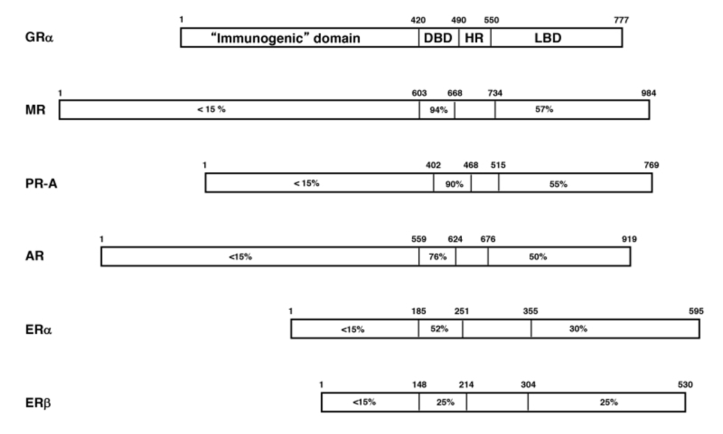
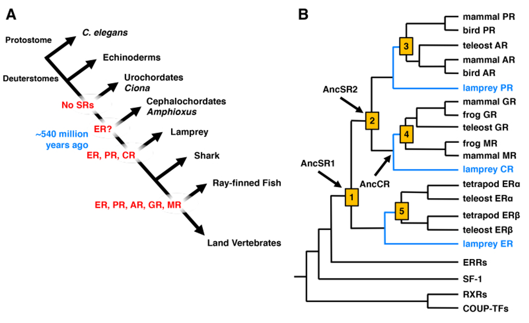
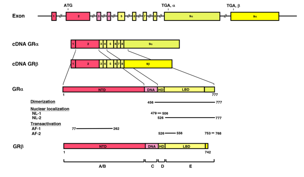
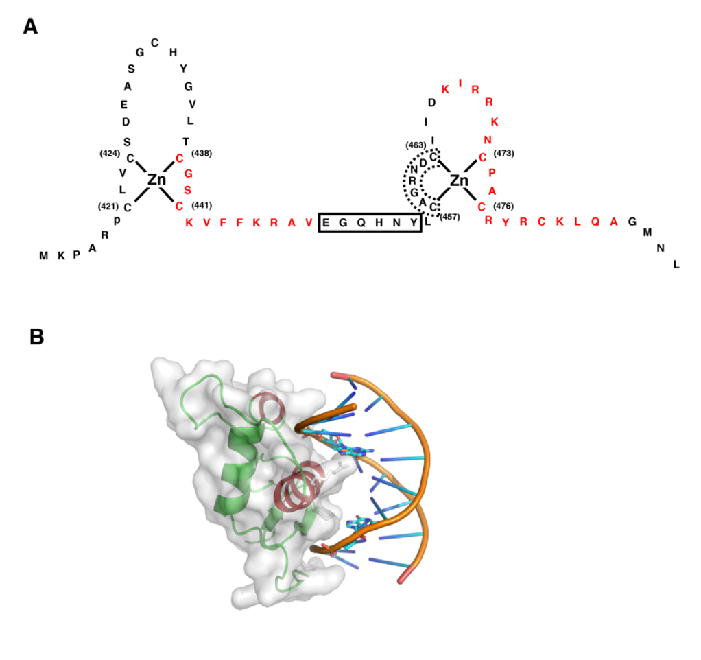
The mineralocorticoid receptor (MR) is found in the cytoplasm and nucleus and the sodium channels are expressed in the apical membrane of epithelial cells of the distal convoluted tubule as well as in cells of other tissues involved with conservation of salt, such as colon, sweat glands, lung, and tongue. MR is a member of the nuclear receptor superfamily. Together with the glucocorticoid, progesterone, and androgen receptors, MR forms the steroid receptor subfamily (30). Steroid receptors display a modular structure comprised of five regions (A-E). The N-terminal A/B region harbors an autonomous activation function. The central C region, corresponding to the DNA-binding domain, is highly conserved and is composed of two zinc fingers involved in DNA binding and receptor dimerization. The D region is a hydrophilic region and it forms a hinge between DNA-binding domain and ligand-binding domain. The E region corresponds to the C-terminal ligand-binding domain and mediates numerous functions, including ligand binding, interaction with heat-shock proteins, dimerization, nuclear targeting, and hormone-dependent activation (31) (Figure 2). The human MR (hMR) and human glucocorticoid receptor (hGR) have almost identical DNA-binding domains (94% homology in the amino acid) and very similar ligand-binding domains (57%), but divergent N-terminal A/B regions (<15%) (32). The hMR gene was mapped on chromosome 4q31.1-31.2 (33,34) and hMR cDNA encodes a 107 kilodalton polypeptide with 984 amino acids (32). The hMR gene consists of 10 exons, including two exons 1 that encode different 5'-untranslated sequences (35). Expression of the two different hMR variants is under the control of two different promoters that contain no obvious TATA element, but multiple GC boxes. Both hMRα and hMRβ mRNAs are expressed at approximately the same level in the mineralocorticoid target tissues (36).

Figure 2. The linearized structures of the mineralocorticoid receptor gene, mRNAs and protein. The MR gene consists of 10 exons. The MR has two exons 1 (exon 1α and exon 1β), each with an alternative promoter; however, the finally translated MR protein is the same. Exons 1 are untranslated regions, exon 2 codes for the immunogenic domain (A/B), exons 3 and 4 for the DNA-binding domain (C), and exons 5-9 for the hinge region (D) and the ligand-binding domain (E) (37)

Molecular and Cellular Mechanisms of the Aldosterone Action

MRs in its unliganded state is located in the cytoplasm, as part of hetero-oligomeric complexes containing heat shock proteins 90, 70 and 50 (38). Upon binding with their ligand, the receptor-ligand complex dissociates from the heat shock proteins, homo- or heterodimerizes and translocates into the nucleus. Homodimers or heterodimers of the MR interact with hormone-responsive elements (HRE) and/or other transcription factors in the promoter regions of target genes, including the subunits of the ENaC or other proteins related to this channel and sodium transport in general, and modulates the transcription rates of these genes (39) (Figure 3).

Figure 3. Mechanism of aldosterone action on sodium reabsorption at the distal convoluted tubule of the nephron. Aldosterone binds to the MR, which is located in the cytoplasm in complex with heat shock proteins 90, 70 and 50. After binding, the receptor-ligand complex translocates into the nucleus, binds to hormone-responsive elements (HRE) of target genes where it modulates their transcription rate. Amiloride-sensitive sodium channel (ENaC) subunits or other related proteins may be targets of such regulation (40).

Pre-Receptor Regulation

Since cortisol circulates at plasma concentrations several orders of magnitude higher than those of aldosterone does, and since it has a high affinity for the MR, it would be expected to overwhelm this receptor in mineralocorticoid target tissues and cause mineralocorticoid excess. A local enzyme, 11β-hydroxysteroid dehydrogenase type 2 (11β-HSD2), however, converts active cortisol to inactive cortisone, and protects the MRs from the effects of cortisol (40) 11β-HSD catalyzes the inter-conversion of hormonally active C11-hydroxylated corticosteroids (cortisol in humans or corticosterone in rodents) and their inactive C11-keto metabolites (cortisone in humans or 11-dehydrocorticosterone in rodents). Two isozymes of 11β-HSD have been identified, 11β-HSD type 1 (11β-HSD1) and 11β-HSD2, which differ in their biological properties and tissue distributions. 11β-HSD2, a potent NAD-dependent 11β-hydrogenase, rapidly inactivates glucocorticoids. The human 11β-HSD2 gene encodes 405 amino acids and its molecular weight is approximately 40-kilodalton (41). 11β-HSD2 has a hydrophilic N-terminal domain that is thought to anchor the protein into membranes (42). 11β-HSD2 is localized as a dimer in the nucleus and cytoplasm of cells of the cortical collecting duct and colon (42,43). Prednisolone and prednisone are substrates for both 11β-HSD isozymes (44,45) and dexamethasone is metabolized slightly by 11β-HSD2 (46). Licorice derivatives, such as glycyrrhizic acid, and the hemisuccinate derivative carbenoxolone are inhibitors of 11β-HSD2. Inhibition of 11β-HSD2 with such agents, confers mineralocorticoid potency to physiologic concentrations of endogenous glucocorticoids in the kidney and colon (47). Thus, in normal physiology, 11β-HSD2 protects the MR by converting cortisol to the inactive cortisone and allows aldosterone-selective access to the inherently nonselective MR in mineralocorticoid target tissues.

Amiloride-Sensitive Sodium Channel (Epithelial Sodium Channel; ENaC)

The cDNA of the α-subunit of the ENaC (αENaC) was cloned from the rat colon in 1993 (48) and soon after the cDNAs of the β- and γ-subunits of this channel were cloned for the same species (49). The human α-, β- and γ-subunits of ENaC were also cloned (50,51). In vitro studies demonstrated that the α subunit of the ENaC itself had the majority of Na channel function, while, the β- and γ- subunits alone were not shown to play as major a role in sodium transport (48). However, the β- and γ-subunits enhanced the function of the α-subunit and all subunits are required for full ENaC activity (52). It appears then that this channel consists of the α-, β- and γ-subunits and an amiloride-binding protein (Figure 4). Aldosterone increases transcription of αENaC but not β- and γ-subunits, resulting enhanced channel assembly and transported from endoplasmic reticulum to Golgi (53). In Golgi, furin proteolytically cleaves specific sites in the extracellular domains of α- and γ-ENaC, resulting in channel activation. At the cell surface, Nedd4-2 binds to ENaC, increasing endocytosis and degeneration (54).The proline-rich region of the C-terminal of the αENaC is important for binding to α-spectrin and for stabilization of the sodium channel in the membrane (55). Recently, several studies demonstrated abnormalities of the β- and γ-subunits of the ENaC in patients with Liddle's syndrome, characterized by mineralocorticoid excess (hypertension and hypokalemic alkalosis), and suppressed aldosterone secretion (56–59). The truncation caused by these mutations influenced the PY motif at the N-terminal of the molecule. This motif is responsible for the binding of the channel subunits with NEDD4, a carrier protein facilitating clearance of the channel (60). Moreover, a point mutation of the αENaC gene, located close to the N-terminal of the protein, was reported to cause a decrease of the probability of an open sodium channel, resulting in defective reabsorption (40,61).

The ENaC-Regulatory Complexes in Aldosterone-Mediated Sodium Transport

Aldosterone-induced trans-epithelial Na+ transport via ENaC involves the coordinate functioning of stimulatory signaling proteins such as serum- and glucocorticoid-induce kinase-1 (SGK1) (23,62), glucocorticoid-induced leucine zipper protein-1 (GILZ1) (63) and connector enhancer of kinase suppressor of Ras 3 (CNK3) (64), with inhibitory proteins, such as neural precursor cell expressed, developmentally downregulated protein (Nedd4-2) (24) and extracellular signal-regulated kinase (ERK) 1/2 (23,24,62,65).


SGK1 is an aldosterone-regulated protein kinase that stimulates renal ENaC through many mechanisms. First, SGK1 phosphorylates the E3 ubiquitin ligase and Nedd4-2, and inhibits its actions. Nedd4-2 interacts with the C-terminal tail of ENaC subunits, decrease surface expression of the channel via channel ubiquitinoylation (23,24,62). Second, SGK1 phosphorylates kinase with no lysine (WNK) 4 and prevents ENaC endocytosis (66). Third, SGK1 directly phosphorylates alpha ENaC and transforms silent ENaC channels to active ones (67). Then, SGK1 alters ENaC expression, trafficking and activity, and stimulates Na+ transport in the kidney cortical collecting duct (CCD) (68). However, SGK1 is a short-lived protein. Following synthesis, SGK1 is rapidly targeted to the endoplasmic reticulum (ER), where ER-associated ubiquitin ligases CHIP and HRD1 aid in its ubiquitinoylation and subsequent proteasome-mediated degradation (69). Another aldosterone-induced ENaC-regulator, GILZ, which protects SGK1 from rapid ER-associated degradation by controlling protein-protein interaction (53.6). In kidney CCD, GILZ1 is robustly induced by aldosterone (70). GILZ1 stimulates ENaC cell surface expression and activity at least in part by inhibiting ERK1/2, which abrogates ENaC function (65,71,72).

The recently identified MR target gene CNKSR3 (connector enhancer of kinase suppressor of Ras 3), commonly referred as CNK3, is highly expressed in the connecting tubule (CNT) and the CCD (73). CNK3, like SGK1 and GILZ1, is rapidly induced by physiological concentrations of aldosterone (64). CNK3 acts to assembly various ENaC-regulatory components in close vicinity of the channel and thereby exerts its stimulatory effects on channel function (74).

Epigenetic Control of  ENaC Transcription by Aldosterone-Sensitive Dot1A-Af9 Complex

Chromatin regulates gene transcription by the post-translational modification of histone N-terminal tails such as acetylation and methylation. The histone H3 Lys 79 methyltransferase disruptor of telomeric silencing alternative splice variant a (Dot1a) methylates histone H3 Lys79, which resides in the globular domain (75). ALL-1 fused gene from chromatin 9 (Af9), putative transcription factor, physically and functionally interact with Dot1a to form a nuclear repressor complex that directly or indirectly binds specific site of the alpha ENaC promoter. Aldosterone reduces the level of Af9 mRNA and protein. Then, Af9 overexpression induces hypermethylation of histone H3 Lys 79 and repression of alpha ENaC transcription (76). Aldosterone impairs the formation of Dot1a -Af9 complex associated with alpha ENaC promoter by 1) decreasing abundance of Dot1a and Af9; 2) attenuating the interaction between Dot1a and Af9 via Sgk-1-catalyzed phosphorylation of Af9 at Ser 435; 3) counterbalancing the repression through binding to mineralocorticoid receptor (MR) and facilitating its translocation into the cell nucleus, where MR and Dot1a compete for binding to Af9.  These are aldosterone-dependent and -independent mechanisms for Dot1a-Af9-mediated repression of alpha ENaC transcription. While aldosterone -independent de-repression achieved through the action of ALL-1 fused gene from chromatin 17 (Af17), Af17 upregulates alpha ENaC transcription by decreasing Af9 binding to Dot1a and relieving Dot1a-Af9-mediated repression of ENaC (77). 4) SGK1 phosphorylates Af9, thus, down-regulating Dot1a-Af9 complex, and relieving the basal repression on alpha ENaC transcription (67,78).

Figure 4. Model of a putative amiloride-sensitive sodium channel (ENaC). The amiloride-sensitive sodium channel appears to consist of the α-, β- and γ- subunits and an amiloride-binding protein. This channel is located at the apical site of the renal epithelium and plays a role in passive sodium transport, which is mainly regulated by mineralocorticoids (79).

THE RENIN-ANGIOTENSIN-ALDOSTERONE SYSTEM IN NEWBORNS AND INFANTS                

Aldosterone secretion rate of newborns and infants was similar to that of older children and adults. Therefore, the aldosterone secretion rate corrected by body surface was much higher in infancy than later in life (80). Urinary aldosterone at birth depends on gestational age and increases progressively, concurrently with the levels of plasma aldosterone. Plasma renin activity, plasma aldosterone and urinary excretion rate of aldosterone decrease with age (81). At  birth, human kidneys display tubular immaturity leading to sodium wasting and impaired ability to reabsorb water. Past studies showed that plasma potassium concentrations were significantly higher in newborns than in their respective mothers, while neonatal and maternal plasma sodium concentrations were closely related. Aldosterone and renin levels in newborns differs significantly from the corresponding maternal concentrations (82). The aldosterone-renin ratio significantly increases with gestational age. Thus, neonatal partial aldosterone resistance was previously suggested because of the high urinary sodium loss in the presence of hyperactivity of the renin-angiotensin-aldosterone system (83). Previous study found that the highest aldosterone levels detected in the cord blood originated from de novo synthesis by the fetal adrenal glands (84). In addition, neonatal aldosterone resistance was associated with weak or undetectable renal MR expression at birth. MR mRNA is transiently expressed between 15 and 24 weeks of gestation, but it is undetectable in late gestational age and neonatal kidney (85). 11 beta-hydroxysteroid dehydrogenase type 2 (11 beta HSD2) and alpha ENaC are closely correlated with cyclic MR expression.

CLASSIFICATION OF HYPOALDOSTERONISM

Various syndromes are characterized by or associated with hypoaldosteronism. Hypoaldosteronism is classified in three large categories, defective stimulation of aldosterone secretion, primary defects in adrenal synthesis or secretion of aldosterone, and aldosterone resistance, according to their pathophysiology and summarized in Table 1.

Table 1. Causes of Hypoaldosteronism and Hormonal Profiles

Causes of Hypoaldosteronism

Hormonal Profiles

DEFECTIVE STIMULATION OF ALDOSTERONE

v  Congenital keep tablehyporeninemic hypoaldosteronism

v  Acquired hyporeninemic hypoaldosteronism

Ø Associated with diabetes mellitus

Ø Associated with nephropathy

Ø Glomerulonephritis

Ø Gouty nephritis

Ø Pyelonephritis

Ø Nephropathy associated with multiple myeloma

Ø Nephropathy associated with systemic lupus erythematosa

Ø Mixed cryoglobulinemia

Ø Nephrolithiasis

Ø Analgesic nephropathy

Ø Renal amyloidosis

Ø Iga nephropathy

v  Associated with autonomic insufficiency

v  Associated with liver cirrhosis

v  Associated with sickle cell anemia

v  Associated with acquired immune deficiency syndrome

v  Associated with polyneuropathy, organomegaly, endocrinopathy, m protein and skin changes syndrome

v  Lead poisning

v  Excess sodium bicarbonate

v  Sjogren's syndrome

v  Drugs interfering with renin production

Ø Β-blocker

Ø Prostaglandin synthetase inhibitors

Ø Non-steroidal anti-inflammatory drugs

Ø Calcium channel blocker

v  Other drugs

Ø Cyclosporin a

Ø Mitomycin c

Ø Cosyntropin

Low plasma renin;

Low plasma and urinary aldosterone

Drugs interfering with angiotensin ii production

Ø  Angiotensin ii converting enzyme inhibitors

High plasma renin; low plasma aldosterone; low angiotensin ii

PRIMARY DEFECTS IN ADRENAL SECRETION OF ALDOSTERONE

Combined with defective cortisol synthesis

a) Congenital causes

Ø Congenital adrenal hypoplasia (dax-1 mutation)

Ø Congenital adrenal hyperplasia

§  Cholesterol desmolase deficiency (lipoid adrenal hyperplasia)

§  3β-hydroxysteroid dehydrogenase deficiency

§  21-hydroxylase deficiency

§  11β-hydroxylase deficiency


Adrenoleukodystrophy, adrenomyeloneuropathy

Low plasma renin; low plasma aldosterone; low plasma cortisol


High plasma deoxycorticosteorne

b) Acquired causes

Ø  Autoimmune adrenal destruction

·       Addison's disease

·       Multiple autoimmune endocrinopathy

Ø  Infectious adrenal destruction

·       Bacterial infection

·       Fungal infection

Ø  Infiltration of adrenal glands

·       Amyloidosis

·       Hemochromatosis

·       Sarcoidosis

·       Metastatic or infiltrative malignant disease

Ø  Bilateral adrenalectomy

Ø  Drug induced

§  Mitotane

§  Aminoglutethimide

§  Torilostane

§  Ketoconazole

Low plasma renin; low plasma aldosterone; low plasma cortisol

v  Isolated deficiency of aldosterone secretion

Ø Congenital causes

§  Cyp11b2 (aldosterone syntase) deficiency

¨     Corticosterone methyloxidase type i (cmo i) deficiency


¨     Corticosterone methyloxidase type ii (cmo ii) deficiency

¨      

High plasma renin; low plasma aldosterone


Normal plasma 18-hydroxycorticosterone/aldosterone ratio

High plasma 18-hydroxycorticosterone/aldosterone ratio

Ø Acquired causes

§  Critically ill patients associated with hypotension or hypovolemia

¨     Sepsis

¨     Pneumonia

¨     Peritonitis

¨     Cholangitis

¨     Liver failure

·       After removal of mineralocorticoid secreting adrenal tumor

·       Discontinuation of agents with mineralocorticod activity

·       Heparin or chlorbutol administration


Low plasma aldosterone concentration; inappropriate elevated plasma renin

DEFECTIVE ALDOSTERONE ACTION

v  Pseudohypoaldosteronism (pha) type 1

Ø Renal (autosomal dominant pha)

Ø Systemic pha (autosomal recessive pha)

v  Secondary pseudohypoaldosteronism

§  Associated with urinary tract infection

§  Associated with medication that blocks epithelial sodium channel (enac)

¨     Amiloride

¨     Triamterene

¨     Trimethoprim

¨     Pentamidine

§  Administration of aldosterone antagonists

¨     Spironolactone

¨     Progesterone

¨     17-hydroxyprogesterone

¨     Synthetic progestin

§  Drugs that may lead to aldosterone resistance

                Caludinerin inhibitor (cyclosporin a, tacrolimus)

High plasma renin; high plasma and urinary aldosterone


Defective Stimulation of Aldosterone

The first category of conditions, which is characterized by defective stimulation of aldosterone secretion, includes the syndromes of congenital and acquired hyporeninemic hypoaldosteronism. One of these conditions is due to a defect of renin secretion such as hyporeninemia resulting from β-blockers, prostaglandin synthetase inhibitors, and calcium channel blockers. Another condition is due to decrease in the conversion of angiotensin I to angiotensin II mediated by converting enzyme inhibitor medications and is associated with hyperreninemia.

Primary Defects in Adrenal Biosynthesis or Secretion of Aldosterone

The second category of conditions, which are characterized by primary defects in adrenal synthesis or secretion of aldosterone, includes all causes of primary adrenal insufficiency and primary hypoaldosteronism caused by aldosterone synthase (CYP11B2) deficiency or as an acquired state. Primary adrenal insufficiency causes include congenital adrenal hypoplasia, congenital adrenal hyperplasia, adrenoleukodystrophy/ adrenomyeloneuropathy, acquired adrenal insufficiency due to autoimmune, infectious and infiltrative disease, bilateral adrenalectomy and use of adrenolytic agents and enzyme inhibitors that block cortisol and aldosterone biosynthesis. These conditions are usually combined with defective cortisol synthesis. Aldosterone synthase (CYP11B2) deficiency (ASD) leads to reduced aldosterone production associated with low or high levels of 18-hydroxycorticosterone, referred to as CMO I or CMO II deficiency, respectively. Several conditions may be associated with aldosterone biosynthetic activity. Heparin suppresses aldosterone synthesis. Critically ill patients with persistent hypovolemia and hypotension also have inappropriately low plasma aldosterone concentrations in relation to the activity of the renin-angiotensin system. Isolated primary hypoaldosteronism in occasionally associated with metastatic cancer of the adrenal gland.

Defective Aldosterone Actions

The third category which is characterized by defective aldosterone action includes syndromes of aldosterone resistance such as pseudohypoaldosteronism type 1 and sodium-wasting states resulting from excessive amounts of circulating mineralocorticoid antagonists, such as spironolactone and its analogues, and synthetic progestin or natural agonists, such as progesterone or 17-hydroxyprogesterone. These mineralocorticoid antagonists may antagonize aldosterone at the levels of mineralocorticoid receptor (86) and frequently, these states are compensated for by elevated concentrations of plasma aldosterone.

HYPORENINEMIC HYPOALDOSTERONISM

The most common form of isolated hypoaldosteronism is caused by impaired renin release from the kidney. Hudson et al. first described this syndrome in 1957 (87), however, hyporeninemia was first recognized in 1972 (88) (89). The typical patient is 50 to 70 years old and usually presents with chronic and asymptomatic hyperkalemia and mild to moderate renal insufficiency with a 40-70% decrease in the glomerular filtration rate when compared to that of age matched healthy subjects. Hyperchloremic metabolic acidosis is seen in approximately half of the patients. This acidosis is classified as a renal tubular acidosis type IV (90). The acidosis is a consequence of decreased renal ammonia neogenesis, reduced hydrogen ion-secretory capacity in the distal nephron, and mild reduction in the proximal tubular threshold for bicarbonate reabsorption. Occasionally, muscle weakness or cardiac arrhythmias are present in some patients. More than a half of the patients have diabetes mellitus (91). Other frequently associated states include autonomic neuropathy, hypotension, and various nephropathies such as glomerulonephritis, gouty nephropathy, and pyelonephritis. Also, this syndrome is associated with nephropathies associated with multiple myeloma and systemic lupus erythematosus, mixed cryoglobulinemia, nephrolithiasis, analgesic nephropathy, renal amyloidosis, IgA nephropathy, cirrhosis, sickle cell anemia, acquired immune deficiency syndrome (AIDS), polyneuropathy, organomegaly, endocrinopathy, M protein and skin changes (POEMS) syndrome, lead poisoning, excess sodium bicarbonate, and Sjogren’s syndrome (90,92–101) . Moreover, this syndrome occurs transiently in association with use of non-steroidal anti-inflammatory drugs, cyclosporin A, mitomycin C, cosyntropin, and other agents in susceptible individuals (102–104).

Pathophysiology

Urinary aldosterone excretion is low under basal conditions and fails to increase after sodium restriction. Plasma renin activity is also low and does not increase appropriately during sodium restriction, periods of prolonged upright posture, or diuretic administration (88). Interstitial renal disease and damage to the juxtaglomerular apparatus seems the most likely cause for the primary defect in renin generation or release and secondary deficiency of aldosterone. However, in some patients with this syndrome there is an absent or blunted aldosterone response to angiotensin II (94,104), suggesting a coexisting primary defect in aldosterone secretion or it reflects atrophy of the zona glomerulosa caused by chronic renin deficiency.

There are various mechanisms to be explained for the hyporeninemia. First possible mechanism is the hypervolemia. The expanded extracellular fluid volume due to hypertension may suppress renin. In fact, long-term sodium restriction and diuretic administration increase plasma renin activity in these patients, however, the increments of plasma renin activity are less than those of normal subjects (97). A second possible mechanism is insufficiency of the autonomic nervous system, particularly in patients with diabetic neuropathy. Impaired adrenergic response to postural change may contribute to insufficient renin release. Besides, these patients exhibit decreased sensitivity to β-adrenergic agonists, suggesting defects in both production and action of catecholamines (96). A third proposed mechanism is secretion of abnormal forms of renin, such as a defect in the conversion of prorenin to renin. Insufficiency of autonomic nervous system may be associated with impaired conversion of prorenin to renin. Indeed, patients with diabetes mellitus and autonomic neuropathy have elevated plasma levels of prorenin (105). A fourth possibility is prostaglandin deficiency. Production of prostaglandin I2 (prostacyclin), which mediates renin release, is apparently diminished in patients with hyporeninemic hypoaldosteronism as assessed by measurement of the stable urinary metabolite 6-keto-prostaglandin F1α (95). Furthermore, the prostaglandin I2 in these patients was unresponsive to the potent stimulator’s norepinephrine and calcium. Prostaglandin I2 deficiency may cause hyporeninemic hypoaldosteronism by causing defects in the conversion of prorenin to renin and renin release (106).

Diagnosis

The diagnosis of hyporeninemic hypoaldosteronism must be considered in any patient with unexplained hyperkalemia. Excess potassium intake from food or drugs does not cause sustained hyperkalemia, if renal function is normal. Renal function should be evaluated and drugs that impair renal potassium excretion should be excluded as a cause. The clinical diagnosis is confirmed by low plasma renin activity and low plasma concentrations or urinary aldosterone excretion under conditions that activate the renin-angiotensin-aldosterone axis by maintenance of upright posture and/or furosemide administration. A low random plasma renin concentration associated with a normal ratio of aldosterone to plasma renin activity is also useful for the diagnosis (94).

Therapy

The therapeutic approach should be chosen after taking into consideration the age of the patients and other concurrent disorders. Only monitoring potassium concentrations is enough for patients with moderate hyperkalemia and without electro-cardiographic changes. Drugs that promote hyperkalemia, such as β-adrenergic antagonists, cyclooxygenase inhibitors, angiotensin-converting enzyme inhibitors, heparin, and potassium-sparing diuretics, should be avoided. Dietary potassium intake should be reduced, if possible. Diuretics are the initial treatment for patients who have disorders associated with sodium retention, such as hypertension and congestive heart failure. Mineralocorticoid replacement with fludrocortisone is reserved for patients with severe hyperkalemia without hypertension and congestive heart failure.

PRIMARY HYPOALDOSTERONISM- ALDOSTERONE SYNTHASE DEFICIENCY (ASD)

Congenital hypoaldosteronism is a rare inherited disorder transmitted as either an autosomal recessive or autosomal dominant trait with mixed penetrance. This disorder was previously termed "corticosterone methyloxidase (CMO)” deficiency and subdivided into two types according to the relative levels of aldosterone and its precursors in an affected person. Patients with "corticosterone methyloxidase I (CMO I)" deficiency have elevated serum levels of corticosterone and low levels of 18-hydroxycorticosterone and aldosterone. In contrast, patients with "corticosterone methyloxidase II (CMO II)" deficiency have high levels of 18-hydroxycorticosterone, the immediate precursor of aldosterone (107). With greater understanding of structure-activity relationships in the CYP11B2 enzyme, this disorder may be better considered a spectrum of hormonal deficiencies, depending on the nature of the CYP11B2 gene defect (108). Two steps of aldosterone biosynthesis from corticosterone previously proposed to be catalyzed by separate enzymes, CMO I and II, previously, are known to involve only one enzyme substrate interaction (6). Isolated aldosterone deficiency results from loss of activity of aldosterone synthase encoded by CYP11B2 gene (109–118). Therefore, the term aldosterone synthase deficiency type 1 (ASD1) and type 2 (ASD2) reflects more appropriately the molecular basis of this disease. In both ASD1 and 2, glomerulosa zone corticosterone is increased and aldosterone decreased, but 18-hydroxycorticosterone is increased in ASD2 (108).  ASD1 is associated with loss of both 18-hydroxilation and 18-oxidation enzyme activities. In ASD2, the ability to convert corticosterone (B) to 18-hydorxytetrahydro11-dehydrocorticosterone (18-OH-B) is preserved with failure of further oxidation of 18-hhdroxicorticosrerone to aldosterone (119). The deficiency of aldosterone is much more severe in ASD1. In contrast, aldosterone may reach normal levels under intense stimulation of renin-angiotensin system in ASD2 (108). The clinical presentations of these deficiencies are otherwise similar.

Clinical Presentation

The clinical presentation is typical of aldosterone deficiency, including electrolyte abnormalities such as a variable degree of hyponatremia, hyperkalemia and metabolic acidosis, with poor growth in childhood, but there are usually no symptoms in adults (107,120). Miao et al. reviewed 45 ASD patients (20 of ASD1, 12 of ASD2, 13 of undefined subtype) (121).  From their review, 95% of the patients having ASD1 and all of having ASD2 and an undefined subtype had hyponatremia, while 89% showed hyperkalemia. In infants, it is characterized by recurrent dehydration, salt wasting and failure to thrive. These symptoms are present generally within the first 3 months of life, and most often after the first 5 days of life. A modest uremia with a normal creatinine level reflects dehydration in the presence of intrinsically normal renal function. Plasma renin activity might vary, while elevated plasma renin activity levels were more likely to be found in the ASD1 (121).

Diagnosis and Therapy

The diagnosis can be established by measuring the appropriate corticosteroids or their major metabolic products, such as 11-deoxycorticosterone (DOC), corticosterone, 18-hydroxycorticosterone, 18-hydroxy-DOC, and aldosterone levels in plasma. The ratio of plasma 18-hydroxycorticosterone to plasma aldosterone differentiates the two disorders; it is less than 10 in ASD1 (CMO I deficiency) and more than 100 in ASD2 (CMO II deficiency) (121,122). Patients with ASD2 (CMO II deficiency) tend to have increased plasma cortisol levels that may result from increased adrenal sensitivity to ACTH induced by the increased plasma angiotensin II levels in response to sodium depletion (123).

Both forms of the syndrome are treated by replacement of mineralocorticoid with the usual dosage of fludrocortisone (0.1-0.3 mg/ day). Almost infants and children require oral sodium supplementation (2 g/day as NaCl alone or in combination with NaHCO3), although some infants with severe symptom need intravenous fluids. Oral sodium supplementation may be discontinued once plasma rennin activity has decreased to normal, but mineralocorticoid replacement is usually maintained through childhood.

Molecular Mechanism of CYP11B2 Deficiency

ASD has been identified in Jews of European, North American, and Iranian descent (119). In Asians, it was reported in the Thai (124), Indian (124), Japanese (125) and Chinese populations (120,126).

To date, approximately 40 mutations, such as missense and nonsense mutations, splicing mutations, small insertions/deletions, gross deletions, and complex rearrangements, in the CYP11B2 have been reported in cases of ASD; the most common mutations were missense and nonsense (121). Some variants, such as p.Q170X, p.E198D, c.1398+2T>A, p. F233fsX*295, p.L462R, p.Q337X and p.Q272W, were identified in patients without an ASD classification subtype (121). A majority of mutations led to complete loss of enzyme activity, while in some mutations, such as V386A and R181W, double homozygosity was required for clinical phenotype (112,113,121).

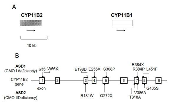
Some patients with ASD1 (CMO I deficiency) have a homozygous 5 nucleotide deletion in exon 1 which leads to a frameshift and premature stop codon, resulting in the complete lack of enzyme production (109,110). A male Caucasian patient with ASD1 (CMO I deficiency) had a homozygous point mutation causing a R384P substitution, resulting in complete loss of 11 β- and 18-hydroxylase activity (111) (Figure 5). This suggests that the arginine-384 in aldosterone synthase is highly conserved and apparently quite important for enzyme activity.

A male infant of Turkish parents who presented with ASD1 had a homozygous missense mutation (L451F) in exon 8 of CYP11B2 gene. The L451F mutant protein in vitro showed complete aldosterone deficiency with 11-deoxycirticosterone or corticosterone as substrates. The L451F mutation located immediately adjacent to the highly conserved heme-binding C450 of the cytochrome P450 (117). Computer modeling of the molecule suggested that this substitute my lead a steric effect resulting in preventing the activity of CYP11B2 (117).

Three siblings of Pakistan origin who presented with ASD1 had a homozygous mutation (S308P) in exon 5 of CYP11B2 gene. The S308P mutant protein in vitro showed complete loss of enzyme activity. This mutated residue is likely to locate within the a-helix I, close to the heme-binding, active site of the enzyme. This structural change may be the cause of this disorder in this family (118). 

A large number of kindreds with ASD2 (CMO II deficiency) have been identified among Jews originally from Isfahan, Iran. Such patients are all homozygous for two mutations, R181W in exon 3 and V386A in exon 7 (109,112,113) (Figure 5). These mutations together reduce aldosterone synthase activity to 0.2 % of normal without affecting 11 β-hydroxylase activity (112,113). However, one non-Iranian patient with ASD2 (CMO II deficiency) carries mutations in the paternal allele, including V386A and T318A mutations, and maternal allele, including R181W and a deletion/frameshift mutation, resulting in complete loss of enzyme activity (113). This suggests that the high levels of 18-hydroxycorticosterone seen in ASD2 (CMO II deficiency) can be synthesized by CYP11B1, which has some 18-hydroxylase activity, and not by CYP11B2. A patient with apparent ASD 1 was homozygous for the mutations E198A and V386A, yet when assayed in vitro the double mutant enzyme behaved similarly to the mutant enzyme found in the Iranian Jewish ASD 2 patients (127). Thus, a difference in expression of CYP11B1 rather than allelic variation of CYP11B2 may be involved in the mechanism underlying the different levels of 18-hydroxycorticosterone between ASD1 and 2 (CMO I and CMO II deficiency). The distinction between ASD 1 and ASD 2 is not precise, and these disorders should be regarded as different degrees of severity on a continuous clinical spectrum.

 A male Japanese patient with ASD1 (CMO I) was a compound heterozygous for W56X in exon 1 and R384W in exon 7. W56X was inherited from his mother and R384X was from his father. Since both alleles contain nonsense mutations, a lack of CYP11B2 activity was speculated to cause his condition (125).

Two male Japanese patients with ASD2 (CMO II) had homozygous missense mutation (G435S) in the exon 8 of CYP11B2 gene. The expression studies indicated that the steroid 18-hydroxylase/oxidase activities of mutant enzyme were substantially reduced.

A female infant of Albanian origin with ASD2 (CMO II) revealed homozygosity for a pathogenic T185I mutation in Exon 3 of the CYP11B2 gene and two other homozygous polymorphisms F168F and K1738 in Exon3 (128). Both healthy parents revealed heterozygous for all three substitutions.

Another female Italian Caucasian patient was diagnosed with a compound heterozygous mutation located in exon 4 causing a premature stop codon (E255X) and a further mutation in exon 5, also causing a premature stop codon (Q272X). The patient’s CYP11B2 encoded two truncated forms of aldosterone synthase predicted to be inactive because they lack critical active site residues as well as the hormone-binding site. However, this case displays biochemical features intermediate between those of ASD1 and 2 (CMO I and II).

Some cases of ASD without causative mutations in CYP11B2 have also been reported (116,119).

Figure 5. Relative positions of CYP11B1 and CYP11B2 on chromosome 8 and mutations of CYP11B2. A, The relative positions of CYP11B1 and CYP11B2 on chromosome 8q22. Arrows indicate direction of transcription. B, Mutations of CYP11B2 in reported patients with CYP11B2 deficiency are summarized in the figure (109,121,126,128).

ACQUIRED FORMS OF PRIMARY HYPOALDOSTERONISM  

Several conditions may be associated with aldosterone biosynthetic defects. The administration of heparin causes natriuresis and hyperkalemia (129). Heparin preparations suppress aldosterone synthesis, leading to a compensatory rise in plasma renin activity. However, it has been demonstrated that this suppression of enzyme activity is attributable to chlorbutol (1,1,1-trichloro-2-methyl-2-propanol), the preservative used in commercial heparin, rather than to pure heparin (130).

Persistently hypotensive, critically ill patients with sepsis, pneumonia, peritonitis, cholangitis and liver failure, also have inappropriately low plasma aldosterone concentrations in relation to elevated plasma renin activity (131). The defect is at the level of the adrenal but has not been associated with any particular disease or therapy. Plasma cortisol levels are high, reflecting the stressed state. The response to angiotensin infusion is impaired, and the ratio of plasma 18-hydroxycorticosterone to aldosterone is increased, suggesting selective insufficiency of CMO II. It is possible that the hypoxia causes a relative zona glomerulosa insufficiency (132).

ALDOSTERONE RESISTANCE

Pseudohypoaldosteronism (PHA) Type 1

Mineralocorticoid resistance (pseudohypoaldosteronism type 1, PHA1) results from inability of aldosterone to exert its effect on its target tissues and was first reported by Cheek and Perry as a sporadic occurrence in 1958 (133). This disease, usually presents in infancy with severe salt-wasting and failure to thrive, accompanied by profound urinary sodium loss, severe hyponatremia, hyperkalemia, acidosis, hyperreninemia, and paradoxically markedly elevated plasma and urinary aldosterone concentrations. Usually, renal and adrenal functions are normal. This disease has been reported in over 70 patients (134). The prevalence, as estimated from recruitment through a genetic laboratory at the Hôpital Européen Georges Pompidou in France, which is a national reference center for a rare disease, is ~1 per 80,000 newborns (135)(136). Approximately one fifth of these cases are familial, and both an autosomal dominant and a recessive form of genetic transmission have been observed. A previous study found that all patients had renal tubular unresponsiveness to aldosterone, while some had involvement of other mineralocorticoid target-tissues, including the sweat and salivary glands, and the colonic epithelium, as well. Autosomal recessive PHA1 presents in the neonatal period with hyponatremia caused by multi-organ salt loss, including kidney, colon, and sweat and salivary glands. Autosomal recessive PHA1 persists into adulthood and shows no improvement over time. However, literature regarding follow-up of these patients after diagnosis is insufficient.  In contrast, autosomal dominant PHA1 is characterized by an isolated renal resistance to aldosterone, leading to renal salt loss. Particularly autosomal dominant form of PHA1 typically shows a gradual clinical improvement during childhood, allowing the cessation of sodium supplementation. 

PATHOPHYSIOLOGY

The mechanism(s) by which aldosterone controls sodium transport in its target tissues involves the mineralocorticoid receptor (MR) and proteins that are associated with the amiloride-sensitive sodium channel (ENaC). The latter proteins are expressed in the apical membrane of epithelial cells of the distal convoluted tubule and in the membranes of cells of other tissues involved in the conservation of salt, such as colon, sweat gland, lung and tongue. Thus, the MR and the ENaC were considered as potential candidate molecules for the pathogenesis of PHA1. In fact, mutations of α- and β-subunits of the ENaC were reported in PHA patients from autosomal recessive kindreds (61,137). Mutations of the MR were also reported in the patients with autosomal dominant PHA1 (138,139). However, no molecular defects were found in either MR or ENaC in some patients with PHA1, especially in those with the sporadic form PHA1, which suggests molecular heterogeneity in PHA1 (79,140–144).

DIAGNOSIS

Electrolyte profiles suggest mineralocorticoid deficiency or end-organ resistance, along with hyperkalemia, hyponatremia and metabolic acidosis associated with profound urinary salt loss. Renal and adrenal function is normal. The diagnosis is confirmed by the markedly elevated plasma aldosterone concentrations and plasma renin activity.

The differential diagnosis of PHA1 includes salt-wasting states due to hypoaldosteronism, including several forms of congenital adrenal hyperplasia, isolated hypoaldosteronism due to corticosterone methyloxidase (CMO) I and II deficiencies and congenital adrenal hypoplasia. Normal cortisol and excessive aldosterone responses to adrenocorticotropin (ACTH) are expected in patients with congenital PHA.

THERAPY         

The standard treatment of PHA has been replacement with high doses of salt, with a variable response among patients (134). Recently, carbenoxolone, an 11β-hydroxysteroid dehydrogenase inhibitor, was employed as therapy in PHA1 and an ameliorating effect was observed which was attributed to mediation by the MR (140). We studied a 17-yr-old male patient with congenital multifocal target-organ resistance to aldosterone. We examined his clinical response to carbenoxolone, expected to increase the intracellular level of cortisol in the kidney by preventing local conversion of cortisol to cortisone, and to high doses of fludrocortisone, a synthetic mineralocorticoid. Subsequently, and for a brief period of time, we administered dexamethasone, which has no intrinsic salt-retaining activity, in addition to carbenoxolone, to suppress endogenous cortisol, along with its intrinsic mineralocorticoid activity.

Figure 6. Effect of carbenoxolone, carbenoxolone plus dexamethasone, and fludrocortisone (top panel) on the serum sodium (middle panel) and potassium (bottom panel) concentrations of a patient with PHA. Carbenoxolone normalized plasma electrolytes, addition of dexamethasone reversed this effect, while fludrocortisone at high doses also normalized plasma electrolytes (140).

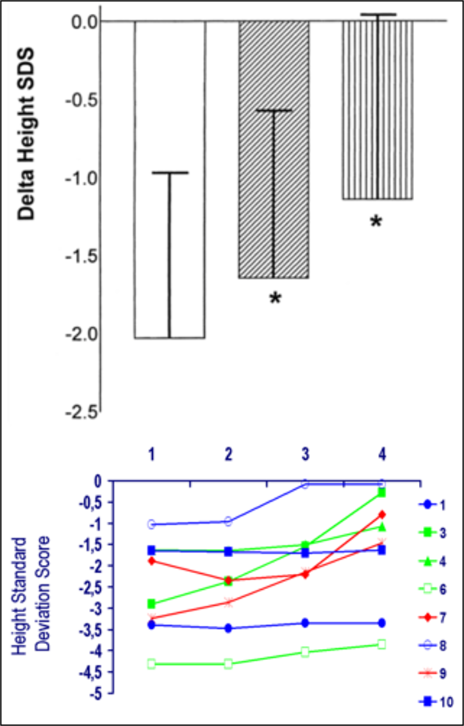
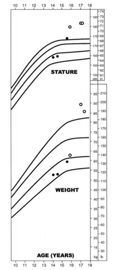
Carbenoxolone normalized the patient's serum electrolyte concentrations and decreased his urinary excretion of sodium within a week (Figure 6). Subsequent long-term therapy of this patient with carbenoxolone (450 mg/day p.o.) maintained his electrolyte concentrations within the normal range. His urinary 24 h free cortisol was increased during carbenoxolone therapy. Addition of dexamethasone suppressed his urinary free cortisol excretion and reversed the beneficial effect of carbenoxolone on serum and urinary electrolytes (Figure 6). These data suggest that an increase in urinary free cortisol observed during carbenoxolone therapy was due to a localized effect of this drug on the kidney rather than on tissues involved in the negative feedback effect of glucocorticoids. The effect of carbenoxolone does not seem to be mediated by GR but seems to be exerted purely via the MR (Figure 7). There were no adverse effects of long-term carbenoxolone therapy in this patient. He also reported increased stamina, a better ability to concentrate and less anxiety. On treatment, the patient grew 6 cm/y and progressed from -4SD to -3SD scores for mean height for age. He also progressed in his pubertal development from Tanner stage III to IV for pubic hair, while his bone age advanced from 12 to 14 y.

Figure 7. Mechanism of the effect of carbenoxolone. Carbenololone inhibits of conversion of cortisol to cortisone in the kidney, resulting in the enhancement of the effect of cortisol as a ligand for MR. Dexamethasone suppressed cortisol production and reversing the beneficial effect of carbenoxolone in our patient with PHA1.

Both carbenoxolone and fludrocortisone normalized the serum electrolytes of our patient, suggesting the presence of a functional, albeit possibly defective, renal MR. Interestingly, the same patient was unresponsive to intravenous infusion of aldosterone and fludrocortisone (up to 3 mg/day) when studied in infancy (145), suggesting that the clinical improvement that has been noted in the majority of PHA patients with age may be related to changes in their responsiveness to mineralocorticoid.

On the other hand, another study reported that carbenoxolone did not show any significant salt-retaining effect in two patients with multiple PHA, while carbenoxolone significantly suppressed the renin-aldosterone system in a patient with renal-form PHA (146).  This difference of responsiveness to carbenoxolone may be due to an age-dependent change on mineralocorticoid responsiveness. Additionally, the different mineralocorticoid responsiveness of renal and multisystem PHA patients indicates a difference in their MR function. The partial response to carbenoxolone in renal PHA suggests that there is at least a partly functional MR. This is also supported by the observation that spironolactone, a mineralocorticoid antagonist, aggravated sodium loss in several patients with renal PHA (147).

MOLECULAR MECHANISM(S) OF PSEUDOHYPOALDOSTERONISM TYPE 1          

In 1996, a study reported homozygous mutations introducing a stop codon or frame shift in the αENaC gene of affected members of families with autosomal recessive PHA (61).  To date, worldwide more than 40 different mutations have been described in the coding region of ENaC subunit genes (148–150). The majority of mutations appear in the αENaC gene SCNN1A, most frequently in exon 8 (61,150–152). Mutations are nonsense, single base deletions or insertions, or splice-site mutations, leading to abnormal length of mRNA and protein. Few missense mutations in αENaC gene have also been reported (149,153). Only a few cases of mutations in β and gamma ENaC genes have been reported (149,154,155). Phenotype and genotype correlations have been noted with more severe phenotype in nonsense, frameshift, and abnormal splicing mutations than patients with missense mutations (148,154,155).

A Swedish study regarding families with autosomal recessive PHA, homozygous or compound heterozygous mutations showed that a stop codon or a frame shift in the αENaC gene was associated with pulmonary disease as well (150). The truncation caused by these mutations influenced the PY motif at the N-terminal region of the molecule. This motif is responsible for the binding of the channel subunits with Nedd4, a carrier protein facilitating clearance of the channel (60). Moreover, a point mutation of the αENaC gene, located close to the N-terminal of the protein, was reported to cause a decrease of the probability of an open sodium channel, resulting in defective reabsorption (61,153). In the other four families with autosomal recessive PHA, insertion of a T in exon 8 and nonsense mutation (R508X) in exon 11 of the αENaC gene, resulting in a truncated αENaC subunit, was found (156). A splice site mutation in intron 12 of the βENaC gene, which preventing correct splicing of the mRNA was found in a Scottish patient (156). Also, other autosomal recessive families with PHA had a homozygous splice-site mutation in the γENaC, while a Japanese sporadic patient with the systemic form of PHA was a compound heterozygote for mutations in the αENaC, which resulted in the generation of a truncated channel subunit (137,157) . Compound heterozygous mutations (Q217X in exon 4 and Y306X in exon 6) of βENaC have been reported in the patient with multi-organ PHA1 of Ashkenazi family in Israel (154). These mutations produce shortened βENaC subunits with 253 and 317 residues respectively instead of the 640 residues present in βENaC subunit. Expression of cRNA carrying these mutations in Xenopas oocytes showed that the either mutation drastically reduced to only 3% of normal ENaC activity (154).  An African American female with PHA, who had persistent and symptom hyperkalemia, had compound heterozygous mutation in the βENaC gene: c.1288delC in exon 9, a one-base deletion that generated a frameshift mutation, and c.1466+1 G>A, an intronic base substitution in intron11 that leaded to a splice site mutation (158).

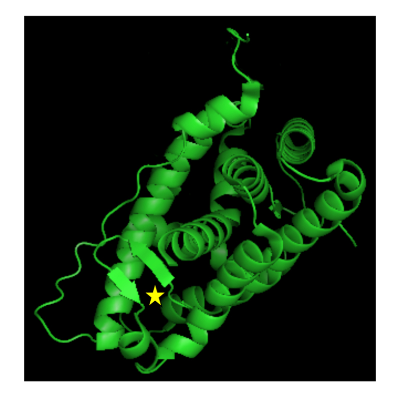
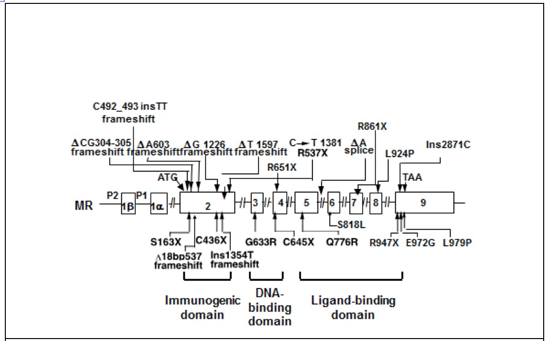
To date more than 50 different mutations in the human MR gene (NR3C2) causing autosomal dominant PHA1 have been described. NR3C2 mutations were found in 62% of patients with renal PHA1 referred to a genetics laboratory at the Hôpital Européen Georges Prompidou in France (135). Nonsense mutations, frameshift mutations, splice site mutations, and deletions of whole or part of the gene lead to gross change of the MR protein. Nonsense mutations are found in all exons and lead to truncated MR protein. A past study. reported families with autosomal dominant PHA, who had molecular defects of the MR resulting in non-expression of one of the 2 alleles (138) (Figure 8). In addition, another study reported a sporadic patient with PHA who had a heterozygous mutation in exon 9 of the MR that introduced a premature stop codon (144) (Figure 8). These results, may suggest that expression of only one allele of the MR is insufficient to prevent salt loss. Another case study did not identify any abnormalities of the MR in PHA patients from two families with the autosomal dominant form of the disease (144), while other authors reported a heterozygous missense mutation in exon 8 of the MR gene identified in PHA patients from a Japanese autosomal dominant family (139) (Figure 8). A heterozygous nonsense mutation in exon 2 (S163X, C436X) and in exon 9 (R947X) of the MR, leading to a premature stop codon of the MR gene were found in other patients with autosomal dominant PHA (159–161). It was previously reported a heterozygous splice acceptor site mutation, which results in exon 7 skipping and subsequently in premature termination in exon 8 of MR with Japanese female patients with PHA1 (162). This study showed that RT-PCR products of mRNA with that patient showed both wiled-type and mutated mRNA, suggesting that haploinsufficiency due to nonsense mediated mRNA decay with premature termination is not sufficient to give rise to the PHA phenotype (162). It was also reported that Q776R mutation in exon 5 or L979P mutation in exon 9, which is located in the ligand-binding domain of the MR, presented reduced or absent aldosterone binding, respectively (163). Three-dimensional structure of MR suggests that the residue Q776 is located in helix 3 and is locking aldosterone in the ligand-binding pocket (163). A study examined patients with PHA1 presenting isolated renal salt loss from six families in Italy and Germany and found one nonsense mutation (E378X), one frameshift mutation (A958R) and two missense mutations (S818L and E972G) (164). S818L does not bind aldosterone or activate transcription or translocate into the nucleus. Three-dimensional molecular structure showed that S818 was located in helix H5 and S818 was speculated to be necessary to stabilize helix H5 and the -sheet 1 via hydrogen bond to Y828. E972G mutation showed a significantly lower ligand-binding affinity and only 9% of wild-type transcriptional activity. Three-dimensional molecular structure showed that E972 is involved in a hydrogen-bond network with R947 anchoring helix H12 to H10. Thus, substitute of E972G suggested to be open up the hydrophobic core and displace helix H10, causing the decreased ligand-binding ability (164).

A Japanese study reported four sporadic patients and two siblings with a renal form of PHA (165). Two siblings and one sporadic patient had R651X of NR3C2 (MR) gene. One sporadic patient had R947X, another two patients had 603A deletion and 304-305CG deletion, respectively, both resulting in frameshift mutations (165).

Another study reported two female Japanese infants with the renal form of PHA1 and identified two heterozygous mutations. One had a c.4932_493insTT in Exon 2, resulting in a premature stop codon (p.Met166 LeufsX8) and another had a nonsense mutation of R861X in exon 7 (166).  These mutations resulted in haploinsufficiency of the MR and were the cause of aldosterone resistance in the kidney.

From the study of the genetics laboratory at the Hôpital Européen Georges Pompidou in France, 20 mutations were found in exon 2; all of them led to truncated receptors, Of the 22 mutations identified in exon 3 and 4, coding for the MR DBD, 11 were nonsense or frameshift mutations, the reminder missense mutations. Thirty variants were located in exon 5-9 and affected LBD; the majority were missense mutations. Nine were splice variants in different introns, 19 were large deletions encompassing single or multiple exons and the flanking intronic regions of the NR3C2 gene (135) (figure 8).

These studies suggest major molecular heterogeneity in PHA.

Figure 8. Mutations of the MR in patients with PHA1. Mutations of the MR that have been reported in patients with PHA1 are summarized in the figure (135,138,139,144,166)

Another study investigated 5 unrelated cases of sporadic PHA (79,140,143). The researchers found a nonconservative homozygous mutation (A241V) in the MR of 4 of the patients and a conservative heterozygous mutation (I180V) in one of these patients and his asymptomatic father, while no abnormalities were found in the DNA- or ligand-binding domains of the MR. The Val241 and Val180 substitutions were found also in the norm 6al population. The heterozygosity and homozygosity frequencies of the Val241 and Val180 mutations were 48%, 38%, 22% and 1.5%, respectively. Another finding was a nonconservative amino acid substitution (T663A) in the αENaC, which was located close to the C-terminal (79). Of the 5 patients, 2 were homozygous and 3 heterozygous for this variation, respectively. This amino acid substitution was also present at high frequency in apparently normal controls. The homozygosity and heterozygosity frequencies of the αENaC Ala663 were 31% and 64%, respectively. Three of the 4 (75%) patients with multiple tissue resistance to aldosterone had both αENaC (heterozygous or homozygous) and MR (homozygous) mutations as described above, while only 7% of our controls with apparently normal salt conservation had the same concurrent abnormalities (Table 2, p < 0.025).

Table 2. MR and aENaC Polymorphisms in PHA and Normal Subjects


 MR

 αENaC

 Target organ


 I180V

 A241V

 T663A


 Homo

 hetero

 homo

 Hetero

 homo

 Hetero


 Pt.1


+


+


+


 Multiple

 Pt.2


 +


 Multiple

 Pt.3


 +


 +


 Multiple


 Pt.4


+


 Multiple


 Pt.5


+


+


 Isolated


controls


1.5%


22%


38%


48%


31%


64%


controls


+


+


+


controls


+


+


controls


+


+


                (79) with permission

The researchers identified, in a Japanese patient with sporadic PHA, three homozygous substitutions in the MR gene: G215C, I180V or A241V, which had previously reported to occur in healthy populations. Luciferase activities induced by MR with either G215C, I180V or A241V substitution were significantly lower than those for wild-type MR with aldosterone at concentrations ranging from 10-11 to 10-9 M, 10-8M, or 10-11 to 10-6M, respectively. A homozygous A to G substitution of the donor splice site of αENaC intron 4 was found in the patient. These results suggest that each of three MR polymorphisms identified in our patient is functionally and structurally heterogeneous (167).

The authors suggested that the above polymorphisms may confer vulnerability in salt conservation, which might be expressed fully only when concurrently present with other genetic defects of the MR or other proteins that participate in sodium homeostasis, such as Nedd4 (168). This hypothesis, if true, would be compatible with a sporadic presentation or a digenic or multigenic expression and heredity as previously described in retinitis pigmentosa (169). In this case, hereditary transmission might be complex and appear either as a dominant and/or recessive trait with variable penetrance.

Secondary Pseudohypoaldosteronism (PHA)

Secondary PHA is a form of renal resistance to aldosterone. The cause of secondary PHA is either renal disease or medication. The clinical and laboratory findings resemble those of a transient PHA. Since Rodriguez-Soriano et al. reported the first case in 1983 (169), more than 68 cases have been reported. Secondary PHA may occur mainly in neonates and young infants with urinary tract infections, such as pyelonephritis, and/or malformation of urinary system causing obstructive uropathy, tubulointerstitial nephritis, sickle cell nephropathy, and systemic lupus erythematosus(170). Secondary PHA has been also related to drugs like non-steroidal anti-inflammatory agents and potassium-sparing diuretics (170–172). This state occurs in male infants more frequently than female infants because of the higher incidence of urinary tract infections and obstructive uropathy in male infants rather than in female infants(169). Patients present poor feeding, poor weight gain or failure to thrive, vomiting, diarrhea, polyuria, and dehydration. Acute worsening of their general condition may occur, with severe weight loss, peripheral circulatory failure, rise in serum urea and creatinine levels, and occasional life-threatening hyperkalemia (169). The laboratory features are hyponatremia, hyperkalemia, metabolic acidosis, elevation of plasma aldosterone concentrations and plasma renin activity, and inappropriately increased sodium and decreased potassium excretion in urine (173).  The aldosterone resistance of secondary PHA is transient and usually reverts with the resolution of the infection.

PATHOPHYSIOLOGY

The very high ratio of plasma aldosterone to potassium, together with diminished urinary K/Na values, strongly suggests that hyponatremia and hyperkalemia result from a lack of response of the renal tubule to endogenous mineralocorticoids (174). The intrarenal expression of several cytokines, such as tumor necrosis factor alpha, interleukin (IL) 1, IL-6, transforming growth factor beta-1, angiotensin II, endothelin, thromboxane A2, and prostaglandins, are increased in cases of urinary tract infections. These changes result in inhibition of aldosterone action through reduction of its expression and/or impairment of its receptor, vasoconstriction and reduction of glomerular filtration rate, increased natriuresis and/or decreased Na+-K+-ATPase activity(173) . A past study found that the number of mineralocorticoid receptors in obstructive uropathy were low in the acute phase but returned to normal after successful surgical correction of the obstruction (175). This suggests that a reduced aldosterone effect can also reflect down-regulation of the receptor sites, due to highly elevated aldosterone levels (175).

THERAPY

The clinical and laboratory findings improve within one or two days and disappear after the completion of medical treatment of urinary tract infection and/or surgical correction of obstructive uropathy, usually within a few days to one week after beginning of treatment (173). However, in some patients, sodium bicarbonate and/or sodium chloride supplementation may be necessary for a week or month (173)

REFERENCES

  1. Miller WL. Molecular biology of steroid hormone synthesis. Endocrine Reviews 1988;9(3):295–318.
  2. Chung BC, Matteson KJ, Voutilainen R, Mohandas TK, Miller WL. Human cholesterol side-chain cleavage enzyme, P450scc: cDNA cloning, assignment of the gene to chromosome 15, and expression in the placenta. Proceedings of the National Academy of Sciences of the United States of America 1986;83(23):8962–8966.
  3. White PC, Chaplin DD, Weis JH, Dupont B, New MI, Seidman JG. Two steroid 21-hydroxylase genes are located in the murine S region. Nature 1984;312(5993):465–467.
  4. White PC, New MI, Dupont B. HLA-linked congenital adrenal hyperplasia results from a defective gene encoding a cytochrome P-450 specific for steroid 21-hydroxylation. Proceedings of the National Academy of Sciences of the United States of America 1984;81(23 I):7505–7509.
  5. Curnow KM, Tusie-Lunaf MT, Pascoe L, Natarajan R, Gu JL, Nadler JL, Whitef PC. The product of the CYP11B2 gene is required for aldosterone biosynthesis in the human adrenal cortex. Molecular Endocrinology 1991;5(10):1513–1522.
  6. Kawamoto T, Mitsuuchi Y, Toda K, Yokoyama Y, Miyahara K, Miura S, Ohnishi T, Ichikawa Y, Nakao K, Imura H, Ulick S, Shizuta Y. Role of steroid 11β-hydroxylase and steroid 18-hydroxylase in the biosynthesis of glucocorticoids and mineralocorticoids in humans. Proceedings of the National Academy of Sciences of the United States of America 1992;89(4):1458–1462.
  7. Chua SC, Szabo P, Vitek A, Grzeschik KH, John M, White PC. Cloning of cDNA encoding steroid 11 beta-hydroxylase (P450c11). Proceedings of the National Academy of Sciences of the United States of America 1987;84(20):7193–7197.
  8. Rainey WE. Adrenal zonation: Clues from 11β-hydroxylase and aldosterone synthase. Molecular and Cellular Endocrinology 1999;151(1–2):151–160.
  9. Demura M, Bulun SE. CpG dinucleotide methylation of the CYP19 I.3/II promoter modulates cAMP-stimulated aromatase activity. Molecular and Cellular Endocrinology 2008;283(1–2):127–132.
  10. Takeda Y, Demura M, Wang F, Karashima S, Yoneda T, Kometani M, Hashimoto A, Aono D, Horike SI, Meguro-Horike M, Yamagishi M, Takeda Y. Epigenetic regulation of aldosterone synthase gene by sodium and angiotensin II. Journal of the American Heart Association 2018;7(10). doi:10.1161/JAHA.117.008281.
  11. Gibbons GH, Dzau VJ, Farhi ER, Barger AC. Interaction of Signals Influencing Renin Release. Annual Review of Physiology 1984;46(1):291–308.
  12. Quinn SJ, Williams GH. Regulation of aldosterone secretion. Annual Review of Physiology 1988;50:409–426.
  13. Kramer E, Gallant S, Brownie AC. Actions of Angiotensin 11 on Aldosterone Adrenal Cortex Biosynthesis in the Rat AND LATE PATHWAY* Animals and Tissue Preparation-Female Sprague-Dawley rats.
  14. Bassett MH, White PC, Rainey WE. The regulation of aldosterone synthase expression. In: Molecular and Cellular Endocrinology.Vol 217. Mol Cell Endocrinol; 2004:67–74.
  15. Kojima I, Kojima K, Rasmussen H. Role of calcium and cAMP in the action of adrenocorticotropin on aldosterone secretion. Journal of Biological Chemistry 1985;260(7):4248–4256.
  16. Woodcock EA, McLeod JK, Johnston CI. Vasopressin stimulates phosphatidylinositol turnover and aldosterone synthesis in rat adrenal glomerulosa cells: Comparison with angiotensin ii. Endocrinology 1986;118(6):2432–2436.
  17. Hollenberg NK, Chenitz WR, Adams DF, Williams GH. Reciprocal influence of salt intake on adrenal glomerulosa and renal vascular responses to angiotensin II in normal man. Journal of Clinical Investigation 1974;54(1):34–42.
  18. Carey RM. Acute Dopaminergic Inhibition of Aldosterone Secretion Is Independent of Angiotensin II and Adrenocorticotropin. Journal of Clinical Endocrinology and Metabolism 1982;54(2):463–469.
  19. Missale C, Liberini P, Memo M, Carruba MO, Spano P. Characterization of dopamine receptors associated with aldosterone secretion in rat adrenal glomerulosa. Endocrinology 1986;119(5):2227–2232.
  20. Chartier L, Schiffrin EL. Role of calcium in effects of atrial natriuretic peptide on aldosterone production in adrenal glomerulosa cells. American Journal of Physiology - Endocrinology and Metabolism 1987;252(4 (15/4)). doi:10.1152/ajpendo.1987.252.4.e485.
  21. Jorgensen PL. Structure, function and regulation of Na,K-ATPase in the kidney. Kidney International 1986;29(1):10–20.
  22. Oguchi A, Ikeda U, Kanbe T, Tsuruya Y, Yamamoto K, Kawakami K, Medford RM, Shimada K. Regulation of Na-K-ATPase gene expression by aldosterone in vascular smooth muscle cells. American Journal of Physiology - Heart and Circulatory Physiology 1993;265(4 34-4). doi:10.1152/ajpheart.1993.265.4.h1167.
  23. Pearce D. SGK1 regulation of epithelial sodium transport. Cellular Physiology and Biochemistry 2003;13(1):13–20.
  24. Bhalla V, Daidié D, Li H, Pao AC, LaGrange LP, Wang J, Vandewalle A, Stockand JD, Staub O, Pearce D. Serum- and glucocorticoid-regulated kinase 1 regulates ubiquitin ligase neural precursor cell-expressed, developmentally down-regulated protein 4-2 by inducing interaction with 14-3-3. Molecular Endocrinology 2005;19(12):3073–3084.
  25. Kornel L, Smoszna-Konaszewska B. Aldosterone (ALDO) increases transmembrane influx of Na+ in vascular smooth muscle (VSM) cells through increased synthesis of Na+ channels. Steroids 1995;60(1):114–119.
  26. Mick VE, Itani OA, Loftus RW, Husted RF, Schmidt TJ, Thomas CP. The α-Subunit of the Epithelial Sodium Channel Is an Aldosterone-Induced Transcript in Mammalian Collecting Ducts, and This Transcriptional Response Is Mediated via Distinct cis -Elements in the 5′-Flanking Region of the Gene . Molecular Endocrinology 2001;15(4):575–588.
  27. Yoo D, Kim BY, Campo C, Nance L, King A, Maouyo D, Welling PA. Cell surface expression of the ROMK (Kir 1.1) channel is regulated by the aldosterone-induced kinase, SGK-1, and protein kinase A. Journal of Biological Chemistry 2003;278(25):23066–23075.
  28. Gros R, Ding Q, Sklar LA, Prossnitz EE, Arterburn JB, Chorazyczewski J, Feldman RD. GPR30 expression is required for the mineralocorticoid receptor-independent rapid vascular effects of aldosterone. Hypertension 2011;57(3):442–451.
  29. Gros R, Ding Q, Liu B, Chorazyczewski J, Feldman RD. Aldosterone mediates its rapid effects in vascular endothelial cells through GPER activation. American Journal of Physiology - Cell Physiology 2013;304(6). doi:10.1152/ajpcell.00203.2012.
  30. Mangelsdorf DJ, Thummel C, Beato M, Herrlich P, Schütz G, Umesono K, Blumberg B, Kastner P, Mark M, Chambon P, Evans RM. The nuclear receptor superfamily: The second decade. Cell 1995;83(6):835–839.
  31. Hellal-Levy C, Fagart J, Souque A, Rafestin-Oblin ME. Mechanistic aspects of mineralocorticoid receptor activation. In: Kidney International.Vol 57. Blackwell Publishing Inc.; 2000:1250–1255.
  32. Arriza JL, Weinberger C, Cerelli G, Glaser TM, Handelin BL, Housman DE, Evans RM. Cloning of human mineralocorticoid receptor complementary DNA: Structural and functional kinship with the glucocorticoid receptor. Science 1987;237(4812):268–275.
  33. Fan YS, Eddy RL, Byers MG, Haley LL, Henry WM, Novvak NJ, Shows TB. The human mineralocorticoid receptor gene (MLR) is located on chromosome 4 at q31.2. Cytogenetic and Genome Research 1989;52(1–2):83–84.
  34. Morrison N, Harrap SB, Arriza JL, Boyd E, Connor JM. Regional chromosomal assignment of the human mineralocorticoid receptor gene to 4q31.1. Human Genetics 1990;85(1):130–132.
  35. Zennaro MC, Keightley MC, Kotelevtsev Y, Conway GS, Soubrier F, Fuller PJ. Human mineralocorticoid receptor genomic structure and identification of expressed isoforms. Journal of Biological Chemistry 1995;270(36):21016–21020.
  36. Zennaro MC, Farman N, Bonvalet JP, Lombès M. Tissue-specific expression of α and β messenger ribonucleic acid isoforms of the human mineralocorticoid receptor in normal and pathological states. Journal of Clinical Endocrinology and Metabolism 1997;82(5):1345–1352.
  37. Arai K, Zachman K, Shibasaki T, Chrousos GP. Polymorphisms of Amiloride-Sensitive Sodium Channel Subunits in Five Sporadic Cases of Pseudohypoaldosteronism: Do They Have Pathologic Potential? 1 . The Journal of Clinical Endocrinology & Metabolism 1999;84(7):2434–2437.
  38. Beato M, Sánchez-Pacheco A. Interaction of steroid hormone receptors with the transcription initiation complex. Endocrine Reviews 1996;17(6):587–609.
  39. Bamberger CM, Bamberger AM, Wald M, Chrousos GP, Schulte HM. Inhibition of mineralocorticoid activity by the β isoform of the human glucocorticoid receptor. Journal of Steroid Biochemistry and Molecular Biology 1997;60(1–2):43–50.
  40. Arai K, Chrousos GP. Aldosterone Deficiency and Resistance. MDText.com, Inc.; 2000. Available at: http://www.ncbi.nlm.nih.gov/pubmed/25905305. Accessed August 18, 2020.
  41. Funder JW, Pearce PT, Myles K, Roy LP. Apparent mineralocorticoid excess, pseudohypoaldosteronism, and urinary electrolyte excretion: toward a redefinition of mineralocorticoid action. The FASEB Journal 1990;4(14):3234–3238.
  42. Albiston AL, Obeyesekere VR, Smith RE, Krozowski ZS. Cloning and tissue distribution of the human 1 lβ-hydroxysteroid dehydrogenase type 2 enzyme. Molecular and Cellular Endocrinology 1994;105(2). doi:10.1016/0303-7207(94)90176-7.
  43. Obeyesekere VR, Li KXZ, Ferrari P, Krozowski Z. Truncation of the N- and C-terminal regions of the human 11β-hydroxysteroid dehydrogenase type 2 enzyme and effects on solubility and bidirectional enzyme activity. Molecular and Cellular Endocrinology 1997;131(2):173–182.
  44. Bujalska I, Shimojo M, Howie A, Stewart PM. Human 11β-hydroxysteroid dehydrogenase: Studies on the stably transfected isoforms and localization of the type 2 isozyme within renal tissue. In: Steroids.Vol 62. Elsevier Inc.; 1997:77–82.
  45. Murphy BEP. Specificity of human 11β-hydroxysteroid dehydrogenase. Journal of Steroid Biochemistry 1981;14(8):807–809.
  46. Frey FJ. Kinetics and dynamics of prednisolone. Endocrine Reviews 1987;8(4):453–473.
  47. Ferrari P, Smith RE, Funder JW, Krozowski ZS. Substrate and inhibitor specificity of the cloned human 11β-hydroxysteroid dehydrogenase type 2 isoform. American Journal of Physiology - Endocrinology and Metabolism 1996;270(5). doi:10.1152/ajpendo.1996.270.5.E900.
  48. Souness GW, Morris DJ. The antinatriuretic and kaliuretic effects of the glucocorticoids corticosterone and cortisol following pretreatment with carbenoxolone sodium (a liquorice derivative) in the adrenalectomized rat. Endocrinology 1989;124(3):1588–1590.
  49. Canessa CM, Horisberger JD, Rossier BC. Epithelial sodium channel related to proteins involved in neurodegeneration. Nature 1993;361(6411):467–470.
  50. Canessa CM, Schild L, Buell G, Thorens B, Gautschi I, Horisberger JD, Rossier BC. Amiloride-sensitive epithelial Na+ channel is made of three homologous subunits. Nature 1994;367(6462):463–467.
  51. Voilley N, Lingueglia E, Champigny G, Mattéi MG, Waldmann R, Lazdunski M, Barbry P. The lung amiloride-sensitive Na+ channel: Biophysical properties, pharmacology, ontogenesis, and molecular cloning. Proceedings of the National Academy of Sciences of the United States of America 1994;91(1):247–251.
  52. McDonald FJ, Price MP, Snyder PM, Welsh MJ. Cloning and expression of the β- and γ-subunits of the human epithelial sodium channel. American Journal of Physiology - Cell Physiology 1995;268(5 37-5). doi:10.1152/ajpcell.1995.268.5.c1157.
  53. Snyder PM. Minireview: Regulation of epithelial Na+ channel trafficking. Endocrinology 2005;146(12):5079–5085.
  54. Masilamani S, Kim GH, Mitchell C, Wade JB, Knepper MA. Aldosterone,mediated regulation of ENaC α, β, and γ subunit proteins in rat kidney. Journal of Clinical Investigation 1999;104(7). doi:10.1172/JCI7840.
  55. Snyder PM, Steines JC, Olson DR. Relative Contribution of Nedd4 and Nedd4-2 to ENaC Regulation in Epithelia Determined by RNA Interference. Journal of Biological Chemistry 2004;279(6):5042–5046.
  56. Rotin D, Bar-Sagi D, O’Brodovich H, Merilainen J, Lehto VP, Canessa CM, Rossier BC, Downey GP. An SH3 binding region in the epithelial Na+ channel (alpha rENaC) mediates its localization at the apical membrane. The EMBO journal 1994;13(19):4440–50.
  57. Shimkets RA, Warnock DG, Bositis CM, Nelson-Williams C, Hansson JH, Schambelan M, Gill JR, Ulick S, Milora R v., Findling JW, Canessa CM, Rossier BC, Lifton RP. Liddle’s syndrome: Heritable human hypertension caused by mutations in the β subunit of the epithelial sodium channel. Cell 1994;79(3):407–414.
  58. Hansson JH, Nelson-Williams C, Suzuki H, Schild L, Shimkets R, Lu Y, Canessa C, Iwasaki T, Rossier B, Lifton RP. Hypertension caused by a truncated epithelial sodium channel γ subunit: Genetic heterogeneity of Liddle syndrome. Nature Genetics 1995;11(1):76–82.
  59. Hansson JH, Schild L, Lu Y, Wilson TA, Gautschi I, Shimkets R, Nelson-Williams C, Rossier BC, Lifton RP. A de novo missense mutation of the β subunit of the epithelial sodium channel causes hypertension and Liddle syndrome, identifying a proline-rich segment critical for regulation of channel activity. Proceedings of the National Academy of Sciences of the United States of America 1995;92(25):11495–11499.
  60. Tamura H, Schild L, Enomoto N, Matsui N, Marumo F, Rossier BC, Sasaki S. Liddle disease caused by a missense mutation of β subunit of the epithelial sodium channel gene. Journal of Clinical Investigation 1996;97(7):1780–1784.
  61. Schild L, Lu Y, Gautschi I, Schneeberger E, Lifton RP, Rossier BC. Identification of a PY motif in the epithelial Na channel subunits as a target sequence for mutations causing channel activation found in Liddle syndrome. EMBO Journal 1996;15(10):2381–2387.
  62. Chang SS, Grunder S, Hanukoglu A, Rösler A, Mathew PM, Hanukoglu I, Schild L, Lu Y, Shimkets RA, Nelson-Williams C, Rossier BC, Lifton RP. Mutations in subunits of the epithelial sodium channel cause salt wasting with hyperkalaemic acidosis, pseudohypoaldosteronism type 1. Nature Genetics 1996;12(3):248–253.
  63. McCormick JA, Bhalla V, Pao AC, Pearce D. SGK1: A rapid aldosterone-induced regulator of renal sodium reabsorption. Physiology 2005;20(2):134–139.
  64. Muller OG, Parnova RG, Centeno G, Rossier BC, Firsov D, Horisberger JD. Mineralocorticoid effects in the kidney: Correlation between αENaC, GILZ, and Sgk-1 mRNA expression and urinary excretion of Na+ and K+. Journal of the American Society of Nephrology 2003;14(5):1107–1115.
  65. Ziera T, Irlbacher H, Fromm A, Latouche C, Krug SM, Fromm M, Jaisser F, Borden SA. Cnksr3 is a direct mineralocorticoid receptor target gene and plays a key role in the regulation of the epithelial sodium channel. The FASEB Journal 2009;23(11):3936–3946.
  66. Booth RE, Stockand JD. Targeted degradation of ENaC in response to PKC activation of the ERK1/2 cascade. American Journal of Physiology - Renal Physiology 2003;284(5 53-5). doi:10.1152/ajprenal.00373.2002.
  67. Ring AM, Leng Q, Rinehart J, Wilson FH, Kahle KT, Hebert SC, Lifton RP. An SGK1 site in WNK4 regulates Na+ channel and K+ channel activity and has implications for aldosterone signaling and K + homeostasis. Proceedings of the National Academy of Sciences of the United States of America 2007;104(10):4025–4029.
  68. Valinsky WC, Touyz RM, Shrier A. Aldosterone, SGK1, and ion channels in the kidney. Clinical Science 2018;132(2):173–183.
  69. Debonneville C, Flores SY, Kamynina E, Plant PJ, Tauxe C, Thomas MA, Münster C, Chraïbi A, Pratt JH, Horisberger JD, Pearce D, Loffing J, Staub O. Phosphorylation of Nedd4-2 by Sgk1 regulates epithelial Na+ channel cell surface expression. EMBO Journal 2001;20(24):7052–7059.
  70. Arteaga MF, Wang L, Ravid T, Hochstrasser M, Canessa CM. An amphipathic helix targets serum and glucocorticoid-induced kinase 1 to the endoplasmic reticulum-associated ubiquitin-conjugation machinery. Proceedings of the National Academy of Sciences of the United States of America 2006;103(30):11178–11183.
  71. Robert-Nicoud M, Flahaut M, Elalouf JM, Nicod M, Salinas M, Bens M, Doucet A, Wincker P, Artiguenave F, Horisberger JD, Vandewalle A, Rossier BC, Firsov D. Transcriptome of a mouse kidney cortical collecting duct cell line: Effects of aldosterone and vasopressin. Proceedings of the National Academy of Sciences of the United States of America 2001;98(5):2712–2716.
  72. Soundararajan R, Melters D, Shih IC, Wang J, Pearce D. Epithelial sodium channel regulated by differential composition of a signaling complex. Proceedings of the National Academy of Sciences of the United States of America 2009;106(19):7804–7809.
  73. Soundararajan R, Wang J, Melters D, Pearce D. Glucocorticoid-induced leucine zipper 1 stimulates the epithelial sodium channel by regulating serum- and glucocorticoid-induced kinase 1 stability and subcellular localization. Journal of Biological Chemistry 2010;285(51):39905–39913.
  74. Loffing J, Zecevic M, Féraille E, Kaissling B, Asher C, Rossier BC, Firestone GL, Pearce D, Verrey F. Aldosterone induces rapid apical translocation of ENaC in early portion of renal collecting system: Possible role of SGK. American Journal of Physiology - Renal Physiology 2001;280(4 49-4). doi:10.1152/ajprenal.2001.280.4.f675.
  75. Soundararajan R, Pearce D, Ziera T. The role of the ENaC-regulatory complex in aldosterone-mediated sodium transport. Molecular and Cellular Endocrinology 2012;350(2):242–247.
  76. Zhang W, Xia X, Jalal DI, Kuncewicz T, Xu W, Lesage GD, Kone BC. Aldosterone-sensitive repression of ENaCα transcription by a histone H3 lysine-79 methyltransferase. American Journal of Physiology - Cell Physiology 2006;290(3). doi:10.1152/ajpcell.00431.2005.
  77. Zhang W, Xia X, Reisenauer MR, Hemenway CS, Kone BC. Dot1a-AF9 complex mediates histone H3 Lys-79 hypermethylation and repression of ENaCα in an aldosterone-sensitive manner. Journal of Biological Chemistry 2006;281(26):18059–18068.
  78. Reisenauer MR, Anderson M, Huang L, Zhang Z, Zhou Q, Kone BC, Morris AP, LeSage GD, Dryer SE, Zhang W. AF17 competes with AF9 for binding to Dot1a to up-regulate transcription of epithelial Na+ channel α. Journal of Biological Chemistry 2009;284(51):35659–35669.
  79. Zhang W, Xia X, Reisenauer MR, Rieg T, Lang F, Kuhl D, Vallon V, Kone BC. Aldosterone-induced Sgk1 relieves Dot1a-Af9-mediated transcriptional repression of epithelial Na+ channel α. Journal of Clinical Investigation 2007;117(3):773–783.
  80. Arai K, Zachman K, Shibasaki T, Chrousos GP. Polymorphisms of Amiloride-Sensitive Sodium Channel Subunits in Five Sporadic Cases of Pseudohypoaldosteronism: Do They Have Pathologic Potential? 1 . The Journal of Clinical Endocrinology & Metabolism 1999;84(7):2434–2437.
  81. Kowarski A, Katz H, Mlgeon CJ. Plasma aldosterone concentration in normal subjects from infancy to adulthood. Journal of Clinical Endocrinology and Metabolism 1974;38(3):489–491.
  82. van Acker KJ, Scharpe SL, Deprettere AJR, Neels HM. Renin-angiotensin-aldosterone system in the healthy infant and child. Kidney International 1979;16(2):196–203.
  83. Laetitia M, Eric P, Laurence FLH, Francois P, Claudine C, Pascal B, Lombès M. Physiological partial aldosterone resistance in human newborns. Pediatric Research 2009;66(3):323–328.
  84. Bizzarri C, Pedicelli S, Cappa M, Cianfarani S. Water balance and “salt wasting” in the first year of life: The role of aldosterone-signaling defects. Hormone Research in Paediatrics 2016;86(3):143–153.
  85. Coulter CL, Jaffe RB. Functional maturation of the primate fetal adrenal in vivo: 3. Specific zonal localization and developmental regulation of CYP21A2 (P450c21) and CYP11B1/CYP11B2 (P450c11/aldosterone synthase) lead to integrated concept of zonal and temporal steroid biosynthesis. Endocrinology 1998;139(12):5144–5150.
  86. Martinerie L, Viengchareun S, Delezoide AL, Jaubert F, Sinico M, Prevot S, Boileau P, Meduri G, Lombes M. Low renal mineralocorticoid receptor expression at birth contributes to partial aldosterone resistance in neonates. Endocrinology 2009;150(9):4414–4424.
  87. LANDAU RL, LUGIBIHL K. Inhibition of the sodium-retaining influence of aldosterone by progesterone. The Journal of clinical endocrinology and metabolism 1958;18(11):1237–1245.
  88. HUDSON JB, CHOBANIAN A v., RELMAN AS. Hypoaldosteronism; a clinical study of a patient with an isolated adrenal mineralocorticoid deficiency, resulting in hyperkalemia and Stokes-Adams attacks. The New England journal of medicine 1957;257(12):529–536.
  89. Schambelan M, Stockigt JR, Biglieri EG. Isolated Hypoaldosteronism in Adults: A Renin-Deficiency Syndrome. New England Journal of Medicine 1972;287(12):573–578.
  90. Perez G, Siegel L, Schreiner GE. Selective hypoaldosteronism with hyperkalemia. Annals of internal medicine 1972;76(5):757–763.
  91. Sebastian A, Schambelan M, Lindenfeild S, Morris RC. Amelioration of Metabolic Acidosis with Fludrocortisone Therapy in Hyporeninemic Hypoaldosteronism. New England Journal of Medicine 1977;297(11):576–583.
  92. Perez GO, Lespier L, Jacobi J, Oster JR, Katz FH, Vaamonde CA, Fishman LM. Hyporeninemia and Hypoaldosteronism in Diabetes Mellitus. Archives of Internal Medicine 1977;137(7):852–855.
  93. Kalin MF, Poretsky L, Seres DS, Zumoff B. Hyporeninemic hypoaldosteronism associated with acquired immune deficiency syndrome. The American Journal of Medicine 1987;82(5):1035–1038.
  94. Onozaki A, Katoh T, Watanabe T. Hyporeninemic hypoaldosteronism associated with Sjogren’s syndrome [4]. American Journal of Medicine 2002;112(3):245–246.
  95. DeFronzo RA. Hyperkalemia and hyporeninemic hypoaldosteronism. Kidney International 1980;17(1):118–134.
  96. Nadler JL, Lee FO, Hsueh W, Horton R. Evidence of Prostacyclin Deficiency in the Syndrome of Hyporeninemic Hypoaldosteronism. New England Journal of Medicine 1986;314(16):1015–1020.
  97. Tuck ML, Sambhi MP, Levin L. Hyporeninemic hypoaldosteronism in diabetes mellitus. Studies of the autonomic nervous system’s control of renin release. Diabetes 1979;28(3):237–241.
  98. Perez GO, Lespier LE, Oster JR, Vaamonde CA. Effect of alterations of sodium intake in patients with hyporeninemic hypoaldosteronism. Nephron 1977;18(5):259–265.
  99. Escarce JJ. Hyporeninemic Hypoaldosteronism in a Patient With Cirrhosis and Ascites. Archives of Internal Medicine 1986;146(12):2407–2408.
  100. Nakamoto Y, Imai H, Hamanaka S, Yoshida K, Akihama T, Miura AB. IgM monoclonal gammopathy accompanied by nodular glomerulosclerosis, urine-concentrating defect, and hyporeninemic hypoaldosteronism. American Journal of Nephrology 1985;5(1):53–58.
  101. Yoshino M, Amerian R, Brautbar N. Hyporeninemic hypoaldosteronism in sickle cell disease. Nephron 1982;31(3):242–244.
  102. Kiley J, Zager P. Hyporeninemic Hypoaldosteronism in Two Patients With Systemic Lupus Erythematosus. American Journal of Kidney Diseases 1984;4(1):39–43.
  103. Masud T, Winocour P, Clarke F. Reversible hyporeninaemic hypoaldosteronism and life-threatening cardiac dysrhythmias: The interaction of non-steroidal anti-inflammatory drugs and autonomic dysfunction. Postgraduate Medical Journal 1993;69(813):593–594.
  104. Motoo Y, Sawabu N, Takemori Y, Ohta H, Okai T, Ikeda K, Yokoyama H. Long-Term Follow-Up of Mitomycin C Nephropathy. Internal Medicine 1994;33(3):180–184.
  105. Sunderlin FF, Anderson GH, Streeten DHP, Blumenthal SA. The renin-angiotensin-aldosterone system in diabetic patients with hyperkalemia. Diabetes 1981;30(4):335–340.
  106. Deleiva A, Christlieb AR, Melby JC, Graham CA, Day RP, Luetscher JA, Zager PG. Big Renin and Biosynthetic Defect of Aldosterone in Diabetes Mellitus. New England Journal of Medicine 1976;295(12):639–643.
  107. FitzGerald GA, Hossmann V, Hummerich W, Konrads A. The renin - kallikrein - prostaglandin system: Plasma active and inactive renin and urinary kallikrein during prostacyclin infusion in man. Prostaglandines and Medicine 1980;5(6):445–456.
  108. Ulick S, Wang JZ, Morton DH. The biochemical phenotypes of two inborn errors in the biosynthesis of aldosterone. Journal of Clinical Endocrinology and Metabolism 1992;74(6):1415–1420.
  109. Ulick S. Correction of the nomenclature and mechanism of the aldosterone biosynthetic defects. The Journal of Clinical Endocrinology & Metabolism 1996;81(3):1299–1300.
  110. Mitsuuchi Y, Kawamoto T, Miyahara K, Ulick S, Morton DH, Naiki Y, Kuribayashi I, Toda K, Hara T, Orii T, Yasuda K, Miura K, Yamamoto Y, Imura H, Shizuta Y. Congenitally defective aldosterone biosynthesis in humans: Inactivation of the P450C18 gene (CYP11B2) due to nucleotide deletion in CMO I deficient patients. Biochemical and Biophysical Research Communications 1993;190(3):864–869.
  111. Shizuta Y, Kawamoto T, Mitsuuchi Y, Miyahara K, Rösler A, Ulick S, Imura H. Inborn errors of aldosterone biosynthesis in humans. Steroids 1995;60(1):15–21.
  112. Geley S, Jöhrer K, Peter M, Denner K, Bernhardt R, Sippell WG, Kofler R. Amino acid substitution R384P in aldosterone synthase causes corticosterone methyloxidase type I deficiency. Journal of Clinical Endocrinology and Metabolism 1995;80(2):424–429.
  113. Pascoe L, Curnow KM, Slutsker L, Rosler A, White PC. Mutations in the human CYP11B2 (aldosterone synthase) gene causing corticosterone methyloxidase II deficiency. Proceedings of the National Academy of Sciences of the United States of America 1992;89(11):4996–5000.
  114. Zhang G, Rodriguez H, Fardella CE, Harris DA, Miller WL. Mutation T318M in the CYP11B2 gene encoding P450c11AS (aldosterone synthase) causes corticosterone methyl oxidase II deficiency. American Journal of Human Genetics 1995;57(5):1037–1043.
  115. Kuribayashi I, Kuge H, Santa RJ, Mutlaq AZ, Yamasaki N, Furuno T, Takahashi A, Chida S, Nakamura T, Endo F, Doi Y, Onishi S, Shizuta Y. A missense mutation (GGC[435Gly]→AGC[ser]) in exon 8 of the CYP11B2 gene inherited in Japanese patients with congenital hypoaldosteronism. Hormone Research 2003;60(5):255–260.
  116. Williams TA, Mulatero P, Bosio M, Lewicka S, Palermo M, Veglio F, Armanini D. A particular phenotype in a girl with aldosterone synthase deficiency. In: Journal of Clinical Endocrinology and Metabolism.Vol 89. J Clin Endocrinol Metab; 2004:3168–3172.
  117. Leshinsky-Silver E, Landau Z, Unlubay S, Bistrizer T, Zung A, Tenenbaum-Rakover Y, DeVries L, Lev D, Hanukoglu A. Congenital hyperreninemic hypoaldosteronism in Israel: Sequence analysis of CYP11B2 gene. Hormone Research 2006;66(2):73–78.
  118. Nguyen HH, Hannemann F, Hartmann MF, Wudy SA, Bernhardt R. Aldosterone synthase deficiency caused by a homozygous L451F mutation in the CYP11B2 gene. Molecular Genetics and Metabolism 2008;93(4):458–467.
  119. Løvås K, McFarlane I, Nguyen HH, Curran S, Schwabe J, Halsall D, Bernhardts R, Wallace AM, Chatterjee VKK. A novel CYP11b2 gene mutation in an asian family with aldosterone synthase deficiency. Journal of Clinical Endocrinology and Metabolism 2009;94(3):914–919.
  120. White PC. Aldosterone synthase deficiency and related disorders. In: Molecular and Cellular Endocrinology.Vol 217. Mol Cell Endocrinol; 2004:81–87.
  121. Rösler A. The natural history of salt-wasting disorders of adrenal and renal origin. Journal of Clinical Endocrinology and Metabolism 1984;59(4):689–700.
  122. Miao H, Yu Z, Lu L, Zhu H, Auchus RJ, Liu J, Jiang J, Pan H, Gong F, Chen S, Lu Z. Analysis of novel heterozygous mutations in the CYP11B2 gene causing congenital aldosterone synthase deficiency and literature review. Steroids 2019;150. doi:10.1016/j.steroids.2019.108448.
  123. Peter M, Partsch CJ, Sippell WG. Multisteroid analysis in children with terminal aldosterone biosynthesis defects. Journal of Clinical Endocrinology and Metabolism 1995;80(5):1622–1627.
  124. Ulick S. Cortisol as mineralocorticoid. The Journal of Clinical Endocrinology & Metabolism 1996;81(4):1307–1308.
  125. Klomchan T, Supornsilchai V, Wacharasindhu S, Shotelersuk V, Sahakitrungruang T. Novel CYP11B2 mutation causing aldosterone synthase (P450c11AS) deficiency. European Journal of Pediatrics 2012;171(10):1559–1562.
  126. Kondo E, Nakamura A, Homma K, Hasegawa T, Yamaguchi T, Narugami M, Hattori T, Aoyagi H, Ishizu K, Tajima T. Two novel mutations of the CYP11B2 gene in a Japanese patient with aldosterone deficiency type 1. Endocrine Journal 2013;60(1):51–55.
  127. Hui E, Yeung MCW, Cheung PT, Kwan E, Low L, Tan KCB, Lam KSL, Chan AOK. The clinical significance of aldosterone synthase deficiency: Report of a novel mutation in the CYP11B2 gene. BMC Endocrine Disorders 2014;14. doi:10.1186/1472-6823-14-29.
  128. Portrat-Doyen S, Tourniaire J, Richard O, Mulatero P, Aupetit-Faisant B, Curnow KM, Pascoe L, Morel Y. Isolated Aldosterone Synthase Deficiency Caused by Simultaneous E198D and V386A Mutations in the CYP11B2 Gene 1 . The Journal of Clinical Endocrinology & Metabolism 1998;83(11):4156–4161.
  129. Jessen CL, Christensen JH, Birkebæk NH, Rittig S. Homozygosity for a mutation in the CYP11B2 gene in an infant with congenital corticosterone methyl oxidase deficiency type II. Acta Paediatrica, International Journal of Paediatrics 2012;101(11). doi:10.1111/j.1651-2227.2012.02823.x.
  130. O’Kelly R, Magee F, McKenna J. Routine heparin therapy inhibits adrenal aldosterone production. Journal of Clinical Endocrinology and Metabolism 1983;56(1):173–176.
  131. Sequeira SJ, McKenna TJ. Chlorbutol, a new inhibitor of aldosterone biosynthesis identified during examination of heparin effect on aldosterone production. Journal of Clinical Endocrinology and Metabolism 1986;63(3):780–784.
  132. Zipser RD, Davenport MW, Martin KL, Tuck ML, Warner NE, Swinney RR, Davis CL, Horton R. Hyperreninemic hypoaldosteronism in the critically 111: A new entity. Journal of Clinical Endocrinology and Metabolism 1981;53(4):867–873.
  133. Davenport MW, Zipser RD. Association of hypotension with hyperreninemic hypoaldosteronism in the critically ill patient. Archives of internal medicine 1983;143(4):735–7.
  134. Cheek DB, Perry JW. A salt wasting syndrome in infancy. Archives of Disease in Childhood 1958;33(169):252–256.
  135. Speiser PW, Stoner E, New MI. Pseudohypoaldosteronism: a review and report of two new cases. Advances in experimental medicine and biology 1986;196:173–195.
  136. Zennaro MC, Fernandes-Rosa F. Mineralocorticoid receptor mutations. Journal of Endocrinology 2017;234(1):T93–T106.
  137. Zennaro MC, Lombès M. Mineralocorticoid resistance. Trends in Endocrinology and Metabolism 2004;15(6):264–270.
  138. Strautnieks SS, Thompson RJ, Gardiner RM, Chung E. A novel splice-site mutation in the γ subunit of the epithelial sodium channel gene in three pseudohypoaldosteronism type 1 families. Nature Genetics 1996;13(2):248–250.
  139. Geller DS, Rodriguez-Soriano J, Vallo Boado A, Schifter S, Bayer M, Chang SS, Lifton RP. Mutations in the mineralocorticoid receptor gene cause autosomal dominant pseudohypoaldosteronism type I. Nature Genetics 1998;19(3):279–281.
  140. Tajima T, Kitagawa H, Yokoya S, Tachibana K, Adachi M, Nakae J, Suwa S, Katoh S, Fujieda K. A novel missense mutation of mineralocorticoid receptor gene in one Japanese family with a renal form of pseudohypoaldosteronism type 1. Journal of Clinical Endocrinology and Metabolism 2000;85(12):4690–4694.
  141. Arai K, Tsigos C, Suzuki Y, Irony I, Karl M, Listwak S, Chrousos GP. Physiological and molecular aspects of mineralocorticoid receptor action in pseudohypoaldosteronism: a responsiveness test and therapy. The Journal of Clinical Endocrinology & Metabolism 1994;79(4):1019–1023.
  142. Zennaro MC, Borensztein P, Jeunemaitre X, Armanini D, Soubrier F. No alteration in the primary structure of the mineralocorticoid receptor in a family with pseudohypoaldosteronism. The Journal of Clinical Endocrinology & Metabolism 1994;79(1):32–38.
  143. Komesaroff PA, Verity K, Fuller PJ. Pseudohypoaldosteronism: molecular characterization of the mineralocorticoid receptor. The Journal of Clinical Endocrinology & Metabolism 1994;79(1):27–31.
  144. Arai K, Tsigos C, Suzuki Y, Listwak S, Zachman K, Zangeneh F, Rapaport R, Chanoine JP, Chrousos GP. No apparent mineralocorticoid receptor defect in a series of sporadic cases of pseudohypoaldosteronism. Journal of Clinical Endocrinology and Metabolism 1995;80(3):814–817.
  145. Viemann M, Peter M, López-Siguero JP, Simic-Schleicher G, Sippell WG. Evidence for genetic heterogeneity of pseudohypoaldosteronism type 1: Identification of a novel mutation in the human mineralocorticoid receptor in one sporadic case and no mutations in two autosomal dominant kindreds. Journal of Clinical Endocrinology and Metabolism 2001;86(5):2056–2059.
  146. Oberfield SE, Levine LS, Carey RM, Bejar R, New MI. Pseudohypoaldosteronism: multiple target organ unresponsiveness to mineralocorticoid hormones. The Journal of clinical endocrinology and metabolism 1979;48(2):228–34.
  147. Hanukoglu A, Omana J, Steinitz M, Rosler A, Hanukoglu I. Pseudohypoaldosteronism due to renal and multisystem resistance to mineralocorticoids respond differently to carbenoxolone. Journal of Steroid Biochemistry and Molecular Biology 1997;60(1–2):105–112.
  148. Postel-Vinay MC, Alberti GM, Ricour C, Limal JM, Rappaport R, Royer P. Pseudohypoaldosteronism: Persistence of hyperaldosteronism and evidence for renal tubular and intestinal responsiveness to endogenous aldosterone. Journal of Clinical Endocrinology and Metabolism 1974;39(6):1038–1044.
  149. Hanukoglu I, Hanukoglu A. Epithelial sodium channel (ENaC) family: Phylogeny, structure-function, tissue distribution, and associated inherited diseases. Gene 2016;579(2):95–132.
  150. Dirlewanger M, Huser D, Zennaro MC, Girardin E, Schild L, Schwitzgebel VM. A homozygous missense mutation in SCNN1A is responsible for a transient neonatal form of pseudohypoaldosteronism type 1. American Journal of Physiology - Endocrinology and Metabolism 2011;301(3). doi:10.1152/ajpendo.00066.2011.
  151. Schaedel C, Marthinsen L, Kristoffersson AC, Kornfält R, Nilsson KO, Orlenius B, Holmberg L. Lung symptoms in pseudohypoaldosteronism type 1 are associated with deficiency of the α-subunit of the epithelial sodium channel. Journal of Pediatrics 1999;135(6):739–745.
  152. Wang J, Yu T, Yin L, Li J, Yu L, Shen Y, Yu Y, Shen Y, Fu Q. Novel Mutations in the SCNN1A Gene Causing Pseudohypoaldosteronism Type 1. PLoS ONE 2013;8(6). doi:10.1371/journal.pone.0065676.
  153. Edelheit O, Hanukoglu I, Gizewska M, Kandemir N, Tenenbaum-Rakover Y, Yurdakök M, Zajaczek S, Hanukoglu A. Novel mutations in epithelial sodium channel (ENaC) subunit genes and phenotypic expression of multisystem pseudohypoaldosteronism. Clinical Endocrinology 2005;62(5):547–553.
  154. Gründer S, Firsov D, Chang SS, Jaeger NF, Gautschi I, Schild L, Lifton RP, Rossier BC. A mutation causing pseudohypoaldosteronism type 1 identifies a conserved glycine that is involved in the gating of the epithelial sodium channel. EMBO Journal 1997;16(5):899–907.
  155. Edelheit O, Hanukoglu I, Shriki Y, Tfilin M, Dascal N, Gillis D, Hanukoglu A. Truncated beta epithelial sodium channel (ENaC) subunits responsible for multi-system pseudohypoaldosteronism support partial activity of ENaC. Journal of Steroid Biochemistry and Molecular Biology 2010;119(1–2):84–88.
  156. Hanukoglu A, Edelheit O, Shriki Y, Gizewska M, Dascal N, Hanukoglu I. Renin-aldosterone response, urinary Na/K ratio and growth in pseudohypoaldosteronism patients with mutations in epithelial sodium channel (ENaC) subunit genes. Journal of Steroid Biochemistry and Molecular Biology 2008;111(3–5):268–274.
  157. Saxena A, Hanukoglu I, Saxena D, Thompson RJ, Mark Gardiner R, Hanukoglu A. Novel mutations responsible for autosomal recessive multisystem pseudohypoaldosteronism and sequence variants in epithelial sodium channel α-, β-, and γ-subunit genes. Journal of Clinical Endocrinology and Metabolism 2002;87(7):3344–3350.
  158. Adachi M, Tachibana K, Asakura Y, Abe S, Nakae J, Tajima T, Fujieda K. Compound Heterozygous Mutations in the γ Subunit Gene of ENaC (1627delG and 1570-1G→A) in One Sporadic Japanese Patient with a Systemic Form of Pseudohypoaldosteronism Type 1. The Journal of Clinical Endocrinology & Metabolism 2001;86(1):9–12.
  159. Nobel YR, Lodish MB, Raygada M, del Rivero J, Faucz FR, Abraham SB, Lyssikatos C, Belyavskaya E, Stratakis CA, Zilbermint M. Pseudohypoaldosteronism type 1 due to novel variants of SCNN1B gene. Endocrinology, Diabetes and Metabolism Case Reports 2016;2016. doi:10.1530/EDM-15-0104.
  160. Riepe FG, Krone N, Morlot M, Ludwig M, Sippell WG, Partsch CJ. Identification of a novel mutation in the human mineralocorticoid receptor gene in a german family with autosomal-dominant pseudohypoaldosteronism type 1: Further evidence for marked interindividual clinical heterogeneity. Journal of Clinical Endocrinology and Metabolism 2003;88(4):1683–1686.
  161. Riepe FG, Krone N, Morlot M, Peter M, Sippell WG, Partsch CJ. Autosomal-Dominant Pseudohypoaldosteronism Type 1 in a Turkish Family Is Associated with a Novel Nonsense Mutation in the Human Mineralocorticoid Receptor Gene. Journal of Clinical Endocrinology and Metabolism 2004;89(5):2150–2152.
  162. Nyström AM, Bondeson ML, Skanke N, Mårtensson J, Strömberg B, Gustafsson J, Annerén G. A Novel Nonsense Mutation of the Mineralocorticoid Receptor Gene in a Swedish Family with Pseudohypoaldosteronism Type I (PHA1). Journal of Clinical Endocrinology and Metabolism 2004;89(1):227–231.
  163. Kanda K, Nozu K, Yokoyama N, Morioka I, Miwa A, Hashimura Y, Kaito H, Iijima K, Matsuo M. Autosomal dominant pseudohypoaldosteronism type 1 with a novel splice site mutation in MR gene. BMC Nephrology 2009;10(1). doi:10.1186/1471-2369-10-37.
  164. Sartorato P, Lapeyraque AL, Armanini D, Kuhnle U, Khaldi Y, Salomon R, Abadie V, di Battista E, Naselli A, Racine A, Bosio M, Caprio M, Poulet-Young V, Chabrolle JP, Niaudet P, de Gennes C, Lecornec MH, Poisson E, Fusco AM, Loli P, Lombès M, Zennaro MC. Different inactivating mutations of the mineralocorticoid receptor in fourteen families affected by type I pseudohypoaldosteronism. Journal of Clinical Endocrinology and Metabolism 2003;88(6):2508–2517.
  165. Riepe FG, Finkeldei J, de Sanctis L, Einaudi S, Testa A, Karges B, Peter M, Viemann M, Grötzinger J, Sippell WG, Fejes-Toth G, Krone N. Elucidating the underlying molecular pathogenesis of NR3C2 mutants causing autosomal dominant pseudohypoaldosteronism type 1. Journal of Clinical Endocrinology and Metabolism 2006;91(11):4552–4561.
  166. Hatta Y, Nakamura A, Hara S, Kamijo T, Iwata J, Hamajima T, Abe M, Okada M, Ushio M, Tsuyuki K, Tajima T. Clinical and molecular analysis of six Japanese patients with a renal form of pseudohypoaldosteronism type 1. Endocrine Journal 2013;60(3):299–304.
  167. Morikawa S, Komatsu N, Sakata S, Nakamura-Utsunomiya A, Okada S, Tajima T. Two Japanese patients with the renal form of pseudohypoaldosteronism type 1 caused by mutations of NR3C2. Clinical Pediatric Endocrinology 2015;24(3):135–138.
  168. Arai K, Nakagomi Y, Iketani M, Shimura Y, Amemiya S, Ohyama K, Shibasaki T. Functional polymorphisms in the mineralocorticoid receptor and amirolide-sensitive sodium channel genes in a patient with sporadic pseudohypoaldosteronism. Human Genetics 2003;112(1):91–97.
  169. Warnock DG. Accessory factors and the regulation of epithelial sodium channel activity. Journal of Clinical Investigation 1999;103(5):593.
  170. Rodríguez-Soriano J, Vallo A, Oliveros R, Castillo G. Transient pseudohypoaldosteronism secondary to obstructive uropathy in infancy. The Journal of Pediatrics 1983;103(3):375–380.
  171. Bizzarri C, Olivini N, Pedicelli S, Marini R, Giannone G, Cambiaso P, Cappa M. Congenital primary adrenal insufficiency and selective aldosterone defects presenting as salt-wasting in infancy: A single center 10-year experience. Italian Journal of Pediatrics 2016;42(1). doi:10.1186/s13052-016-0282-3.
  172. Geller DS. Mineralocorticoid resistance. Clinical Endocrinology 2005;62(5):513–520.
  173. Riepe FG. Clinical and Molecular Features of Type 1 Pseudohypoaldosteronism. Hormone Research 2009;72(1):1–9.
  174. Bogdanović R, Stajić N, Putnik J, Paripović A. Transient type 1 pseudo-hypoaldosteronism: Report on an eight-patient series and literature review. Pediatric Nephrology 2009;24(11):2167–2175.
  175. Adam WR. Hypothesis: A simple algorithm to distinguish between hypoaldosteronism and renal aldosterone resistance in patients with persistent hyperkalemia. Nephrology 2008;13(6):459–464.
  176. Kuhnle U, Guariso G, Sonega M, Hinkel GK, Hubl W, Armanini D. Transient pseudohypoaldosteronism in obstructive renal disease with transient reduction of lymphocytic aldosterone receptors. Hormone Research in Paediatrics 1993;39(3–4):152–155.

AIDS AND HPA Axis

ABSTRACT

The Acquired Immunodeficiency Syndrome (AIDS), caused by infection with the Human Immunodeficiency Virus Type-1 (HIV), is characterized by profound immunosuppression, particularly of the innate, and T-helper (Th) 1-directed immunity. AIDS causes multisystem dysfunction, including impairment of the hypothalamic-pituitary-adrenal (HPA) axis, a major system coordinating the resting state and the adaptive response to stress. This neuroendocrine axis consists of three components: the hypothalamus, the pituitary gland, and the adrenal cortex with its end-effector molecules, the glucocorticoids. AIDS/HIV influence the HPA axis directly, through modulation of the host immune activity and alterations of the cellular biological pathways via HIV-encoded proteins, as well as indirectly, through immunodeficiency-associated opportunistic infections and various side effects of the therapeutic compounds employed, including those used in the highly active antiretroviral therapy (HAART). In this chapter, the interaction between AIDS/HIV and the HPA axis is reviewed and discussed.

INTRODUCTION

Patients with Acquired Immunodeficiency Syndrome (AIDS), caused by infection with the Human Immunodeficiency Virus Type-1 (HIV), develop profound immunosuppression, particularly of their innate and T-helper (Th) 1-directed cellular immunity (1). These patients may also present with dysfunction of many organ systems, including the hypothalamic-pituitary-adrenal (HPA) axis (2). During the last 25 years, numerous reports have provided evidence for alterations of the HPA axis and its influence on target tissues in HIV-infected patients (Table 1). Indeed, AIDS has been associated with adrenalitis caused by opportunistic infections, adrenal dysfunction secondary to neoplastic infiltration into the adrenal cortices, and changes related to circulating cytokines and other bioactive molecules known to influence functions of the HPA axis (3). Glucocorticoid hormones secreted from the adrenal cortex act as end-effectors of the HPA axis and have strong anti-inflammatory effects (4). Thus, these hormones were considered for reversing HIV-mediated depletion of circulating CD4+ lymphocytes and slowing progression to AIDS, as well as to subside complications associated with HIV infection (5) (Table 2).

Table 1. Impact of HIV infection on the HPA Axis/Glucocorticoid/GR Signaling System

Manifestations

Virus-mediated

Treatment-mediated

Adrenalitis (Common) and adrenal insufficiency (Rare)

Ö


Pituitary (corticotroph) dysfunction

Ö


GR affinity-dependent generalized glucocorticoid resistance

Ö?


Modulation of glucocorticoid metabolism

Ö

Ö

Modulation of GR activity

Ö

Ö

AIDS-related insulin resistance/lipodystrophy syndrome

Ö

Ö

Fatigue/muscle wasting

Ö?


Table 2. Conditions/Manifestations in which Glucocorticoid Treatment is Considered in HIV-Infected Patients

Conditions/manifestations

Types of conditions/manifestations

AIDS-related lymphoma (Hodgkin and non-Hodgkin)

Complication

HIV-associated nephropathy

Complication

Kaposi sarcoma*

Complication

Appetite loss/fatigue

Complication

Opportunistic infections (mycobacterium tuberculosis, cryptococcus)

Complication

HIV-associated immune reconstitution inflammatory syndrome

Complication

Slowing of AIDS progression (increase of CD4+ counts)

Direct effect on HIV replication

*Acceleration of Kaposi sarcoma by glucocorticoids (110)


Although development of HIV vaccines targeting components of the viral particles is still challenging, establishment and clinical introduction of the highly active antiretroviral therapy (HAART) that employs combinatory use of the three different types of antiretroviral drugs, such as nucleoside and non-nucleoside analogues acting as reverse transcriptase inhibitors, non-peptidic viral protease inhibitors (PIs) and the compounds blocking entry of HIV to CD4+ lymphocytes, efficiently suppress HIV replication in infected patients and have dramatically improved clinical course and life expectancy of AIDS patients (6-9). However, the prolongation of lives with long-term use of the above antiretroviral agents have generated novel morbidities and complications, which influence the patients’ quality of life and add new risk factors for premature death. Central among them is the quite common AIDS-related insulin resistance and lipodystrophy syndrome (ARIRLS), which is characterized by a striking phenotype and marked metabolic disturbances that are reminiscent of Cushing syndrome (10). In agreement with above-indicated clinical background, acquired alterations in the sensitivity of tissues to glucocorticoids were originally hypothesized in AIDS patients, and this concept was further extended to other nuclear receptor (NR) family proteins. In addition, some PIs inhibit the cytochrome p450 enzyme CYP3A4, which is necessary to metabolize glucocorticoids into inactive forms (11). Thus, the pharmacologic action of glucocorticoids used in the AIDS patients treated with these compounds is pronounced due to slowing of their metabolism, and “iatrogenic” Cushing syndrome is subsequently developed in these patients (12).

AIDS patients frequently develop several different types of malignancies, such as lymphoma and Kaposi sarcoma, in part due to profound destruction of host immune system by HIV (13,14). Glucocorticoids are among the central compounds for the treatment of the patients harboring these malignancies (13,15). Glucocorticoids are also pivotal for the treatment of HIV-associated nephropathy, which is observed in about 10% of AIDS patients (16). Use of glucocorticoids is further considered for the patients with HIV-associated tuberculosis and other opportunistic infections as part of the immunoadjuvant therapy (17,18).

In this chapter, we will explain known interactions between HIV infection and the HPA axis, particularly focusing on glucocorticoid hormones. We also present our understanding on some emerging concepts of such interactions, and discuss their possible mechanisms and relevance to HIV pathogenesis. 

HPA AXIS AND GLUCOCORTICOID ACTIONS

Humans face unforeseen short- and long-term environmental changes called “stressors”, which can be external (e.g. excessive heat or cold, food deprivation, trauma and invasion by pathogens) or internal (e.g. hurtful memories, splachnic injuries, neoplasia’s) (19-22). To adapt to these changes, humans have the stress-responsive system, which senses such stressors through various peripheral sensory organs, processes them in the central nervous system (CNS), and adjusts the CNS and peripheral organ activities (19-22). The hypothalamic-pituitary-adrenal (HPA) axis with its end-effectors glucocorticoids is one of the two arms of this regulatory system, together with the locus caeruleus/norepinephrine-autonomic system and their end-effectors, norepinephrine and epinephrine (19,21,22). At baseline, activity of the HPA axis and circulating glucocorticoid levels are in a typical diurnal rhythm, reaching their zenith in the early morning and their nadir in the late evening in diurnal animals including humans through input from a circadian rhythm center, the suprachiasmatic nucleus (SCN), and they participate in the maintenance of internal homeostasis (20,23,24). Upon exposure to stressors, the HPA axis is liberated from this regular circadian rhythm, and is strongly activated to modulate many biological activities including those of the CNS, intermediary metabolism, immunity and reproduction via highly elevated circulating glucocorticoids (4,19-25). However, this stress-induced activation of the HPA axis may also exert an array of adverse effects when its response is not properly tailored to the stressful stimuli (25). For example, acute hyper-activation of the HPA axis has been associated with development of post-traumatic stress disorder, while chronic activation of the HPA axis, and consequently prolonged elevation of serum glucocorticoid levels, induce visceral-type obesity and insulin resistance/dyslipidemia, which are represented as metabolic syndrome (19,21-25).

The HPA axis consists of the hypothalamic PVN parvocellular corticotropin-releasing hormone (CRH)- and arginine vasopressin (AVP)-secreting neurons, the corticotrophs of the pituitary gland, and the adrenal gland cortex (3,21-24) (Figure 1A). The PVN neurons release CRH and AVP into the hypophyseal portal system located under the median eminence of the hypothalamus in response to stimulatory signals from higher brain regulatory centers (3,21-24). Secreted CRH and AVP reach the pituitary gland and synergistically stimulate secretion of the adrenocorticotropic hormone (ACTH) from corticotrophs (19,21-24,26). ACTH released into systemic circulation finally stimulates both production and secretion of glucocorticoids (cortisol in humans and corticosterone in rodents) from the zona fasciculata of the adrenal cortex (25). Secreted glucocorticoids modulate activity of virtually all organs and tissues to adjust their functions. In addition, these hormones suppress higher regulatory centers of the HPA axis, the PVN and the pituitary gland, ultimately forming a closed negative feedback loop that aims to reset the activated HPA axis and restore its homeostasis (19).

Figure 1. The HPA axis and intracellular actions of GR

Organization of the HPA Axis

The HPA axis consists of 3 components: the PVN of hypothalamus, the anterior pituitary gland and the adrenal cortex. Neurons residing in PVN produce CRH and AVP and release them into the pituitary portal vein under the control of upper centers, including the central circadian rhythm center, hypothalamic suprachiasmatic nucleus (SCN). Released CRH and AVP stimulate secretion of ACTH from corticotrophs of the anterior pituitary gland. ACTH then stimulates the production and secretion of glucocorticoids (cortisol in humans and corticosterone in rodents) from adrenocortical cells located in zona fasciculata of the adrenal gland. Circulating glucocorticoids suppress upper regulatory centers including PVN and pituitary gland, forming a closed regulatory loop.

Intracellular Actions of GR

In the absence of glucocorticoids, GR resides in the cytoplasm forming a heterocomplex with several heat shock proteins (HSP). Upon binding to glucocorticoids, GR releases HSPs and translocates into the nucleus. In the nucleus, GR directly binds its specific sequence called glucocorticoid response elements (GREs) located in the promoter/enhancer region of glucocorticoid-responsive genes as a homodimer, and stimulates transcription by attracting many transcriptional cofactors and the RNA polymerase II complex. GR also modulates transcriptional activity of other transcription factors through physical protein-protein interaction without associating directly to DNA. After regulating transcription of glucocorticoid-responsive genes, GR moves back into the cytoplasm with help of the nuclear export system and returns to its ligand friendly condition by reforming a heterocomplex with HSPs. This complex regulatory system for the GR intracellular activity is sensitive to many inputs from other intracellular regulatory systems in order to adjust net GR actions upon local needs. [modified from (27)]

Infection of pathogens including HIV potently activates the HPA axis and induces subsequent secretion of glucocorticoids from the adrenal cortex (28,29). Pathogens generally stimulate central part of this regulatory system (e.g., brain hypothalamus and pituitary corticotrophs) directly with their structural and genetic components, and indirectly with cytokines and inflammatory mediators, such as the tumor necrosis factor a (TNFa), interleukin (IL)-1 and IL-6, secreted from activated immune cells and/or infected tissues (30). Secreted glucocorticoids in turn subside inflammation, functioning as a counter regulatory mechanism for otherwise overshooting immune response (31). Glucocorticoids do this mainly by suppressing release of humoral inflammatory mediators, granulocyte migration, cellular immunity and production of Th1 cytokines, such as IL-12, TNFa and the interferon (IFN) g, while they stimulate humoral immunity and secretion of Th2-inducing anti-inflammatory cytokines, including IL-4, IL-10 and the transforming growth factor b (4,32,33).

Glucocorticoids exert profound influences on many physiologic functions by virtue of their diverse roles in growth, development, and maintenance of cardiovascular, metabolic and immune homeostasis (4,34,35). Excess amounts of glucocorticoids have strong effects on intermediary metabolism, developing insulin resistance/overt diabetes mellitus and hyperlipidemia (especially triglycerides and free fatty acids) through modulation of their broad target regulatory systems/molecules (4). As glucocorticoids possess potent anti-inflammatory and immunosuppressive activities, they are used as invaluable therapeutic means in inflammatory and autoimmune diseases (36). In addition, glucocorticoids are central components of the anti-cancer treatment especially for hematologic malignancies, such as leukemia and lymphoma (4).

Glucocorticoids exert their effects on their target cells through the glucocorticoid receptor (GR), a ligand-specific and -dependent transcription factor, ubiquitously expressed in almost all tissues and cells (21-24,28). There are two 3’ splicing variants, GRa and GRb, through alternative use of a different terminal exon termed 9a or 9b. GRa is the classic glucocorticoid receptor, which binds glucocorticoids and transactivates or transrepresses glucocorticoid-responsive genes (37). GRa shuttles between the cytoplasm and the nucleus; Binding of glucocorticoids to GRa causes it to dissociate from the cytoplasmic hetero-oligomer containing heat shock proteins and to translocate into the nucleus through the nuclear pore (28) (Figure 1B). Ligand-bound and nucleus-translocated GRa binds as a homo-dimer to specific DNA sequences called glucocorticoid response elements (GREs) located in the promoter/enhancer regions of glucocorticoid-responsive genes to modulate their transcription (28). On the other hand, GRb does not bind glucocorticoids and functions as a dominant negative inhibitor of GRa on GRE-containing glucocorticoid-responsive promoters, together with its intrinsic transcriptional activity on the genes not related to glucocorticoid transcriptional activity (37,38). However, its physiologic and pathophysiologic roles have not been fully determined as yet (37).

The GRE-bound GR (hereafter for GRa) attracts to the promoter regions of glucocorticoid-responsive genes numerous “coactivator complexes”, including those with histone acetyltransferase (HAT) activity, such as the NR coactivator [p160, p300/CREB-binding protein (CBP) and p300/CBP-associated factor (p/CAF)] complex, the SWI/SNF and the vitamin D receptor-interacting protein/thyroid hormone receptor-associated protein (DRIP/TRAP) chromatin-remodeling complexes (39). Among them, p160-type NR coactivators bind first to the ligand-activated and DNA-bound GR through their coactivator LxxLL motifs, and attract other coactivators and chromatin modulatory complexes including p300/CBP to the promoter/enhancer region of glucocorticoid-responsive genes. Through these proteins and protein complexes, GR alters chromatin structure and facilitates access of other transcription factors, RNA polymerase II and its ancillary factors to the promoter region of glucocorticoid-responsive genes, and ultimately changes their transcription rates (28). In addition to these protein molecules, recent research identified that several long non-protein-coding RNA molecules, such as the steroid receptor RNA coactivator (SRA) and the growth arrest-specific 5 (Gas5), regulate the transcriptional activity of GR (40,41).

Complexity of the GR-signaling system residing in glucocorticoid target organs/tissues suggests that it provides potential regulatory “windows” to the GR-induced transcriptional network, which further indicates that glucocorticoid activity is under the tight regulation of numerous factors to adjust its activity upon local needs (25,28) (Figure 1B). This peripheral modulation of the glucocorticoid-signaling system is referred to as “sensitivity of tissues to glucocorticoids”, which determines effectiveness of circulating glucocorticoids in local tissues (30). Depending on its directions -decreased or increased-, it is categorized into two subgroups; resistance and hypersensitivity. Both states may be generalized or tissue-specific, as well as congenital or acquired. The generalized, congenital form of glucocorticoid resistance, namely the familial/sporadic glucocorticoid resistance syndrome or Chrousos syndrome, was described and established approximately 30 years ago (42-45). It is characterized by partial, relatively well-compensated resistance of all tissues to glucocorticoids and is mostly caused by inactivating mutations in the GR gene (42-45). On the other hand, tissue-specific, acquired forms of glucocorticoid resistance/hypersensitivity have been inferred but not fully confirmed or elucidated (46). Such states may be limited to certain tissues, as for instance leukocytes or adipocytes, and present with the manifestations associated with deficiency or excess glucocorticoids specific to respective tissues (25). Allergic, inflammatory or autoimmune diseases, such as glucocorticoid resistant asthma, Crohn’s disease, rheumatoid arthritis and systemic lupus erythematosus, may be glucocorticoid resistant states found in the components of the immune system (25,46). Conditions associated with chronic deprivation of energy resources, such as severe lean and anorexia nervosa, are considered as glucocorticoid resistance specific in the liver, fat and/or muscles, in part through activation of several kinases including the AMP-activated protein kinase and subsequent cytoplasmic segregation of a newly-identified GR coactivator, the CREB-regulated transcription coactivator 2 and/or induction of the repressive molecules, such as the RNA corepressor Gas5 (41,46-49). In contrast, the conditions associated with excess energy resources, such as central obesity-associated insulin resistance, hyperlipidemia and hypertension, may be glucocorticoid hypersensitivity states in adipose and/or vascular tissues (46).

INTERACTION OF THE HPA AXIS AND HIV INFECTION

Pathologic Conditions Associated with the Adrenal Glands in AIDS Patients

The adrenal gland is one of the organs frequently found damaged by HIV infection at autopsy, mostly caused by the opportunistic infection of other pathogens due to immunodeficiency of AIDS patients (50-52). Incidence of adrenalitis has significantly dropped recently, because the immune function of AIDS patients is much better preserved and the incidence of opportunistic infection has been dramatically reduced due to introduction of HAART (53). Pathologic findings of AIDS-associated adrenalitis are intra-adrenal inflammatory lesions with or without necrosis, thrombosis, and/or fibrosis, as well as metastases of Kaposi sarcoma. Cytomegalovirus adrenalitis is the most common cause of the adrenal insufficiency seen in AIDS patients (51,52), while cryptococcus, mycobacteria, histoplasma, Toxoplasma gondii, and Pneumocystis carinii also affect the adrenal glands (50,53,54). Pathologic findings vary from mild focal inflammation to extensive hemorrhagic necrosis. Although several cases with extensive adrenal necrosis and profound adrenal dysfunction have been reported (55-57), infectious adrenalitis does not usually cause clinical adrenal insufficiency in most of the AIDS patients (2). Indeed, 17% of 74 hospitalized AIDS patients demonstrated abnormal response of serum cortisol against ACTH injection in one early study, whereas only 4% of these patients developed adrenal insufficiency (58). However, a report on 60 advanced AIDS patients with less than 50 cells/ml of peripheral CD4+ lymphocyte counts demonstrated that over 25% of these patients had abnormally low and high levels of respectively serum cortisol and plasma ACTH, reduced excretion of urinary free cortisol and/or blunted response of serum cortisol to exogenous ACTH (59). Thirty-eight (63.3%) patients had cytomegalovirus antigenemia. Furthermore, 16 out of the 36 patients followed up for at least one year developed overt adrenal insufficiency and half of them were treated with corticosteroid replacement. In conclusion, pathologic findings of the adrenal glands are frequently encountered at autopsy, yet these are mild and are not associated with overt primary adrenal insufficiency in the majority of cases. Presence of adrenal insufficiency, and hence, glucocorticoid replacement therapy should be considered in some end-stage AIDS patients with special caution. Indeed, one fatal case with severe adrenal insufficiency due to cytomegalovirus infection even under the treatment with pharmacologic doses of glucocorticoids was reported (60).

Change of the HPA Axis/Pituitary Gland in AIDS Patients

           
Because the adrenal glands are frequently affected in AIDS patients and common manifestations of these patients, such as weakness, fatigue and body weight loss, mimic those of adrenal insufficiency, many studies have examined basal and/or reserve activity of the HPA axis (2,61-63). A majority of publications indicates that basal levels of serum cortisol and plasma ACTH are normal or slightly elevated and their circadian rhythm is preserved in AIDS patients (54,64-68). Elevations of serum cortisol have been reported both in the early stages of AIDS and in severely affected, terminal patients (63,69,70). Twenty four-hour urinary free cortisol excretion was increased depending on severity of the AIDS-associated manifestations (71). The adrenocortical reserve capacity evaluated with a standard ACTH stimulation test is preserved in the majority of patients, while it is reduced in advanced cases (59). In a large study of 350 patients with HIV infection, 30.9% of participants displayed serum cortisol levels below 100 µg/L with a median value of 55.48 µg/L (11.36-99.96 µg/L); however, only 16.3% of participants had stimulated serum cortisol levels below 180 µg/L with median of 118 µg/L (19.43-179.62) (60). Importantly, the authors found a high prevalence of hypocortisolism among HIV patients, especially in those who had been on ART for a longer time (72). Secretion of ACTH in response to CRH is blunted, especially in terminal-stage AIDS patients (62,63,73,74). Altered profiles of circulating cytokines are suggested as a cause of low responsiveness of the pituitary gland to CRH (62). Significant blunting of the ACTH response in AIDS patients was also reported in the cold immersion stress test (66).

Focal to widespread necrosis and/or fibrosis of the anterior pituitary gland was observed at autopsy in 10 out of the 88 AIDS patients; 5 showed apparent signs of cytomegalovirus infection in the absence of apparent inflammatory reaction, and one demonstrated severe cryptococcus infection (75). Based on the above evidence, it appears that the function of the pituitary gland (corticotrophs) for secretion of ACTH is generally preserved in AIDS patients. Hyponatremia and hypovolemia observed in AIDS patients at the end-stage of their disease is likely to be a result of the adrenal insufficiency due to dysfunction of the adrenal gland caused by specific adrenal lesions, such as infectious adrenalitis or neoplastic infiltration (51).

GLUCOCORTICOIDS IN THE TREATMENT OF AIDS PATIENTS

Protease Inhibitor-Mediated Inhibition of Glucocorticoid Metabolism and Development of Iatrogenic Cushing Syndrome

PIs, which inhibit activity of the viral-encoded protease and are widely used as part of HAART, act as inhibitors of one of the cytochrome P450 (CYP) enzymes, CYP3A4, which is necessary for metabolizing glucocorticoids into inactive forms in the liver (11). Ritonavir is the strongest suppressor of CYP3A4-mediated 6b-hydroxylation of steroids, while indinavir and nelfinavir are moderate suppressors and saquinavir is the weakest (11). All these PIs cause full-blown Cushing syndrome in AIDS patients treated even with inhaled or intranasal synthetic glucocorticoids (e.g., fluticasone, budesonide, mometasone and belclomethasone) by extremely reducing their metabolic clearance (12,76-82). Duration of the glucocorticoid-PI co-administration prior to the development of iatrogenic Cushing syndrome is highly variable, from 10 days to 5 years (mean: 7.1 years), while mean doses of administered glucocorticoids (e.g., fluticasone) are around 200-800 mg/day (mean: 400mg/day) in adults (12). Thus, glucocorticoids, even applied topically, should be used with caution in the patients treated with PIs. Changing ritonavir to other PIs or use of different classes of anti-viral drugs may help reducing this characteristic side effect.

Other Therapeutic Compounds That Potentially Affect Glucocorticoid Metabolism in AIDS Patients

Some other medications used for the treatment of AIDS patients are known to affect glucocorticoid metabolism and contribute to the development of adrenal insufficiency or Cushing syndrome. Ketoconazole, an anti-fungal compound frequently used for fungal skin infections especially in immunocompromised patients, such as those with HIV infection and those on chemotherapy, can suppress steroidogenesis by inhibiting the steroidogenic enzymes P450 side-chain cleavage enzyme and 17b-hydroxylase, and cause cortisol deficiency (83,84). This effect of ketoconazole is not observed with other similar compounds, such as fluconazole and itraconazole, and imidazole derivatives. Phenytoin and rifampicin, which are respectively an anticonvulsant and an antibiotic used for the treatment of tuberculosis, can accelerate cortisol metabolism, and thus, potentially cause adrenal insufficiency particularly in AIDS patients with reduced adrenal reserve (53). Megestrol acetate, a progesterone derivative also known as 17α-acetoxy-6-dehydro-6-methylprogesterone, is often used at relatively high doses to boost appetite and to induce weight gain in AIDS patients with cachexia (85). This compound has some glucocorticoid actions, therefore, causes glucocorticoid excess and subsequent adrenal insufficiency upon its withdrawal or under stress (86).

Potential Use of Glucocorticoids for Slowing AIDS Progression and Treatment of AIDS Complications

Current therapeutic regimens, including HAART, have enabled us to control viremia and viral replication in HIV-infected patients, and thus, have expanded their life expectancy significantly (6,8). However, these therapeutic regimens are expensive and their adherence rates are sometimes low (87-89). In addition, compounds used for the treatment of AIDS often have chronic toxic side effects, such as the characteristic AIDS-related insulin resistance and lipodystrophy syndrome (ARIRLS), which will be discussed in a later section, as well as mitochondrial toxicity, lactic acidosis, hepatotoxicity, and cardiomyopathy (90). Thus, other antiretroviral agents have been developed, including inhibitors of viral integrase, host CXCR4 and CCR5, and fusion of HIV to CD4+ lymphocytes (91). In addition to these compounds that directly interfere with viral activities, immunosuppressive agents, such as glucocorticoids and cyclosporine A, have been tested in HIV-infected patients, as these agents may suppress HIV-mediated immune activation, which is one of the major factors for AIDS progression and reduction of peripheral CD4+ lymphocytes (5,92-95); The synthetic glucocorticoid prednisone at 0.3-0.5 mg/kg/day successfully increases peripheral CD4+ lymphocyte counts and prevents their reduction for up to 10 years (5,96). It also suppresses circulating levels of TNFa and IL-6, known indicators of HIV-mediated host immune activation and possible causative agents for AIDS-associated wasting syndrome (92,97,98). These cytokines may also participate in HIV replication by potentiating Tat-mediated activation of the HIV long terminal repeat (LTR) promoter via stimulation of the nuclear factor-kB (NF-kB) (99). This beneficial effect of glucocorticoids is more obvious in patients whose immune system is less damaged (5,95). Glucocorticoids do not alter peripheral viral load in the patients who have already been treated with antiretroviral drugs, and thus, have low viral load before initiation of therapy (5,94,95). However, one case report indicated that high doses of prednisone (100 mg for 9 consecutive days) demonstrated extremely strong suppression on the circulating virus titer of the patient infected with multi-drug-resistant HIV (100). The synthetic glucocorticoid dexamethasone inhibits elimination of CD4+ lymphocytes by macrophages isolated from HIV-infected patients in vitro (101). Glucocorticoids reduce circulating mature monocytes in monkeys (sooty magabey) infected with the simian immunodeficiency virus, a model virus of HIV used in animal studies (102). These monocytes act as the HIV reservoirs due to their ability to transfer the virus to CD4+ lymphocytes and their relatively long life (103). Furthermore, reduced diurnal amplitude of circulating cortisol in HIV-infected patients is correlated with their greater T cell immune activation, which is a known risk factor for immunologic and clinical progression of AIDS (104). This evidence suggests that healthy diurnal cortisol production is beneficial for slowing down the AIDS progression. Thus, at treatment-naïve or equivalent states, glucocorticoids appear to inhibit viral replication by suppressing HIV-mediated inflammation, subsequent production of inflammatory cytokines and viral transmission from monocytes to CD4+ lymphocytes. However, glucocorticoids are also risk factors for AIDS-associated complications, including sarcopenia, osteoporosis and/or osteonecrosis of the hip, and are reported to accelerate development of human herpes virus-8 (HHV8)-associated Kaposi sarcoma in the patients with pleural tuberculosis, interstitial pneumonia and glomerulonephritis (105-113). Indeed, HHV8 encodes the latency-associated nuclear antigen (LANA), which functions as a coactivator of GR through direct physical interaction (114). Glucocorticoids are also risk factors for elective hip surgery (total hip arthroplasty and resurfacing), and may be a potential factor for the development of CD8 encephalitis in HIV-infected patients (111,115).


Thus, the therapeutic use of glucocorticoids in AIDS patients appears to be quite limited by several factors, particularly in the era of improved HAART, which can control viral replication with less side effects. Selective glucocorticoids or other non-steroidal compounds, with immunosuppressive actions but not metabolic side effects, might be beneficial in the treatment of AIDS patients. Indeed, some of such compounds (e.g., Compound Abbott-Ligand (AL)-438, ZK216348 and the hydroxyl phenyl aziridine precursor analogue Compound A) are under investigation for their selective glucocorticoid effects (116) (please see Endotext chapter in the Adrenal Diseases and Function section entitled “Glucocorticoid Receptor”).

In addition to the effect on circulating CD4+ lymphocyte counts, glucocorticoids act as central components in the treatment regimens for HIV-associated lymphoma (such as Hodgkin and non-Hodgkin lymphoma and latter’s subtypes Burkitt lymphoma and plasmablastic lymphoma), multi-centric, HHV8-associated Castleman’s disease (also known as giant or angiofollicular lymph node hyperplasia, lymphoid hamartoma, angiofollicular lymph node hyperplasia) and HIV-associated nephropathy (13,16,117-120). Glucocorticoids are also used to subside some complications of opportunistic infections, such as those by Pneumocystis carinii and mycobacteria (pleuritis and pericarditis), and those associated with immune reconstitution inflammatory syndrome (IRIS), which sometimes happens in AIDS patients upon recovery of their immune system with antiretroviral treatment (121-124). One clinical study examining the beneficial effects of glucocorticoids for the treatment of AIDS-associated cryptococcal meningitis was performed (18). Moreover, a recent double-blind, placebo-controlled, cross-over study investigated the effects of a single low-dose administration of hydrocortisone (10 mg oral) on cognition in 36 HIV-infected women (125). The authors found that this low dose had beneficial effects in verbal learning and delayed memory, working memory, visuospatial abilities and behavioral inhibition (125). Further larger studies are clearly needed to verify these promising results. Finally, glucocorticoids are prescribed empirically for AIDS patients to treat their fatigue and appetite loss (126-130).

Adverse Effects of the Contraceptive Medroxyprogesterone Acetate for Increasing the Chance of HIV Infection through GR Activation

It is important for the HIV endemic area whether contraceptives increase/reduce the chance of HIV infection, therefore several clinical studies were previously performed to address this possibility (131). These compounds, regularly mixtures of progestins and estrogens, stimulate the progesterone (PR) and estrogen receptor for mimicking the hormonal profiles of pregnancy (132). There are 2 types of contraceptives with regard to their routes of administration; injection and oral intake (131). Recent studies revealed that one of the injectable contraceptives, medroxyprogesterone acetate (MPA), a compound widely used in sub-Saharan Africa, increases a chance of HIV infection particularly in young women with high exposure to this virus (131). Subsequent research revealed that MPA can bind GR in addition to PR with high affinity in contrast to other progestins, such as progesterone and norethisterone acetate, and strongly suppresses inflammatory response in endocervical cells by activating local GR (131,133). Moreover, medroxyprogesterone acetate was found to increase HIV-1 replication in human peripheral blood mononuclear cells through mechanisms involving the glucocorticoid receptor, increased CD4/CD8 ratios and CCR5 levels (134). Further, this compound enhances Vpr-mediated apoptosis of human CD4+ lymphocytes by cooperating with GR, which further affects clinical course of HIV-infected patients (133).


GLUCOCORTICOIDS RESISTANCE/HYPERSENSITIVITY ASSOCIATED WITH AIDS PATIENTS

Glucocorticoid Resistance with Reduced GR Affinity to its Ligands

Norbiato et al. reported a distinct subgroup of AIDS patients who showed apparent adrenal insufficiency with fatigue, weakness, body weight loss, hypotension, and skin and mucosal hyperpigmentation associated with markedly elevated levels of serum cortisol and moderately increased levels of plasma ACTH (135). In these patients, affinity of the GR to its ligand was markedly decreased in peripheral leukocytes with concurrent elevations of receptor numbers, suggesting that the apparent adrenal insufficiency seen in these patients might be caused by decreased sensitivity of peripheral tissues to glucocorticoids. This research group estimated that up to 17 % of AIDS patients are likely to have altered GR actions (136).

Pathologic mechanism(s) underlying this characteristic condition with markedly reduced receptor affinity has(have) not been elucidated as yet. A similar glucocorticoid resistance state associated with reduced receptor affinity was previously reported in glucocorticoid resistant asthma patients. In the latter patients, the affinity change is limited to immune tissues, such as peripheral leukocytes, and is progressively reverted to normal when cells are incubated ex vivo (137). Since incubation of patients’ peripheral lymphocytes with IL-2 and IL-4 preserves the decrease in receptor affinity (137,138), and since elevation of these cytokine levels is generally observed in asthma patients (139), it is likely that cytokine-related mechanisms are involved in the development/maintenance of the receptor affinity change observed in AIDS patients. It was subsequently reported that glucocorticoid resistant asthma was also associated with increased expression of the GRb isoform, suggesting that this splicing variant receptor might participate in the pathogenesis of the glucocorticoid resistance of AIDS patients as well (140). Because many kinases and other molecules important for the cytokine and growth factor signaling potentially modulate GR activity (28,30), and cytomegalovirus alters GR transcriptional activity by phosphorylating this receptor through activation of the extracellular signal-regulated kinases (141), it is possible that some of such molecules might also contribute to the alteration of the receptor affinity in AIDS patients.

The exact prevalence of this glucocorticoid resistance associated with reduced receptor affinity observed in AIDS patients is not known. Although similar patients were reported by another group just after appearance of the initial cases (142), very few reports followed subsequently, suggesting that this characteristic AIDS-related pathologic condition may be rare and/or associated with some special condition of AIDS patients, which may be disappeared after introduction of HAART. In this instance, severe uncontrollable immune dysregulation and/or inflammation by HIV observed at an early and/or specific period may be required for developing this characteristic phenotype. 

In late ’90s, an acquired form of lipodystrophy, which partially mimics the clinical presentation of Cushing syndrome, was reported in AIDS patients (10,143-146). The patients had a characteristic redistribution of their adipose tissue, with an enlargement of their dorsocervical fat pad (“buffalo hump”), axial fat pads (bilateral symmetric lipomatosis), lipomastia, and expansion in their abdominal girth ("Crix-belly" or "protease paunch") [lipohypertrophy in trunk and abdomen]. Since these manifestations are reminiscent of the typical phenotype of chronic glucocorticoid excess or Cushing syndrome, this condition was initially referred as a pseudo-Cushing state, a term reserved for obese, depressive or alcoholic patients with biochemical hypercortisolism who are frequently hard to differentiate from true Cushing syndrome (31). In addition to these initial characteristic manifestations, some patients develop lipoatrophy in face, buttocks and limbs (147). Furthermore, they frequently demonstrate metabolic complications, such as severe insulin resistance, hyperlipidemia and hepatic steatosis, similar to some of the congenital lipodystrophy syndromes (29,46,147). Taken together, this AIDS-related characteristic syndrome has 3 major components in its manifestations, lipohypertrophy, lipoatrophy and metabolic complication, such as insulin resistance and dyslipidemia.

Pathologic causes of ARIRLS are not known, but appear to be multifactorial. ARIRLS patients demonstrate manifestations shared with or district from those of other lipodystrophies unrelated to HIV infection, suggesting that it is caused by the pathologic mechanisms somewhat different from the latter conditions (148). Alteration of the HPA axis and/or the glucocorticoid/GR signaling system appear(s) to be involved in the development of certain part of this syndrome, as we will discuss below. 

Factors Contributing to the Development of ARIRLS

ANTIRETROVIRAL DRUGS

Protease Inhibitors (PIs)

Possible mechanisms contributing to this characteristic syndrome are listed in Table 3 and summarized in Figure 2. As several previous reports indicated, one of the earlier suggestions was that the syndrome was outcome of adverse effects of antiretroviral drugs including PIs, nucleoside reverse transcriptase inhibitors (NRTIs) and/or non-nucleoside reverse transcriptase inhibitors (NNRTIs) (147,149). PIs interfere with viral replication by efficiently inhibiting the activity of the viral-encoded protease, which normally digests the Gag-Pol p160 kDa precursor protein, producing several polypeptide fragments with distinct functions (149,150). NRTIs and NNRTIs, on the other hand, inhibit viral replication by suppressing the activity of the reverse transcriptase also encoded by HIV (149). The effects of various antiretroviral drugs on the development of lipodystrophy and metabolic complications are listed in Table 4. Since prototype drugs were significantly associated with the development of ARIRLS, new compounds with less association were subsequently developed.

Figure 2. Major proposed mechanisms in the genesis of ARIRLS. Three major components, antiretroviral drugs, viral factors and host factors differentially contribute to the development of ARIRLS by respectively modulating adipogenesis, lipogenesis, and tissue insulin action through induction of/responsiveness to inflammatory cytokines, damage to adipocytes (e.g. by mitochondrial toxicity and reactive oxygen species) and/or through modulation of host cellular mechanisms, such as NR (GR, PPAR, PXR and LXR) signaling systems and inhibition/modulation of p450 enzyme activity (such as CYP3A and steroidogenic enzymes). Some changes can also alter tissue glucocorticoid action (glucocorticoid sensitivity) through expression of the GR and/or 11bHSD1 that converts inactive cortisone to active cortisol. As sum of these changes, major manifestations, lipohypertrophy, lipoatrophy and insulin resistance/dyslipidemia are finally developed in which modulation of the glucocorticoid metabolism/signaling system play a significant part. Their specific actions on visceral and subcutaneous fat may contribute to the development of lipohypertrophy and lipoatrophy in different body areas. [from (29,46,147,151)]

Table 3. Potential Contributing Factors to AIDS-Related Insulin Resistance and Lipodystrophy Syndrome (ARIRLS) Before and After Treatment with Antiretroviral Drugs


Before Rx

After Rx

Nonspecific, disease-related


Sickness-related starvation

+

Refeed

Sickness-related change in body composition

Lean body mass loss*

Fat mass gain*

Infection-induced hypercytokinemia

+


Cytokine-induced adipose tissue 11bHSD1 stimulation

+

-

Stress- and starvation-induced hypercortisolism          

+

-

Specific, HIV-related


Virally-induced muscle, liver, and fat glucocorticoid hypersensitivity

+

+

Virally-induced adipose tissue PPARg inhibition

+

+

Virally-induced adipose tissue 11bHSD1 stimulation

+

+

Antiretroviral drug-related


Rx-induced-insulin resistance/dyslipidemia

-

+

Alteration of glucocorticoid clearance through hepatic CYP3A inhibition

-

+

Modulation of NR activity (PXR and LVR) by acting as ligands

-

+

Genetic/constitutional predisposition      

+

+

+: presence, -: absence, ?: unknown, * During stress and starvation, both fat and lean body mass are lost. Post stress and starvation body weight gain is primarily due to fat accumulation. 

Table 4. Differential Effects of Antiretroviral Drugs on Fat and Metabolism Associated with AIDS-Related Insulin Resistance and Lipodystrophy Syndrome (ARIRLS)*

Class of drugs

Name of drug

Abbreviation

Lipo-atrophy

Lipo-hypertrophy

Dyslipidemia

Insulin

resistance

PIs

Ritonavir

RTV

+/-

+

+++

++


Indinavir

IDV

+/-

+

+

+++


Nelfinavir

NFV

+/-

+

++

+


Lopinavir

LPV

+/-

+

++

++


Amprenavir Fosamprenavir

APV FPV

+/-

+

+

+/-


Saquinavir

SQV

+/-

+

+/-

+/-


Atazanavir

ATV

-

++

+/-

-


Darunavir

DRV

-

+

+/-

+/-


NRTIs

Stavudine

D4T

+++

++

++

++


Zidovudine

AZT, ZDV

++

+

+

++


Didanosine

ddI

+/-

+/-

+

+


Lamivudine

3TC

-

-

+

-


Abacavir

ABC

-

-

+

-


Tenofovir

TDF

-

-

-

-


Emtricitabine

FTC

-

-

-

-


NNTRIs

Efavirenz

EFV

+/-

+/-

++, increased HDL

+


Nevirapine

NVP

-

-

++, increased HDL

_


CCR5 inhibitor

Maraviroc

MVC

?

?

-

-


Integrase inhibitor

Raltegravir

RAL

?

?

-

-


Fusion inhibitor

Enfuvirtide

T20

?

?

-

-

Modified from (147) (Permission for re-use was obtained from Elsevier with the license number: 3012541054207)

* These data should be considered with caution because discrepancies exist among studies that cannot be presented in one table. 

Mechanistically, PIs act as inhibitors of the CYP3A4 enzyme, which metabolizes and inactivates glucocorticoids as we discussed above (11). Thus, these compounds may slightly increase circulating levels of endogenously produced cortisol by reducing its clearance in the liver, and participate in the development of ARIRLS. PIs also decrease hepatic lipase activity and modulate differentiation of pre-adipocytes (152-154). A possible underlying mechanism for this PI-mediated modulation of adipocyte activity is that these compounds change the expression levels of the peroxisome proliferation receptor (PPAR) g and the CAAT/enhancer-binding protein (C/EBP) a (148). PPARg is a NR family protein and acts as a pivotal regulator of glucose and lipid metabolism and development/differentiation of adipocytes (155). C/EBPa is a bZip family transcription factor, and plays also a key role in adipogenesis and adipocyte differentiation (156). In addition, PIs increase IL-6 and TNFa production by activating the NF-kB pathway in subcutaneous fat (157). These cytokines are known to play important roles in local inflammation and lipid accumulation in adipose tissue (158). The adverse effect of PIs may also result from induction of the endoplasmic reticulum stress or inhibition of the proteosomes (159,160).

NRTIs and NNRTIs

In addition to PIs, these classes of antiretroviral drugs are also associated strongly with development of ARIRLS. Among them, thymidine NRTI (tNRTI) stavudine and zidovudine cause severe lipoatrophy in AIDS patients, thus they were removed from the list of the first-line antiretroviral compounds in Western countries (161). These compounds demonstrate mitochondrial toxicity by inhibiting the mitochondrial DNA polymerase g, facilitating generation of the reactive oxygen species in adipose tissues and possibly causing lipoatrophy in AIDS patients (147). Although weak, NNRTIs, such as efavirenz and nevirapine, also have an activity to develop lipodystrophy and dyslipidemia (147).

Modulation of NR Activity by Antiretroviral Drugs

In addition to above-indicated potential actions of antiretroviral drugs on the development of ARIRLS, some of these compounds can modulate the transcriptional activity of several NRs, such as the pregnane X receptor (PXR), constitutive androstane receptor (CAR), liver X receptors (LXRs) and the estrogen receptor a (ERa), and directly stimulate their transcriptional activity. Interestingly, these antiretroviral drugs were demonstrated to act potentially as ligands for the receptors in in silico structural analysis on the ligand-binding pocket of these receptors (154). PXR and CAR act as xenobiotic sensing receptors and induce drug metabolizing enzymes with broad ligand specificity for many chemical compounds, and several PIs can stimulate CYP3A4 and CYP2B6 promoter activity through activation of these receptors (154). Activation of PXR, either by its known ligands or transgenic expression of PXR, increases production of glucocorticoids in the adrenal glands by stimulating expression of the steroidogenic enzymes, such as CYP11A, CYP11B1, CYP11B2 and 3b-hydroxysteroid dehydrogenase, and develops Cushingoid manifestations in rodents (162), suggesting that PIs may increase cortisol production and participate in the development of ARIRLS indirectly through activation of PXR. Furthermore, PIs (ritonavir, atazanavir and darunavir) and maraviroc (CCR5 antagonist) activate the transcriptional activity of LXRa and/or LXRb, while NNRTIs (tenofovir and efavirenz) stimulate ERa (but not ERb) (154). Since LXRs are the receptors for regulating cholesterol/fatty acid metabolism and insulin actions, activation of these receptors by antiretroviral drugs may underlie pathophysiology of ARIRLS (163). In addition, LXRs and ERa cooperate with GR for expression of glucocorticoid-responsive genes, thus it is likely that these antiretroviral drugs enhance glucocorticoid actions indirectly through stimulating these NRs (164,165).

VIRAL FACTORS

Although antiretroviral drugs are generally accepted for causing ARIRLS, a small percentage of HIV-infected patients develop characteristic features of this syndrome prior to their introduction; HIV-infected patients who are not receiving antiretroviral therapy often have lipid abnormality, including elevated triglyceride levels, a high proportion of small and dense LDL particles, and low HDL cholesterol levels, similar to ARIRLS patients (166). Furthermore, different classes of chemical compounds that target different components of HIV/adipocyte biological pathways can develop similar ARIRLS manifestations in AIDS patients (147). These pieces of evidence thus suggest that the HIV infection itself could nonspecifically, -in part via inflammatory cytokine elevations and stress induced cortisol hypersecretion-, induce an insulin resistant phenotype (31). Pro-inflammatory cytokines, such as TNFa, IL-1 and IL-6, which are released from the HIV-infected macrophages localized in adipose tissues, do cause resistance to insulin and fat accumulation in neighboring adipocytes (158). In addition, these cytokines indirectly activate GR in adipose tissues by stimulating expression of the 11b-hydroxysteroid dehydrogenase-1 (11bHSD1), which converts inactive cortisone into active cortisol (167). Moreover, increased expression of GR is also reported in subcutaneous fat of zidovudine-treated AIDS patients (168). In this context, antiretroviral drugs might just exacerbate already present, smoldering insulin resistance and lipodystrophy, not expressed because of the known malnutrition of sick AIDS patients and the absence of sufficient calories to build visceral and other fat deposits (10,30,169). As manifestations of the sickness syndrome subside with treatment, the emaciated patient goes through refeeding with body weight gain of mostly fat, tilting the ratio of fat to lean body mass upward, further worsening insulin resistance.

HIV, in its 9.8 kb genomic information, encodes and produces 3 precursor proteins, the Gag, RNA polymerase and envelope polypeptides, whose processed products are the reverse transcriptase, protease, integrase, matrix, and capsid, as well as 6 accessory proteins, Tat, Rev, Nef, Vif, Vpr and Vpu (170) (Figure 3). Some of these polypeptides are virion-associated proteins incorporated in the viral particle and others are expressed in host cells where they direct viral replication and gene expression and several host cell functions. Since infection with HIV has a dramatic impact on host target cells, it is quite possible that some of these viral proteins modulate host cell glucose and lipid metabolism by changing the activity of GR in local tissues, such as in adipose tissue, skeletal muscles and liver, and participate in the development of ARIRLS. Indeed, there are several pieces of evidence indicating that AIDS patients have altered tissue sensitivity to glucocorticoids. First of all, they all develop reduction of innate and Th1-directed cellular immunity. Levels of plasma IL-2, IL-12 and IFN-g, which direct cellular immunity, are suppressed in AIDS patients, while levels of IL-4 are increased (171,172). All changes can be induced by exogenously introduced glucocorticoids and are seen in hypercortisolemic patients with classic Cushing syndrome (173). AIDS patients also frequently present with muscle wasting and myopathy, as well as dyslipidemia and visceral obesity-related insulin resistance (174-176). Therefore, some unknown viral factor(s) might modulate tissue sensitivity to glucocorticoids in AIDS patients in a tissue-specific fashion, sparing their HPA axis preserving normal negative feedback sensitivity to glucocorticoids.

Figure 3. Lineralized structure of the HIV genome and localization of vpr and tat coding region (shown in black boxes). HIV, in its 9.8 kb genomic information, encodes and produces 3 precursor proteins, the Gag (gag), RNA polymerase (pol) and envelope polypeptides (env), whose processed products are the reverse transcriptase, protease, integrase, matrix, and capsid, as well as 6 accessory proteins, Tat, Rev, Nef, Vif, Vpr and Vpu. LTR: long terminal repeat [modified from (170,177)]

In agreement with these reported findings, one of the HIV proteins, Vpr, which is a 96-amino acid virion-associated accessory protein with multiple functions, including influencing transcriptional activity and having a cell cycle-arresting effect, increases the action of GR by several fold, functioning as a potent GR coactivator (178). The GR coactivator activity of Vpr is biologically evident in the suppression of IL-12 production from monocytes and the expression of activated NF-kB ligand (RANKL) in lymphocytes (179,180). Similar to host p160 type coactivators, Vpr contains one LxxLL coactivator motif through which it binds to the ligand-activated and promoter-bound GR (178). GR-bound Vpr then attracts p300/CBP, and ultimately potentiates the transcriptional activity of GR by acting as a molecular adaptor between GR and p300/CBP (177,181) (Figure 4). p300/CBP are HAT coactivators also known as integrators or regulatory “platforms” for many signal transduction cascades by providing docking sites for many transcription factors, including NRs, CRE-binding protein (CREB), activator protein-1 (AP-1), NF-kB and the signal transducers and activators of transcription (STATs) (182) (Figure 4). Vpr easily penetrates the cell membrane to exert its biologic effects (183,184), thus its effects may be extended to tissues not infected with HIV.

Figure 4. Linearized Vpr, Tat and p300 molecules and their mutual interaction domains. Vpr interacts with cellular molecules, such as NR, p300/CBP coactivators and 14-3-3, while Tat is physically associated with pTEFb elongation factor through its component Cyclin T1. Tat also binds p300/CBP and p160 type coactivators. Numerous transcription factors, transcriptional regulators and viral molecules bind the transcriptional coactivator p300. Binding sites of p160 NR coactivators and Vpr overlap with each other and they both bind NRs and p300/CBP. Thus, Vpr mimics the host p160 NR coactivators and enhances NR transcriptional activity. p300 facilitates attraction of transcription factors, cofactors and general transcription complexes by loosening the histone/DNA interaction through acetylation of histone tails by its histone acetyltransferase (HAT) domain. [modified from (29,30)]. CREB: CRE-binding protein, HAT: histone acetyltransferase, NF-kB: nuclear factor-kB, NR: nuclear hormone receptor, p/CAF: p300/CBP-associating factor, pTEFb: positive-acting transcription elongation factor b, Rb: retinoblastoma protein, SF-1: steroidogenic factor-1, STAT2: signal transducer and activator of transcription 2, TFIIB: transcription factor IIB.

Another HIV accessory protein, Tat, the most potent transactivator of the HIV long terminal repeat promoter, also moderately potentiates GR-induced transcriptional activity, possibly through accumulation of the positive-acting transcription elongation factor b (pTEFb) complex, that is comprised by the cyclin-dependent kinase 9 and its partner molecule cyclin T, on glucocorticoid responsive promoters (185) (Figure 4). Because Tat, like Vpr, also circulates in blood and exerts its actions as an auto/paracrine or endocrine factor by penetrating the cell membrane (186), it is possible that Tat modulates tissue sensitivity to glucocorticoids irrespectively of a cell’s infection by HIV. Concomitantly with Vpr, Tat may induce tissue hypersensitivity to glucocorticoids that might contribute to viral proliferation indirectly, by suppressing local immune system activity and by altering the host’s metabolic balance, with both functions being governed by glucocorticoids (30,46).

Vpr reduces tissue sensitivity to insulin not only through potentiating the actions of glucocorticoids, but also by modulating insulin’s transcriptional activity via interaction with the protein of the 14-3-3 family, which participates in the cell cycle arrest activity of Vpr (29,187). Insulin uses the forkhead transcription factors (FoxOs) to control gene induction; baseline unphosphorylated FoxOs are active, reside in the nucleus, and bind to their responsive sequences in the promoter region of insulin-responsive genes; in contrast, insulin activates Akt kinase, which phosphorylates specific serine and threonine residues of FoxOs rendering it inactive (188). Indeed, once FoxOs are phosphorylated at specific residues, they lose their transcriptional activity, by binding with 14-3-3 through phosphorylated residues and subsequently segregated into the cytoplasm (188). We found that Vpr moderately inhibited insulin-induced translocation of FoxO3a into the cytoplasm through inhibiting its association with 14-3-3 (187). Thus, Vpr may participate in the induction of insulin resistance by interfering with the insulin signaling through FoxOs/14-3-3 (29,151,177). 

We further found that Vpr-mediated insulin resistance might be compounded by the ability of the viral protein to interfere with the signal transduction of PPARg (183). Indeed, Vpr suppresses the c-Cbl associating protein (CAP) mRNA expression in pre-adipocyte cells and associated with the PPAR-binding site located in the promoter region of this gene. CAP is predominantly expressed in insulin-sensitive tissues and positively regulates insulin action, directly associating with both the insulin receptor and the c-Cbl proto-oncogene product (189). Vpr delivered either by exogenous expression or as a peptide added to media suppresses PPARg agonist-induced adipocyte differentiation (183). Thus, circulating Vpr, or alternatively Vpr produced as a consequence of direct infection of adipocytes, may suppress differentiation of preadipocytes by acting as a corepressor of PPARg-mediated gene transcription (29,183,190). We further found that Vpr regulates the transcriptional activity of PPARb/d as well, and alters cellular energy metabolism organized by mitochondria (191). Vpr disturbs the insulin signaling and induces hepatic steatosis by disrupting the transcriptional program of PPARs in the liver and adipose tissue in the animal models, such as the transgenic mice expressing Vpr specifically in these organ and tissue and the mice inoculated with the pump that continuously releases the synthetic Vpr peptide into circulation (192). Moreover, Vpr was demonstrated to induce fatty liver in mice via LXRα and PPARα dysregulation (193). Taken together, based on these pieces of evidence, Vpr may be a key factor for the development of lipodystrophy, insulin resistance and hyperlipidemia observed in HIV-infected patients through modulation of the GR/PPARs/LXR and FoxOs/14-3-3 activities.

HOST FACTORS

Several host factors may influence susceptibility and manifestation of ARIRLS. Variant alleles of APOC3, APOE contribute to an unfavorable lipid profile in patients with HIV infection, while application of antiretroviral therapy further worsens it (194). Another study identified that APOE polymorphism is also associated with the dyslipidemia seen in AIDS patients treated with PIs (195). One recent study demonstrated that polymorphisms of the genes involved in apoptosis and adipocyte metabolism are significantly related to the development of ARIRLS (196). Among the polymorphisms examined, ApoC3-455 variant is associated with lipoatrophy, while two variants of the adrenergic receptor b2 influence fat accumulation in ARIRLS patients (196). A polymorphism in the TNFa gene promoter is associated with development of lipodystrophy in one study, while this association was not confirmed in larger studies (194). Stavudine-induced lipoatrophy is associated with the HLA-B100*4001 allele among the genetic variants of HLA-A, HLA-B HLA-C, HLA-DRB1, HLA-DQB1 and HLA-DPB1 (190). A newly identified polymorphism (Tth111I) in the GR gene is negatively associated with the development of some manifestations of ARIRLS in the African-American population (197). Finally, toxicity of antiretroviral drugs depends on their metabolism in each patient, which is partly determined genetically (196).

Summary for ARIRLS

Above pieces of evidence indicate that ARIRLS is most likely caused by multiple factors, including the infection itself, - via nonspecific inflammatory cytokine - and stress-induced hypercortisolism causing insulin resistance-, several HIV products disturbing the cellular functions of the host, and antiretroviral drugs, all acting on a genetic and constitutional background of variable predisposition to the syndrome. It is highly possible that alteration of glucocorticoid/GR signaling system by any of the above indicated factors contributes to the development of ARIRLS. Further studies are necessary to characterize this syndrome further, to better define the mechanisms involved in its development, and devise ways to prevent it from occurring or for reversing it.

ACKNOWLEDGEMENTS

This literary work was supported by the intramural fund of the Sidra Medical and Research Center to T. Kino.

REFERENCES

  1. Graziosi C, Soudeyns H, Rizzardi GP, Bart PA, Chapuis A, Pantaleo G. Immunopathogenesis of HIV infection. AIDS Res Hum Retroviruses 1998; 14 Suppl 2:S135-142
  2. Sellmeyer DE, Grunfeld C. Endocrine and metabolic disturbances in human immunodeficiency virus infection and the acquired immune deficiency syndrome. Endocr Rev 1996; 17:518-532
  3. Chrousos GP. The hypothalamic-pituitary-adrenal axis and immune-mediated inflammation. N Engl J Med 1995; 332:1351-1362
  4. Glucocorticoids, Overview. Nicolaides NC, Charmandari E, Chrousos GP. In Encyclopedia of Endocrine Diseases (2nd Edition), edited by Ilpo Huhtaniemi and Luciano Martini, New York 2018, pp. 64-71.
  5. Andrieu JM, Lu W. Long-term clinical, immunologic and virologic impact of glucocorticoids on the chronic phase of HIV infection. BMC Med 2004; 2:17
  6. Morris AB, Cu-Uvin S, Harwell JI, Garb J, Zorrilla C, Vajaranant M, Dobles AR, Jones TB, Carlan S, Allen DY. Multicenter review of protease inhibitors in 89 pregnancies. J Acquir Immune Defic Syndr 2000; 25:306-311
  7. Nannini EC, Okhuysen PC. HIV1 and the gut in the era of highly active antiretroviral therapy. Curr Gastroenterol Rep 2002; 4:392-398
  8. Tashima KT, Flanigan TP. Antiretroviral therapy in the year 2000. Infect Dis Clin North Am 2000; 14:827-849
  9. Collins SE, Grant PM, Shafer RW. Modifying Antiretroviral Therapy in Virologically Suppressed HIV-1-Infected Patients. Drugs 2016; 76:75-98
  10. Lo JC, Mulligan K, Tai VW, Algren H, Schambelan M. "Buffalo hump" in men with HIV-1 infection. Lancet 1998; 351:867-870
  11. Malaty LI, Kuper JJ. Drug interactions of HIV protease inhibitors. Drug Saf 1999; 20:147-169
  12. Saberi P, Phengrasamy T, Nguyen DP. Inhaled corticosteroid use in HIV-positive individuals taking protease inhibitors: a review of pharmacokinetics, case reports and clinical management. HIV Med 2013; 14:519-529
  13. Weiss R, Mitrou P, Arasteh K, Schuermann D, Hentrich M, Duehrsen U, Sudeck H, Schmidt-Wolf IG, Anagnostopoulos I, Huhn D. Acquired immunodeficiency syndrome-related lymphoma: simultaneous treatment with combined cyclophosphamide, doxorubicin, vincristine, and prednisone chemotherapy and highly active antiretroviral therapy is safe and improves survival -results of the German Multicenter Trial. Cancer 2006; 106:1560-1568
  14. Carbone A, Cesarman E, Spina M, Gloghini A, Schulz TF. HIV-associated lymphomas and g-herpesviruses. Blood 2009; 113:1213-1224
  15. Dezube BJ. Acquired immunodeficiency syndrome-related Kaposi's sarcoma: clinical features, staging, and treatment. Semin Oncol 2000; 27:424-430
  16. Pope SD, Johnson MD, May DB. Pharmacotherapy for human immunodeficiency virus-associated nephropathy. Pharmacotherapy 2005; 25:1761-1772
  17. Mayanja-Kizza H, Jones-Lopez E, Okwera A, Wallis RS, Ellner JJ, Mugerwa RD, Whalen CC. Immunoadjuvant prednisolone therapy for HIV-associated tuberculosis: a phase 2 clinical trial in Uganda. J Infect Dis 2005; 191:856-865
  18. Day J, Imran D, Ganiem AR, Tjahjani N, Wahyuningsih R, Adawiyah R, Dance D, Mayxay M, Newton P, Phetsouvanh R, Rattanavong S, Chan AK, Heyderman R, van Oosterhout JJ, Chierakul W, Day N, Kamali A, Kibengo F, Ruzagira E, Gray A, Lalloo DG, Beardsley J, Binh TQ, Chau TT, Chau NV, Cuc NT, Farrar J, Hien TT, Van Kinh N, Merson L, Phuong L, Tho LT, Thuy PT, Thwaites G, Wertheim H, Wolbers M. CryptoDex: a randomised, double-blind, placebo-controlled phase III trial of adjunctive dexamethasone in HIV-infected adults with cryptococcal meningitis: study protocol for a randomised control trial. Trials 2014; 15:441
  19. Chrousos GP. Stress and disorders of the stress system. Nat Rev Endocrinol 2009; 5:374-381
  20. Nader N, Chrousos GP, Kino T. Interactions of the circadian CLOCK system and the HPA axis. Trends Endocrinol Metab 2010; 21:277-286
  21. Nicolaides NC, Kyratzi E, Lamprokostopoulou A, Chrousos GP, Charmandari E. Stress, the stress system and the role of glucocorticoids. Neuroimmunomodulation. 2015; 22(1-2):6-19
  22. Stavrou S, Nicolaides NC, Critselis E, Darviri C, Charmandari E, Chrousos GP. Paediatric stress: from neuroendocrinology to contemporary disorders. Eur J Clin Invest 2017; 47(3):262-269
  23. Nicolaides NC, Charmandari E, Kino T, Chrousos GP. Stress-Related and Circadian Secretion and Target Tissue Actions of Glucocorticoids: Impact on Health. Front Endocrinol (Lausanne). 2017; 8:70
  24. Agorastos A, Nicolaides NC, Bozikas VP, Chrousos GP, Pervanidou P. Multilevel Interactions of Stress and Circadian System: Implications for Traumatic Stress. Front Psychiatry. 2020; 10:1003
  25. Chrousos GP, Kino T. Glucocorticoid action networks and complex psychiatric and/or somatic disorders. Stress 2007; 10:213-219
  26. Bao AM, Meynen G, Swaab DF. The stress system in depression and neurodegeneration: focus on the human hypothalamus. Brain Res Rev 2008; 57:531-553
  27. Kino T. Stress, glucocorticoid hormones, and hippocampal neural progenitor cells: implications to mood disorders. Front Physiol 2015; 6:230
  28. Chrousos GP, Kino T. Intracellular glucocorticoid signaling: a formerly simple system turns stochastic. Sci STKE 2005; 2005:pe48
  29. Kino T, Chrousos GP. Virus-mediated modulation of the host endocrine signaling systems: clinical implications. Trends Endocrinol Metab 2007; 18:159-166
  30. Kino T. Tissue glucocorticoid sensitivity: beyond stochastic regulation on the diverse actions of glucocorticoids. Horm Metab Res 2007; 39:420-424
  31. Chrousos GP. Stress, chronic inflammation, and emotional and physical well-being: Concurrent effects and chronic sequelae. J Allergy Clin Immunol 2000; 106:S275-S291
  32. Elenkov IJ. Glucocorticoids and the Th1/Th2 balance. Ann N Y Acad Sci 2004; 1024:138-146
  33. Elenkov IJ, Chrousos GP. Stress hormones, proinflammatory and antiinflammatory cytokines, and autoimmunity. Ann N Y Acad Sci 2002; 966:290-303
  34. Clark JK, Schrader WT, O'Malley BW. Mechanism of steroid hormones. In: Wilson JD, Foster DW, eds. Williams Textbook of Endocrinology. Philadelphia: WB Sanders Co.; 1992:35-90.
  35. Munck A, Guyre PM, Holbrook NJ. Physiological functions of glucocorticoids in stress and their relation to pharmacological actions. Endocr Rev 1984; 5:25-44
  36. Boumpas DT, Chrousos GP, Wilder RL, Cupps TR, Balow JE. Glucocorticoid therapy for immune-mediated diseases: basic and clinical correlates. Ann Intern Med 1993; 119:1198-1208
  37. Kino T, Su YA, Chrousos GP. Human glucocorticoid receptor isoform b: recent understanding of its potential implications in physiology and pathophysiology. Cell Mol Life Sci 2009; 66:3435-3448
  38. Kino T, Manoli I, Kelkar S, Wang Y, Su YA, Chrousos GP. Glucocorticoid receptor (GR) b has intrinsic, GRa-independent transcriptional activity. Biochem Biophys Res Commun 2009; 381:671-675
  39. McKenna NJ, Lanz RB, O'Malley BW. Nuclear receptor coregulators: cellular and molecular biology. Endocr Rev 1999; 20:321-344
  40. Lanz RB, McKenna NJ, Onate SA, Albrecht U, Wong J, Tsai SY, Tsai MJ, O'Malley BW. A steroid receptor coactivator, SRA, functions as an RNA and is present in an SRC-1 complex. Cell 1999; 97:17-27
  41. Kino T, Hurt DE, Ichijo T, Nader N, Chrousos GP. Noncoding RNA gas5 is a growth arrest- and starvation-associated repressor of the glucocorticoid receptor. Sci Signal 2010; 3:ra8
  42. Charmandari E, Kino T. Chrousos syndrome: a seminal report, a phylogenetic enigma and the clinical implications of glucocorticoid signalling changes. Eur J Clin Invest 2010; 40:932-942
  43. Charmandari E, Kino T, Ichijo T, Chrousos GP. Generalized glucocorticoid resistance: clinical aspects, molecular mechanisms, and implications of a rare genetic disorder. J Clin Endocrinol Metab 2008; 93:1563-1572
  44. Nicolaides NC, Charmandari E. Chrousos syndrome: from molecular pathogenesis to therapeutic management. Eur J Clin Invest 2015; 45(5):504-514
  45. Nicolaides NC, Charmandari E. Novel insights into the molecular mechanisms underlying generalized glucocorticoid resistance and hypersensitivity syndromes. Hormones (Athens) 2017; 16(2):124-138
  46. Kino T, De Martino MU, Charmandari E, Mirani M, Chrousos GP. Tissue glucocorticoid resistance/hypersensitivity syndromes. J Steroid Biochem Mol Biol 2003; 85:457-467
  47. Nader N, Ng SS, Lambrou GI, Pervanidou P, Wang Y, Chrousos GP, Kino T. AMPK regulates metabolic actions of glucocorticoids by phosphorylating the glucocorticoid receptor through p38 MAPK. Mol Endocrinol 2010; 24:1748-1764
  48. Laue L, Gold PW, Richmond A, Chrousos GP. The hypothalamic-pituitary-adrenal axis in anorexia nervosa and bulimia nervosa: pathophysiologic implications. Adv Pediatr 1991; 38:287-316
  49. Hill MJ, Suzuki S, Segars JH, Kino T. CRTC2 Is a coactivator of GR and couples GR and CREB in the regulation of hepatic gluconeogenesis. Mol Endocrinol 2016; 30:104-117
  50. Bricaire F, Marche C, Zoubi D, Regnier B, Saimot AG. Adrenocortical lesions and AIDS. Lancet 1988; 1:881
  51. Glasgow BJ, Steinsapir KD, Anders K, Layfield LJ. Adrenal pathology in the acquired immune deficiency syndrome. Am J Clin Pathol 1985; 84:594-597
  52. Nassoro DD, Mkhoi ML, Sabi I, Meremo AJ, Lawala PS, Mwakyula IH. Adrenal Insufficiency: A Forgotten Diagnosis in HIV/AIDS Patients in Developing Countries. Int J Endocrinol. 2019; 2019:2342857
  53. Lo J, Grinspoon SK. Adrenal function in HIV infection. Curr Opin Endocrinol Diabetes Obes 2010; 17:205-209
  54. Hawken MP, Ojoo JC, Morris JS, Kariuki EW, Githui WA, Juma ES, Gathua SN, Kimari JN, Thiong'o LN, Raynes JG, Broadbent P, Gilks CF, Otieno LS, McAdam KP. No increased prevalence of adrenocortical insufficiency in human immunodeficiency virus-associated tuberculosis. Tuber Lung Dis 1996; 77:444-448
  55. Seel K, Guschmann M, van Landeghem F, Grosch-Worner I. Addison-disease - an unusual clinical manifestation of CMV-end organ disease in pediatric AIDS. Eur J Med Res 2000; 5:247-250
  56. Angulo JC, Lopez JI, Flores N. Lethal cytomegalovirus adrenalitis in a case of AIDS. Scand J Urol Nephrol 1994; 28:105-106
  57. Greene LW, Cole W, Greene JB, Levy B, Louie E, Raphael B, Waitkevicz J, Blum M. Adrenal insufficiency as a complication of the acquired immunodeficiency syndrome. Ann Intern Med 1984; 101:497-498
  58. Membreno L, Irony I, Dere W, Klein R, Biglieri EG, Cobb E. Adrenocortical function in acquired immunodeficiency syndrome. J Clin Endocrinol Metab 1987; 65:482-487
  59. Hoshino Y, Yamashita N, Nakamura T, Iwamoto A. Prospective examination of adrenocortical function in advanced AIDS patients. Endocr J 2002; 49:641-647
  60. Uno K, Konishi M, Yoshimoto E, Kasahara K, Mori K, Maeda K, Ishida E, Konishi N, Murakawa K, Mikasa K. Fatal cytomegalovirus-associated adrenal insufficiency in an AIDS patient receiving corticosteroid therapy. Intern Med 2007; 46:617-620
  61. Verges B, Chavanet P, Desgres J, Vaillant G, Waldner A, Brun JM, Putelat R. Adrenal function in HIV infected patients. Acta Endocrinol (Copenh) 1989; 121:633-637
  62. Biglino A, Limone P, Forno B, Pollono A, Cariti G, Molinatti GM, Gioannini P. Altered adrenocorticotropin and cortisol response to corticotropin-releasing hormone in HIV-1 infection. Eur J Endocrinol 1995; 133:173-179
  63. Lortholary O, Christeff N, Casassus P, Thobie N, Veyssier P, Trogoff B, Torri O, Brauner M, Nunez EA, Guillevin L. Hypothalamo-pituitary-adrenal function in human immunodeficiency virus-infected men. J Clin Endocrinol Metab 1996; 81:791-796
  64. Coodley GO, Loveless MO, Nelson HD, Coodley MK. Endocrine function in the HIV wasting syndrome. J Acquir Immune Defic Syndr 1994; 7:46-51
  65. Findling JW, Buggy BP, Gilson IH, Brummitt CF, Bernstein BM, Raff H. Longitudinal evaluation of adrenocortical function in patients infected with the human immunodeficiency virus. J Clin Endocrinol Metab 1994; 79:1091-1096
  66. Kumar M, Kumar AM, Morgan R, Szapocznik J, Eisdorfer C. Abnormal pituitary-adrenocortical response in early HIV-1 infection. J Acquir Immune Defic Syndr 1993; 6:61-65
  67. Malone JL, Oldfield ECd, Wagner KF, Simms TE, Daly R, O'Brian J, Burke DS. Abnormalities of morning serum cortisol levels and circadian rhythms of CD4+ lymphocyte counts in human immunodeficiency virus type 1-infected adult patients. J Infect Dis 1992; 165:185-186
  68. Yanovski JA, Miller KD, Kino T, Friedman TC, Chrousos GP, Tsigos C, Falloon J. Endocrine and metabolic evaluation of human immunodeficiency virus-infected patients with evidence of protease inhibitor-associated lipodystrophy. J Clin Endocrinol Metab 1999; 84:1925-1931
  69. Christeff N, Gherbi N, Mammes O, Dalle MT, Gharakhanian S, Lortholary O, Melchior JC, Nunez EA. Serum cortisol and DHEA concentrations during HIV infection. Psychoneuroendocrinology 1997; 22:S11-18
  70. de la Torre B, von Krogh G, Svensson M, Holmberg V. Blood cortisol and dehydroepiandrosterone sulphate (DHEAS) levels and CD4 T cell counts in HIV infection. Clin Exp Rheumatol 1997; 15:87-90
  71. Stolarczyk R, Rubio SI, Smolyar D, Young IS, Poretsky L. Twenty-four-hour urinary free cortisol in patients with acquired immunodeficiency syndrome. Metabolism 1998; 47:690-694
  72. Akase IE, Habib AG, Bakari AG, Muhammad H, Gezawa I, Nashabaru I, Iliyasu G, Mohammed AA. Occurrence of hypocortisolism in HIV patients: Is the picture changing? Ghana Med J. 2018; 52(3):147-152
  73. Dobs AS, Dempsey MA, Ladenson PW, Polk BF. Endocrine disorders in men infected with human immunodeficiency virus. Am J Med 1988; 84:611-616
  74. Raffi F, Brisseau JM, Planchon B, Remi JP, Barrier JH, Grolleau JY. Endocrine function in 98 HIV-infected patients: a prospective study. AIDS 1991; 5:729-733
  75. Ferreiro J, Vinters HV. Pathology of the pituitary gland in patients with the acquired immune deficiency syndrome (AIDS). Pathology 1988; 20:211-215
  76. Rouanet I, Peyriere H, Mauboussin JM, Vincent D. Cushing's syndrome in a patient treated by ritonavir/lopinavir and inhaled fluticasone. HIV Med 2003; 4:149-150
  77. Chen F, Kearney T, Robinson S, Daley-Yates PT, Waldron S, Churchill DR. Cushing's syndrome and severe adrenal suppression in patients treated with ritonavir and inhaled nasal fluticasone. Sex Transm Infect 1999; 75:274
  78. Hillebrand-Haverkort ME, Prummel MF, ten Veen JH. Ritonavir-induced Cushing's syndrome in a patient treated with nasal fluticasone. AIDS 1999; 13:1803
  79. Dubrocq G, Estrada A, Kelly S, Rakhmanina N. Acute development of Cushing syndrome in an HIV-infected child on atazanavir/ritonavir based antiretroviral therapy. Endocrinol Diabetes Metab Case Rep. 2017; 2017:17-0076
  80. Colpitts L, Murray TB, Tahhan SG, Boggs JP. Iatrogenic Cushing Syndrome in a 47-Year-Old HIV-Positive Woman on Ritonavir and Inhaled Budesonide. J Int Assoc Provid AIDS Care. 2017; 16(6):531-534
  81. Alidoost M, Conte GA, Agarwal K, Carson MP, Lann D, Marchesani D. Iatrogenic Cushing's Syndrome Following Intra-Articular Triamcinolone Injection in an HIV-Infected Patient on Cobicistat Presenting as a Pulmonary Embolism: Case Report and Literature Review. Int Med Case Rep J. 2020; 13:229-235
  82. Figueiredo J, Serrado M, Khmelinskii N, do Vale S. Iatrogenic Cushing syndrome and multifocal osteonecrosis caused by the interaction between inhaled fluticasone and ritonavir. BMJ Case Rep. 2020; 13(5):e233712
  83. Veytsman I, Nieman L, Fojo T. Management of endocrine manifestations and the use of mitotane as a chemotherapeutic agent for adrenocortical carcinoma. J Clin Oncol 2009; 27:4619-4629
  84. Fassnacht M, Hahner S, Beuschlein F, Klink A, Reincke M, Allolio B. New mechanisms of adrenostatic compounds in a human adrenocortical cancer cell line. Eur J Clin Invest 2000; 30 Suppl 3:76-82
  85. Chidakel AR, Zweig SB, Schlosser JR, Homel P, Schappert JW, Fleckman AM. High prevalence of adrenal suppression during acute illness in hospitalized patients receiving megestrol acetate. J Endocrinol Invest 2006; 29:136-140
  86. Gonzalez Villarroel P, Fernandez Perez I, Paramo C, Gentil Gonzalez M, Carnero Lopez B, Vazquez Tunas ML, Carrasco Alvarez JA. Megestrol acetate-induced adrenal insufficiency. Clin Transl Oncol 2008; 10:235-237
  87. d'Arminio Monforte A, Lepri AC, Rezza G, Pezzotti P, Antinori A, Phillips AN, Angarano G, Colangeli V, De Luca A, Ippolito G, Caggese L, Soscia F, Filice G, Gritti F, Narciso P, Tirelli U, Moroni M. Insights into the reasons for discontinuation of the first highly active antiretroviral therapy (HAART) regimen in a cohort of antiretroviral naive patients. I.CO.N.A. Study Group. Italian Cohort of Antiretroviral-Naive Patients. AIDS 2000; 14:499-507
  88. Deloria-Knoll M, Chmiel JS, Moorman AC, Wood KC, Holmberg SD, Palella FJ. Factors related to and consequences of adherence to antiretroviral therapy in an ambulatory HIV-infected patient cohort. AIDS Patient Care STDS 2004; 18:721-727
  89. Brook MG, Dale A, Tomlinson D, Waterworth C, Daniels D, Forster G. Adherence to highly active antiretroviral therapy in the real world: experience of twelve English HIV units. AIDS Patient Care STDS 2001; 15:491-494
  90. Hawkins T. Understanding and managing the adverse effects of antiretroviral therapy. Antiviral Res 2010; 85:201-209
  91. Tang MW, Shafer RW. HIV-1 antiretroviral resistance: scientific principles and clinical applications. Drugs 2012; 72:e1-25
  92. Tavel JA, Sereti I, Walker RE, Hahn B, Kovacs JA, Jagannatha S, Davey RT, Jr., Falloon J, Polis MA, Masur H, Metcalf JA, Stevens R, Rupert A, Baseler M, Lane HC. A randomized, double-blinded, placebo-controlled trial of intermittent administration of interleukin-2 and prednisone in subjects infected with human immunodeficiency virus. J Infect Dis 2003; 188:531-536
  93. Rizzardi GP, Harari A, Capiluppi B, Tambussi G, Ellefsen K, Ciuffreda D, Champagne P, Bart PA, Chave JP, Lazzarin A, Pantaleo G. Treatment of primary HIV-1 infection with cyclosporin A coupled with highly active antiretroviral therapy. J Clin Invest 2002; 109:681-688
  94. McComsey GA, Whalen CC, Mawhorter SD, Asaad R, Valdez H, Patki AH, Klaumunzner J, Gopalakrishna KV, Calabrese LH, Lederman MM. Placebo-controlled trial of prednisone in advanced HIV-1 infection. AIDS 2001; 15:321-327
  95. Wallis RS, Kalayjian R, Jacobson JM, Fox L, Purdue L, Shikuma CM, Arakaki R, Snyder S, Coombs RW, Bosch RJ, Spritzler J, Chernoff M, Aga E, Myers L, Schock B, Lederman MM. A study of the immunology, virology, and safety of prednisone in HIV-1-infected subjects with CD4 cell counts of 200 to 700 mm(-3). J Acquir Immune Defic Syndr 2003; 32:281-286
  96. Ulmer A, Muller M, Bertisch-Mollenhoff B, Frietsch B. Low-dose prednisolone has a CD4-stabilizing effect in pre-treated HIV-patients during structured therapy interruptions (STI). Eur J Med Res 2005; 10:227-232
  97. Dudgeon WD, Phillips KD, Carson JA, Brewer RB, Durstine JL, Hand GA. Counteracting muscle wasting in HIV-infected individuals. HIV Med 2006; 7:299-310
  98. Belec L, Meillet D, Hernvann A, Gresenguet G, Gherardi R. Differential elevation of circulating interleukin-1b, tumor necrosis factor a, and interleukin-6 in AIDS-associated cachectic states. Clin Diagn Lab Immunol 1994; 1:117-120
  99. Osborn L, Kunkel S, Nabel GJ. Tumor necrosis factor a and interleukin 1 stimulate the human immunodeficiency virus enhancer by activation of the nuclear factor kB. Proc Natl Acad Sci U S A 1989; 86:2336-2340
  100. Ansari AW, Schmidt RE, Heiken H. Prednisolone mediated suppression of HIV-1 viral load strongly correlates with C-C chemokine CCL2: In vivo and in vitro findings. Clin Immunol 2007; 125:1-4
  101. Orlikowsky TW, Wang ZQ, Dudhane A, Dannecker GE, Niethammer D, Wormser GP, Hoffmann MK, Horowitz HW. Dexamethasone inhibits CD4 T cell deletion mediated by macrophages from human immunodeficiency virus-infected persons. J Infect Dis 2001; 184:1328-1330
  102. Moniuszko M, Liyanage NP, Doster MN, Parks RW, Grubczak K, Lipinska D, McKinnon K, Brown C, Hirsch V, Vaccari M, Gordon S, Pegu P, Fenizia C, Flisiak R, Grzeszczuk A, Dabrowska M, Robert-Guroff M, Silvestri G, Stevenson M, McCune J, Franchini G. Glucocorticoid treatment at moderate doses of SIVmac251-infected rhesus macaques decreases the frequency of circulating CD14+CD16++ monocytes but does not alter the tissue virus reservoir. AIDS Res Hum Retroviruses 2015; 31:115-126
  103. Ellery PJ, Tippett E, Chiu YL, Paukovics G, Cameron PU, Solomon A, Lewin SR, Gorry PR, Jaworowski A, Greene WC, Sonza S, Crowe SM. The CD16+ monocyte subset is more permissive to infection and preferentially harbors HIV-1 in vivo. J Immunol 2007; 178:6581-6589
  104. Patterson S, Moran P, Epel E, Sinclair E, Kemeny ME, Deeks SG, Bacchetti P, Acree M, Epling L, Kirschbaum C, Hecht FM. Cortisol patterns are associated with T cell activation in HIV. PLoS One 2013; 8:e63429
  105. Miller KD, Masur H, Jones EC, Joe GO, Rick ME, Kelly GG, Mican JM, Liu S, Gerber LH, Blackwelder WC, Falloon J, Davey RT, Polis MA, Walker RE, Lane HC, Kovacs JA. High prevalence of osteonecrosis of the femoral head in HIV-infected adults. Ann Intern Med 2002; 137:17-25
  106. Scribner AN, Troia-Cancio PV, Cox BA, Marcantonio D, Hamid F, Keiser P, Levi M, Allen B, Murphy K, Jones RE, Skiest DJ. Osteonecrosis in HIV: a case-control study. J Acquir Immune Defic Syndr 2000; 25:19-25
  107. Glesby MJ, Hoover DR, Vaamonde CM. Osteonecrosis in patients infected with human immunodeficiency virus: a case-control study. J Infect Dis 2001; 184:519-523
  108. Panayotakopoulos GD, Day S, Peters BS, Kulasegaram R. Severe osteoporosis and multiple fractures in an AIDS patient treated with short-term steroids for lymphoma: a need for guidelines. Int J STD AIDS 2006; 17:567-568
  109. Elliott AM, Luzze H, Quigley MA, Nakiyingi JS, Kyaligonza S, Namujju PB, Ducar C, Ellner JJ, Whitworth JA, Mugerwa R, Johnson JL, Okwera A. A randomized, double-blind, placebo-controlled trial of the use of prednisolone as an adjunct to treatment in HIV-1-associated pleural tuberculosis. J Infect Dis 2004; 190:869-878
  110. Jinno S, Goshima C. Progression of Kaposi sarcoma associated with iatrogenic Cushing syndrome in a person with HIV/AIDS. AIDS Read 2008; 18:100-104
  111. Kerr E, Middleton A, Churchill D, Reading I, Walker-Bone K. A case-control study of elective hip surgery among HIV-infected patients: exposure to systemic glucocorticoids significantly increases the risk. HIV Med 2014; 15:182-188
  112. Joo M, Soon Lee S, Jin Park H, Shin HS. Iatrogenic Kaposi's sarcoma following steroid therapy for nonspecific interstitial pneumonia with HHV-8 genotyping. Pathol Res Pract 2006; 202:113-117
  113. Agbaht K, Pepedil F, Kirkpantur A, Yilmaz R, Arici M, Turgan C. A case of Kaposi's sarcoma following treatment of membranoproliferative glomerulonephritis and a review of the literature. Ren Fail 2007; 29:107-110
  114. Togi S, Nakasuji M, Muromoto R, Ikeda O, Okabe K, Kitai Y, Kon S, Oritani K, Matsuda T. Kaposi's sarcoma-associated herpesvirus-encoded LANA associates with glucocorticoid receptor and enhances its transcriptional activities. Biochem Biophys Res Commun 2015; 463:395-400
  115. Lescure FX, Moulignier A, Savatovsky J, Amiel C, Carcelain G, Molina JM, Gallien S, Pacanovski J, Pialoux G, Adle-Biassette H, Gray F. CD8 encephalitis in HIV-infected patients receiving cART: a treatable entity. Clin Infect Dis 2013; 57:101-108
  116. Rosen J, Miner JN. The search for safer glucocorticoid receptor ligands. Endocr Rev 2005; 26:452-464
  117. Mounier N, Spina M, Gabarre J, Raphael M, Rizzardini G, Golfier JB, Vaccher E, Carbone A, Coiffier B, Chichino G, Bosly A, Tirelli U, Gisselbrecht C. AIDS-related non-Hodgkin lymphoma: final analysis of 485 patients treated with risk-adapted intensive chemotherapy. Blood 2006; 107:3832-3840
  118. Castillo J, Pantanowitz L, Dezube BJ. HIV-associated plasmablastic lymphoma: lessons learned from 112 published cases. Am J Hematol 2008; 83:804-809
  119. Jacomet C, Lesens O, Villemagne B, Darcha C, Tournilhac O, Henquell C, Cormerais L, Gourdon F, Peigue-Lafeuille H, Travade P, Beytout J, Laurichesse H. Non Hodgkin's and Hodgkin's lymphomas and HIV: frequency, outcome and immune response under HAART; Clermont-Ferrand University Hospital, 1991-2003. Med Mal Infect 2006; 36:157-162
  120. Soumerai JD, Sohani AR, Abramson JS. Diagnosis and management of Castleman disease. Cancer Control 2014; 21:266-278
  121. Martin-Blondel G, Mars LT, Liblau RS. Pathogenesis of the immune reconstitution inflammatory syndrome in HIV-infected patients. Curr Opin Infect Dis 2012; 25:312-320
  122. Newsome SD, Nath A. Varicella-zoster virus vasculopathy and central nervous system immune reconstitution inflammatory syndrome with human immunodeficiency virus infection treated with steroids. J Neurovirol 2009; 15:288-291
  123. Mayosi BM, Ntsekhe M, Bosch J, Pandie S, Jung H, Gumedze F, Pogue J, Thabane L, Smieja M, Francis V, Joldersma L, Thomas KM, Thomas B, Awotedu AA, Magula NP, Naidoo DP, Damasceno A, Chitsa Banda A, Brown B, Manga P, Kirenga B, Mondo C, Mntla P, Tsitsi JM, Peters F, Essop MR, Russell JB, Hakim J, Matenga J, Barasa AF, Sani MU, Olunuga T, Ogah O, Ansa V, Aje A, Danbauchi S, Ojji D, Yusuf S. Prednisolone and Mycobacterium indicus pranii in tuberculous pericarditis. N Engl J Med 2014; 371:1121-1130
  124. Cotton MF, Rabie H, Nemes E, Mujuru H, Bobat R, Njau B, Violari A, Mave V, Mitchell C, Oleske J, Zimmer B, Varghese G, Pahwa S; P1073 team. A prospective study of the immune reconstitution inflammatory syndrome (IRIS) in HIV-infected children from high prevalence countries. PLoS One. 2019; 14(7):e0211155
  125. Rubin LH, Phan KL, Keating SM, Maki PM. A single low dose of hydrocortisone enhances cognitive functioning in HIV-infected women. AIDS. 2018; 32(14):1983-1993
  126. Dichter JR, Lundgren JD, Nielsen TL, Jensen BN, Schattenkerk J, Benfield TL, Lawrence M, Shelhamer J. Pneumocystis carinii pneumonia in HIV-infected patients: effect of steroid therapy on surfactant level. Respir Med 1999; 93:373-378
  127. Thwaites GE, Nguyen DB, Nguyen HD, Hoang TQ, Do TT, Nguyen TC, Nguyen QH, Nguyen TT, Nguyen NH, Nguyen TN, Nguyen NL, Nguyen HD, Vu NT, Cao HH, Tran TH, Pham PM, Nguyen TD, Stepniewska K, White NJ, Tran TH, Farrar JJ. Dexamethasone for the treatment of tuberculous meningitis in adolescents and adults. N Engl J Med 2004; 351:1741-1751
  128. Aster RH. Thrombocytopenia in the HIV-infected patient. Hosp Pract (Off Ed) 1994; 29:81-86
  129. Koduri PR, Singa P, Nikolinakos P. Autoimmune hemolytic anemia in patients infected with human immunodeficiency virus-1. Am J Hematol 2002; 70:174-176
  130. Levy R, Colonna P, Tourani JM, Gastaut JA, Brice P, Raphael M, Taillan B, Andrieu JM. Human immunodeficiency virus associated Hodgkin's disease: report of 45 cases from the French Registry of HIV-Associated Tumors. Leuk Lymphoma 1995; 16:451-456
  131. Hapgood JP, Ray RM, Govender Y, Avenant C, Tomasicchio M. Differential glucocorticoid receptor-mediated effects on immunomodulatory gene expression by progestin contraceptives: implications for HIV-1 pathogenesis. Am J Reprod Immunol 2014; 71:505-512
  132. Sech LA, Mishell DR, Jr. Oral steroid contraception. Womens Health (Lond Engl) 2015; 11:743-748
  133. Govender Y, Avenant C, Verhoog NJ, Ray RM, Grantham NJ, Africander D, Hapgood JP. The injectable-only contraceptive medroxyprogesterone acetate, unlike norethisterone acetate and progesterone, regulates inflammatory genes in endocervical cells via the glucocorticoid receptor. PLoS One 2014; 9:e96497
  134. Maritz MF, Ray RM, Bick AJ, Tomasicchio M, Woodland JG, Govender Y, Avenant C, Hapgood JP. Medroxyprogesterone acetate, unlike norethisterone, increases HIV-1 replication in human peripheral blood mononuclear cells and an indicator cell line, via mechanisms involving the glucocorticoid receptor, increased CD4/CD8 ratios and CCR5 levels. PLoS One. 2018 Apr 26;13(4):e0196043
  135. Norbiato G, Bevilacqua M, Vago T, Baldi G, Chebat E, Bertora P, Moroni M, Galli M, Oldenburg N. Cortisol resistance in acquired immunodeficiency syndrome. J Clin Endocrinol Metab 1992; 74:608-613
  136. Norbiato G, Bevilacqua M, Vago T, Clerici M. Glucocorticoids and Th-1, Th-2 type cytokines in rheumatoid arthritis, osteoarthritis, asthma, atopic dermatitis and AIDS. Clin Exp Rheumatol 1997; 15:315-3123
  137. Sher ER, Leung DY, Surs W, Kam JC, Zieg G, Kamada AK, Szefler SJ. Steroid-resistant asthma. Cellular mechanisms contributing to inadequate response to glucocorticoid therapy. J Clin Invest 1994; 93:33-39
  138. Kam JC, Szefler SJ, Surs W, Sher ER, Leung DY. Combination IL-2 and IL-4 reduces glucocorticoid receptor-binding affinity and T cell response to glucocorticoids. J Immunol 1993; 151:3460-3466
  139. Leung DY, Martin RJ, Szefler SJ, Sher ER, Ying S, Kay AB, Hamid Q. Dysregulation of interleukin 4, interleukin 5, and interferon g gene expression in steroid-resistant asthma. J Exp Med 1995; 181:33-40
  140. Leung DYM, Hamid Q, Vottero A, Szefler SJ, Surs W, Minshall E, Chrousos GP, Klemm DJ. Association of glucocorticoid insensitivity with increased expression of glucocorticoid receptor b. J Exp Med 1997; 186:1567-1574
  141. Ng SS, Li A, Pavlakis GN, Ozato K, Kino T. Viral infection increases glucocorticoid-induced interleukin-10 production through ERK-mediated phosphorylation of the glucocorticoid receptor in dendritic cells: potential clinical implications. PLoS One 2013; 8:e63587
  142. Vagnucci AH, Winkelstein A. Circadian rhythm of lymphocytes and their glucocorticoid receptors in HIV-infected homosexual men. J Acquir Immune Defic Syndr 1993; 6:1238-1247
  143. Christeff N, Melchior JC, de Truchis P, Perronne C, Nunez EA, Gougeon ML. Lipodystrophy defined by a clinical score in HIV-infected men on highly active antiretroviral therapy: correlation between dyslipidaemia and steroid hormone alterations. AIDS 1999; 13:2251-2260
  144. Saint-Marc T, Touraine JL. "Buffalo hump" in HIV-1 infection. Lancet 1998; 352:319-320
  145. De Luca A, Murri R, Damiano F, Ammassari A, Antinori A. "Buffalo hump" in HIV-1 infection. Lancet 1998; 352:320
  146. Darvay A, Acland K, Lynn W, Russell-Jones R. Striae formation in two HIV-positive persons receiving protease inhibitors. J Am Acad Dermatol 1999; 41:467-469
  147. Caron-Debarle M, Lagathu C, Boccara F, Vigouroux C, Capeau J. HIV-associated lipodystrophy: from fat injury to premature aging. Trends Mol Med 2010; 16:218-229
  148. Nolis T. Exploring the pathophysiology behind the more common genetic and acquired lipodystrophies. J Hum Genet 2014; 59:16-23
  149. De Clercq E. Antiretroviral drugs. Curr Opin Pharmacol 2010; 10:507-515
  150. Carr A, Samaras K, Chisholm DJ, Cooper DA. Pathogenesis of HIV-1-protease inhibitor-associated peripheral lipodystrophy, hyperlipidaemia, and insulin resistance. Lancet 1998; 351:1881-1883
  151. Kino T, Chrousos GP. Human immunodeficiency virus type-1 accessory protein Vpr: a causative agent of the AIDS-related insulin resistance/lipodystrophy syndrome? Ann N Y Acad Sci 2004; 1024:153-167
  152. Lenhard JM, Furfine ES, Jain RG, Ittoop O, Orband-Miller LA, Blanchard SG, Paulik MA, Weiel JE. HIV protease inhibitors block adipogenesis and increase lipolysis in vitro. Antiviral Res 2000; 47:121-129
  153. Zhang B, MacNaul K, Szalkowski D, Li Z, Berger J, Moller DE. Inhibition of adipocyte differentiation by HIV protease inhibitors. J Clin Endocrinol Metab 1999; 84:4274-4277
  154. Svard J, Blanco F, Nevin D, Fayne D, Mulcahy F, Hennessy M, Spiers JP. Differential interactions of antiretroviral agents with LXR, ER and GR nuclear receptors: potential contributing factors to adverse events. Br J Pharmacol 2014; 171:480-497
  155. Ahmadian M, Suh JM, Hah N, Liddle C, Atkins AR, Downes M, Evans RM. PPARg signaling and metabolism: the good, the bad and the future. Nat Med 2013; 19:557-566
  156. Ramji DP, Foka P. CCAAT/enhancer-binding proteins: structure, function and regulation. Biochem J 2002; 365:561-575
  157. Vatier C, Leroyer S, Quette J, Brunel N, Capeau J, Antoine B. HIV protease inhibitors differently affect human subcutaneous and visceral fat: they induce IL-6 production and later lipid storage capacity in subcutaneous but not visceral adipose tissue explants. Antiviral Therapy 2008; 13:A3
  158. Ouchi N, Parker JL, Lugus JJ, Walsh K. Adipokines in inflammation and metabolic disease. Nat Rev Immunol 2011; 11:85-97
  159. Flint OP, Noor MA, Hruz PW, Hylemon PB, Yarasheski K, Kotler DP, Parker RA, Bellamine A. The role of protease inhibitors in the pathogenesis of HIV-associated lipodystrophy: cellular mechanisms and clinical implications. Toxicol Pathol 2009; 37:65-77
  160. Djedaini M, Peraldi P, Drici MD, Darini C, Saint-Marc P, Dani C, Ladoux A. Lopinavir co-induces insulin resistance and ER stress in human adipocytes. Biochem Biophys Res Commun 2009; 386:96-100
  161. Nolan D, Mallal S. Complications associated with NRTI therapy: update on clinical features and possible pathogenic mechanisms. Antivir Ther 2004; 9:849-863
  162. Zhai Y, Pai HV, Zhou J, Amico JA, Vollmer RR, Xie W. Activation of pregnane X receptor disrupts glucocorticoid and mineralocorticoid homeostasis. Mol Endocrinol 2007; 21:138-147
  163. Lee SD, Tontonoz P. Liver X receptors at the intersection of lipid metabolism and atherogenesis. Atherosclerosis 2015; 242:29-36
  164. Nader N, Ng SS, Wang Y, Abel BS, Chrousos GP, Kino T. Liver x receptors regulate the transcriptional activity of the glucocorticoid receptor: implications for the carbohydrate metabolism. PLoS One 2012; 7:e26751
  165. Miranda TB, Voss TC, Sung MH, Baek S, John S, Hawkins M, Grontved L, Schiltz RL, Hager GL. Reprogramming the chromatin landscape: interplay of the estrogen and glucocorticoid receptors at the genomic level. Cancer Res 2013; 73:5130-5139
  166. Riddler SA, Smit E, Cole SR, Li R, Chmiel JS, Dobs A, Palella F, Visscher B, Evans R, Kingsley LA. Impact of HIV infection and HAART on serum lipids in men. Jama 2003; 289:2978-2982
  167. Sutinen J, Kannisto K, Korsheninnikova E, Nyman T, Ehrenborg E, Andrew R, Wake DJ, Hamsten A, Walker BR, Yki-Jarvinen H. In the lipodystrophy associated with highly active antiretroviral therapy, pseudo-Cushing's syndrome is associated with increased regeneration of cortisol by 11b-hydroxysteroid dehydrogenase type 1 in adipose tissue. Diabetologia 2004; 47:1668-1671
  168. Boothby M, McGee KC, Tomlinson JW, Gathercole LL, McTernan PG, Shojaee-Moradie F, Umpleby AM, Nightingale P, Shahmanesh M. Adipocyte differentiation, mitochondrial gene expression and fat distribution: differences between zidovudine and tenofovir after 6 months. Antivir Ther 2009; 14:1089-1100
  169. Miller KK, Daly PA, Sentochnik D, Doweiko J, Samore M, Basgoz NO, Grinspoon SK. Pseudo-Cushing's syndrome in human immunodeficiency virus-infected patients. Clin Infect Dis 1998; 27:68-72
  170. Pavlakis GN. The molecular biology of HIV-1. In: DeVita VT, Hellman S, Rosenberg SA, eds. AIDS: Diagnosis, Treatment and Prevention. 4 ed. Philadelphia: Lippincott Raven; 1996:45-74.
  171. Norbiato G, Bevilacqua M, Vago T, Taddei A, Clerici. Glucocorticoids and the immune function in the human immunodeficiency virus infection: a study in hypercortisolemic and cortisol-resistant patients. J Clin Endocrinol Metab 1997; 82:3260-3263
  172. Norbiato G, Bevilacqua M, Vago T, Clerici M. Glucocorticoids and interferon-a in the acquired immunodeficiency syndrome. J Clin Endocrinol Metab 1996; 81:2601-2606
  173. Elenkov IJ, Chrousos GP. Stress Hormones, Th1/Th2 patterns, Pro/Anti-inflammatory Cytokines and Susceptibility to Disease. Trends Endocrinol Metab 1999; 10:359-368
  174. Hadigan C, Miller K, Corcoran C, Anderson E, Basgoz N, Grinspoon S. Fasting hyperinsulinemia and changes in regional body composition in human immunodeficiency virus-infected women. J Clin Endocrinol Metab 1999; 84:1932-1937
  175. Belec L, Mhiri C, DiCostanzo B, Gherardi R. The HIV wasting syndrome. Muscle Nerve 1992; 15:856-857
  176. Simpson DM, Bender AN, Farraye J, Mendelson SG, Wolfe DE. Human immunodeficiency virus wasting syndrome may represent a treatable myopathy. Neurology 1990; 40:535-538
  177. Kino T, Pavlakis GN. Partner molecules of accessory protein Vpr of the human immunodeficiency virus type 1. DNA Cell Biol 2004; 23:193-205
  178. Kino T, Gragerov A, Kopp JB, Stauber RH, Pavlakis GN, Chrousos GP. The HIV-1 virion-associated protein vpr is a coactivator of the human glucocorticoid receptor. J Exp Med 1999; 189:51-62
  179. Mirani M, Elenkov I, Volpi S, Hiroi N, Chrousos GP, Kino T. HIV-1 protein Vpr suppresses IL-12 production from human monocytes by enhancing glucocorticoid action: potential implications of Vpr coactivator activity for the innate and cellular immunity deficits observed in HIV-1 infection. J Immunol 2002; 169:6361-6368
  180. Fakruddin JM, Laurence J. HIV-1 Vpr enhances production of receptor of activated NF-kB ligand (RANKL) via potentiation of glucocorticoid receptor activity. Arch Virol 2005; 150:67-78
  181. Kino T, Gragerov A, Slobodskaya O, Tsopanomichalou M, Chrousos GP, Pavlakis GN. Human immunodeficiency virus type-1 (HIV-1) accessory protein Vpr induces transcription of the HIV-1 and glucocorticoid-responsive promoters by binding directly to p300/CBP coactivators. J Virol 2002; 76:9724-9734
  182. Goodman RH, Smolik S. CBP/p300 in cell growth, transformation, and development. Genes Dev 2000; 14:1553-1577
  183. Shrivastav S, Kino T, Cunningham T, Ichijo T, Schubert U, Heinklein P, Chrousos GP, Kopp JB. Human immunodeficiency virus (HIV)-1 viral protein R suppresses transcriptional activity of peroxisome proliferator-activated receptor g and inhibits adipocyte differentiation: implications for HIV-associated lipodystrophy. Mol Endocrinol 2008; 22:234-247
  184. Sherman MP, Schubert U, Williams SA, de Noronha CM, Kreisberg JF, Henklein P, Greene WC. HIV-1 Vpr displays natural protein-transducing properties: implications for viral pathogenesis. Virology 2002; 302:95-105
  185. Kino T, Slobodskaya O, Pavlakis GN, Chrousos GP. Nuclear receptor coactivator p160 proteins enhance the HIV-1 long terminal repeat promoter by bridging promoter-bound factors and the Tat-P-TEFb complex. J Biol Chem 2002; 277:2396-2405
  186. Fawell S, Seery J, Daikh Y, Moore C, Chen LL, Pepinsky B, Barsoum J. Tat-mediated delivery of heterologous proteins into cells. Proc Natl Acad Sci U S A 1994; 91:664-668
  187. Kino T, De Martino MU, Charmandari E, Ichijo T, Outas T, Chrousos GP. HIV-1 accessory protein Vpr inhibits the effect of insulin on the Foxo subfamily of forkhead transcription factors by interfering with their binding to 14-3-3 proteins: potential clinical implications regarding the insulin resistance of HIV-1-infected patients. Diabetes 2005; 54:23-31
  188. Barthel A, Schmoll D, Unterman TG. FoxO proteins in insulin action and metabolism. Trends Endocrinol Metab 2005; 16:183-189
  189. Baumann CA, Chokshi N, Saltiel AR, Ribon V. Cloning and characterization of a functional peroxisome proliferator activator receptor-g-responsive element in the promoter of the CAP gene. J Biol Chem 2000; 275:9131-9135
  190. Wangsomboonsiri W, Mahasirimongkol S, Chantarangsu S, Kiertiburanakul S, Charoenyingwattana A, Komindr S, Thongnak C, Mushiroda T, Nakamura Y, Chantratita W, Sungkanuparph S. Association between HLA-B*4001 and lipodystrophy among HIV-infected patients from Thailand who received a stavudine-containing antiretroviral regimen. Clin Infect Dis 2010; 50:597-604
  191. Shrivastav S, Zhang L, Okamoto K, Lee H, Lagranha C, Abe Y, Balasubramanyam A, Lopaschuk GD, Kino T, Kopp JB. HIV-1 Vpr enhances PPARb/d-mediated transcription, increases PDK4 expression, and reduces PDC activity. Mol Endocrinol 2013; 27:1564-1576
  192. Agarwal N, Iyer D, Patel SG, Sekhar RV, Phillips TM, Schubert U, Oplt T, Buras ED, Samson SL, Couturier J, Lewis DE, Rodriguez-Barradas MC, Jahoor F, Kino T, Kopp JB, Balasubramanyam A. HIV-1 Vpr induces adipose dysfunction in vivo through reciprocal effects on PPAR/GR co-regulation. Sci Transl Med 2013; 5:213ra164
  193. Agarwal N, Iyer D, Gabbi C, Saha P, Patel SG, Mo Q, Chang B, Goswami B, Schubert U, Kopp JB, Lewis DE, Balasubramanyam A. HIV-1 viral protein R (Vpr) induces fatty liver in mice via LXRα and PPARα dysregulation: implications for HIV-specific pathogenesis of NAFLD. Sci Rep. 2017; 7(1):13362
  194. Tarr PE, Taffe P, Bleiber G, Furrer H, Rotger M, Martinez R, Hirschel B, Battegay M, Weber R, Vernazza P, Bernasconi E, Darioli R, Rickenbach M, Ledergerber B, Telenti A. Modeling the influence of APOC3, APOE, and TNF polymorphisms on the risk of antiretroviral therapy-associated lipid disorders. J Infect Dis 2005; 191:1419-1426
  195. Behrens GM, Stoll M, Schmidt RE. Lipodystrophy syndrome in HIV infection: what is it, what causes it and how can it be managed? Drug Saf 2000; 23:57-76
  196. Zanone Poma B, Riva A, Nasi M, Cicconi P, Broggini V, Lepri AC, Mologni D, Mazzotta F, Monforte AD, Mussini C, Cossarizza A, Galli M. Genetic polymorphisms differently influencing the emergence of atrophy and fat accumulation in HIV-related lipodystrophy. Aids 2008; 22:1769-1778
  197. Manenschijn L, Scherzer R, Koper JW, Danoff A, van Rossum EF, Grunfeld C. Association of glucocorticoid receptor haplotypes with body composition and metabolic parameters in HIV-infected patients from the FRAM study. Pharmacogenet Genomics 2014; 24:156-161


Normal Physiology of ACTH and GH Release in the Hypothalamus and Anterior Pituitary in Man

ABSTRACT


This chapter summarizes the intimate relationship between the hypothalamus and the anterior pituitary with respect to the secretion of ACTH and GH from the physiological viewpoint. Other chapters in Endotext cover the hormones prolactin, LH, FSH, TSH and the posterior pituitary. Adrenocorticotropic hormone (ACTH) and growth hormone (GH) are both peptide hormones secreted from the anterior pituitary. ACTH is derived from cleavage of the precursor hormone pro-opiomelanocortin (POMC) by prohormone convertase enzymes. Classically, it activates the production and release of cortisol from the zona fasciculata of the adrenal cortex via the melanocortin receptor MC2R. The major hypophysiotropic factor controlling ACTH expression and secretion is corticotropin-releasing hormone (CRH), in conjunction with arginine vasopressin (AVP). Key physiological features of the hypothalamo-pituitary-adrenal (HPA) axis are discussed, including the ultradian pulsatility of CRH, AVP and ACTH secretion, the circadian pattern of secretion, the negative feedback of cortisol on the HPA axis, the stress response, and the effects of aging and gender. GH is secreted mainly by somatotrophs in the anterior pituitary but it is also expressed in other parts of the brain. Similarly, to ACTH, the release of GH is pulsatile with diurnal variation, under a negative feedback auto-regulatory loop, and can be affected by various factors. Activities that affect secretion of GH include sleep and exercise, and physical stresses such as fasting and hypoglycemia, hyperglycemia, hypovolemic shock and surgery. GH secretion demonstrates differences between the sexes, with male ‘pulsatile’ secretion versus female ‘continuous’ secretion. In addition, the level of secretion also declines with age, a phenomenon termed the ‘somatopause’. All these are discussed in detail in this chapter.


THE HYPOTHALAMO-PITUITARY INTERFACE


The hypothalamus and pituitary serve as the body’s primary interface between the nervous system and the endocrine system. This interface takes the form of:


  • Amplification from femto (10-15) and pico (10-12)-molar concentrations of hypophysiotropic hormones to nano (10-9) molar concentrations of pituitary hormones;
  • Temporal smoothing from ultradian pulsed secretion of hypophysiotropic hormones to circadian rhythms of pituitary hormone secretion (1).


The function of this interface is modified by feedback, usually negative, via the nervous system and via the endocrine system.


REGULATION OF ACTH


Cells of Origin


ACTH is released from corticotrophs in the human pituitary, constituting 15-20% of the cells of the anterior pituitary (see Endotext chapter- Development and Microscopic Anatomy of the Pituitary Gland). They are distributed in the median wedge, anteriorly and laterally, and posteriorly adjacent to the pars nervosa. These cells are characteristically identified from their basophil staining and PAS-positivity due to the high glycoprotein content of the N-terminal glycopeptide of pro-opiomelanocortin (vide infra), as well as ACTH immunopositivity. Scattered ACTH-positive cells are also present in the human homologue of the intermediate lobe. Some of these appear to extend into the posterior pituitary, the so-called “basophilic invasion” (2).


ACTH/POMC


POMC GENE STRUCTURE  


ACTH is derived from a 266 amino acid precursor, pro-opiomelanocortin (POMC: Figure 1). POMC is encoded by a single-copy gene on chromosome 2p23.3 over 8 kb (3). It contains a 5′ promoter and three exons. Apart from the hydrophobic signal peptide and 18 amino acids of the N-terminal glycopeptide, the rest of POMC is encoded by the 833 bp exon 3.

Figure 1. POMC and its derivatives

POMC PROMOTER


The promoter of POMC has most extensively been studied in rodents (4). Common transcription elements such as a TATA box, a CCAAT box, and an AP-1 site are found within the promoter (5,6). Corticotroph and melanotroph-specific transcription of POMC appears to be dependent on a CANNTG element motif synergistically binding corticotroph upstream transcription element-binding (CUTE) proteins (7). These include neurogenic differentiation 1 factor (NeuroD1) (8), pituitary homeobox 1 (Pitx1 or Ptx1) (9), and Tpit (10,11). NeuroD1 is a member of the NeuroD family and forms heterodimers with other basic-helix-loop-helix (bHLH) proteins, activating transcription of genes that contain an E-box, in this case POMC. This highly restricted pattern of expression in the nervous and endocrine systems is important during development. NeuroD1 is expressed in corticotrophs but not melanotrophs, thus indicating that there are some differences between the operations of the transcriptional mechanisms of these two POMC-expressing cell types (8). Tpit is a transcription factor of the T-box family and it plays an important role in late-stage cell determination of corticotrophs and melanotrophs (10). Pitx1 is a homeoprotein belonging to a class of transcription factors that are involved in organogenesis and cell differentiation. Both Tpit and Pitx1 bind to their respective responsive elements and are involved in controlling the late differentiation of POMC gene expression, maintaining a basal level of POMC transcription and participating in hormone-induced POMC expression (12). To summarize the respective roles of the CUTE proteins, Pitx1 confers pituitary specificity in the broadest sense, Tpit confers the POMC lineage identity common to corticotrophs and melanotrophs, whereas NeuroD1 expression confers corticotroph identity (4). However, CUTE proteins are not the only method by which POMC expression is differentiated between corticotrophs and melanotrophs. The Pax7 transcription factor has been shown to be a key determinant of melanotroph identity, and it works by remodeling chromatin prior to Tpit expression, opening key areas of chromatin to allow Tpit and other transcription factors access to enhancers, resulting in melanotroph specification (13).


Ikaros transcription factors, which had previously been characterized as being essential for B and T cell development, have been demonstrated to bind and regulate the POMC gene in mice. Moreover, Ikaros knockout mice demonstrate impaired corticotroph development in their pituitaries, as well as reduced circulating ACTH, MSH, and corticosterone levels (14), suggesting a role in corticotroph development.


POMC transcription is positively regulated by corticotrophin releasing hormone (CRH). CRH acts via its G-protein coupled receptor to activate adenylate cyclase, increase intracellular cAMP and stimulate protein kinase-A (15). Transcription stimulation is mediated by an upstream element (PCRH-RE) binding a novel transcription factor (PCRH-REB) containing protein kinase-A phosphorylation sites (16). CRH also stimulates the transcription of c-Fos, FosB and JunB, as well as binding to the POMC AP-1 site (17). Another secondary messenger pathway that controls POMC expression involves intracellular Ca2+ ions (18). Both cAMP and intracellular Ca2+ pathways cross-talk with each other(19). These findings further support the importance of cAMP and Ca2+ in the intracellular signaling of corticotrophs and melanotrophs. Interestingly, there is a remarkable absence of cAMP-responsive elements (CRE) and Ca2+responsive elements (CaRE) in the promoter region of POMC despite the demonstrated importance of cAMP and Ca2+ in the intracellular signaling of corticotrophs and melanotrophs. Other, more indirect strategies have evolved to translate cAMP signals into changes in POMC gene expression involving a CREB/c-Fos/AP-1 signaling cascade activating POMC transcription via an activator protein-1 (AP-1) site in exon 1. Similarly, intracellular Ca2+ may signal via the Ca2+ binding repressor DREAM (downstream response element-antagonist modulator) and modulation of c-Fos expression (20).


CRH also activates POMC expression through a Nur response element which binds the related orphan nuclear receptors Nur77, Nurr1, and NOR1 (21). The pituitary adenylate cyclase-activating peptide (PACAP) also stimulates cAMP synthesis and POMC transcription, presumably through a common pathway with CRH (22).


The effect of Nuclear transcription factor kappa B (NF-κB) on POMC expression is unclear. Although NF-κB is mostly associated with an activation of gene expression, it has been shown to inhibit POMC gene expression by binding to the promoter region (23). In keeping with this finding, CRH treatment blocks this binding, leading to an increase in POMC expression. On the contrary, it has also been shown that more pertinent high glucose (metabolic stress condition) elevates POMC transcription in AtT-20 cells through, or at least in part, the NF-κB responsive element and AP-1 sites (24).


POMC mRNA transcription in corticotrophs is negatively regulated by glucocorticoids (25), although glucocorticoids increase expression of POMC in the hypothalamus (26). The inhibitory effect of glucocorticoids on corticotroph POMC expression appears, in the rat POMC promoter, to be dependent on a glucocorticoid response element partially overlapping the CCAAT box (27). The element binds the glucocorticoid receptor as a homodimer plus a monomer on the other side of the DNA helix (28). Glucocorticoid regulation of corticotroph POMC transcription is also indirectly mediated via other mechanisms such as down-regulation of c-jun expression and direct protein-protein mediated inhibition of CRH-induced AP-1 binding (29), inhibition of CRH receptor transcription (30), inhibition of CRH/cAMP induced activation of Tpit/Pitx1, inhibition of CRH action via the Nur response element (12), and suppression of NeuroD1 expression which in turn inhibits the positive NeuroD1/E-box interaction in the POMC promoter (31).


There are also some other nuclear receptors and respective ligands that show potential roles in POMC regulation. All-trans retinoic acid (ATRA), a stereoisomeric form of retinoic acid, has been shown to inhibit POMC transactivation and ACTH secretion in murine corticotroph tumor AtT20 cells via inhibition of AP-1 and Nur transcriptional activities (32). Mutations in the retinoic acid receptor-related orphan receptors (ROR) also result in enhanced corticosterone secretion and ACTH response as well as a lack of diurnal variation compared to wild-type mice (33). As for the thyroid hormone and its receptor, there appears to be no reported direct interaction with the POMC promoter, although POMC-/- animals are known to display primary hyperthyroidism (34). More studies are needed to elucidate the potential roles of different nuclear receptors and ligands in POMC regulation. It is also important to note that most of these studies were conducted using tumor cells or in vitro models, as some of the global knockout models can be lethal or difficult.


Leukemia Inhibitory Factor (LIF), a pro-inflammatory cytokine expressed in corticotrophs, has also been shown to stimulate POMC transcription via activation of the Jak-STAT pathway (35,36). This stimulation is synergistic with CRH. Deletional analysis of the POMC promoter has identified a LIF-responsive region from –407 to –301. A STAT binding site that stimulates POMC transcription and which partly overlaps with the Nur response element has been identified within the POMC promoter (37). This pathway might form an interface between the immune system and regulation of the pituitary-adrenal axis, particularly during chronic inflammation, where pro-inflammatory cytokines such as LIF might stimulate STAT3 expression and therefore POMC transcription (38). Another interface between the immune system and POMC expression involves Toll-like receptor (most likely TLR4) recognition of lipopolysaccharide, which is a component of the bacterial cell wall. This appears to act via activation of c-Fos and AP-1 expression (39).


The POMC promoter sits within a CpG island, defined as the regions in the genome which the G and C content exceeds 50%. These genomic regions are important controllers of gene expression as hypermethylation of the cytosine leads to silencing of gene expression via remodeling of the chromatin structure to favor heterochromatinization (40). Hypermethylation of the POMC promoter leads to repression of POMC expression in non-expressing tissues. In contrast, hypomethylation leads to de-repression of the POMC promoter in POMC expressing tissues (e.g. corticotrophs). Notably, a small cell lung carcinoma cell line, which expresses POMC and ACTH, possesses a hypomethylated POMC promoter, suggesting that ectopic ACTH secretion by tumors may be due to hypomethylation at a relatively early stage in carcinogenesis (41).


BIOGENESIS OF ACTH


Prohormone convertase enzymes PC1 and PC2 process POMC at pairs of basic residues (Lys-Lys or Lys-Arg). This generates ACTH, the N-terminal glycopeptide, joining peptide, and beta-lipotropin (beta-LPH) (Figure 1). ACTH can be further processed to generate alpha-melanocyte stimulating hormone (alpha-MSH) and corticotropin-like intermediate lobe peptide (CLIP), whereas beta-LPH can be processed to generate gamma-LPH and beta-endorphin (42). In corticotrophs, POMC is mainly processed to the N-terminal glycopeptide, joining peptide, ACTH, and beta-LPH; smaller amounts of the other peptides are present (43). Other post-translational modifications include glycosylation of the N-terminal glycopeptide (44), C-terminal amidation of N-terminal glycopeptide, joining peptide and alpha-MSH (45,46), and N-terminal acetylation of ACTH, alpha-MSH and beta-endorphin (47,48).


HYPOPHYSIOTROPIC HORMONES AFFECTING ACTH RELEASE


Corticotropin Releasing Hormone (CRH)


This 41 amino acid neuropeptide (49) is derived from a 196-amino acid prohormone (50). CRH is likely to be involved in all the three types of stress responses: behavioral, autonomic and hormonal. CRH immunoreactivity is mainly found in the paraventricular nuclei (PVN) of the hypothalamus, often co-localized with AVP (51). CRH is part of a family of neuropeptides together with the urocortins 1, 2 and 3 (52).


CRH binds to G-protein coupled seven-transmembrane domain receptors (53,54), which are classically coupled to adenylate cyclase via Gs, stimulating cAMP synthesis and PK-A activity. However, it is increasingly clear that CRH receptors also couple to Gi (inhibiting adenylate cyclase) and Gq (stimulating phospholipase C, the processing of phosphatidylinositol 4,5-bisphosphate into inositol trisphosphate and diacylglycerol and intracellular Ca2+ release), as well as the recruitment of beta-arrestins which counter-regulate CRH-R function via G-protein decoupling and receptor internalization/desensitization (52).


To date, two CRH receptor genes have been identified in humans. CRH-R1 mediates the action of CRH at corticotrophs by binding to CRH; it also binds urocortin 1. CRH-R1 is most extensively expressed in the CNS. CRH-R2 binds to all three urocortins, while binding CRH at a far lower affinity (52). CRH-R2 is predominantly expressed in the heart and has profound effects on the regulation of the cardiovascular system and blood pressure (55,56).


Besides stimulating POMC transcription and ACTH biogenesis, CRH stimulates the release of ACTH from corticortophs via CRH-R1 leading to a biphasic response with the fast release of a pre-synthesized pool of ACTH, and the slower and sustained release of newly-synthesized ACTH (57). Figure 2 describes the stimulation of ACTH release by CRH (58). It is clear that CRH and CRH-R1 is the ‘main line’ of the HPA axis with major defects in this axis with CRH (59) and CRH-R1 knockout (60). Although urocortin 1 can also activate CRH-R1, urocortin 1 knockout mice appear to have normal HPA axis function, suggesting that urocortin 1 does not have a significant regulatory role on the axis (61). Indeed, knocking out all three urocortins does not have any major effect on basal corticosterone levels (62)although female urocortin 2 knockout mice exhibit a more subtle dysregulation with elevated basal ACTH and corticosterone secretion which is modulated by their estrogen status (63).


Figure 2. Diagram showing the release of ACTH from corticotroph cells. CRH binds to a particular receptor that leads to activation of cAMP. The rise in cAMP inhibits TREK-1, thus leading to the depolarization of the cell and subsequently influx of calcium via VGCC. The rise in intracellular calcium leads to the exocytosis and release of ACTH.

CRH secretion is also regulated by other neurotransmitters and cytokines. These include acetylcholine, norepinephrine/noradrenaline, histamine, serotonin, gamma-aminobutyric acid (GABA), interleukin-1beta, and tumor necrosis factor.  All of these factors increase hypothalamic CRH expression, except for GABA which is inhibitory.


Arginine Vasopressin (AVP)


In the anterior pituitary, AVP principally binds to the seven-transmembrane domain V1b receptor, also known as the V3 receptor (64). The receptor is coupled to phospholipase C, phosphatidyl inositol generation, and activation of protein kinase-C (65,66) and not via adenylate cyclase and cAMP (15). AVP stimulates ACTH release weakly by itself, but synergizes with the effects of CRH on ACTH release (67). Downregulation of protein kinase C by phorbol ester treatment abolishes the synergistic effect of AVP on ACTH release by CRH (68). AVP does not stimulate POMC transcription either by itself or in synergism with CRH (69). Between the two neuropeptide effects on ACTH release, CRH is the more dominant effect although there is some residual HPA axis activation in female CRH knockout mice (59).


The association between AVP and ACTH release suggests that measurement of AVP levels might be useful for assessing anterior pituitary function. However, direct measurement of plasma AVP is technically difficult due to its small molecular size and binding to platelets. Copeptin is a 39-amino acid glycosylated peptide which is derived from the C-terminal part of the AVP precursor at an equimolar amount to AVP. It remains stable for several days at room temperature in serum or plasma, and its measurement is reliable and reproducible, making it a biomarker of AVP release (70). The copeptin increment during glucagon stimulation testing correlates well with the ACTH increment in healthy controls, but not in patients with pituitary disease (71). Interestingly, there appears to be a sexual dimorphism in terms of the correlation between copeptin and ACTH/cortisol release under the conditions of insulin tolerance testing, with a positive correlation observed in women but no significant correlation in men, i.e. copeptin cannot be used as a universal marker of HPA axis stimulation (72).


Other Influences on ACTH Release


Oxytocin and AVP have been co-localized to the PVN and supraoptic nuclei of the hypothalamus (73). Oxytocin controversially inhibits ACTH release in man (74-76) by competing for AVP receptor binding (77), but its more dominant effect seems to be a potentiation of the effects of CRH on ACTH release (78,79).


Vasoactive intestinal peptide (VIP) and its relative, peptide histidine isoleucine (PHI), have been shown to activate ACTH secretion (80). This is most probably mediated indirectly via CRH (81).


Atrial natriuretic peptide (ANP) 1-28 has been localized to the PVN and supraoptic nuclei (82). In healthy males, infusion of ANP 1-28 was reported to attenuate the ACTH release induced by CRH (83,84), but this only occurs under highly specific conditions and is not readily reproducible. In physiological doses, ANP 1-28 does not appear to affect CRH-stimulated ACTH release (85).


Opiates and opioid peptides inhibit ACTH release (86). There does not seem to be a direct action at the pituitary level. It is likely that these act by modifying release of CRH at the hypothalamic level (87).  Opiate receptor antagonists such as naloxone or naltrexone cause ACTH release by blocking tonic inhibition by endogenous opioid peptides (88).


The endocannabinoid system has recently appeared as a key player in regulating the baseline tone and stimulated peaks of ACTH release. The seven-transmembrane cannabinoid receptor type 1 (CB1) is found on corticotrophs, and the endocannabinoids anandamide and 2-arachidonoylglycerol can be detected in normal pituitaries (89). Antagonism of CB1 causes a dose-dependent rise in corticosterone levels in mice (90). CB1-/- knockout mice demonstrate higher corticosterone levels compared to wild-type CB1+/+ littermates, although the circadian rhythm is preserved. Treatment of the CB1-/- mice with low-dose dexamethasone did not significantly suppress their corticosterone levels and surprisingly caused a paradoxical rise in ACTH levels when compared to the wild-type, although high-dose dexamethasone suppressed corticosterone and ACTH to the same degree in both CB1-/- and CB1+/+ mice. These CB1-/- mice have: (1) higher CRH mRNA expression in the PVN; (2) lower glucocorticoid receptor mRNA expression in the CA1 hippocampal region, but not in the dentate gyrus or the PVN; (3) significantly higher baseline ACTH secretion from primary pituitary cell cultures as well as augmented ACTH responses to stimulation with CRH or forskolin (91). It has also been known for some time that the administration of the cannabinoid agonist delta-9-tetrahydrocannabinol (THC) for 14 days suppresses the cortisol response to hypoglycemia in normal humans (92). Thus, the endocannabinoids appear to negatively regulate basal and stimulated ACTH release at multiple levels of the hypothalamo-pituitary-adrenal axis.


Catecholamines act centrally via alpha1-adrenergic receptors to stimulate CRH release. Peripheral catecholamines do not affect ACTH release at the level of the pituitary in humans (93).


Nitric oxide (NO) and carbon monoxide negatively modulate the HPA axis by reducing CRH release, at least in vitro(94,95). Endotoxin administered into isolated rat hypothalamus led to generation of NO and CO, which subsequently led to significant decrease in CRH and vasopressin secretion (95).


GH secretagogues such as ghrelin and the synthetic GH secretagogue hexarelin stimulate ACTH release, probably via stimulating AVP release with a much lesser effect on CRH (96-99). GH-releasing peptide-2 (GHRP-2) has also been shown to cause ACTH release in humans (100,101). GH releasing hormone (GHRH) has been shown to potentiate the ACTH and cortisol response to insulin-induced hypoglycemia, but not to potentiate the ACTH and cortisol response after administration of CRH/AVP (102).


Obestatin, a 23 amino acid amidated peptide, is derived from preproghrelin, which is the same precursor as ghrelin (Figure 3). Obestatin is found to suppress food intake and have opposing metabolic effects to ghrelin when administered intraperitoneally in mice (103). An early study showed that intravenous or intracerebroventricular obestatin had no effects on pituitary hormone release (GH, prolactin, ACTH and TSH) in male rats (104), consistent with the fact that the obestatin receptor GPR39 is not expressed in the pituitary (103,105,106). A study in mice and non-human primates (baboon) again showed no effects of obestatin on prolactin, LH, FSH and TSH expression and release. However, obestatin was shown to stimulate POMC expression and ACTH release in vitro and in vivo, and in this study the authors found GPR39 expression in pituitary tissue and primary pituitary cell cultures, contrary to the above-mentioned studies. This effect was mediated by the adenylyl cyclase and MAPK pathways. The increase in ACTH release was also associated with an increase in pituitary CRH receptor expression. Interestingly, obestatin did not inhibit the stimulatory effect of ghrelin on ACTH release (107). Therefore, the effects of obestatin on pituitary hormone secretions remain controversial.

Figure 3. Schematic diagram showing the synthesis of ghrelin and obestatin from the same precursor, preproghrelin. Preproghrelin is a 117 amino acid precursor encoded at chromosome 3. Cleavage of this protein leads to the production of ghrelin, a 28 amino acid peptide, and obestatin, a 23 amino acid protein. Ghrelin can be present as both des-acyl- and acyl-ghrelin (figure modified from (291)).

Angiotensin II (Ang II) is able to stimulate ACTH release in vitro from pituitary cells (108). Central Ang II is likely to stimulate CRH release via its receptors in the median eminence, as passive immunization with anti-CRH can abolish the effect of Ang II (109). Intracerebroventricular Ang II can stimulate ACTH release in rats (110) and is able to stimulate the synthesis of CRH and POMC mRNA (111). Conversely, blockade of Ang II subtype 1 (AT1) receptors with candesartan is able to decrease the CRH, ACTH, and cortisol response to isolation stress in rats (112,113). There is some controversy as to whether peripheral Ang II can modulate ACTH secretion. It is likely that the ACTH rise seen after Ang II infusion into rats is mediated via circumventricular organ stimulation, as blockade of Ang II effects on the circumventricular organs with simultaneous infusion of saralasin blocks this rise (110).


In vitro studies have shown an inhibitory effect of somatostatin on ACTH release in AtT-20 pituitary cell lines from rats, which is mediated via somatostatin receptor (SSTR) subtypes 2 and 5 (114). This inhibitory effect is dependent on the absence of glucocorticoids in the culture medium, but is more prominent when somatostatin analogues targeting SSTR 5 are used (115,116). In rodents, pasireotide, a somatostatin analogue capable of activating SSTRs 1, 2, 3, and 5, is capable of inhibiting CRH-stimulated ACTH release in contrast to octreotide (selective for SSTRs 2 and 5), which was less efficacious (117). Early in vivo studies in humans showed no effect of somatostatin on basal or CRH-stimulated ACTH release (118), although somatostatin does decrease basal secretion in the context of Addison’s disease (119). It is unlikely, therefore, that somatostatin itself is an inhibitor of ACTH release in normal human physiology. Corticotroph adenomas express the somatostatin receptor (SSTR) subtype 5 (120) and ACTH secretion from cultured corticotroph adenomas is inhibited by pasireotide (121). This is the basis for the use of pasireotide to treat Cushing’s disease (122). Octreotide is clinically ineffective in this context (123), but may be effective if glucocorticoids are lowered.


The role of TRH in ACTH release is in dispute. Although there is evidence that prepro-TRH 178-199 can inhibit both basal and CRH-stimulated ACTH release in AtT-20 cell lines and rat anterior pituitary cells (124,125), other investigators have not been able to confirm this (126). There has also been another study showing that TRH is able to induce ACTH release from AtT-20/NYU-1 cells (127), but no in vivo studies exist to substantiate a physiological role.


Tumor necrosis factor-alpha (TNFalpha) is a macrophage-derived pleiotropic cytokine that has been shown to stimulate plasma ACTH and corticosterone secretion in a dose-dependent manner (128). The primary site of action of TNFalpha effect on the HPA axis is likely to be on hypothalamic CRH-secreting neurons. The effects are abolished with CRH antiserum treatment, thus suggesting that CRH is a major mediator of the HPA axis response to TNFalpha.


Interleukins IL-1, IL-6 and possibly IL-2 appear to stimulate ACTH release (129-131). There seem to be multiple mechanisms for interleukins to stimulate ACTH release, but most of the acute effects of these agents are almost certainly via the hypothalamus (132).


Leukemia Inhibitory Factor is able to stimulate POMC synthesis, as noted above.


Endothelial Growth Factor (EGF) is a pituitary cell growth factor that is previously known to induce production of prolactin (133). Both EGF and its receptor (EGFR) are expressed in normal pituitary tissue (134). More recently, EGF has been found to regulate the transcription of POMC and production of ACTH (135-137). The mechanism behind this is still unclear, although mutations in ubiquitin-specific protease 8 (USP8), a deubiquitinase enzyme with various targets including EGFR, leading to hyperactivation of this enzyme and subsequent increased EGFR deubiquitination and recirculation to the cell surface, enhance the release of ACTH (135,138). A significant percentage of corticotroph adenomas harbor somatic mutations in USP8, and a germline mutation case have also been described and can develop Cushing’s disease (135,138,139). These findings further provide evidence that EGF and EGFR can regulate production of ACTH.     


PHYSIOLOGY OF ACTH RELEASE


Pulsatility of ACTH Release


Frequent sampling of ACTH with deconvolution analysis reveals that it is secreted in pulses from the corticotroph with 40 pulses ± 1.5 measured per 24 hours, on analysis of 10-minute sampling data. These pulses temporally correlate with the pulsed secretion of cortisol, allowing for a 15 minute delay in secretion, and correlate in amplitude (140). Pulse concordance has been measured at 47% (ACTH to cortisol) and 60% (cortisol to ACTH) in one study (141), and 90% (ACTH to cortisol) in another (142). Although the pulsatility of ACTH secretion may result from pulsatile CRH release, there is evidence that isolated human pituitaries intrinsically release ACTH in a pulsatile fashion (143).


Circadian Rhythm


In parallel with cortisol, ACTH levels vary in an endogenous circadian rhythm, reaching a peak between 06.00-09.00h, declining through the day to a nadir between 23.00h-02.00h, and beginning to rise again at about 02.00-03.00h. An increase in ACTH pulse amplitude rather than frequency is responsible for this rhythm (140). The circadian rhythm in glucocorticoid secretion is a key mechanism for re-entraining behavior in the face of external perturbations such as an abrupt phase shift of light conditions, i.e. a model of ‘jet lag’ (144).


The circadian rhythm is mediated via a master oscillator in the supra-chiasmatic nucleus (SCN). A lesion in the SCN eliminates the glucocorticoid circadian rhythm (145). An autoregulatory negative transcription-translation loop feedback system involving cyclical synthesis of the period proteins Per1-3, Clock/BMAL1, and Cry1/2 acts as the basic molecular oscillator, where the Clock/BMAL1 heterodimer acts to activate the transcription of Per and Cry proteins (the so-called ‘positive limb’). In turn, the Per and Cry proteins complex together, translocate back into the nucleus and inhibit Clock/BMAL1-mediated transcription (the so-called ‘negative limb’). The system is reset by phosphorylation, ubiquitination and proteasomal degradation of the Per/Cry repressor complexes (146,147). Entrainment of the oscillator is achieved by light input from the retina, mediated via the retino-hypothalamic tract. Light-activated transcription of immediate-early genes such as c-fos and JunB (148,149) causes activation of PER1 gene transcription as well as modification of the acetylation pattern of histone tails. The latter are implicated in the control of chromatin structure and accessibility of genes to transcription (150). The impact of a period protein gene deletion on circulating glucocorticoids depends on which side of the clock feedback loop is affected (147). Knockout mice with mutations in the components of positive limb of the oscillator (Clock or BMAL1) suffer from hypocortisolism and lose circadian cyclicity (151,152). The deletion of Per2, which affects the negative limb of the oscillator, also results in hypocortisolism (153). However, Cry1 knockout (also affecting the negative limb) leads to hypercortisolism (154,155).


Is a circadian rhythm in CRH secretion responsible for the ACTH rhythm? Although there is a report of a circadian rhythm in CRH secretion (156), and in situ hybridization studies show that there is a circadian rhythm in CRH expression in the suprachiasmatic nucleus (157), other reports do not confirm this (158). Moreover, the circadian rhythm persists despite a continuous infusion of CRH, suggesting that other factors are responsible for the modulation of ACTH pulses (159). The most likely alternative candidate is AVP: immunocytochemical studies show a circadian rhythm in AVP expression (160) and Clock knockout mice show a loss of the circadian rhythm in AVP RNA expression in the SCN (161). In addition, metyrapone and CRH infusion in normal individuals showed a persistence of the HPA circadian rhythm, thus further supporting the role of AVP in regulating ACTH rhythm (159).


However, rhythmic HPA axis activity is not the be-all and end-all of the circadian rhythm of glucocorticoid release. For example, the adrenal rhythm of cortisol secretion persists after hypophysectomy (162). Indeed, light pulses can induce glucocorticoid secretion independent of ACTH secretion. This HPA axis-independent pathway is mediated by the sympathetic nervous system innervation of the adrenals (163). The adrenal glands also possess an independent circadian oscillator: oscillatory Clock/BMAL1, Per1-3 and Cry1 expression is seen in the outer adrenal cortex (zona glomerulosa and zona fasciculata). This adrenal circadian clock appears to ‘gate’ the response to ACTH, i.e. it defines a time window during which ACTH is most able to stimulate glucocorticoid secretion (164). Exogenous ACTH is capable of phase-dependently resetting glucocorticoid rhythms (165), suggesting that the adrenal circadian clock can be entrained by the ACTH rhythm. This illustrates a general principle of circadian system organization, namely that there is a hierarchical system with the SCN master clock entraining and coordinating peripheral and non-SCN tissue clocks via endocrine and neuronal signals.


Stress


Stress, both physical and psychological, induces the release of ACTH and cortisol, particularly via CRH and AVP (166,167), and increases the turnover of these neurohypophysiotropic factors by increasing the transcription of CRH and AVP (168).


During acute stress, an immediate activation of the autonomic nervous system takes place, followed by a delayed response via the HPA axis-mediated release of glucocorticoids (147). During the initial stage, there is an immediate increase of catecholamines via activation of the sympathetic preganglionic neurons in the spinal cord, which in turn stimulates adrenal medulla production of catecholamines via splanchnic nerve innervation. The catecholamines released will also collectively affect peripheral effector organs where they are translated into the classical fight-or-flight response. The delayed response of stress involves activation of the HPA axis, leading to an increase in glucocorticoid level, which in turn can terminate the effects of the sympathetic response together with the reflex parasympathetic activation. It is important to note that this neurohormonal stress response has an additional endocrine leg in the form of glucagon: together, one of the important effects of this trio is to enhance the release of glucose, amino acids and fatty acids, a coordinated catabolic response to stress (169).


Stress paradigms studied in humans include hypoglycemia during the insulin tolerance test (Figure 4), and venipuncture (170). Elective surgery has also long been used as a paradigm of the stress response in humans (171-173): the magnitude of cortisol rise correlates positively with the severity of surgery (174). Experimentally, other stress paradigms such as hemorrhage, oxidative stress, intraperitoneal hypertonic saline, restraint/immobilization, foot shock, forced swimming, or shaking are used to study the stress responses in animals. Importantly, different stress paradigms can have differential effects on CRH and AVP. In situ hybridization with intronic and exonic probes can be used to study the transcription of heterogenous nuclear RNA (hnRNA), followed by its processing (including splicing, capping and polyadenylation) to messenger RNA (mRNA) within 1-2 hours. CRH and AVP hnRNA levels in rats subjected to restraint show significant increases at 1 and 2 hours after the induction of stress, followed by significant increases in mRNA levels at 4 hours (175). In contrast, intraperitoneal hypertonic saline causes a rapid 8.6-fold increase in CRH hnRNA and mRNA within 15 minutes, returning to basal levels by 1 hour. AVP hnRNA responses are slower, peaking at 11.5-fold increase by 2 hours, followed by a prolonged elevation of AVP mRNA levels from 4 hours onwards (176). As previously noted, serum copeptin can be used as a more stable biomarker of AVP secretion and copeptin increments correlate well with cortisol secretion in a glucagon stimulation test paradigm (71), but exhibit a sexual dimorphism in the context of the insulin tolerance test (72).

Figure 4. Typical response to hypoglycemia (≤2.2 mmol/l) induced by 0.15 U/kg Actrapid i.v. in a normal subject. Peak cortisol is ≥550 nmol/l.

Various stressors are known to stimulate oxytocin release which in turn, at least acutely, appears to potentiate CRH-induced ACTH secretion and therefore cortisol release (78). There are also roles for endogenous nitric oxide (NO) and carbon monoxide (CO) in modulating the ACTH response to stress (177). Neuronal NO synthase co-localizes with AVP and to some extent CRH in paraventricular neurons (178,179). Knockout mice lacking wild-type and neuronal NO synthase have much reduced quantities of POMC immunoreactivity in their arcuate nuclei and pituitaries compared to wild-type mice (178,180).  In general, inflammatory stressors appear to activate an endogenous inhibitory pathway, whereby NO and CO attenuate the stimulated secretion of CRH and AVP. These effects can also be seen in terms of circulating AVP. However, the regulation of the pituitary-adrenal axis by other stressors may involve an activating role for these gaseous neurotransmitters. CRH-R2, as noted above, binds the urocortins 1, 2 and 3, and appears to mediate a down-regulatory role in the HPA response to stress: knockout mice exhibit a ‘hypersensitive’ acute ACTH and corticosterone response (181) and a defective recovery from stress with a slower drop in corticosterone (182).


Repetitive stress causes variable effects, enhancement or desensitization, on ACTH responses, depending on the stress paradigm involved. This appears to be positively correlated with changes in AVP binding to V1b receptors, reflecting changes in the number of binding sites and not their affinities. It is at present unclear whether this is due to changes in transcription of the V1b gene, alterations in mRNA stability, translational control or recruitment of receptors from intercellular pools (183). With chronic stress, oxytocin is thought to have a longer term stress-antagonistic function, partially via cortisol-mediated negative feedback on CRH, partially via GABAergic inhibition of CRH neuron function and partially via a direct inhibitory effect of oxytocin on CRH expression (78).


As noted above, circadian rhythms in adrenal ACTH responsiveness, controlled by local oscillator circuits, ‘gate’ the glucocorticoid output in response to a certain level of ACTH. In the case of stress, this leads to markedly different glucocorticoid responses depending on when (during the active or inactive phase) the experimental stress is applied to experimental animals. Moreover, the timing of repetitive stress application can lead to differences in the behavioral and metabolic responses to repetitive/chronic stress. Lastly, it is also known that stress can influence clock function at the level of the SCN and also at the level of the adrenal circadian oscillator leading to phase shifts (147). In humans, stressors such as illness leads to abolition of the diurnal variation of cortisol, which appears to be ACTH independent (184,185). This change in the diurnal regulation of cortisol secretion is linked to regulation of immune responses which is likely to be adaptive in the acute context, but which may be maladaptive with chronic stress (186).  


FEEDBACK REGULATION OF THE HPA AXIS


Glucocorticoid feedback occurs at multiple levels: at the pituitary, at the hypothalamus, and most importantly, centrally at the level of the hippocampus, which contains the highest concentration of glucocorticoid receptors in the central nervous system. Multiple effects mediate this feedback (Figure 5), including:


  • inhibition of CRH and AVP synthesis and release in the PVN (187,188).
  • inhibition of POMC transcription (as outlined above)
  • inhibition of ACTH release induced by CRH and AVP (189).

Figure 5. Regulation of ACTH. Green arrows denote stimulatory influences, red arrows denote inhibitory influences.

Fast feedback occurs within seconds to minutes and involves inhibition of ACTH release by the corticosteroids, mediated through the glucocorticoid receptor (GR). For example, an injection of prednisolone inhibits ovine CRH-stimulated ACTH release within 20 minutes (190). In vitro this appears to involve inhibition of CRH-stimulated ACTH release, and CRH release, but basal secretion is not affected. Protein synthesis is not required, implying that the glucocorticoid effect is non-genomic (191,192). Cell membrane-associated GR has recently been shown to directly mediate fast feedback inhibition by inhibition of Src phosphorylation in corticotrophs (193), but other work implicates the GC-induced secretion of annexin 1/lipocortin1 from folliculostellate cells as a paracrine mechanism for inhibition of ACTH release (194). In addition, receptors for ACTH (MC2R) are present in normal corticotrophs, allowing ‘ultra-fast’ feedback regulation of the HPA axis (195). The receptor expression is lost in the corticotroph adenomas of patients with Cushing’s disease, which could be the potential mechanism of resistance to feedback of the HPA axis seen in these patients (195).


Intermediate feedback occurs within 4 hours’ time frame and involves inhibition of CRH synthesis and release from CRH neurons, not affecting ACTH synthesis (192). However, it is thought that this is a relatively minor contributor to negative feedback (196). Slow feedback occurs over longer timeframes and involves inhibition of POMC transcription (192), via GR antagonism of Nur response element activation of POMC transcription by CRH. The molecular mechanism involves a GR-dependent recruitment of the histone deacetylase HDAC2 to a trans-repressor complex with Brg1, histone H4 deacetylation, and chromatin remodeling (197,198).


There is evidence that ACTH can inhibit CRH synthesis in the context of elevated CRH levels due to Addison’s disease or hypopituitarism, although not in the context of normal human subjects (199). Immunohistochemical studies of the paraventricular nuclei in adrenalectomized or hypophysectomized rats show a reduction of CRH and AVP positive cells when these rats are given ACTH infusions (200).


Glucocorticoids have also been shown to control the cell cycle in corticotrophs. This occurs via feedback repression of the positive cell-cycle regulators L-Myc, N-Myc, and E2F2, plus activation of the negative cell-cycle regulators Gadd45b, GADD45g, and Cables1. In this way, glucocorticoids negatively regulate corticotroph proliferation, a key influence which appears to be lost in corticotroph adenomas (201).


Eating


Cortisol is well known to rise after eating (202,203). This rise is provoked by two mechanisms: (i) by direct stimulation of the HPA axis; and (ii) via regeneration of cortisone to cortisol by stimulation of 11β-hydroxysteroid dehydrogenase type 1 (11βHSD1) (204). The postprandial rise in cortisol has been shown to be mediated via increased pituitary ACTH secretion, which is in turn is modulated by central stimulant alpha-1 adrenoreceptors (205). The cortisol response to food is also enhanced in obese subjects compared to normal BMI individuals (206).


There also appear to be key differences between the effects of individual macronutrients, where carbohydrates lead to equal stimulation of the HPA axis and 11βHSD1, and where fat and protein led to greater stimulation of the HPA axis compared to 11βHSD1. Direct intravenous infusion of macronutrients such as Intralipid and amino acids does not stimulate cortisol secretion (207,208). The most likely candidates for the factors that mediate stimulation of the HPA axis after eating are the gut hormones which are released in response to enteral nutrients. For example, glucagon-like peptide-17-36 (GLP-17-36) has been shown to stimulate cortisol and ACTH secretion, suggesting a direct effect on the hypothalamus/pituitary (209-211). Gastric inhibitory peptide (GIP), however, has not been shown to stimulate cortisol secretion except in the special case of ectopic GIP receptors in bilateral adrenal hyperplasia, causing food-stimulated Cushing’s syndrome (212). 11bHSD1 activity appears to be inhibited by GIP (213), therefore suggesting the GIP is not a key player in mediating the post-prandial rise in cortisol. Although ghrelin has been shown to increase cortisol secretion when given in infusion (96-98), ghrelin is suppressed after eating, making it an unlikely mediator of the post-prandial cortisol response.


AGING OF THE HPA AXIS


Studies in humans and experimental animals have shown evidence that hyperactivity of the HPA axis contributes to neuronal and peripheral deterioration associated with aging (214,215). Hyperactivity of the HPA axis can be caused by stress and is necessary as part of the physiological adaptation. However, there must be mechanisms to limit the stress response, especially during chronic stress, in order to avoid the damaging effects of prolonged exposure to stress hormones such as CRH and corticosterone.


High basal levels of glucocorticoids and loss of circadian rhythm have been associated with greater cognitive decline at a given age (216). Aging is associated with high basal levels of circulating corticosteroids, although there is not always a correlation between plasma ACTH and corticosteroids (217-219). In addition, there is also an alteration to the circadian rhythm of the HPA axis, as demonstrated by studies using a feeding-associated circadian rhythm paradigm. It was found that it took 1 week for young rats and 3 weeks for older rats to entrain the secretion of corticosterone in response to a restricted feeding schedule where they were fed for 2 hours per day. After the rats were shifted to a different pattern of feeding, the entrained circadian rhythm of corticosterone secretion persisted much longer in young rats than in older rats. This suggests that the aged HPA axis appears to take longer to adjust to changes in circadian rhythm, but such adjustments do not ‘stick’ as well as compared to the younger HPA axis (220,221).


When the expression of CRH in the SCN was examined using in situ hybridization, younger 3-4-month-old Sprague-Dawley rats exposed to light from 04.00h to 18.00h have a clear diurnal rhythm with higher expression seen in samples taken at 03.00h versus 23.00h. This rhythm was lost in older 17-20 month old rats with equal expression seen in samples from 03.00h and 23.00h (157).  Fetal grafts containing the SCN have been shown to restore the circadian rhythm in old Sprague-Dawley rats, thereby suggesting that the altered diurnal variation of HPA axis probably involves alterations in the function of the suprachiasmatic nuclei (222).


Aging is also associated with an increase in expression of 11bHSD1 both in brain and peripheral tissues (223,224). Such changes could conceivably expose tissues to elevated levels of glucocorticoids and contribute to the aging process.


The effects of aging on CRH regulation and whether CRH influences the course of aging are still unclear. Studies have reported increased, unchanged, or reduced hypothalamic CRH release and expression during aging (216).


GENDER DIFFERENCES IN HPA AXIS REGULATION


Endogenous glucocorticoid responses to stress are significantly elevated (in an estrogen-dependent fashion) in females as compared with males (225-228). This estrogen dependence is likely mediated through estrogen-response elements within the promoter regions of CRH (229). As previously noted, there is also a sexual differential in the relationship between AVP release and the ACTH/cortisol response during insulin tolerance testing where the serum levels of copeptin (as a marker of AVP release) positively correlate with ACTH/cortisol release in women but not men (72). However, the sexual dimorphism of the stress response is not seen with exercise-induced stress (230) nor acute psychological stress (231).


REGULATION OF GH RELEASE

Somatotroph Development and Differentiation


Somatotrophs make up approximately 50% of the cell population of the anterior pituitary, and generally are concentrated in the lateral wings of the pituitary gland. These cells are characteristically acidophilic, polyhedral and immunopositive for GH and Pit-1. A smaller number of such cells are mammo-somatotrophs, i.e. immunopositive for GH and prolactin (232).


During the process of cell differentiation in the Rathke’s pouch primordium, a cascade of transcription factors is activated to specify anterior pituitary cell types. The two factors particularly involved in differentiation of the lactotroph, somatotroph, and thyrotroph lineages are Prop-1 (Prophet of Pit-1) and Pit-1, also known as GHF-1 and Pou1f1. Prop-1 is a paired-like homeodomain transcription factor; mutations in this gene cause combined GH, prolactin, and TSH deficiency. Mutations of Prop-1 will also give abnormalities of gonadotroph function and, occasionally, corticotroph reserve. Interestingly, these deficiencies are often progressive over time. Pit-1 is part of the POU homeodomain family of transcription factors that includes unc-86, Oct-1, and Oct-2 (233). Pit-1 is a key transcription factor that activates GH gene transcription in the somatotroph (vide infra).


The transcription factor Foxo1 (forkhead box transcription factor) is expressed in 40% of somatotrophs. Foxo1 is involved in the development of various other tissues slow-twitch muscle fibers, bone and pancreas, and a global knockout is lethal. A pituitary-specific knockout of Foxo1 causes a delay in the terminal differentiation of somatotrophs but does not affect commitment of pituitary progenitor cells to the somatotroph lineage (234). Foxo1 exerts its effect via stimulation of NeuroD4 expression which is also important to the terminal differentiation of somatotrophs (235).


Growth Hormone (GH)


GH GENOMIC LOCUS


Human GH was first isolated in 1956 (236) and the structure of the peptide was elucidated fifteen years later (237). Human GH is a 191 amino acids single chain peptide with two disulphide bonds and molecular weight of 22,000 daltons. The GH locus, a 66 kb region of DNA, is located on chromosome 17q22-q24 and consists of 5 homologous genes, which appear to have been duplicated from an ancestral GH-like gene (Table 1) (238,239).


Table 1. The Five Genes in the GH Locus

Gene

Product

Variant(s)

Expressed in

References

hGH-N or GH1

Normal GH

2 alternatively spliced variants (97):

22 kDa (full-length 191 aa);

20 kDa (lacking residues 32-46)

Anterior pituitary

(240)

hGH-V or GH2

Variant GH detectable in pregnancy from mid-term to delivery (241,242)

20 kDa

Placental syncytiotrophoblast cells

(243)

CSH-1, CSH-2

Chorionic somatotropin/human placental lactogen

22 kDa

Placental syncytiotrophoblast cells

(244,245)

CSH-like gene CSHL-1

Non-functional proteins

Many alternatively spliced variants


(246)


STRUCTURE OF THE GH PROMOTER


Because of their origin from an ancestral GH-like gene, all five genes in the GH genomic locus share 95% sequence identity including their promoters (247): proximal elements in the promoter bind Pit-1/GHF-1 (248-251). Pit-1 plays a central role in controlling the expression of hGH-NN gene. Inactivation or lack of functional Pit-1 expression in both mice and human inhibits the differentiation and proliferation of the pituitary cells (252). Although Pit-1 is necessary for transcription of transfected GH1 genes in rat pituitary cells, it is not sufficient (253). Other transcription factors such as Sp1, CREB, and the thyroid hormone receptor are involved (250,254,255).


A placenta-specific enhancer found downstream of the CSH genes (256) as well as pituitary-specific repressor sequences found upstream of GH2, CSH-1 and -2, and CSHL-1  may serve to limit transcription of these particular genes to the placenta (257).


A locus control region consisting of two DNase-I hypersensitive regions (HS), specifically HG-I site, 14.5 and 30 kb upstream of GH1 appears to be required for pituitary-specific GH1 expression (258). This region, which also binds Pit-1 (259), activates histone acetyltransferase, which controls chromatin structure and the accessibility of the GH locus to transcription factors (260,261). The acetylated histone domain potentiates GH transcription and, more recently, HS-I was also shown to be crucial for establishing a domain of non-coding polymerase II transcription necessary for gene activation (262).


Pit-1 is mainly expressed in the pituitary somatotrophs, but it has also notably been demonstrated to be expressed in extrapituitary tissues. Pit-1 regulates local GH expression in the mammary gland and may be involved in mammary development and possibly the pathogenesis of breast carcinoma (263).


GROWTH HORMONE STRUCTURE


This is a 191 amino acid single chain polypeptide hormone that occurs in various modified forms in the circulation. During spontaneous pulses of secretion, the majority full-length isoform of 22 kDa makes up 73%, the alternatively spliced 20 kDa isoform contributes 16%, while the ‘acidic’ desamido and N-alpha acylated isoforms make up 10%. During basal secretion between pulses other forms (30 kDa, 16 kDa and 12 kDa) can also be identified which consist of immunoreactive fragments of GH (264-266).


Higher molecular weight forms of GH exist in the circulation, representing GH bound to growth hormone binding proteins (GHBP) (267). The high-affinity GHBP consists of the extracellular domain of the hepatic GH receptor, and this binds the 22 kDa GH isoform preferentially (268). This high-affinity GHBP is released into circulation by proteolytic processing of the GH receptor by the metalloprotease TACE/ADAM-17 (269). The low-affinity GHBP binds the 20 kDa isoform preferentially (270). Binding of GH to GHBP prolongs the circulation time of GH as the complex is not filtered by the glomeruli (265). GH/GHBP interactions may also compete for GH binding to its surface receptors (271).


GH is also expressed in other areas of the brain, such as the cortex, hippocampus, cortex, caudate nucleus, and retinal areas (272), as is the GH receptor, IGF-1, and the IGF-1 receptor, where it is thought that these mediate neuroprotective and regenerative functions (273).


HYPOPHYSIOTROPIC HORMONES AFFECTING GH RELEASE


GHRH


GHRH was originally isolated from a pancreatic tumor taken from a patient that presented with acromegaly and somatotroph hyperplasia (274). GHRH is derived from a 108 amino acid prepro-hormone to give GHRH (1-40) and (1-44) (Figure 6), which are both found in the human hypothalamus (275,276). The C-terminal 30-44 residues appear to be dispensable, as residues 1-29 show full bioactivity. GHRH binds to a seven-transmembrane domain G-protein coupled receptor that activates adenylate cyclase (277), which stimulates transcription of the GH gene as well as release of GH from intracellular pools (278,279). No other hormone is released by GHRH, although GHRH has homology to other neuropeptides such as PHI, glucagon, secretin and GIP (280).

Figure 6. Hypophysiotrophic hormones influencing GH release. The pathway of GPR101 leading to GH release is currently unclear therefore not shown on this figure.

Somatostatin


Somatostatin (a.k.a. somatotropin release inhibitory factor or SRIF) is derived from a 116 amino acid prohormone to give rise to two principal forms, somatostatin-28 and -14 (281). Both of these are cyclic peptides due to an intramolecular disulphide bond (Figure 6). Somatostatin has multiple effects on anterior pituitary as well as pancreatic, liver and gastrointestinal function:


  • It inhibits GH secretion directly from somatotrophs (282,283) and antagonizes the GH secretagogue activity of ghrelin (284).
  • It inhibits GH secretion indirectly via antagonizing GHRH secretion.
  • It inhibits GH secretion indirectly via inhibiting the secretion of ghrelin from the stomach (285-287).
  • It inhibits secretion of TSH and TRH stimulation of TSH secretion from the pituitary (288,289).
  • It inhibits the secretion of CCK, glucagon, gastrin, secretin, GIP, insulin and VIP from the pancreas (290).


Somatostatin binds to specific seven-transmembrane domain G-protein coupled receptors (SSTRs), of which there are at least 5 subtypes. SSTRs 2 and 5 are the most abundant in the pituitary (291). An immunohistochemical study on fetal pituitaries has shown that SSTR 2 is present from 13 weeks gestation, mainly on thyrotrophs and gonadotrophs. SSTR 5 is mainly found on somatotrophs and develops relatively late in gestation at 35-38 weeks of gestation, suggesting that SSTR 2 regulates TSH, LH and FSH whereas SSTR 5 regulates GH (292). The somatostatin receptors couple to various 2nd messenger systems such as adenylate cyclase, protein phosphatases, phospholipase C, cGMP dependent protein kinases, potassium, and calcium ion channels (293).


Ghrelin


Ghrelin is an orexigenic (appetite-stimulatory) peptide that was isolated from stomach and can stimulate the release of GH. It is derived from preproghrelin, a 117 amino acid peptide, by cleavage and n-octanoylation at the third residue to give a 28 amino acid active peptide (Figure 3 and Figure 6). Ghrelin is the endogenous ligand of the GH secretagogue receptor (GHS-R) 1a, another member of the seven-transmembrane receptor family G-protein coupled to the phospholipase C-phosphoinositide pathway (294,295). This variant of GHS-R has been shown to transduce the GH-releasing effect of synthetic growth hormone secretagogues (GHSs) as well as ghrelin, and also plays a role in neuroendocrine and appetite-stimulating activities centrally. Both ghrelin and GHS-R1a have corresponding widespread tissue expression (296). The other GHS-R variant, GHS-R1b, is a 289 amino acid G-protein coupled receptor with five transmembrane domains. The biological function of GHS-R1b is unclear. It has widespread expression throughout the body (296) but does not bind to ghrelin or other GHSs. However, it was shown to have counter-regulatory attenuating role on GHS-R1a signaling, possibly via the formation of heterodimers with GHS-R1a (297).


The majority of circulating ghrelin exists as the des-octanoylated (des-acyl) form: octanoylated ghrelin constitutes approximately 1.8% of the total amount of circulating ghrelin (298). Octanoylation appears to be essential for GH secretagogue activity, as des-acyl ghrelin is inactive for GH release (294). The enzyme that octanoylates ghrelin has recently been identified as ghrelin O-acyltransferase (GOAT) (299). GOAT is a porcupine-like enzyme belonging to the super-family of membrane-bound O-acyltransferase 4 (MBOAT4) and has widespread tissue expression corresponding to ghrelin (300). Historically, the earliest GH secretagogues discovered such as GHRP-1, GHRP-2, GHRP-6, and hexarelin were synthetic and derived from the enkephalins (301).


In the circulation, ghrelin appears to be bound to a subfraction of HDL particles containing clusterin and the A-esterase paraoxonase. It has been suggested that paraoxonase may be responsible for catalyzing the conversion of ghrelin to des-acyl ghrelin (302). However, inhibition of paraoxonase in human serum does not inhibit the de-acylation of ghrelin, and there is a negative correlation in these sera between the paraoxonase activity and ghrelin degradation. Instead, it is more likely that butyrylcholinesterase and other B-esterases are responsible for this activity (303).


Ghrelin is present in the arcuate nucleus of the hypothalamus and in the anterior pituitary (304). Immunofluorescence studies show that ghrelin is localized in somatotrophs, thyrotrophs, and lactotrophs, but not in corticotrophs or gonadotrophs, suggesting that ghrelin may be acting in a paracrine fashion in the anterior pituitary (305). It stimulates GH release in vitro directly from somatotrophs (294) and also when infused in vivo, although the latter action appears to require the participation of an intact GHRH system (284). Ghrelin stimulates GH secretion in a synergistic fashion when co-infused with GHRH (98). Both GHS and ghrelin have been shown to stimulate the release of GH in a dose-related pattern which is more marked in humans than in animals (306,307).


Besides its GH releasing activity, ghrelin has orexigenic activity (308,309), and stimulates insulin secretion (310), ACTH and prolactin release (311). Knocking out the preproghrelin gene in mice does not seem to affect their size, growth rate, food intake, body composition, and reproduction, indicating that proghrelin products (acyl- or desacyl-ghrelin, obestatin) are not dominantly and critically involved in mouse viability, appetite regulation, and fertility (312), although subtle reductions in the amplitude of secretory GH peaks can be detected in these knockout mice during their youth: these differences recede with aging (313). Ghrelin null mice show an increased utilization of fat as an energy substrate when placed on a high-fat diet, which may indicate that ghrelin is involved in modulating the use of metabolic substrates (314). GHS-R knockout mice have the same food intake and body composition as their wild-type littermates, although their body weight is decreased in comparison. However, treatment of GHS-R null mice with ghrelin does not stimulate GH release or food intake, confirming that these properties of ghrelin are mediated through the GHS-R (315).


Although it is clear that acyl-ghrelin activates GH secretion when injected into mice and men, the specific contribution of acyl-ghrelin to physiological pulsatile GH release is less clear. This question has been studied by knocking out GOAT: these mice showed an overall decline in the amount of GH release compared to age matched wild-type mice. The alteration of the GH release observed did not coincide with alterations in the pituitary GH content and GHRH, somatostatin, neuropeptide Y, or GHS-R mRNA expression. However, an increase in pulse number and greater irregularity of GH pulses was observed in these mice. Although other mutations that cause derangement of GH secretion have been previously associated with the ‘feminization’ of the expression of GH-dependent sexually divergent liver genes in male animals, there was no evidence of this in the Goat-/- mice. An increase in IGF-1 in the circulation, in the liver and also in the muscle was observed in the Goat-/- mice, either as a result of the disordered GH pulse pattern, or because there was a failure of the elevated IGF-1 levels to feedback on GH release. Overall, the data suggest that acyl-ghrelin has a regulatory role in the patterning of GH secretion, but the absence of acyl-ghrelin does not fatally knock out GH production (316).


To complicate things further, des-acyl ghrelin may have biological effects of its own. It has been shown to inhibit apoptosis and cell death in primary cardiomyocyte and endothelial cell cultures (317), to have varying effects on the proliferation of various prostate carcinoma cell lines (318), to inhibit isoproterenol-induced lipolysis in rat adipocyte cultures (319), and to induce hypotension and bradycardia when injected into the nucleus tractus solitarii of rats (320). More controversially, intracerebroventricular or peripherally administered des-acyl ghrelin causes a decrease in food consumption in fasted mice and inhibits gastric emptying. Des-acyl ghrelin overexpression in transgenic mice causes a decrease in body weight, food intake, fat pad mass weight, and decreased linear growth compared to normal littermates (321).  These observations were not replicated by other researchers, who found no effect of des-acyl ghrelin on feeding (322). The effects of des-acyl ghrelin appear not to be mediated via the type 1a or 1b GHS-R (317-319). The effects of peripherally administered des-acyl ghrelin on stomach motility can be inhibited by intracerebrovascular CRH receptor type 2 antagonists, suggesting that CRH receptor type 2 is involved, but there is no direct evidence that des-acyl ghrelin binds this receptor (323)


As noted above, the GH-stimulatory actions of ghrelin in vivo seem to require an intact GHRH system, as immunoneutralization of GHRH blocks ghrelin-induced GH secretion (284). The actions of GH secretagogues are blocked by hypothalamo-pituitary disconnection, which suggests that in vivo ghrelin’s stimulatory actions are indirect and mediated by GHRH (324). However, GHRH cannot be the sole mediator of ghrelin’s actions as the GH response to ghrelin is greater than that to GHRH (325), and, as noted above, ghrelin synergistically potentiates GH release by a maximal dose of GHRH (98). There is no evidence to suggest that ghrelin decreases somatostatinergic tone as immunoneutralization of somatostatin does not block ghrelin’s ability to release GH (284). There may therefore be another mediator, the so-called ‘U’ factor, released by ghrelin, which causes GH secretion (326).


LEAP2


Liver-expressed antimicrobial peptide 2 (LEAP2) has recently been discovered as an endogenous antagonist to ghrelin receptor (GHSR) (327). It is produced in the small intestines, mainly in the jejunum (327). Level of LEAP2 declines with fasting, as opposed to the level of ghrelin which goes up (327,328). In addition, the expression of LEAP2 is significantly upregulated following bariatric surgery, which is currently the most effective treatment for obesity (327).In vivo studies have shown that LEAP2 is capable of inhibiting the effects of ghrelin on GH secretion and food intake(327). LEAP2 is also shown to bind to GHSR in a non-competitive manner to ghrelin, thereby suggesting the presence of an allosteric site on the receptor (327).


Obestatin


As mentioned earlier, the effects of obestatin on pituitary hormones release remain controversial. Initial study has shown that intravenous or intracerebrovascular treatment of obestatin did not affect the release of growth hormone in male rats (104). However, a more recent study has shown that obestatin treatment inhibits both basal and ghrelin-induced GH release and expression, both in vitro and in vivo in non-human primates and in mice (107). This inhibitory effect is mediated by the adenylyl-cyclase and MAPK pathways. Obestatin treatment causes a reduction in Pit-1 and GHRH-R mRNA levels in the pituitary as well as a decrease in hypothalamic GHRH and ghrelin expression. Obestatin also reduces the expression of pituitary somatostatin receptors, namely SSTR subtypes 1 and 2 (107).  


OTHER INFLUENCES ON GROWTH HORMONE RELEASE


Glucocorticoids and Sex Hormones


Glucocorticoid treatment has a biphasic effect on GH secretion: an initial acute stimulation in 3 hours, followed by suppression within 12 hours (329,330). The latter is the clinically important effect, as excess endogenous and exogenous glucocorticoids are well known to suppress growth in children (331). The inhibitory effect of glucocorticoids on GH release is possibly mediated by increase in expression of somatostatin (332).


Sex hormones are also involved in regulating GH release particularly during puberty and also later in life. They affect GH release by acting at hypothalamic, pituitary, and peripheral levels. Both estrogen and testosterone increase GH secretion in humans by amplifying secretory burst mass and reduce the orderliness of GH secretion (333). Estrogen affects GH secretion mainly by interacting with the estrogen receptor-alpha expressed in the GHRH neurons and in the GH-secreting pituitary cells. The stimulatory effects of estrogen on GH secretion are possibly mediated by the release of GHRH and/or by enhancing the sensitivity to ghrelin released from the hypothalamus (334).  Estrogen increases the irregularity in pulsatility and lowering total and free IGF-1. Although estrogen increases the secretion of GH, it is also known to counter-regulate itself by reducing GH sensitivity in the liver and other peripheral organs, hence decreasing the secretion of IGF-1. The mechanism of this effect is via upregulating the SOCS-2 protein which in turn inhibits the JAK1-STAT5 signal transduction pathway of the GHR (335). GH deficient patients started on estrogen therapy therefore require a higher dose of GH replacement therapy to achieve a particular target IGF-1 level (336). The route of estrogen replacement is an important influence on GH requirement and those on oral estrogen are clearly more GH resistant than women using transdermal preparations (337,338). Testosterone, on the other hand, increases basal GH secretion and IGF-1 concentrations, thus relieving the negative feedback on GH secretion (333).


Leptin


Leptin is a 167 amino acid anorexigenic peptide primarily produced by white adipose tissue (339), regulates body fat mass (340) by feedback inhibition of the appetite centers of the hypothalamus (341). Leptin and its receptor have been detected both by RT-PCR and immunohistochemistry in surgical pituitary adenoma specimens and in normal pituitary tissue (342,343). However, pituitary adenoma cells in culture do not secrete GH in response to leptin treatment (343,344).


Leptin increases GH secretion in the short term, mainly via an increase in GHRH secretion and decrease in somatostatin expression. In the long term, it leads to a decrease in GH secretion, probably reducing GHRH sensitivity (345). In obese subjects, in whom which plasma leptin levels are persistently elevated, GH secretion and responsiveness are reduced in both animals and humans (346). However, if leptin-deficient obese subjects are studied in parallel with sex and BMI-matched leptin-replete obese subjects, it is found that their GH responses to GHRH and GHRP-6 are equally blunted suggesting that the leptin is not influential in mediating the hyposomatotropinism of obesity (347).


IGSF1


IGSF1 (X-linked immunoglobulin superfamily, member 1) gene encodes a transmembrane immunoglobulin superfamily glycoprotein that is highly expressed in the Rathke’s pouch, adult anterior pituitary cells, and the hypothalamus. Loss of function mutations in IGSF1 result in a variable spectrum of anterior pituitary dysfunction, including central hypothyroidism and hypoprolactinemia (348,349). More recently, effects of IGSF1 deficiency on somatroph function were characterized in adult males harboring hemizygous IGSF1 loss-of-function mutations and Igsf1-deficient mice (350). It was shown that IGFS1-deficient patients develop acromegaloid facial features accompanied with elevated IGF-1 concentrations and GH profile. Similar biochemical profiles were also observed in the male Igsf1-deficient mice. The exact mechanism of how IGSF1 regulates or influence GH secretion has not been elucidated.


Kisspeptin


Kisspeptin is a peptide hormone that binds to the G-protein coupled receptor GPR54. Although it was originally characterized as a ‘metastasis suppressor’ gene, its most well-characterized role is in stimulating the secretion of GnRH from GnRH neurons, in turn leading to gonadotrophin production from pituitary gonadotrophs. In addition to this, kisspeptin stimulates GH release from somatotrophs (351,352). These positive effects of kisspeptin are seen when given in vivo to cows or sheep (353), but so far have not been seen when given intravenously in small studies in human volunteers (354), although this may be because the GH stimulatory effects are only observed with central administration.


Catecholamines


In general, alpha-adrenergic pathways stimulate GH secretion, by stimulation of GHRH release and inhibition of somatostatinergic tone, while beta-adrenergic pathways inhibit secretion by increasing somatostatin release (355,356). The alpha2-adrenoceptor agonist clonidine can therefore be used as a provocative test of GH secretion (357,358) although clinical experience suggests that this is an unreliable stimulatory test for GH secretion in practice. L-dopa stimulates GH secretion; however, this action does not appear to be mediated via dopamine receptors as specific blockade of these receptors with pimozide does not alter the GH response to L-dopa (359). Instead, L-dopa’s effects appear to depend on conversion to noradrenaline or adrenaline, as alpha-adrenoceptor blockade with phentolamine disrupts the GH response to L-dopa (360).


Acetylcholine


Muscarinic pathways are known to stimulate GH secretion, probably by modulating somatostatinergic tone (361). Pyridostigmine, an indirect agonist which blocks acetylcholinesterase, increases the 24 hour secretion of GH by selectively increasing GH pulse mass (362). On the other hand, the muscarinic antagonist atropine is able to blunt the GH release associated with slow wave sleep (363) and that associated with GHRH administration (364). Passive immunization with anti-somatostatin antibodies abolishes the pyridostigmine induced rise in GH in rats, but not immunization with anti-GHRH antibodies, supporting the central role of somatostatinergic tone in mediating this response (365).


Dopamine


Continuous infusion of dopamine into normal healthy men leads to an increase in mean GH secretion comparable to that observed with GHRH. When given together, dopamine and GHRH have additive effects on GH secretion, and similarly the dopamine agonist bromocriptine augments the effects of GHRH (366).


Endogenous Opioids


Endorphins and enkephalins are able to stimulate GH secretion in man (367), and blockade with opiate antagonists can attenuate the GH response to exercise (368). Passive immunization against GHRH in rats inhibits GH release in response to an enkephalin analogue, which argues for stimulation of GHRH in response to these compounds (369). In keeping with this, a recent study demonstrated close juxtapositions between the enkephalinergic/ endorphinergic/ dynorphinergic axonal varicosities and GHRH-immunoreactive perikarya in the human hypothalamus (370). Morphologically, the majority of contacts between the GHRH perikarya and endogenous opiates were enkephalinergic while only few dynorphin- and endorphin-GHRH interactions were detected. Enkephalinergic-GHRH interactions and fibers are known to be densely populated in the infundibular nucleus and anterior periventricular area, thereby suggesting that enkephalin regulates not only the activity of GHRH- but also somatostatin-synthesizing neurons (371). The balance between the activation of GHRH and somatostatin neuronal systems may determine if enkephalin stimulates or inhibits or has no effect on pituitary GH secretion. Unfortunately, the study was unable to detect the presence of synapses between the enkephalinergic/ endorphinergic/ dynorphinergic and GHRH neurons because the immunocytochemistry was carried out under light microscope. Electron microscopy was not applied in the study due to the long post-mortem period. Nevertheless, these findings demonstrated the presence of intimate associations between the endogenous opioid and GHRH systems in the human hypothalamus, as well as indicated the significant differences between the regulatory roles of endogenous opioids on growth in human.


Stimulation of GHRH by endorphins and enkephalins cannot be the only mechanism increasing GH release, however, as the met-enkephalin analogue DAMME is able to increase GH release over and above the levels released during maximal stimulation by a GHRH analogue (372). It is possible that the actions of endogenous opioids occur via an interaction with the GHS-R, as the original GH secretagogues characterized were derived from the enkephalins (301).


Endocannabinoids


As with ACTH/cortisol, the endocannabinoids may also influence the release of GH. Somatotroph cells bear the CB1 receptor (89). The administration of THC for 14 days suppresses GH secretion in response to hypoglycemia in healthy human subjects (92). Oddly enough, THC and anandamide appear to have opposing effects on GH levels in ovariectomized rats: THC increases and anandamide decreases GH secretion in this context (373). However, the treatment of anterior pituitary cells in primary culture with THC does not seem to influence the release of GH and prolactin to GHRH and TRH, suggesting that the effects of THC are mediated via the hypothalamus and not directly on the anterior pituitary (374), perhaps by stimulating somatostatin release (375).


Ghrelin and the Endocannabinoid System


Ghrelin and the endocannabinoid system interact in a bidirectional fashion. The intraperitoneal administration of cannabinoids results in increased plasma ghrelin levels and stomach ghrelin expression in rats (376) and CB1 receptor antagonism with rimonabant reduces ghrelin levels (377), suggesting that the orexigenic effects of cannabinoids may also be connected to an increase in ghrelin secretion from the gastric X/A-like cells. The effects of ghrelin on appetite were also abolished in CB1 knockout or in the presence of the CB1 antagonist rimonabant (378-380). In addition, the effects of cannabinoids are also abolished in the absence of the ghrelin receptor GHS-R1a (381). These findings confirm that both ghrelin and cannabinoid signaling pathways have to intact to mediate the effects of these two systems on appetite. Interestingly, in vivo and in vitro GH release is intact in response to ghrelin in CB1-knockout animals (379). These findings are intriguing because they suggest that the effects of ghrelin on GH release are somehow modulated differently at the receptor-binding stage of the pathway compared to its orexigenic and metabolic effects. Moreover, it has also been proposed that the bidirectional relationship of the ghrelin and endocannabinoid system might be potentially mediated by the interaction (e.g. heterodimerization) between GHS-R1a and CB1 receptors (381). However, further molecular and functional studies are needed to elucidate the exact mechanism of interaction between these two systems.


Other Neuropeptides and Factors Affecting GH Secretion


Many neuropeptides, including the ones in the following paragraphs, have been shown to influence GH secretion in various contexts. For the most part, however, their physiological role in man is not well characterized.


Infusion of galanin, a 29 amino acid peptide originally isolated from the small intestine, causes stimulation of GH secretion when infused alone and also enhances GHRH-stimulated GH secretion (382).


Calcitonin, the 32 amino acid peptide secreted from the C cells of the thyroid gland, appears to inhibit the stimulated secretion of GH by GHRH, arginine, and insulin-induced hypoglycemia (383,384).


Neuropeptide Y (NPY) is an orexigenic peptide that has been shown to inhibit GH secretion in rats (385-387), from human somatotroph tumor cells in culture (388), and from rat hypothalamic explants (389). When infused into patients with prolactin-secreting pituitary adenomas, 9 out of 15 patients showed a paradoxical rise in GH levels (390). However, when infused into healthy young men overnight, NPY did not have any significant effect on GH secretion (391).


Pituitary adenylate cyclase-activating polypeptide (PACAP) is a hypothalamic C-terminally amidated 38 residue peptide hormone originally characterized on the basis of its ability to stimulate cAMP accumulation from anterior pituitary cells (392). In rats, PACAP stimulates GH release from pituitary cell lines and also when infused in vivo (393-395). When infused into human volunteers, however, GH levels do not appear to be affected (396).


Klotho, a transmembrane protein that is classically known for its ‘co-receptor’ activity with fibroblast growth hormone receptors, has recently been characterized as a possible secretagogue for GH. Although it is usually attached to membranes, the extracellular region can be shed from the cell surface, and there is some evidence for endocrine activity. Klotho knockout mice exhibit reduced growth in the context of a ‘early aging’ phenotype, and histopathological examination of their somatotrophs demonstrate reduced numbers of secretory granules. Klotho treatment of somatotrophs in vitro has been demonstrated to increase GH secretion, but at present its physiological role is yet to be fully elucidated (397).


GPR101, an orphan GPCR that is constitutively coupled to Gs, has been shown to induce GH secretion through the activation of protein kinase A and protein kinase C in the Gs and Gq/11 pathways (398). Transgenic mice with overexpression of pituitary-specific Gpr101 develops gigantism phenotype and has hypersecretion of GH, in the absence of pituitary hyperplasia or tumorigenesis, thereby indicating that the role of Gpr101 in the pituitary enhances secretion rather than enhancing proliferation (398). In humans, duplication of the GPR101 gene and thus, overexpression of GPR101, leads to a severe form of pituitary gigantism known as X-linked acrogigantism (X-LAG) (399-402). X-LAG is characterized by infant-onset somatotroph tumors or hyperplasia with high levels of GH and in most cases prolactin as well.


FEEDBACK LOOPS OF GH SECRETION


Multiple negative feedback loops exist to autoregulate the GH axis (Figure 7).


  • Somatostatin auto-inhibits its own secretion (403).
  • GHRH auto-inhibits its own secretion by stimulating somatostatin release (404).
  • GH auto-regulates its own secretion in short term by stimulating somatostatin release and inhibiting GHRH-stimulated GH release (405-407). There is also a negative feedback on stomach ghrelin release by GH (408). More recently, it is demonstrated that in long-term feedback situation, the inhibition of GH release is most likely due to feedback inhibition by IGF-1 (409).
  • IGF-1, whose production is stimulated by GH, inhibits GH release in a biphasic manner: (1) by stimulating hypothalamic somatostatin release early, and (2) by inhibiting GH release after 24 hours, probably by inhibiting GH mRNA transcription (410,411). Interestingly, IGF-1 infusion suppresses GHRH-induced GH release in males but not in females, suggesting a sexually dimorphic effect (409).

Figure 7. Regulation of GH. Green arrows denote stimulatory influences, red arrows denote inhibitory influences.

PHYSIOLOGY OF GH SECRETION


Pulsatility of GH Secretion


The secretory pattern of GH was first elucidated in rats (412). Circulating GH levels are pulsatile, with high peaks separated by valleys where the GH is undetectable by conventional RIAs or IRMAs (Figure 8). The recent development of sensitive chemiluminescent assays for GH with high frequency sampling and deconvolution analysis has allowed the detailed study of GH secretion. This shows that there are detectable levels of basal GH secretion in the ‘valleys’ (413). On average, there are 10 pulses of GH secretion per day lasting a mean of 96.4 mins with 128 mins between each pulse (414). The diurnal secretory pattern of GH in human is fully developed after puberty, demonstrating a major peak at late night/early morning which is associated with NREM (slow wave)-sleep, and a number of peaks during the light hours of the day, but with quite large individual difference (415).

Figure 8. Pulsatility of circulating GH levels in adult men and women.

There is a dynamic interplay of pulsatile GHRH and somatostatin secretion:

  • Via crosstalk: GHRH neurons receive inhibitory inputs from somatostatin neurons, whilst somatostatin neurons receive direct stimulatory inputs from GHRH neurons
  • Via synergistic actions on somatotrophs: Pre-exposure to somatostatin enhances GHRH-stimulated secretion of GH (416).


Further studies in animals have revealed that somatotropin releasing inhibiting factor regulates the magnitude of the troughs of GH as well as the amplitude of the peaks, whereas GHRH functions as the main regulator of the pulsatile pattern (409,417,418). Interestingly, continuous GHRH administration in human volunteers does not affect the pulsatility of GH secretion (419). Moreover, patients with an inactivating mutation of the GHRH receptor continue to show pulsatile GH secretion, suggesting that somatostatin pulsatility is sufficient to determine GH pulsatility (420). These observations suggest that the mechanisms involved in human may differ from the animal models.


GH and Sexual Dimorphism


The technical developments in sensitive detection of GH and deconvolution analysis referred to above have elucidated differences in secretion between men and women. Women have higher mean GH levels throughout the day than men due to higher incremental and maximal GH peak amplitudes (Figure 8), but show no significant difference in GH half-life, interpulse times, or pulse frequency (421). The higher basal GH levels may underlie the higher nadir GH levels seen in normal women after GH suppression with oral glucose (422). Recent evidence suggests that there are sexual differences in the expression of somatostatin and somatostatin receptor subtypes in the rat pituitary, which would clearly cause differences in the physiological regulation of GH release (423).


Differences in GH secretion patterns between the sexes, with male ‘pulsatile’ secretion versus female ‘continuous’ secretion, can cause different patterns of gene activation in target tissues, e.g. induction of linear growth patterns, gain of body weight, induction of liver enzymes and STAT 5b signaling pathway activity (424).


GH and Aging


GH and IGF-1 levels are known to decline continuously with age and to very low levels in those aged ≥60 years (425). This phenomenon, known as the ‘somatopause’, is also seen in other mammals and has led to the speculation that GH treatment can be a potent anti-aging therapy (426). Conversely, decreased GH/IGF-1 signaling has also been shown to extend longevity in a wide variety of species such as worms, fruit flies, mice, and yeast (427), thus raising the question of whether decreased activity of the GH/IGF-1 axis might be beneficial for human longevity. Somatopause might therefore be nature’s way of sustaining the aging individual (428).


It is also suggested that the anorexia associated with aging is due to the decline in the level of acylated ghrelin in older adults. This is supported by a recent study that showed an age-dependent decline in both circulating acyl-ghrelin and growth hormone levels in older adults (aged 62-74 years, BMI range 20.9-29 kg/m2) compared to young adults (aged 18-28 years, BMI range 20.6-26.2 kg/m2) (429). By estimating the correlations between amplitudes of individual GH secretory events and the average acyl-ghrelin concentration in the 60-minute interval preceding each GH burst, the ghrelin/GH association was more than 3-fold lower in the older group compared with the young adults, thus suggesting that with normal aging, endogenous acyl-ghrelin levels are less tightly linked to GH regulation. In addition, ghrelin mimetics have also been shown to be a potential treatment for the musculoskeletal impairment associated with aging (430).


Sleep


The secretion rate of GH shows a circadian pattern, with peak rates measured during sleep. These are approximately triple the daytime rate (431). GH secretion is especially associated with slow wave sleep (SWS – stages 3 and 4) (432). Deep sleep is also shown to enhance the activity of GH axis and has an inhibitory effect on cortisol levels (433). The decline in GH secretion during aging is paralleled by the decreasing proportion of time spent in SWS, although it is unclear which is cause and which is effect (434). In early data from a clinical trial, GH deficient patients have increased sleep fragmentation and decreased total sleep time, and it is conjectured that such alterations in sleep patterns may be responsible for excessive daytime sleepiness in such patients (435).


Sleep deprivation, in the laboratory or due to travel causing ‘jet lag’, causes two alterations in the GH secretory pattern: the magnitude of secretory spikes is augmented: the return to pre-travel levels takes at least 11 days and is slower to recover after westward travel. The major pulse of GH secretion occurring in early sleep is also shifted to late sleep (436). It is also noted that the GH pulses are more equally distributed throughout 24 hours of sleep deprivation compared to a night-time sleep condition, with large individual pulses occurring during the day (437).


Administration of a GHRH antagonist reduces nocturnal GH pulsatility by 75% (438). Normal subjects remain sensitive to GHRH boluses during the night, however, and the lowering of somatostatinergic tone during the night may be responsible for the increase in GH secretion rate (439). Recent work, however, has also demonstrated that ghrelin levels rise through the night in lean men (440). It is likely, therefore, that a combination of increased GHRH, decreased somatostatin and increased ghrelin levels underlie the circadian variation in GH secretion.


Administration of GHRH augments increased nocturnal GH release and promotes SWS. Somatostatin does not change nocturnal GH release, and does not affect the proportion of SWS, but may increase rapid eye movement (REM) sleep density (441). Ghrelin has been shown to promote slow wave sleep at the expense of REM sleep, accompanied by an increase in GH and prolactin release when administered exogenously (442).


Exercise


Exercise is a powerful stimulus to secretion of GH (443), which occurs by about 15 min from the start of exercise (444). The kinetics may vary between subjects, an effect which is likely to be related to differences in age, sex and body composition (445). Ten minutes of high-intensity exercise is required to stimulate a significant rise in GH (446). Anaerobic exercise causes a larger release of GH than aerobic exercise of the same duration (447).


Acetylcholine, adrenaline, noradrenaline, and endogenous opioids have been implicated in exercise-induced GH release (361). However, ghrelin levels do not rise in acute exercise, indicating that ghrelin may not have a role to play in exercise-induced GH release (448).


Recent evidence also indicates that exercise enhances SWS and thus leads to increase in GH release as well as brain-derived neutrotrophic factors (BDNF) and IGF-1 gene expression and protein levels (449,450). This is thought to improve learning and memory performance, especially in the elderly (449,450). Sleep-deprived individuals seem to have a larger exercise-induced GH response, although the reason behind this is still unclear (451).


Hypoglycemia


Insulin-induced hypoglycemia is another powerful stimulus to GH secretion (Figure 9) (452,453). The peak GH levels achieved during insulin stress testing correlate well with those achieved during slow wave sleep (454). The hypoglycemic response is mediated by alpha2-adrenergic receptors (455) to cause inhibition of somatostatin release (361), although other evidence argues for a role of stimulated GHRH release, as a GHRH receptor antagonist significantly suppressed hypoglycemic GH release (456). Ghrelin is unlikely to be involved in the GH response to insulin-induced hypoglycemia as circulating ghrelin levels are suppressed by the insulin bolus (457).

Figure 9. Normal response of GH to insulin-induced hypoglycemia (≤2.2 mmol/l). Peak GH secreted is ≥6.66 µg/L.

Other Stressors


Other physical stresses such as hypovolemic shock (458) and elective surgery (459) cause increased GH release; alpha-adrenergic dependent mechanisms are thought to underly this, as blockade with phentolamine inhibits the response (459).


Hyperglycemia


In contrast to hypoglycemia, ingestion of an oral glucose load causes an initial suppression of plasma GH levels for 1-3 hours (Figure 10), followed by a rise in GH concentrations at 3-5 hours (460). The initial suppression could be mediated by increased somatostatin release as pyridostigmine, a postulated inhibitor of somatostatin release, blocks this suppression (461). Circulating ghrelin levels also fall following ingestion of glucose (462). The GH response to ghrelin and GHRH infusions is blunted by oral glucose, an effect that is probably mediated by somatostatin (463). The later rise in GH levels is postulated to be due to a decline in somatostatinergic tone plus a reciprocal increase in GHRH, leading to a ‘rebound’ rise (361).

Figure 10. GH response to 75g oral glucose in 8 non-acromegalic, non-diabetic women, given at time 0. Error bars denote SD. Note the high variability of the baseline GH level due to the pulsatile nature of GH secretion. GH levels fall to <0.4 µg/L at 120 minutes.

In type I diabetes mellitus, GH dynamics are disordered, with elevated 24 hour release of GH (464). Deconvolution analysis shows that GH pulse frequencies and maximal amplitudes are increased. The latter is accounted for by higher ‘valley’ levels (465). Better glycemic control appears to normalize these disordered dynamics (466). The pathophysiological mechanism appears to involve reduced somatostatinergic tone (361).


There is conflicting evidence for increased, decreased, or normal GH dynamics in type II diabetics. It is likely that this reflects two factors acting in opposite directions: (1) the confounding factor of obesity in these patients, which leads to hyposecretion of GH; and (2) the hyperglycemia, which leads to hypersecretion (361).


Dietary Restriction and Fasting


Dietary restriction and fasting lead to a significant increase in pituitary secretion of GH (467). A 5-day fast in normal healthy men resulted in a significant increase in the pulse frequency as well as pulse amplitude of GH release. This was coupled with a decrease in expression and secretion of IGF-1, which could explain the lack of feedback inhibitory effect on pituitary GH secretion in the fasting state.


Obesity and Malnutrition


Chronic malnutrition states such as marasmus and kwashiorkor cause a rise in GH levels (468). On the other hand, obesity is known to be associated with lower GH levels, partially due to decreased levels of GH binding protein and partially due to decreased frequency of GH pulses (469). Visceral adiposity, as assessed by CT scanning and dual energy X-ray absorptiometry, seems to be especially important, and correlates negatively with mean 24 hour GH concentrations (470). The mechanism of decreased GH release in obesity has been ascribed to increased somatostatinergic tone, as pyridostigmine is able to reverse this, to some extent, by suppressing somatostatin release (471-473). However, this cannot be the full explanation, as pyridostigmine is not able to fully reverse the hyposomatotropinism of obesity, even when combined with GHRH and the GH secretagogue GHRP-6 (474).


The fasting induced elevation in secretion of GH is blunted in obesity (475,476). Nevertheless, fasting in obese volunteers still induces an appreciable increase in GH secretion, with accompanying increase in lipolysis and insulin resistance. Co-administration of pegvisomant (a GH receptor antagonist) abrogated this phenomenon, suggesting that the elevation in GH during fasting is responsible for the insulin resistance induced by fasting (477).


Although leptin has been shown to be influential on GH secretion in rats (478), this may not be so in humans. Leptin-deficient subjects have been compared with obese non-deficient control subjects in their GH responses when stimulated with GHRH plus GHRP-6. Both these groups have decreased GH peaks compared to non-obese control subjects, as expected. There was no significant difference in mean GH peaks between leptin-deficient and leptin-replete controls, suggesting that leptin does not play a significant role in the GH suppression seen in obese humans, and that the decreased GH secretion of obesity is mediated via other mechanisms (347).


Another candidate for the mechanism linking obesity to GH secretion is ghrelin. Its levels correlate negatively with body fat content (479). A comparative study between 5 lean and 5 obese men employed rapid sampling and pulse analysis of ghrelin levels over 24 hours. Ghrelin levels increased at night in the lean controls but did not in the obese group (440). Weight loss caused circulating ghrelin levels to rise in two studies (480,481). Contradicting this, however, Lindeman and colleagues found that ghrelin levels paradoxically correlated positively with visceral fat area, in contrast with 24-hour GH secretion, which correlated negatively. Moreover, in their study, weight loss increased GH secretion but did not affect ghrelin levels (482). More recently, a study comparing subjects with central obesity only with subjects suffering from the metabolic syndrome showed changes in ghrelin levels not to be associated with central obesity per se but with other components of the metabolic syndrome (483). The response of GH secretion to exogenous ghrelin is significantly blunted in obese patients and this response is restored early on after Roux-en-Y gastric bypass (prior to any major weight loss), suggesting that there is an intrinsic resistance to ghrelin in obesity which is reversed with gastric bypass, and which is not linked to weight loss (484). Therefore, there does not appear to be a simple relationship where obesity-induced reduction in ghrelin levels leads to the reduced secretion of GH.


Amino Acids


GH release is stimulated by a protein meal (485). L-arginine, an essential amino acid, can be used as a provocative test for GH secretion (486). Evidence that L-arginine acts through inhibition of somatostatin release includes the observation that L-arginine can still enhance the GH response to GHRH despite the use of maximal doses of GHRH (487). However, a specific GHRH antagonist blunted the GH response to L-arginine, an observation that supports the notion that L-arginine also acts through stimulation of GHRH secretion (456). Unlike oral glucose, L-arginine does not modify the GH response to ghrelin infusion (463).

REFERENCES

1.       Marshall J. Control of Pituitary Hormone Secretion – Role of Pulsatility. In: Besser G, Thorner M, eds. Comprehensive Clinical Endocrinology. Edinburgh, UK: Mosby; 2002:19-34.

2.       Doniach I. Histopathology of the pituitary. Clin Endocrinol Metab. 1985;14(4):765-789.

3.       McKusick V, Phillips JI, Hamosh A, Tiller G, O'Neill M. Pro-opiomelanocortin. In: McKusick V, ed. Online Mendelian Inheritance in Man. Baltimore, MD, USA: Johns Hopkins University; 2007: http://www.ncbi.nlm.nih.gov:80/entrez/dispomim.cgi?id=176830. Accessed 20 Jan 2007

4.       Drouin J. 60 YEARS OF POMC: Transcriptional and epigenetic regulation of POMC gene expression. J Mol Endocrinol. 2016;56(4):T99-T112.

5.       Drouin J, Chamberland M, Charron J, Jeannotte L, Nemer M. Structure of the rat pro-opiomelanocortin (POMC) gene. FEBS Lett. 1985;193(1):54-58.

6.       Therrien M, Drouin J. Pituitary pro-opiomelanocortin gene expression requires synergistic interactions of several regulatory elements. Mol Cell Biol. 1991;11(7):3492-3503.

7.       Therrien M, Drouin J. Cell-specific helix-loop-helix factor required for pituitary expression of the pro-opiomelanocortin gene. Mol Cell Biol. 1993;13(4):2342-2353.

8.       Poulin G, Turgeon B, Drouin J. NeuroD1/beta2 contributes to cell-specific transcription of the proopiomelanocortin gene. Mol Cell Biol. 1997;17(11):6673-6682.

9.       Lamonerie T, Tremblay JJ, Lanctot C, Therrien M, Gauthier Y, Drouin J. Ptx1, a bicoid-related homeo box transcription factor involved in transcription of the pro-opiomelanocortin gene. Genes Dev. 1996;10(10):1284-1295.

10.     Lamolet B, Pulichino AM, Lamonerie T, Gauthier Y, Brue T, Enjalbert A, Drouin J. A pituitary cell-restricted T box factor, Tpit, activates POMC transcription in cooperation with Pitx homeoproteins. Cell. 2001;104(6):849-859.

11.     Langlais D, Couture C, Sylvain-Drolet G, Drouin J. A Pituitary-Specific Enhancer of the POMC Gene with Preferential Activity in Corticotrope Cells. Mol Endocrinol. 2011;25(2):348-359.

12.     Murakami I, Takeuchi S, Kudo T, Sutou S, Takahashi S. Corticotropin-releasing hormone or dexamethasone regulates rat proopiomelanocortin transcription through Tpit/Pitx-responsive element in its promoter. J Endocrinol. 2007;193(2):279-290.

13.     Budry L, Balsalobre A, Gauthier Y, Khetchoumian K, L'Honore A, Vallette S, Brue T, Figarella-Branger D, Meij B, Drouin J. The selector gene Pax7 dictates alternate pituitary cell fates through its pioneer action on chromatin remodeling. Genes Dev. 2012;26(20):2299-2310.

14.     Ezzat S, Mader R, Yu S, Ning T, Poussier P, Asa SL. Ikaros integrates endocrine and immune system development. J Clin Invest. 2005;115(4):1021-1029.

15.     Aguilera G, Harwood JP, Wilson JX, Morell J, Brown JH, Catt KJ. Mechanisms of action of corticotropin-releasing factor and other regulators of corticotropin release in rat pituitary cells. J Biol Chem. 1983;258(13):8039-8045.

16.     Jin WD, Boutillier AL, Glucksman MJ, Salton SR, Loeffler JP, Roberts JL. Characterization of a corticotropin-releasing hormone-responsive element in the rat proopiomelanocortin gene promoter and molecular cloning of its binding protein. Mol Endocrinol. 1994;8(10):1377-1388.

17.     Autelitano DJ, Cohen DR. CRF stimulates expression of multiple fos and jun related genes in the AtT-20 corticotroph cell. Mol Cell Endocrinol. 1996;119(1):25-35.

18.     Loeffler JP, Kley N, Pittius CW, Hollt V. Calcium ion and cyclic adenosine 3',5'-monophosphate regulate proopiomelanocortin messenger ribonucleic acid levels in rat intermediate and anterior pituitary lobes. Endocrinology. 1986;119(6):2840-2847.

19.     Antoni FA. Interactions between intracellular free Ca2+ and cyclic AMP in neuroendocrine cells. Cell Calcium. 2012;51(3-4):260-266.

20.     Jenks BG. Regulation of proopiomelanocortin gene expression: an overview of the signaling cascades, transcription factors, and responsive elements involved. Ann N Y Acad Sci. 2009;1163:17-30.

21.     Martens C, Bilodeau S, Maira M, Gauthier Y, Drouin J. Protein-protein interactions and transcriptional antagonism between the subfamily of NGFI-B/Nur77 orphan nuclear receptors and glucocorticoid receptor. Mol Endocrinol. 2005;19(4):885-897.

22.     Boutillier AL, Monnier D, Koch B, Loeffler JP. Pituitary adenyl cyclase-activating peptide: a hypophysiotropic factor that stimulates proopiomelanocortin gene transcription, and proopiomelanocortin-derived peptide secretion in corticotropic cells. Neuroendocrinology. 1994;60(5):493-502.

23.     Karalis KP, Venihaki M, Zhao J, van Vlerken LE, Chandras C. NF-kappaB participates in the corticotropin-releasing, hormone-induced regulation of the pituitary proopiomelanocortin gene. J Biol Chem. 2004;279(12):10837-10840.

24.     Asaba K, Iwasaki Y, Asai M, Yoshida M, Nigawara T, Kambayashi M, Hashimoto K. High glucose activates pituitary proopiomelanocortin gene expression: possible role of free radical-sensitive transcription factors. Diabetes Metab Res Rev. 2007;23(4):317-323.

25.     Suda T, Tozawa F, Yamada M, Ushiyama T, Tomori N, Sumitomo T, Nakagami Y, Demura H, Shizume K. Effects of corticotropin-releasing hormone and dexamethasone on proopiomelanocortin messenger RNA level in human corticotroph adenoma cells in vitro. J Clin Invest. 1988;82(1):110-114.

26.     Wardlaw SL, McCarthy KC, Conwell IM. Glucocorticoid regulation of hypothalamic proopiomelanocortin. Neuroendocrinology. 1998;67(1):51-57.

27.     Drouin J, Charron J, Gagner JP, Jeannotte L, Nemer M, Plante RK, Wrange O. Pro-opiomelanocortin gene: a model for negative regulation of transcription by glucocorticoids. J Cell Biochem. 1987;35(4):293-304.

28.     Drouin J, Sun YL, Chamberland M, Gauthier Y, De Lean A, Nemer M, Schmidt TJ. Novel glucocorticoid receptor complex with DNA element of the hormone-repressed POMC gene. Embo J. 1993;12(1):145-156.

29.     Autelitano DJ, Lundblad JR, Blum M, Roberts JL. Hormonal regulation of POMC gene expression. Annu Rev Physiol. 1989;51:715-726.

30.     Pozzoli G, Bilezikjian LM, Perrin MH, Blount AL, Vale WW. Corticotropin-releasing factor (CRF) and glucocorticoids modulate the expression of type 1 CRF receptor messenger ribonucleic acid in rat anterior pituitary cell cultures. Endocrinology. 1996;137(1):65-71.

31.     Parvin R, Saito-Hakoda A, Shimada H, Shimizu K, Noro E, Iwasaki Y, Fujiwara K, Yokoyama A, Sugawara A. Role of NeuroD1 on the negative regulation of Pomc expression by glucocorticoid. PLoS One. 2017;12(4):e0175435.

32.     Paez-Pereda M, Kovalovsky D, Hopfner U, Theodoropoulou M, Pagotto U, Uhl E, Losa M, Stalla J, Grubler Y, Missale C, Arzt E, Stalla GK. Retinoic acid prevents experimental Cushing syndrome. J Clin Invest. 2001;108(8):1123-1131.

33.     Frederic F, Chianale C, Oliver C, Mariani J. Enhanced endocrine response to novel environment stress and lack of corticosterone circadian rhythm in staggerer (Rora sg/sg) mutant mice. J Neurosci Res. 2006;83(8):1525-1532.

34.     Martin NM, Small CJ, Sajedi A, Liao XH, Weiss RE, Gardiner JV, Ghatei MA, Bloom SR. Abnormalities of the hypothalamo-pituitary-thyroid axis in the pro-opiomelanocortin deficient mouse. Regul Pept. 2004;122(3):169-172.

35.     Ray DW, Ren SG, Melmed S. Leukemia inhibitory factor (LIF) stimulates proopiomelanocortin (POMC) expression in a corticotroph cell line. Role of STAT pathway. J Clin Invest. 1996;97(8):1852-1859.

36.     Ray DW, Ren SG, Melmed S. Leukemia inhibitory factor regulates proopiomelanocortin transcription. Ann N Y Acad Sci. 1998;840:162-173.

37.     Mynard V, Guignat L, Devin-Leclerc J, Bertagna X, Catelli MG. Different mechanisms for leukemia inhibitory factor-dependent activation of two proopiomelanocortin promoter regions. Endocrinology. 2002;143(10):3916-3924.

38.     Bousquet C, Zatelli MC, Melmed S. Direct regulation of pituitary proopiomelanocortin by STAT3 provides a novel mechanism for immuno-neuroendocrine interfacing. J Clin Invest. 2000;106(11):1417-1425.

39.     Iwasaki Y, Taguchi T, Nishiyama M, Asai M, Yoshida M, Kambayashi M, Takao T, Hashimoto K. Lipopolysaccharide stimulates proopiomelanocortin gene expression in AtT20 corticotroph cells. Endocr J. 2008;55(2):285-290.

40.     Smith ZD, Meissner A. DNA methylation: roles in mammalian development. Nat Rev Genet. 2013;14(3):204-220.

41.     Newell-Price J, King P, Clark AJ. The CpG island promoter of the human proopiomelanocortin gene is methylated in nonexpressing normal tissue and tumors and represses expression. Mol Endocrinol. 2001;15(2):338-348.

42.     Benjannet S, Rondeau N, Day R, Chretien M, Seidah NG. PC1 and PC2 are proprotein convertases capable of cleaving proopiomelanocortin at distinct pairs of basic residues. Proc Natl Acad Sci U S A. 1991;88(9):3564-3568.

43.     Chretien M, Seidah NG. Chemistry and biosynthesis of pro-opiomelanocortin. ACTH, MSH's, endorphins and their related peptides. Mol Cell Biochem. 1981;34(2):101-127.

44.     Seidah NG, Chretien M. Complete amino acid sequence of a human pituitary glycopeptide: an important maturation product of pro-opiomelanocortin. Proc Natl Acad Sci U S A. 1981;78(7):4236-4240.

45.     Seidah NG, Rochemont J, Hamelin J, Benjannet S, Chretien M. The missing fragment of the pro-sequence of human pro-opiomelanocortin: sequence and evidence for C-terminal amidation. Biochem Biophys Res Commun. 1981;102(2):710-716.

46.     Eipper BA, Mains RE. Structure and biosynthesis of pro-adrenocorticotropin/endorphin and related peptides. Endocr Rev. 1980;1(1):1-27.

47.     Bradbury AF, Smyth DG, Snell CR. Prohormones of beta-melanotropin (beta-melanocyte-stimulating hormone, beta-MSH) and corticotropin (adrenocorticotropic hormone, ACTH): structure and activation. Ciba Found Symp. 1976;41:61-75.

48.     Barnea A, Cho G. Acetylation of adrenocorticotropin and beta-endorphin by hypothalamic and pituitary acetyltransferases. Neuroendocrinology. 1983;37(6):434-439.

49.     Vale W, Spiess J, Rivier C, Rivier J. Characterization of a 41-residue ovine hypothalamic peptide that stimulates secretion of corticotropin and beta-endorphin. Science. 1981;213(4514):1394-1397.

50.     Shibahara S, Morimoto Y, Furutani Y, Notake M, Takahashi H, Shimizu S, Horikawa S, Numa S. Isolation and sequence analysis of the human corticotropin-releasing factor precursor gene. Embo J. 1983;2(5):775-779.

51.     Sawchenko PE, Swanson LW. Localization, colocalization, and plasticity of corticotropin-releasing factor immunoreactivity in rat brain. Fed Proc. 1985;44(1 Pt 2):221-227.

52.     Deussing JM, Chen A. The Corticotropin-Releasing Factor Family: Physiology of the Stress Response. Physiol Rev. 2018;98(4):2225-2286.

53.     Perrin M, Donaldson C, Chen R, Blount A, Berggren T, Bilezikjian L, Sawchenko P, Vale W. Identification of a second corticotropin-releasing factor receptor gene and characterization of a cDNA expressed in heart. Proc Natl Acad Sci U S A. 1995;92(7):2969-2973.

54.     Chen R, Lewis KA, Perrin MH, Vale WW. Expression cloning of a human corticotropin-releasing-factor receptor. Proc Natl Acad Sci U S A. 1993;90(19):8967-8971.

55.     Takefuji M, Murohara T. Corticotropin-Releasing Hormone Family and Their Receptors in the Cardiovascular System. Circ J. 2019;83(2):261-266.

56.     Bale TL, Vale WW. CRF and CRF receptors: role in stress responsivity and other behaviors. Annu Rev Pharmacol Toxicol. 2004;44:525-557.

57.     DeBold CR, DeCherney GS, Jackson RV, Sheldon WR, Alexander AN, Island DP, Rivier J, Vale W, Orth DN. Effect of synthetic ovine corticotropin-releasing factor: prolonged duration of action and biphasic response of plasma adrenocorticotropin and cortisol. J Clin Endocrinol Metab. 1983;57(2):294-298.

58.     Tse A, Lee AK, Tse FW. Ca2+ signaling and exocytosis in pituitary corticotropes. Cell Calcium. 2012;51(3-4):253-259.

59.     Muglia L, Jacobson L, Dikkes P, Majzoub JA. Corticotropin-releasing hormone deficiency reveals major fetal but not adult glucocorticoid need. Nature. 1995;373(6513):427-432.

60.     Timpl P, Spanagel R, Sillaber I, Kresse A, Reul JM, Stalla GK, Blanquet V, Steckler T, Holsboer F, Wurst W. Impaired stress response and reduced anxiety in mice lacking a functional corticotropin-releasing hormone receptor 1. Nat Genet. 1998;19(2):162-166.

61.     Wang X, Su H, Copenhagen LD, Vaishnav S, Pieri F, Shope CD, Brownell WE, De Biasi M, Paylor R, Bradley A. Urocortin-deficient mice display normal stress-induced anxiety behavior and autonomic control but an impaired acoustic startle response. Mol Cell Biol. 2002;22(18):6605-6610.

62.     Neufeld-Cohen A, Tsoory MM, Evans AK, Getselter D, Gil S, Lowry CA, Vale WW, Chen A. A triple urocortin knockout mouse model reveals an essential role for urocortins in stress recovery. Proc Natl Acad Sci U S A. 2010;107(44):19020-19025.

63.     Chen A, Zorrilla E, Smith S, Rousso D, Levy C, Vaughan J, Donaldson C, Roberts A, Lee KF, Vale W. Urocortin 2-deficient mice exhibit gender-specific alterations in circadian hypothalamus-pituitary-adrenal axis and depressive-like behavior. J Neurosci. 2006;26(20):5500-5510.

64.     Sugimoto T, Saito M, Mochizuki S, Watanabe Y, Hashimoto S, Kawashima H. Molecular cloning and functional expression of a cDNA encoding the human V1b vasopressin receptor. J Biol Chem. 1994;269(43):27088-27092.

65.     Raymond V, Leung PC, Veilleux R, Labrie F. Vasopressin rapidly stimulates phosphatidic acid-phosphatidylinositol turnover in rat anterior pituitary cells. FEBS Lett. 1985;182(1):196-200.

66.     Abou-Samra AB, Catt KJ, Aguilera G. Involvement of protein kinase C in the regulation of adrenocorticotropin release from rat anterior pituitary cells. Endocrinology. 1986;118(1):212-217.

67.     Plotsky PM. Hypophysiotropic regulation of stress-induced ACTH secretion. Adv Exp Med Biol. 1988;245:65-81.

68.     Bilezikjian LM, Woodgett JR, Hunter T, Vale WW. Phorbol ester-induced down-regulation of protein kinase C abolishes vasopressin-mediated responses in rat anterior pituitary cells. Mol Endocrinol. 1987;1(8):555-560.

69.     Levin N, Blum M, Roberts JL. Modulation of basal and corticotropin-releasing factor-stimulated proopiomelanocortin gene expression by vasopressin in rat anterior pituitary. Endocrinology. 1989;125(6):2957-2966.

70.     Morgenthaler NG, Struck J, Alonso C, Bergmann A. Assay for the measurement of copeptin, a stable peptide derived from the precursor of vasopressin. Clin Chem. 2006;52(1):112-119.

71.     Lewandowski KC, Lewinski A, Skowronska-Jozwiak E, Stasiak M, Horzelski W, Brabant G. Copeptin under glucagon stimulation. Endocrine. 2016;52(2):344-351.

72.     Kacheva S, Kolk K, Morgenthaler NG, Brabant G, Karges W. Gender-specific co-activation of arginine vasopressin and the hypothalamic-pituitary-adrenal axis during stress. Clin Endocrinol (Oxf). 2015;82(4):570-576.

73.     Brownstein MJ, Russell JT, Gainer H. Synthesis, transport, and release of posterior pituitary hormones. Science. 1980;207(4429):373-378.

74.     Legros JJ, Chiodera P, Demey-Ponsart E. Inhibitory influence of exogenous oxytocin on adrenocorticotropin secretion in normal human subjects. J Clin Endocrinol Metab. 1982;55(6):1035-1039.

75.     Legros JJ, Chiodera P, Geenen V, von Frenckell R. Confirmation of the inhibitory influence of exogenous oxytocin on cortisol and ACTH in man: evidence of reproducibility. Acta Endocrinol (Copenh). 1987;114(3):345-349.

76.     Lewis DA, Sherman BM. Oxytocin does not influence adrenocorticotropin secretion in man. J Clin Endocrinol Metab. 1985;60(1):53-56.

77.     Antoni FA, Holmes MC, Kiss JZ. Pituitary binding of vasopressin is altered by experimental manipulations of the hypothalamo-pituitary-adrenocortical axis in normal as well as homozygous (di/di) Brattleboro rats. Endocrinology. 1985;117(4):1293-1299.

78.     Winter J, Jurek B. The interplay between oxytocin and the CRF system: regulation of the stress response. Cell Tissue Res. 2019;375(1):85-91.

79.     Gibbs DM, Vale W, Rivier J, Yen SS. Oxytocin potentiates the ACTH-releasing activity of CRF(41) but not vasopressin. Life Sci. 1984;34(23):2245-2249.

80.     Alexander LD, Evans K, Sander LD. A possible involvement of VIP in feeding-induced secretion of ACTH and corticosterone in the rat. Physiol Behav. 1995;58(2):409-413.

81.     Alexander LD, Sander LD. Involvement of vasopressin and corticotropin-releasing hormone in VIP- and PHI-induced secretion of ACTH and corticosterone. Neuropeptides. 1995;28(3):167-173.

82.     Jirikowski GF, Back H, Forssmann WG, Stumpf WE. Coexistence of atrial natriuretic factor (ANF) and oxytocin in neurons of the rat hypothalamus. Neuropeptides. 1986;8(3):243-249.

83.     King MS, Baertschi AJ. Physiological concentrations of atrial natriuretic factors with intact N-terminal sequences inhibit corticotropin-releasing factor-stimulated adrenocorticotropin secretion from cultured anterior pituitary cells. Endocrinology. 1989;124(1):286-292.

84.     Kellner M, Wiedemann K, Holsboer F. Atrial natriuretic factor inhibits the CRH-stimulated secretion of ACTH and cortisol in man. Life Sci. 1992;50(24):1835-1842.

85.     Ur E, Faria M, Tsagarakis S, Anderson JV, Besser GM, Grossman A. Atrial natriuretic peptide in physiological doses does not inhibit the ACTH or cortisol response to corticotrophin-releasing hormone-41 in normal human subjects. J Endocrinol. 1991;131(1):163-167.

86.     Taylor T, Dluhy RG, Williams GH. beta-endorphin suppresses adrenocorticotropin and cortisol levels in normal human subjects. J Clin Endocrinol Metab. 1983;57(3):592-596.

87.     Tsagarakis S, Navarra P, Rees LH, Besser M, Grossman A, Navara P. Morphine directly modulates the release of stimulated corticotrophin-releasing factor-41 from rat hypothalamus in vitro [published erratum appears in Endocrinology 1989 Dec;125(6):3095]. Endocrinology. 1989;124(5):2330-2335.

88.     Jackson RV, Grice JE, Hockings GI, Torpy DJ. Naloxone-induced ACTH release: mechanism of action in humans. Clin Endocrinol (Oxf). 1995;43(4):423-424.

89.     Pagotto U, Marsicano G, Fezza F, Theodoropoulou M, Grubler Y, Stalla J, Arzberger T, Milone A, Losa M, Di Marzo V, Lutz B, Stalla GK. Normal human pituitary gland and pituitary adenomas express cannabinoid receptor type 1 and synthesize endogenous cannabinoids: first evidence for a direct role of cannabinoids on hormone modulation at the human pituitary level. J Clin Endocrinol Metab. 2001;86(6):2687-2696.

90.     Wade MR, Degroot A, Nomikos GG. Cannabinoid CB1 receptor antagonism modulates plasma corticosterone in rodents. Eur J Pharmacol. 2006;551(1-3):162-167.

91.     Cota D, Steiner MA, Marsicano G, Cervino C, Herman JP, Grubler Y, Stalla J, Pasquali R, Lutz B, Stalla GK, Pagotto U. Requirement of Cannabinoid Receptor Type 1 for the Basal Modulation of Hypothalamic-Pituitary-Adrenal Axis Function. Endocrinology. 2006.

92.     Benowitz NL, Jones RT, Lerner CB. Depression of growth hormone and cortisol response to insulin-induced hypoglycemia after prolonged oral delta-9-tetrahydrocannabinol administration in man. J Clin Endocrinol Metab. 1976;42(5):938-941.

93.     al-Damluji S. Adrenergic control of the secretion of anterior pituitary hormones. Baillieres Clin Endocrinol Metab. 1993;7(2):355-392.

94.     Kostoglou-Athanassiou I, Costa A, Navarra P, Nappi G, Forsling ML, Grossman AB. Endotoxin stimulates an endogenous pathway regulating corticotropin-releasing hormone and vasopressin release involving the generation of nitric oxide and carbon monoxide. J Neuroimmunol. 1998;86(1):104-109.

95.     Pozzoli G, Mancuso C, Mirtella A, Preziosi P, Grossman AB, Navarra P. Carbon monoxide as a novel neuroendocrine modulator: inhibition of stimulated corticotropin-releasing hormone release from acute rat hypothalamic explants. Endocrinology. 1994;135(6):2314-2317.

96.     Korbonits M, Kaltsas G, Perry LA, Grossman AB, Monson JP, Besser GM, Trainer PJ. Hexarelin as a test of pituitary reserve in patients with pituitary disease. Clin Endocrinol (Oxf). 1999;51(3):369-375.

97.     Korbonits M, Kaltsas G, Perry LA, Putignano P, Grossman AB, Besser GM, Trainer PJ. The growth hormone secretagogue hexarelin stimulates the hypothalamo-pituitary-adrenal axis via arginine vasopressin. J Clin Endocrinol Metab. 1999;84(7):2489-2495.

98.     Arvat E, Maccario M, Di Vito L, Broglio F, Benso A, Gottero C, Papotti M, Muccioli G, Dieguez C, Casanueva FF, Deghenghi R, Camanni F, Ghigo E. Endocrine activities of ghrelin, a natural growth hormone secretagogue (GHS), in humans: comparison and interactions with hexarelin, a nonnatural peptidyl GHS, and GH-releasing hormone. J Clin Endocrinol Metab. 2001;86(3):1169-1174.

99.     Mozid AM, Tringali G, Forsling ML, Hendricks MS, Ajodha S, Edwards R, Navarra P, Grossman AB, Korbonits M. Ghrelin is released from rat hypothalamic explants and stimulates corticotrophin-releasing hormone and arginine-vasopressin. Horm Metab Res. 2003;35(8):455-459.

100.   Arvat E, di Vito L, Maccagno B, Broglio F, Boghen MF, Deghenghi R, Camanni F, Ghigo E. Effects of GHRP-2 and hexarelin, two synthetic GH-releasing peptides, on GH, prolactin, ACTH and cortisol levels in man. Comparison with the effects of GHRH, TRH and hCRH. Peptides. 1997;18(6):885-891.

101.   Kimura T, Shimatsu A, Arimura H, Mori H, Tokitou A, Fukudome M, Nakazaki M, Tei C. Concordant and discordant adrenocorticotropin (ACTH) responses induced by growth hormone-releasing peptide-2 (GHRP-2), corticotropin-releasing hormone (CRH) and insulin-induced hypoglycemia in patients with hypothalamopituitary disorders: evidence for direct ACTH releasing activity of GHRP-2. Endocr J. 2010;57(7):639-644.

102.   Perras B, Schultes B, Schwaiger R, Metz C, Wesseler W, Born J, Fehm HL. Growth hormone-releasing hormone facilitates hypoglycemia-induced release of cortisol. Regul Pept. 2002;110(1):85-91.

103.   Zhang JV, Ren PG, Avsian-Kretchmer O, Luo CW, Rauch R, Klein C, Hsueh AJ. Obestatin, a peptide encoded by the ghrelin gene, opposes ghrelin's effects on food intake. Science. 2005;310(5750):996-999.

104.   Yamamoto D, Ikeshita N, Daito R, Herningtyas EH, Toda K, Takahashi K, Iida K, Takahashi Y, Kaji H, Chihara K, Okimura Y. Neither intravenous nor intracerebroventricular administration of obestatin affects the secretion of GH, PRL, TSH and ACTH in rats. Regul Pept. 2007;138(2-3):141-144.

105.   McKee KK, Tan CP, Palyha OC, Liu J, Feighner SD, Hreniuk DL, Smith RG, Howard AD, Van der Ploeg LH. Cloning and characterization of two human G protein-coupled receptor genes (GPR38 and GPR39) related to the growth hormone secretagogue and neurotensin receptors. Genomics. 1997;46(3):426-434.

106.   Holst B, Egerod KL, Schild E, Vickers SP, Cheetham S, Gerlach LO, Storjohann L, Stidsen CE, Jones R, Beck-Sickinger AG, Schwartz TW. GPR39 signaling is stimulated by zinc ions but not by obestatin. Endocrinology. 2007;148(1):13-20.

107.   Luque RM, Cordoba-Chacon J, Ibanez-Costa A, Gesmundo I, Grande C, Gracia-Navarro F, Tena-Sempere M, Ghigo E, Gahete MD, Granata R, Kineman RD, Castano JP. Obestatin plays an opposite role in the regulation of pituitary somatotrope and corticotrope function in female primates and male/female mice. Endocrinology. 2014:en20131728.

108.   Gaillard RC, Favrod-Coune CA, Capponi AM, Muller AF. Corticotropin-releasing activity of the renin-angiotensin system peptides in rat and in man. Neuroendocrinology. 1985;41(6):511-517.

109.   Rivier C, Vale W. Effect of angiotensin II on ACTH release in vivo: role of corticotropin-releasing factor. Regul Pept. 1983;7(3):253-258.

110.   Murakami K, Ganong WF. Site at which angiotensin II acts to stimulate ACTH secretion in vivo. Neuroendocrinology. 1987;46(3):231-235.

111.   Sumitomo T, Suda T, Nakano Y, Tozawa F, Yamada M, Demura H. Angiotensin II increases the corticotropin-releasing factor messenger ribonucleic acid level in the rat hypothalamus. Endocrinology. 1991;128(5):2248-2252.

112.   Armando I, Volpi S, Aguilera G, Saavedra JM. Angiotensin II AT1 receptor blockade prevents the hypothalamic corticotropin-releasing factor response to isolation stress. Brain Res. 2007;1142:92-99.

113.   Armando I, Carranza A, Nishimura Y, Hoe KL, Barontini M, Terron JA, Falcon-Neri A, Ito T, Juorio AV, Saavedra JM. Peripheral administration of an angiotensin II AT(1) receptor antagonist decreases the hypothalamic-pituitary-adrenal response to isolation Stress. Endocrinology. 2001;142(9):3880-3889.

114.   Strowski MZ, Dashkevicz MP, Parmar RM, Wilkinson H, Kohler M, Schaeffer JM, Blake AD. Somatostatin receptor subtypes 2 and 5 inhibit corticotropin-releasing hormone-stimulated adrenocorticotropin secretion from AtT-20 cells. Neuroendocrinology. 2002;75(6):339-346.

115.   Lamberts SW, Zuyderwijk J, den Holder F, van Koetsveld P, Hofland L. Studies on the conditions determining the inhibitory effect of somatostatin on adrenocorticotropin, prolactin and thyrotropin release by cultured rat pituitary cells. Neuroendocrinology. 1989;50(1):44-50.

116.   van der Hoek J, Waaijers M, van Koetsveld PM, Sprij-Mooij D, Feelders RA, Schmid HA, Schoeffter P, Hoyer D, Cervia D, Taylor JE, Culler MD, Lamberts SW, Hofland LJ. Distinct functional properties of native somatostatin receptor subtype 5 compared with subtype 2 in the regulation of ACTH release by corticotroph tumor cells. Am J Physiol Endocrinol Metab. 2005;289(2):E278-287.

117.   Silva AP, Schoeffter P, Weckbecker G, Bruns C, Schmid HA. Regulation of CRH-induced secretion of ACTH and corticosterone by SOM230 in rats. Eur J Endocrinol. 2005;153(3):R7-R10.

118.   Stafford PJ, Kopelman PG, Davidson K, McLoughlin L, White A, Rees LH, Besser GM, Coy DH, Grossman A. The pituitary-adrenal response to CRF-41 is unaltered by intravenous somatostatin in normal subjects. Clin Endocrinol (Oxf). 1989;30(6):661-666.

119.   Fehm HL, Voigt KH, Lang R, Beinert KE, Raptis S, Pfeiffer EF. Somatostatin: a potent inhibitor of ACTH-hypersecretion in adrenal insufficiency. Klin Wochenschr. 1976;54(4):173-175.

120.   de Bruin C, Pereira AM, Feelders RA, Romijn JA, Roelfsema F, Sprij-Mooij DM, van Aken MO, van der Lelij AJ, de Herder WW, Lamberts SW, Hofland LJ. Coexpression of dopamine and somatostatin receptor subtypes in corticotroph adenomas. J Clin Endocrinol Metab. 2009;94(4):1118-1124.

121.   Hofland LJ, van der Hoek J, Feelders R, van Aken MO, van Koetsveld PM, Waaijers M, Sprij-Mooij D, Bruns C, Weckbecker G, de Herder WW, Beckers A, Lamberts SW. The multi-ligand somatostatin analogue SOM230 inhibits ACTH secretion by cultured human corticotroph adenomas via somatostatin receptor type 5. Eur J Endocrinol. 2005;152(4):645-654.

122.   Feelders RA, de Bruin C, Pereira AM, Romijn JA, Netea-Maier RT, Hermus AR, Zelissen PM, van Heerebeek R, de Jong FH, van der Lely AJ, de Herder WW, Hofland LJ, Lamberts SW. Pasireotide alone or with cabergoline and ketoconazole in Cushing's disease. N Engl J Med. 2010;362(19):1846-1848.

123.   de Herder WW, Lamberts SW. Is there a role for somatostatin and its analogs in Cushing's syndrome? Metabolism. 1996;45(8 Suppl 1):83-85.

124.   Redei E, Hilderbrand H, Aird F. Corticotropin release inhibiting factor is encoded within prepro-TRH. Endocrinology. 1995;136(4):1813-1816.

125.   Redei E, Hilderbrand H, Aird F. Corticotropin release-inhibiting factor is preprothyrotropin-releasing hormone-(178-199). Endocrinology. 1995;136(8):3557-3563.

126.   Nicholson WE, Orth DN. Preprothyrotropin-releasing hormone-(178-199) does not inhibit corticotropin release. Endocrinology. 1996;137(5):2171-2174.

127.   Gershengorn MC, Arevalo CO, Geras E, Rebecchi MJ. Thyrotropin-releasing hormone stimulation of adrenocorticotropin production by mouse pituitary tumor cells in culture: possible model for anomalous release of adrenocorticotropin by thyrotropin-releasing hormone in some patients with Cushing's disease and Nelson's syndrome. J Clin Invest. 1980;65(6):1294-1300.

128.   Bernardini R, Kamilaris TC, Calogero AE, Johnson EO, Gomez MT, Gold PW, Chrousos GP. Interactions between tumor necrosis factor-alpha, hypothalamic corticotropin-releasing hormone, and adrenocorticotropin secretion in the rat. Endocrinology. 1990;126(6):2876-2881.

129.   Perlstein RS, Mougey EH, Jackson WE, Neta R. Interleukin-1 and interleukin-6 act synergistically to stimulate the release of adrenocorticotropic hormone in vivo. Lymphokine Cytokine Res. 1991;10(1-2):141-146.

130.   Uehara A, Gottschall PE, Dahl RR, Arimura A. Stimulation of ACTH release by human interleukin-1 beta, but not by interleukin-1 alpha, in conscious, freely-moving rats. Biochem Biophys Res Commun. 1987;146(3):1286-1290.

131.   Uehara A, Gottschall PE, Dahl RR, Arimura A. Interleukin-1 stimulates ACTH release by an indirect action which requires endogenous corticotropin releasing factor. Endocrinology. 1987;121(4):1580-1582.

132.   Besedovsky HO, del Rey A. Immune-neuro-endocrine interactions: facts and hypotheses. Endocr Rev. 1996;17(1):64-102.

133.   Murdoch GH, Potter E, Nicolaisen AK, Evans RM, Rosenfeld MG. Epidermal growth factor rapidly stimulates prolactin gene transcription. Nature. 1982;300(5888):192-194.

134.   Kontogeorgos G, Stefaneanu L, Kovacs K, Cheng Z. Localization of Epidermal Growth Factor (EGF) and Epidermal Growth Factor Receptor (EGFr) in Human Pituitary Adenomas and Nontumorous Pituitaries: An Immunocytochemical Study. Endocr Pathol. 1996;7(1):63-70.

135.   Reincke M, Sbiera S, Hayakawa A, Theodoropoulou M, Osswald A, Beuschlein F, Meitinger T, Mizuno-Yamasaki E, Kawaguchi K, Saeki Y, Tanaka K, Wieland T, Graf E, Saeger W, Ronchi CL, Allolio B, Buchfelder M, Strom TM, Fassnacht M, Komada M. Mutations in the deubiquitinase gene USP8 cause Cushing's disease. Nat Genet. 2015;47(1):31-38.

136.   Araki T, Liu X, Kameda H, Tone Y, Fukuoka H, Tone M, Melmed S. EGFR Induces E2F1-Mediated Corticotroph Tumorigenesis. J Endocr Soc. 2017;1(2):127-143.

137.   Fukuoka H, Cooper O, Ben-Shlomo A, Mamelak A, Ren SG, Bruyette D, Melmed S. EGFR as a therapeutic target for human, canine, and mouse ACTH-secreting pituitary adenomas. J Clin Invest. 2011;121(12):4712-4721.

138.   Perez-Rivas LG, Theodoropoulou M, Ferrau F, Nusser C, Kawaguchi K, Stratakis CA, Faucz FR, Wildemberg LE, Assie G, Beschorner R, Dimopoulou C, Buchfelder M, Popovic V, Berr CM, Toth M, Ardisasmita AI, Honegger J, Bertherat J, Gadelha MR, Beuschlein F, Stalla G, Komada M, Korbonits M, Reincke M. The Gene of the Ubiquitin-Specific Protease 8 Is Frequently Mutated in Adenomas Causing Cushing's Disease. J Clin Endocrinol Metab. 2015;100(7):E997-1004.

139.   Perez-Rivas LG, Osswald A, Knosel T, Lucia K, Schaaf C, Hristov M, Fazel J, Kirchner T, Beuschlein F, Reincke M, Theodoropoulou M. Expression and mutational status of USP8 in tumors causing ectopic ACTH secretion syndrome. Endocr Relat Cancer. 2017;24(9):L73-L77.

140.   Veldhuis JD, Iranmanesh A, Johnson ML, Lizarralde G. Amplitude, but not frequency, modulation of adrenocorticotropin secretory bursts gives rise to the nyctohemeral rhythm of the corticotropic axis in man. J Clin Endocrinol Metab. 1990;71(2):452-463.

141.   Krishnan KR, Ritchie JC, Saunders W, Wilson W, Nemeroff CB, Carroll BJ. Nocturnal and early morning secretion of ACTH and cortisol in humans. Biol Psychiatry. 1990;28(1):47-57.

142.   Desir D, Van Cauter E, Beyloos M, Bosson D, Golstein J, Copinschi G. Prolonged pulsatile administration of ovine corticotropin-releasing hormone in normal man. J Clin Endocrinol Metab. 1986;63(6):1292-1299.

143.   Gambacciani M, Liu JH, Swartz WH, Tueros VS, Rasmussen DD, Yen SS. Intrinsic pulsatility of ACTH release from the human pituitary in vitro. Clin Endocrinol (Oxf). 1987;26(5):557-563.

144.   Kiessling S, Eichele G, Oster H. Adrenal glucocorticoids have a key role in circadian resynchronization in a mouse model of jet lag. J Clin Invest. 2010;120(7):2600-2609.

145.   Moore RY, Eichler VB. Loss of a circadian adrenal corticosterone rhythm following suprachiasmatic lesions in the rat. Brain Res. 1972;42(1):201-206.

146.   Dunlap JC. Molecular bases for circadian clocks. Cell. 1999;96(2):271-290.

147.   Koch CE, Leinweber B, Drengberg BC, Blaum C, Oster H. Interaction between circadian rhythms and stress. Neurobiol Stress. 2017;6:57-67.

148.   Kornhauser JM, Nelson DE, Mayo KE, Takahashi JS. Photic and circadian regulation of c-fos gene expression in the hamster suprachiasmatic nucleus. Neuron. 1990;5(2):127-134.

149.   Kornhauser JM, Nelson DE, Mayo KE, Takahashi JS. Regulation of jun-B messenger RNA and AP-1 activity by light and a circadian clock. Science. 1992;255(5051):1581-1584.

150.   Crosio C, Cermakian N, Allis CD, Sassone-Corsi P. Light induces chromatin modification in cells of the mammalian circadian clock. Nat Neurosci. 2000;3(12):1241-1247.

151.   Leliavski A, Shostak A, Husse J, Oster H. Impaired glucocorticoid production and response to stress in Arntl-deficient male mice. Endocrinology. 2014;155(1):133-142.

152.   Turek FW, Joshu C, Kohsaka A, Lin E, Ivanova G, McDearmon E, Laposky A, Losee-Olson S, Easton A, Jensen DR, Eckel RH, Takahashi JS, Bass J. Obesity and metabolic syndrome in circadian Clock mutant mice. Science. 2005;308(5724):1043-1045.

153.   Yang S, Liu A, Weidenhammer A, Cooksey RC, McClain D, Kim MK, Aguilera G, Abel ED, Chung JH. The role of mPer2 clock gene in glucocorticoid and feeding rhythms. Endocrinology. 2009;150(5):2153-2160.

154.   Barclay JL, Shostak A, Leliavski A, Tsang AH, Johren O, Muller-Fielitz H, Landgraf D, Naujokat N, van der Horst GT, Oster H. High-fat diet-induced hyperinsulinemia and tissue-specific insulin resistance in Cry-deficient mice. Am J Physiol Endocrinol Metab. 2013;304(10):E1053-1063.

155.   Lamia KA, Papp SJ, Yu RT, Barish GD, Uhlenhaut NH, Jonker JW, Downes M, Evans RM. Cryptochromes mediate rhythmic repression of the glucocorticoid receptor. Nature. 2011;480(7378):552-556.

156.   Watabe T, Tanaka K, Kumagae M, Itoh S, Hasegawa M, Horiuchi T, Miyabe S, Ohno H, Shimizu N. Diurnal rhythm of plasma immunoreactive corticotropin-releasing factor in normal subjects. Life Sci. 1987;40(17):1651-1655.

157.   Cai A, Wise PM. Age-related changes in the diurnal rhythm of CRH gene expression in the paraventricular nuclei. Am J Physiol. 1996;270(2 Pt 1):E238-243.

158.   Cunnah D, Jessop DS, Besser GM, Rees LH. Measurement of circulating corticotrophin-releasing factor in man. J Endocrinol. 1987;113(1):123-131.

159.   Ur E, Capstick C, McLoughlin L, Checkley S, Besser GM, Grossman A. Continuous administration of human corticotropin-releasing hormone in the absence of glucocorticoid feedback in man. Neuroendocrinology. 1995;61(2):191-197.

160.   Yamase K, Takahashi S, Nomura K, Haruta K, Kawashima S. Circadian changes in arginine vasopressin level in the suprachiasmatic nuclei in the rat. Neurosci Lett. 1991;130(2):255-258.

161.   Jin X, Shearman LP, Weaver DR, Zylka MJ, de Vries GJ, Reppert SM. A molecular mechanism regulating rhythmic output from the suprachiasmatic circadian clock. Cell. 1999;96(1):57-68.

162.   Fahrenkrug J, Hannibal J, Georg B. Diurnal rhythmicity of the canonical clock genes Per1, Per2 and Bmal1 in the rat adrenal gland is unaltered after hypophysectomy. J Neuroendocrinol. 2008;20(3):323-329.

163.   Ishida A, Mutoh T, Ueyama T, Bando H, Masubuchi S, Nakahara D, Tsujimoto G, Okamura H. Light activates the adrenal gland: timing of gene expression and glucocorticoid release. Cell Metab. 2005;2(5):297-307.

164.   Oster H, Damerow S, Hut RA, Eichele G. Transcriptional profiling in the adrenal gland reveals circadian regulation of hormone biosynthesis genes and nucleosome assembly genes. J Biol Rhythms. 2006;21(5):350-361.

165.   Yoder JM, Brandeland M, Engeland WC. Phase-dependent resetting of the adrenal clock by ACTH in vitro. Am J Physiol Regul Integr Comp Physiol. 2014;306(6):R387-393.

166.   Plotsky PM, Bruhn TO, Vale W. Hypophysiotropic regulation of adrenocorticotropin secretion in response to insulin-induced hypoglycemia. Endocrinology. 1985;117(1):323-329.

167.   Plotsky PM, Bruhn TO, Vale W. Evidence for multifactor regulation of the adrenocorticotropin secretory response to hemodynamic stimuli. Endocrinology. 1985;116(2):633-639.

168.   Berkenbosch F, De Goeij DC, Tilders FJ. Hypoglycemia enhances turnover of corticotropin-releasing factor and of vasopressin in the zona externa of the rat median eminence. Endocrinology. 1989;125(1):28-34.

169.   Jones BJ, Tan T, Bloom SR. Minireview: Glucagon in stress and energy homeostasis. Endocrinology. 2012;153(3):1049-1054.

170.   Meeran K, Hattersley A, Mould G, Bloom SR. Venepuncture causes rapid rise in plasma ACTH. Br J Clin Pract. 1993;47(5):246-247.

171.   Plumpton FS, Besser GM. The adrenocortical response to surgery and insulin-induced hypoglycaemia in corticosteroid-treated and normal subjects. Br J Surg. 1969;56(3):216-219.

172.   Plumpton FS, Besser GM, Cole PV. Corticosteroid treatment and surgery. 1. An investigation of the indications for steroid cover. Anaesthesia. 1969;24(1):3-11.

173.   Plumpton FS, Besser GM, Cole PV. Corticosteroid treatment and surgery. 2. The management of steroid cover. Anaesthesia. 1969;24(1):12-18.

174.   Khoo B, Boshier PR, Freethy A, Tharakan G, Saeed S, Hill N, Williams EL, Moorthy K, Tolley N, Jiao LR, Spalding D, Palazzo F, Meeran K, Tan T. Redefining the stress cortisol response to surgery. Clin Endocrinol (Oxf). 2017.

175.   Ma XM, Levy A, Lightman SL. Rapid changes in heteronuclear RNA for corticotrophin-releasing hormone and arginine vasopressin in response to acute stress. J Endocrinol. 1997;152(1):81-89.

176.   Ma XM, Aguilera G. Transcriptional responses of the vasopressin and corticotropin-releasing hormone genes to acute and repeated intraperitoneal hypertonic saline injection in rats. Brain Res Mol Brain Res. 1999;68(1-2):129-140.

177.   Kim CK, Rivier CL. Nitric oxide and carbon monoxide have a stimulatory role in the hypothalamic-pituitary-adrenal response to physico-emotional stressors in rats. Endocrinology. 2000;141(6):2244-2253.

178.   Bernstein HG, Keilhoff G, Seidel B, Stanarius A, Huang PL, Fishman MC, Reiser M, Bogerts B, Wolf G. Expression of hypothalamic peptides in mice lacking neuronal nitric oxide synthase: reduced beta-END immunoreactivity in the arcuate nucleus. Neuroendocrinology. 1998;68(6):403-411.

179.   Yamada K, Emson P, Hokfelt T. Immunohistochemical mapping of nitric oxide synthase in the rat hypothalamus and colocalization with neuropeptides. J Chem Neuroanat. 1996;10(3-4):295-316.

180.   Keilhoff G, Seidel B, Reiser M, Stanarius A, Huang PL, Bogerts B, Wolf G, Bernstein HG. Lack of neuronal NOS has consequences for the expression of POMC and POMC-derived peptides in the mouse pituitary. Acta Histochem. 2001;103(4):397-412.

181.   Bale TL, Contarino A, Smith GW, Chan R, Gold LH, Sawchenko PE, Koob GF, Vale WW, Lee KF. Mice deficient for corticotropin-releasing hormone receptor-2 display anxiety-like behaviour and are hypersensitive to stress. Nat Genet. 2000;24(4):410-414.

182.   Coste SC, Kesterson RA, Heldwein KA, Stevens SL, Heard AD, Hollis JH, Murray SE, Hill JK, Pantely GA, Hohimer AR, Hatton DC, Phillips TJ, Finn DA, Low MJ, Rittenberg MB, Stenzel P, Stenzel-Poore MP. Abnormal adaptations to stress and impaired cardiovascular function in mice lacking corticotropin-releasing hormone receptor-2. Nat Genet. 2000;24(4):403-409.

183.   Aguilera G, Rabadan-Diehl C. Vasopressinergic regulation of the hypothalamic-pituitary-adrenal axis: implications for stress adaptation. Regul Pept. 2000;96(1-2):23-29.

184.   Michalaki M, Margeli T, Tsekouras A, Gogos CH, Vagenakis AG, Kyriazopoulou V. Hypothalamic-pituitary-adrenal axis response to the severity of illness in non-critically ill patients: does relative corticosteroid insufficiency exist? Eur J Endocrinol. 2010;162(2):341-347.

185.   Tan T, Khoo B, Mills EG, Phylactou M, Patel B, Eng PC, Thurston L, Muzi B, Meeran K, Prevost AT, Comninos AN, Abbara A, Dhillo WS. Association between high serum total cortisol concentrations and mortality from COVID-19. Lancet Diabetes Endocrinol. 2020.

186.   Dumbell R, Matveeva O, Oster H. Circadian Clocks, Stress, and Immunity. Front Endocrinol (Lausanne). 2016;7:37.

187.   Itoi K, Mouri T, Takahashi K, Murakami O, Imai Y, Sasaki S, Yoshinaga K, Sasano N. Suppression by glucocorticoid of the immunoreactivity of corticotropin-releasing factor and vasopressin in the paraventricular nucleus of rat hypothalamus. Neurosci Lett. 1987;73(3):231-236.

188.   Davis LG, Arentzen R, Reid JM, Manning RW, Wolfson B, Lawrence KL, Baldino F, Jr. Glucocorticoid sensitivity of vasopressin mRNA levels in the paraventricular nucleus of the rat. Proc Natl Acad Sci U S A. 1986;83(4):1145-1149.

189.   Abou-Samra AB, Catt KJ, Aguilera G. Biphasic inhibition of adrenocorticotropin release by corticosterone in cultured anterior pituitary cells. Endocrinology. 1986;119(3):972-977.

190.   Russell GM, Henley DE, Leendertz J, Douthwaite JA, Wood SA, Stevens A, Woltersdorf WW, Peeters BW, Ruigt GS, White A, Veldhuis JD, Lightman SL. Rapid glucocorticoid receptor-mediated inhibition of hypothalamic-pituitary-adrenal ultradian activity in healthy males. J Neurosci. 2010;30(17):6106-6115.

191.   Dallman MF, Akana SF, Cascio CS, Darlington DN, Jacobson L, Levin N. Regulation of ACTH secretion: variations on a theme of B. Recent Prog Horm Res. 1987;43:113-173.

192.   Keller-Wood ME, Dallman MF. Corticosteroid inhibition of ACTH secretion. Endocr Rev. 1984;5(1):1-24.

193.   Deng Q, Riquelme D, Trinh L, Low MJ, Tomic M, Stojilkovic S, Aguilera G. Rapid Glucocorticoid Feedback Inhibition of ACTH Secretion Involves Ligand-Dependent Membrane Association of Glucocorticoid Receptors. Endocrinology. 2015;156(9):3215-3227.

194.   Taylor AD, Cowell AM, Flower J, Buckingham JC. Lipocortin 1 mediates an early inhibitory action of glucocorticoids on the secretion of ACTH by the rat anterior pituitary gland in vitro. Neuroendocrinology. 1993;58(4):430-439.

195.   Morris DG, Kola B, Borboli N, Kaltsas GA, Gueorguiev M, McNicol AM, Ferrier R, Jones TH, Baldeweg S, Powell M, Czirjak S, Hanzely Z, Johansson JO, Korbonits M, Grossman AB. Identification of adrenocorticotropin receptor messenger ribonucleic acid in the human pituitary and its loss of expression in pituitary adenomas. J Clin Endocrinol Metab. 2003;88(12):6080-6087.

196.   Evans AN, Liu Y, Macgregor R, Huang V, Aguilera G. Regulation of hypothalamic corticotropin-releasing hormone transcription by elevated glucocorticoids. Molecular endocrinology. 2013;27(11):1796-1807.

197.   Drouin J, Bilodeau S, Vallette S. Of old and new diseases: genetics of pituitary ACTH excess (Cushing) and deficiency. Clin Genet. 2007;72(3):175-182.

198.   Bilodeau S, Vallette-Kasic S, Gauthier Y, Figarella-Branger D, Brue T, Berthelet F, Lacroix A, Batista D, Stratakis C, Hanson J, Meij B, Drouin J. Role of Brg1 and HDAC2 in GR trans-repression of the pituitary POMC gene and misexpression in Cushing disease. Genes Dev. 2006;20(20):2871-2886.

199.   Suda T, Tomori N, Yajima F, Ushiyama T, Sumitomo T, Nakagami Y, Demura H, Shizume K. A short negative feedback mechanism regulating corticotropin-releasing hormone release. J Clin Endocrinol Metab. 1987;64(5):909-913.

200.   Sawchenko PE, Arias C. Evidence for short-loop feedback effects of ACTH on CRF and vasopressin expression in parvocellular neurosecretory neurons. J Neuroendocrinol. 1995;7(9):721-731.

201.   Roussel-Gervais A, Couture C, Langlais D, Takayasu S, Balsalobre A, Rueda BR, Zukerberg LR, Figarella-Branger D, Brue T, Drouin J. The Cables1 Gene in Glucocorticoid Regulation of Pituitary Corticotrope Growth and Cushing Disease. J Clin Endocrinol Metab. 2016;101(2):513-522.

202.   Quigley ME, Yen SS. A mid-day surge in cortisol levels. J Clin Endocrinol Metab. 1979;49(6):945-947.

203.   Follenius M, Brandenberger G, Hietter B. Diurnal cortisol peaks and their relationships to meals. J Clin Endocrinol Metab. 1982;55(4):757-761.

204.   Stimson RH, Mohd-Shukri NA, Bolton JL, Andrew R, Reynolds RM, Walker BR. The postprandial rise in plasma cortisol in men is mediated by macronutrient-specific stimulation of adrenal and extra-adrenal cortisol production. J Clin Endocrinol Metab. 2014;99(1):160-168.

205.   Al-Damluji S, Iveson T, Thomas JM, Pendlebury DJ, Rees LH, Besser GM. Food-induced cortisol secretion is mediated by central alpha-1 adrenoceptor modulation of pituitary ACTH secretion. Clin Endocrinol (Oxf). 1987;26(5):629-636.

206.   Korbonits M, Trainer PJ, Nelson ML, Howse I, Kopelman PG, Besser GM, Grossman AB, Svec F. Differential stimulation of cortisol and dehydroepiandrosterone levels by food in obese and normal subjects: relation to body fat distribution. Clin Endocrinol (Oxf). 1996;45(6):699-706.

207.   Wake DJ, Homer NZ, Andrew R, Walker BR. Acute in vivo regulation of 11beta-hydroxysteroid dehydrogenase type 1 activity by insulin and intralipid infusions in humans. J Clin Endocrinol Metab. 2006;91(11):4682-4688.

208.   Benedict C, Hallschmid M, Scheibner J, Niemeyer D, Schultes B, Merl V, Fehm HL, Born J, Kern W. Gut protein uptake and mechanisms of meal-induced cortisol release. J Clin Endocrinol Metab. 2005;90(3):1692-1696.

209.   Larsen PJ, Tang-Christensen M, Jessop DS. Central administration of glucagon-like peptide-1 activates hypothalamic neuroendocrine neurons in the rat. Endocrinology. 1997;138(10):4445-4455.

210.   Ryan AS, Egan JM, Habener JF, Elahi D. Insulinotropic hormone glucagon-like peptide-1-(7-37) appears not to augment insulin-mediated glucose uptake in young men during euglycemia. J Clin Endocrinol Metab. 1998;83(7):2399-2404.

211.   Gil-Lozano M, Perez-Tilve D, Alvarez-Crespo M, Martis A, Fernandez AM, Catalina PA, Gonzalez-Matias LC, Mallo F. GLP-1(7-36)-amide and Exendin-4 stimulate the HPA axis in rodents and humans. Endocrinology. 2010;151(6):2629-2640.

212.   Lacroix A, Bolte E, Tremblay J, Dupre J, Poitras P, Fournier H, Garon J, Garrel D, Bayard F, Taillefer R, et al. Gastric inhibitory polypeptide-dependent cortisol hypersecretion--a new cause of Cushing's syndrome. N Engl J Med. 1992;327(14):974-980.

213.   Gogebakan O, Andres J, Biedasek K, Mai K, Kuhnen P, Krude H, Isken F, Rudovich N, Osterhoff MA, Kintscher U, Nauck M, Pfeiffer AF, Spranger J. Glucose-dependent insulinotropic polypeptide reduces fat-specific expression and activity of 11beta-hydroxysteroid dehydrogenase type 1 and inhibits release of free fatty acids. Diabetes. 2012;61(2):292-300.

214.   Swaab DF, Bao AM, Lucassen PJ. The stress system in the human brain in depression and neurodegeneration. Ageing Res Rev. 2005;4(2):141-194.

215.   Ferrari E, Magri F. Role of neuroendocrine pathways in cognitive decline during aging. Ageing Res Rev. 2008;7(3):225-233.

216.   Aguilera G. HPA axis responsiveness to stress: implications for healthy aging. Exp Gerontol. 2011;46(2-3):90-95.

217.   Kasckow JW, Lupien SJ, Behan DP, Welge J, Hauger RJ. Circulating human corticotropin-releasing factor-binding protein levels following cortisol infusions. Life Sci. 2001;69(2):133-142.

218.   Tizabi Y, Aguilera G, Gilad GM. Age-related reduction in pituitary corticotropin-releasing hormone receptors in two rat strains. Neurobiol Aging. 1992;13(2):227-230.

219.   Lupien S, Lecours AR, Schwartz G, Sharma S, Hauger RL, Meaney MJ, Nair NP. Longitudinal study of basal cortisol levels in healthy elderly subjects: evidence for subgroups. Neurobiol Aging. 1996;17(1):95-105.

220.   Linkowski P, Van Onderbergen A, Kerkhofs M, Bosson D, Mendlewicz J, Van Cauter E. Twin study of the 24-h cortisol profile: evidence for genetic control of the human circadian clock. Am J Physiol. 1993;264(2 Pt 1):E173-181.

221.   Franz CE, York TP, Eaves LJ, Mendoza SP, Hauger RL, Hellhammer DH, Jacobson KC, Levine S, Lupien SJ, Lyons MJ, Prom-Wormley E, Xian H, Kremen WS. Genetic and environmental influences on cortisol regulation across days and contexts in middle-aged men. Behav Genet. 2010;40(4):467-479.

222.   Cai A, Scarbrough K, Hinkle DA, Wise PM. Fetal grafts containing suprachiasmatic nuclei restore the diurnal rhythm of CRH and POMC mRNA in aging rats. Am J Physiol. 1997;273(5 Pt 2):R1764-1770.

223.   Cooper MS. 11beta-Hydroxysteroid dehydrogenase: a regulator of glucocorticoid response in osteoporosis. J Endocrinol Invest. 2008;31(7 Suppl):16-21.

224.   Holmes MC, Carter RN, Noble J, Chitnis S, Dutia A, Paterson JM, Mullins JJ, Seckl JR, Yau JL. 11beta-hydroxysteroid dehydrogenase type 1 expression is increased in the aged mouse hippocampus and parietal cortex and causes memory impairments. J Neurosci. 2010;30(20):6916-6920.

225.   Lesniewska B, Miskowiak B, Nowak M, Malendowicz LK. Sex differences in adrenocortical structure and function. XXVII. The effect of ether stress on ACTH and corticosterone in intact, gonadectomized, and testosterone- or estradiol-replaced rats. Res Exp Med (Berl). 1990;190(2):95-103.

226.   Lesniewska B, Nowak M, Malendowicz LK. Sex differences in adrenocortical structure and function. XXVIII. ACTH and corticosterone in intact, gonadectomised and gonadal hormone replaced rats. Horm Metab Res. 1990;22(7):378-381.

227.   Jezova D, Kvetnansky R, Vigas M. Sex differences in endocrine response to hyperthermia in sauna. Acta Physiol Scand. 1994;150(3):293-298.

228.   Jezova D, Jurankova E, Mosnarova A, Kriska M, Skultetyova I. Neuroendocrine response during stress with relation to gender differences. Acta Neurobiol Exp (Wars). 1996;56(3):779-785.

229.   Vamvakopoulos NC, Chrousos GP. Evidence of direct estrogenic regulation of human corticotropin-releasing hormone gene expression. Potential implications for the sexual dimophism of the stress response and immune/inflammatory reaction. J Clin Invest. 1993;92(4):1896-1902.

230.   Kirschbaum C, Wust S, Faig HG, Hellhammer DH. Heritability of cortisol responses to human corticotropin-releasing hormone, ergometry, and psychological stress in humans. J Clin Endocrinol Metab. 1992;75(6):1526-1530.

231.   Matthews KA, Gump BB, Owens JF. Chronic stress influences cardiovascular and neuroendocrine responses during acute stress and recovery, especially in men. Health Psychol. 2001;20(6):403-410.

232.   Horvath E, Kovacs K. Fine structural cytology of the adenohypophysis in rat and man. J Electron Microsc Tech. 1988;8(4):401-432.

233.   Cohen LE, Radovick S. Molecular basis of combined pituitary hormone deficiencies. Endocr Rev. 2002;23(4):431-442.

234.   Kapali J, Kabat BE, Schmidt KL, Stallings CE, Tippy M, Jung DO, Edwards BS, Nantie LB, Raeztman LT, Navratil AM, Ellsworth BS. Foxo1 Is Required for Normal Somatotrope Differentiation. Endocrinology. 2016;157(11):4351-4363.

235.   Zhu X, Zhang J, Tollkuhn J, Ohsawa R, Bresnick EH, Guillemot F, Kageyama R, Rosenfeld MG. Sustained Notch signaling in progenitors is required for sequential emergence of distinct cell lineages during organogenesis. Genes Dev. 2006;20(19):2739-2753.

236.   Li CH, Papkoff H. Preparation and properties of growth hormone from human and monkey pituitary glands. Science. 1956;124(3235):1293-1294.

237.   Li CH, Dixon JS. Human pituitary growth hormone. 32. The primary structure of the hormone: revision. Arch Biochem Biophys. 1971;146(1):233-236.

238.   Niall HD, Hogan ML, Sauer R, Rosenblum IY, Greenwood FC. Sequences of pituitary and placental lactogenic and growth hormones: evolution from a primordial peptide by gene reduplication. Proc Natl Acad Sci U S A. 1971;68(4):866-870.

239.   Owerbach D, Rutter WJ, Martial JA, Baxter JD, Shows TB. Genes for growth hormone, chorionic somatommammotropin, and growth hormones-like gene on chromosome 17 in humans. Science. 1980;209(4453):289-292.

240.   McKusick V, Phillips JI, Bottani A, Antonarakis S, O'Neill M, Kniffin C, Hartz P, Gross M. GROWTH HORMONE 1; GH1. In: McKusick V, ed. Online Mendelian Inheritance in Man. Baltimore, MD, USA: Johns Hopkins University; 2007: http://www.ncbi.nlm.nih.gov:80/entrez/dispomim.cgi?id=139250. Accessed 20 Jan 2007

241.   Frankenne F, Rentier-Delrue F, Scippo ML, Martial J, Hennen G. Expression of the growth hormone variant gene in human placenta. J Clin Endocrinol Metab. 1987;64(3):635-637.

242.   Liebhaber SA, Urbanek M, Ray J, Tuan RS, Cooke NE. Characterization and histologic localization of human growth hormone-variant gene expression in the placenta. J Clin Invest. 1989;83(6):1985-1991.

243.   McKusick V, Phillips JI. GROWTH HORMONE 2; GH2. In: McKusick V, ed. Online Mendelian Inheritance in Man. Baltimore, MD: Johns Hopkins University; 2007: http://www.ncbi.nlm.nih.gov:80/entrez/dispomim.cgi?id=139240. Accessed 20 Jan 2007

244.   McKusick V, Phillips JI, Rasooly R. CHORIONIC SOMATOMAMMOTROPIN HORMONE 1; CSH1. In: McKusick V, ed. Online Mendelian Inheritance in Man. Baltimore, MD: Johns Hopkins University; 2007: http://www.ncbi.nlm.nih.gov:80/entrez/dispomim.cgi?id=150200. Accessed 20 Jan 2007

245.   McKusick V, Rasooly R. CHORIONIC SOMATOMAMMOTROPIN HORMONE 2; CSH2. In: McKusick V, ed. Online Mendelian Inheritance in Man. Baltimore, MD: Johns Hopkins University; 2007: http://www.ncbi.nlm.nih.gov:80/entrez/dispomim.cgi?id=118820. Accessed 20 Jan 2007

246.   Rasooly R. CHORIONIC SOMATOMAMMOTROPIN HORMONE-LIKE 1; CSHL1. In: McKusick V, ed. Online Mendelian Inheritance in Man. Baltimore, MD: Johns Hopkins University; 2007: http://www.ncbi.nlm.nih.gov:80/entrez/dispomim.cgi?id=603515. Accessed 20 Jan 2007

247.   Chen EY, Liao YC, Smith DH, Barrera-Saldana HA, Gelinas RE, Seeburg PH. The human growth hormone locus: nucleotide sequence, biology, and evolution. Genomics. 1989;4(4):479-497.

248.   Bodner M, Castrillo JL, Theill LE, Deerinck T, Ellisman M, Karin M. The pituitary-specific transcription factor GHF-1 is a homeobox-containing protein. Cell. 1988;55(3):505-518.

249.   Lemaigre FP, Peers B, Lafontaine DA, Mathy-Hartert M, Rousseau GG, Belayew A, Martial JA. Pituitary-specific factor binding to the human prolactin, growth hormone, and placental lactogen genes. DNA. 1989;8(3):149-159.

250.   Lemaigre FP, Courtois SJ, Durviaux SM, Egan CJ, LaFontaine DA, Rousseau GG. Analysis of cis- and trans-acting elements in the hormone-sensitive human somatotropin gene promoter. J Steroid Biochem. 1989;34(1-6):79-83.

251.   Nachtigal MW, Nickel BE, Klassen ME, Zhang WG, Eberhardt NL, Cattini PA. Human chorionic somatomammotropin and growth hormone gene expression in rat pituitary tumour cells is dependent on proximal promoter sequences. Nucleic Acids Res. 1989;17(11):4327-4337.

252.   Li S, Crenshaw EB, 3rd, Rawson EJ, Simmons DM, Swanson LW, Rosenfeld MG. Dwarf locus mutants lacking three pituitary cell types result from mutations in the POU-domain gene pit-1. Nature. 1990;347(6293):528-533.

253.   Fox SR, Jong MT, Casanova J, Ye ZS, Stanley F, Samuels HH. The homeodomain protein, Pit-1/GHF-1, is capable of binding to and activating cell-specific elements of both the growth hormone and prolactin gene promoters. Mol Endocrinol. 1990;4(7):1069-1080.

254.   Tansey WP, Catanzaro DF. Sp1 and thyroid hormone receptor differentially activate expression of human growth hormone and chorionic somatomammotropin genes. J Biol Chem. 1991;266(15):9805-9813.

255.   Nickel BE, Nachtigal MW, Bock ME, Cattini PA. Differential binding of rat pituitary-specific nuclear factors to the 5'-flanking region of pituitary and placental members of the human growth hormone gene family. Mol Cell Biochem. 1991;106(2):181-187.

256.   Walker WH, Fitzpatrick SL, Saunders GF. Human placental lactogen transcriptional enhancer. Tissue specificity and binding with specific proteins. J Biol Chem. 1990;265(22):12940-12948.

257.   Nachtigal MW, Nickel BE, Cattini PA. Pituitary-specific repression of placental members of the human growth hormone gene family. A possible mechanism for locus regulation. J Biol Chem. 1993;268(12):8473-8479.

258.   Jones BK, Monks BR, Liebhaber SA, Cooke NE. The human growth hormone gene is regulated by a multicomponent locus control region. Mol Cell Biol. 1995;15(12):7010-7021.

259.   Shewchuk BM, Liebhaber SA, Cooke NE. Specification of unique Pit-1 activity in the hGH locus control region. Proc Natl Acad Sci U S A. 2002;99(18):11784-11789.

260.   Elefant F, Cooke NE, Liebhaber SA. Targeted recruitment of histone acetyltransferase activity to a locus control region. J Biol Chem. 2000;275(18):13827-13834.

261.   Ho Y, Elefant F, Cooke N, Liebhaber S. A defined locus control region determinant links chromatin domain acetylation with long-range gene activation. Mol Cell. 2002;9(2):291-302.

262.   Ho Y, Tadevosyan A, Liebhaber SA, Cooke NE. The juxtaposition of a promoter with a locus control region transcriptional domain activates gene expression. EMBO Rep. 2008;9(9):891-898.

263.   Gil-Puig C, Seoane S, Blanco M, Macia M, Garcia-Caballero T, Segura C, Perez-Fernandez R. Pit-1 is expressed in normal and tumorous human breast and regulates GH secretion and cell proliferation. Eur J Endocrinol. 2005;153(2):335-344.

264.   Frohman LA, Burek L, Stachura MA. Characterization of growth hormone of different molecular weights in rat, dog and human pituitaries. Endocrinology. 1972;91(1):262-269.

265.   Baumann G, Winter RJ, Shaw M. Circulating molecular variants of growth hormone in childhood. Pediatr Res. 1987;22(1):21-22.

266.   Baumann G, Stolar MW, Amburn K. Molecular forms of circulating growth hormone during spontaneous secretory episodes and in the basal state. J Clin Endocrinol Metab. 1985;60(6):1216-1220.

267.   Herington AC, Ymer S, Stevenson J. Identification and characterization of specific binding proteins for growth hormone in normal human sera. J Clin Invest. 1986;77(6):1817-1823.

268.   Leung DW, Spencer SA, Cachianes G, Hammonds RG, Collins C, Henzel WJ, Barnard R, Waters MJ, Wood WI. Growth hormone receptor and serum binding protein: purification, cloning and expression. Nature. 1987;330(6148):537-543.

269.   Zhang Y, Jiang J, Black RA, Baumann G, Frank SJ. Tumor necrosis factor-alpha converting enzyme (TACE) is a growth hormone binding protein (GHBP) sheddase: the metalloprotease TACE/ADAM-17 is critical for (PMA-induced) GH receptor proteolysis and GHBP generation. Endocrinology. 2000;141(12):4342-4348.

270.   Baumann G, Shaw MA. Plasma transport of the 20,000-dalton variant of human growth hormone (20K): evidence for a 20K-specific binding site. J Clin Endocrinol Metab. 1990;71(5):1339-1343.

271.   Ross RJ, Esposito N, Shen XY, Von Laue S, Chew SL, Dobson PR, Postel-Vinay MC, Finidori J. A short isoform of the human growth hormone receptor functions as a dominant negative inhibitor of the full-length receptor and generates large amounts of binding protein. Mol Endocrinol. 1997;11(3):265-273.

272.   Gossard F, Dihl F, Pelletier G, Dubois PM, Morel G. In situ hybridization to rat brain and pituitary gland of growth hormone cDNA. Neurosci Lett. 1987;79(3):251-256.

273.   Aberg ND, Brywe KG, Isgaard J. Aspects of growth hormone and insulin-like growth factor-I related to neuroprotection, regeneration, and functional plasticity in the adult brain. ScientificWorldJournal. 2006;6:53-80.

274.   Thorner MO, Perryman RL, Cronin MJ, Rogol AD, Draznin M, Johanson A, Vale W, Horvath E, Kovacs K. Somatotroph hyperplasia. Successful treatment of acromegaly by removal of a pancreatic islet tumor secreting a growth hormone-releasing factor. J Clin Invest. 1982;70(5):965-977.

275.   Mayo K, Vale W, Rivier J, Rosenfeld M, Evans R. Expression-cloning and sequence of a cDNA encoding human growth hormone-releasing factor. Nature. 1983;306(5938):86-88.

276.   Gubler U, Monahan J, Lomedico P, Bhatt R, Collier K, Hoffman B, Bohlen P, Esch F, Ling N, Zeytin F, Brazeau P, Poonian M, Gage L. Cloning and sequence analysis of cDNA for the precursor of human growth hormone-releasing factor, somatocrinin. Proc Natl Acad Sci U S A. 1983;80(14):4311-4314.

277.   Mayo K. Molecular cloning and expression of a pituitary-specific receptor for growth hormone-releasing hormone. Mol Endocrinol. 1992;6(10):1734-1744.

278.   Barinaga M, Yamonoto G, Rivier C, Vale W, Evans R, Rosenfeld M. Transcriptional regulation of growth hormone gene expression by growth hormone-releasing factor. Nature. 1983;306(5938):84-85.

279.   Fukata J, Diamond D, Martin J. Effects of rat growth hormone (rGH)-releasing factor and somatostatin on the release and synthesis of rGH in dispersed pituitary cells. Endocrinology. 1985;117(2):457-467.

280.   Campbell R, Scanes C. Evolution of the growth hormone-releasing factor (GRF) family of peptides. Growth Regul. 1992;2(4):175-191.

281.   Shen L, Pictet R, Rutter W. Human somatostatin I: sequence of the cDNA. Proc Natl Acad Sci U S A. 1982;79(15):4575-4579.

282.   Siler T, VandenBerg G, Yen S, Brazeau P, Vale W, Guillemin R. Inhibition of growth hormone release in humans by somatostatin. J Clin Endocrinol Metab. 1973;37(4):632-634.

283.   Brazeau P, Vale W, Burgus R, Ling N, Butcher M, Rivier J, Guillemin R. Hypothalamic polypeptide that inhibits the secretion of immunoreactive pituitary growth hormone. Science. 1973;179(68):77-79.

284.   Tannenbaum GS, Epelbaum J, Bowers CY. Interrelationship between the Novel Peptide Ghrelin and Somatostatin/Growth Hormone-Releasing Hormone in Regulation of Pulsatile Growth Hormone Secretion. Endocrinology. 2003;144(3):967-974.

285.   Broglio F, Koetsveld Pv P, Benso A, Gottero C, Prodam F, Papotti M, Muccioli G, Gauna C, Hofland L, Deghenghi R, Arvat E, Van Der Lely AJ, Ghigo E. Ghrelin secretion is inhibited by either somatostatin or cortistatin in humans. J Clin Endocrinol Metab. 2002;87(10):4829-4832.

286.   Norrelund H, Hansen TK, Orskov H, Hosoda H, Kojima M, Kangawa K, Weeke J, Moller N, Christiansen JS, Jorgensen JO. Ghrelin immunoreactivity in human plasma is suppressed by somatostatin. Clin Endocrinol (Oxf). 2002;57(4):539-546.

287.   Shimada M, Date Y, Mondal MS, Toshinai K, Shimbara T, Fukunaga K, Murakami N, Miyazato M, Kangawa K, Yoshimatsu H, Matsuo H, Nakazato M. Somatostatin suppresses ghrelin secretion from the rat stomach. Biochem Biophys Res Commun. 2003;302(3):520-525.

288.   Vale W, Rivier C, Brazeau P, Guillemin R. Effects of somatostatin on the secretion of thyrotropin and prolactin. Endocrinology. 1974;95(4):968-977.

289.   Siler TM, Yen SC, Vale W, Guillemin R. Inhibition by somatostatin on the release of TSH induced in man by thyrotropin-releasing factor. J Clin Endocrinol Metab. 1974;38(5):742-745.

290.   Liddle R. Physiology of somatostatin and its analogues. UpToDate version 14.3. Wellesley, MA, United States of America: UpToDate, Inc.; 2006.

291.   Patel Y, Greenwood M, Panetta R, Hukovic N, Grigorakis S, Robertson L, Srikant C. Molecular biology of somatostatin receptor subtypes. Metabolism. 1996;45(8 Suppl 1):31-38.

292.   Peineau S, Guimiot F, Csaba Z, Jacquier S, Fafouri A, Schwendimann L, de Roux N, Schulz S, Gressens P, Auvin S, Dournaud P. Somatostatin receptors type 2 and 5 expression and localization during human pituitary development. Endocrinology. 2014;155(1):33-39.

293.   Reisine T, Bell GI. Molecular biology of somatostatin receptors. Endocr Rev. 1995;16(4):427-442.

294.   Kojima M, Hosoda H, Date Y, Nakazato M, Matsuo H, Kangawa K. Ghrelin is a growth-hormone-releasing acylated peptide from stomach. Nature. 1999;402(6762):656-660.

295.   Howard AD, Feighner SD, Cully DF, Arena JP, Liberator PA, Rosenblum CI, Hamelin M, Hreniuk DL, Palyha OC, Anderson J, Paress PS, Diaz C, Chou M, Liu KK, McKee KK, Pong SS, Chaung LY, Elbrecht A, Dashkevicz M, Heavens R, Rigby M, Sirinathsinghji DJ, Dean DC, Melillo DG, Van der Ploeg LH, et al. A receptor in pituitary and hypothalamus that functions in growth hormone release. Science. 1996;273(5277):974-977.

296.   Gnanapavan S, Kola B, Bustin SA, Morris DG, McGee P, Fairclough P, Bhattacharya S, Carpenter R, Grossman AB, Korbonits M. The tissue distribution of the mRNA of ghrelin and subtypes of its receptor, GHS-R, in humans. J Clin Endocrinol Metab. 2002;87(6):2988.

297.   Chan CB, Cheng CH. Identification and functional characterization of two alternatively spliced growth hormone secretagogue receptor transcripts from the pituitary of black seabream Acanthopagrus schlegeli. Mol Cell Endocrinol. 2004;214(1-2):81-95.

298.   Hosoda H, Kojima M, Matsuo H, Kangawa K. Ghrelin and des-acyl ghrelin: two major forms of rat ghrelin peptide in gastrointestinal tissue. Biochem Biophys Res Commun. 2000;279(3):909-913.

299.   Yang J, Brown MS, Liang G, Grishin NV, Goldstein JL. Identification of the acyltransferase that octanoylates ghrelin, an appetite-stimulating peptide hormone. Cell. 2008;132(3):387-396.

300.   Lim CT, Kola B, Grossman A, Korbonits M. The expression of ghrelin O-acyltransferase (GOAT) in human tissues. Endocr J. 2011;58(8):707-710.

301.   Momany FA, Bowers CY, Reynolds GA, Chang D, Hong A, Newlander K. Design, synthesis, and biological activity of peptides which release growth hormone in vitro. Endocrinology. 1981;108(1):31-39.

302.   Beaumont NJ, Skinner VO, Tan TM, Ramesh BS, Byrne DJ, MacColl GS, Keen JN, Bouloux PM, Mikhailidis DP, Bruckdorfer KR, Vanderpump MP, Srai KS. Ghrelin Can Bind to a Species of High Density Lipoprotein Associated with Paraoxonase. J Biol Chem. 2003;278(11):8877-8880.

303.   De Vriese C, Gregoire F, Lema-Kisoka R, Waelbroeck M, Robberecht P, Delporte C. Ghrelin degradation by serum and tissue homogenates: identification of the cleavage sites. Endocrinology. 2004;145(11):4997-5005.

304.   Korbonits M, Bustin SA, Kojima M, Jordan S, Adams EF, Lowe DG, Kangawa K, Grossman AB. The expression of the growth hormone secretagogue receptor ligand ghrelin in normal and abnormal human pituitary and other neuroendocrine tumors. J Clin Endocrinol Metab. 2001;86(2):881-887.

305.   Caminos JE, Nogueiras R, Blanco M, Seoane LM, Bravo S, Alvarez CV, Garcia-Caballero T, Casanueva FF, Dieguez C. Cellular distribution and regulation of ghrelin messenger ribonucleic acid in the rat pituitary gland. Endocrinology. 2003;144(11):5089-5097.

306.   Kojima M, Hosoda H, Date Y, Nakazato M, Matsuo H, Kangawa K. Ghrelin is a growth-hormone-releasing acylated peptide from stomach. Nature. 1999;402(6762):656-660.

307.   Muccioli G, Tschop M, Papotti M, Deghenghi R, Heiman M, Ghigo E. Neuroendocrine and peripheral activities of ghrelin: implications in metabolism and obesity. Eur J Pharmacol. 2002;440(2-3):235-254.

308.   Wren A, Small C, Abbott C, Dhillo W, Seal L, Cohen M, Batterham R, Taheri S, Stanley S, Ghatei M, Bloom S. Ghrelin causes hyperphagia and obesity in rats. Diabetes. 2001;50(11):2540-2547.

309.   Wren A, Seal L, Cohen M, Brynes A, Frost G, Murphy K, Dhillo W, Ghatei M, Bloom S. Ghrelin enhances appetite and increases food intake in humans. J Clin Endocrinol Metab. 2001;86(12):5992.

310.   Date Y, Nakazato M, Hashiguchi S, Dezaki K, Mondal M, Hosoda H, Kojima M, Kangawa K, Arima T, Matsuo H, Yada T, Matsukura S. Ghrelin is present in pancreatic alpha-cells of humans and rats and stimulates insulin secretion. Diabetes. 2002;51(1):124-129.

311.   Takaya K, Ariyasu H, Kanamoto N, Iwakura H, Yoshimoto A, Harada M, Mori K, Komatsu Y, Usui T, Shimatsu A, Ogawa Y, Hosoda K, Akamizu T, Kojima M, Kangawa K, Nakao K. Ghrelin strongly stimulates growth hormone release in humans. J Clin Endocrinol Metab. 2000;85(12):4908-4911.

312.   Sun Y, Ahmed S, Smith RG. Deletion of ghrelin impairs neither growth nor appetite. Mol Cell Biol. 2003;23(22):7973-7981.

313.   Hassouna R, Zizzari P, Tomasetto C, Veldhuis JD, Fiquet O, Labarthe A, Cognet J, Steyn F, Chen C, Epelbaum J, Tolle V. An early reduction in GH peak amplitude in preproghrelin-deficient male mice has a minor impact on linear growth. Endocrinology. 2014;155(9):3561-3571.

314.   Wortley KE, Anderson KD, Garcia K, Murray JD, Malinova L, Liu R, Moncrieffe M, Thabet K, Cox HJ, Yancopoulos GD, Wiegand SJ, Sleeman MW. Genetic deletion of ghrelin does not decrease food intake but influences metabolic fuel preference. Proc Natl Acad Sci U S A. 2004;101(21):8227-8232.

315.   Sun Y, Wang P, Zheng H, Smith RG. Ghrelin stimulation of growth hormone release and appetite is mediated through the growth hormone secretagogue receptor. Proc Natl Acad Sci U S A. 2004;101(13):4679-4684.

316.   Xie TY, Ngo ST, Veldhuis JD, Jeffery PL, Chopin LK, Tschop M, Waters MJ, Tolle V, Epelbaum J, Chen C, Steyn FJ. Effect of Deletion of Ghrelin-O-Acyltransferase on the Pulsatile Release of Growth Hormone in Mice. J Neuroendocrinol. 2015;27(12):872-886.

317.   Baldanzi G, Filigheddu N, Cutrupi S, Catapano F, Bonissoni S, Fubini A, Malan D, Baj G, Granata R, Broglio F, Papotti M, Surico N, Bussolino F, Isgaard J, Deghenghi R, Sinigaglia F, Prat M, Muccioli G, Ghigo E, Graziani A. Ghrelin and des-acyl ghrelin inhibit cell death in cardiomyocytes and endothelial cells through ERK1/2 and PI 3-kinase/AKT. J Cell Biol. 2002;159(6):1029-1037.

318.   Cassoni P, Ghe C, Marrocco T, Tarabra E, Allia E, Catapano F, Deghenghi R, Ghigo E, Papotti M, Muccioli G. Expression of ghrelin and biological activity of specific receptors for ghrelin and des-acyl ghrelin in human prostate neoplasms and related cell lines. Eur J Endocrinol. 2004;150(2):173-184.

319.   Muccioli G, Pons N, Ghe C, Catapano F, Granata R, Ghigo E. Ghrelin and des-acyl ghrelin both inhibit isoproterenol-induced lipolysis in rat adipocytes via a non-type 1a growth hormone secretagogue receptor. Eur J Pharmacol. 2004;498(1-3):27-35.

320.   Tsubota Y, Owada-Makabe K, Yukawa K, Maeda M. Hypotensive effect of des-acyl ghrelin at nucleus tractus solitarii of rat. Neuroreport. 2005;16(2):163-166.

321.   Asakawa A, Inui A, Fujimiya M, Sakamaki R, Shinfuku N, Ueta Y, Meguid MM, Kasuga M. Stomach regulates energy balance via acylated ghrelin and desacyl ghrelin. Gut. 2005;54(1):18-24.

322.   Neary NM, Druce MR, Small CJ, Bloom SR. Acylated ghrelin stimulates food intake in the fed and fasted states but desacylated ghrelin has no effect. Gut. 2006;55(1):135.

323.   Chen CY, Inui A, Asakawa A, Fujino K, Kato I, Chen CC, Ueno N, Fujimiya M. Des-acyl ghrelin acts by CRF type 2 receptors to disrupt fasted stomach motility in conscious rats. Gastroenterology. 2005;129(1):8-25.

324.   Popovic V, Damjanovic S, Micic D, Djurovic M, Dieguez C, Casanueva FF. Blocked growth hormone-releasing peptide (GHRP-6)-induced GH secretion and absence of the synergic action of GHRP-6 plus GH-releasing hormone in patients with hypothalamopituitary disconnection: evidence that GHRP-6 main action is exerted at the hypothalamic level. J Clin Endocrinol Metab. 1995;80(3):942-947.

325.   Seoane LM, Tovar S, Baldelli R, Arvat E, Ghigo E, Casanueva FF, Dieguez C. Ghrelin elicits a marked stimulatory effect on GH secretion in freely-moving rats. Eur J Endocrinol. 2000;143(5):R7-9.

326.   Bowers CY, Sartor AO, Reynolds GA, Badger TM. On the actions of the growth hormone-releasing hexapeptide, GHRP. Endocrinology. 1991;128(4):2027-2035.

327.   Ge X, Yang H, Bednarek MA, Galon-Tilleman H, Chen P, Chen M, Lichtman JS, Wang Y, Dalmas O, Yin Y, Tian H, Jermutus L, Grimsby J, Rondinone CM, Konkar A, Kaplan DD. LEAP2 Is an Endogenous Antagonist of the Ghrelin Receptor. Cell Metab. 2018;27(2):461-469 e466.

328.   Mani BK, Puzziferri N, He Z, Rodriguez JA, Osborne-Lawrence S, Metzger NP, Chhina N, Gaylinn B, Thorner MO, Thomas EL, Bell JD, Williams KW, Goldstone AP, Zigman JM. LEAP2 changes with body mass and food intake in humans and mice. J Clin Invest. 2019;129(9):3909-3923.

329.   Casanueva FF, Burguera B, Muruais C, Dieguez C. Acute administration of corticoids: a new and peculiar stimulus of growth hormone secretion in man. J Clin Endocrinol Metab. 1990;70(1):234-237.

330.   Burguera B, Muruais C, Penalva A, Dieguez C, Casanueva FF. Dual and selective actions of glucocorticoids upon basal and stimulated growth hormone release in man. Neuroendocrinology. 1990;51(1):51-58.

331.   Strickland AL, Underwood LE, Voina SJ, French FS, Van Wyk JJ. Growth retardation in Cushing's syndrome. Am J Dis Child. 1972;123(3):207-213.

332.   Giustina A, Wehrenberg WB. The role of glucocorticoids in the regulation of Growth Hormone secretion: mechanisms and clinical significance. Trends Endocrinol Metab. 1992;3(8):306-311.

333.   Veldhuis JD, Roemmich JN, Richmond EJ, Bowers CY. Somatotropic and gonadotropic axes linkages in infancy, childhood, and the puberty-adult transition. Endocr Rev. 2006;27(2):101-140.

334.   Kok P, Paulo RC, Cosma M, Mielke KL, Miles JM, Bowers CY, Veldhuis JD. Estrogen supplementation selectively enhances hypothalamo-pituitary sensitivity to ghrelin in postmenopausal women. J Clin Endocrinol Metab. 2008;93(10):4020-4026.

335.   Leung KC, Johannsson G, Leong GM, Ho KK. Estrogen regulation of growth hormone action. Endocr Rev. 2004;25(5):693-721.

336.   Amit Tanna RM, Harinderjeet Sandhu, Jake Powrie, Stephen Thomas, Anna Brackenridge, Louise Breen & Paul Carroll. Oral and transdermal oestrogen treatments have differing effects on GH sensitivity in hypopituitary women receiving GH replacement. Endocrine Abstract. 2010;21 P286

337.   Weissberger AJ, Ho KK, Lazarus L. Contrasting effects of oral and transdermal routes of estrogen replacement therapy on 24-hour growth hormone (GH) secretion, insulin-like growth factor I, and GH-binding protein in postmenopausal women. J Clin Endocrinol Metab. 1991;72(2):374-381.

338.   Isotton AL, Wender MC, Casagrande A, Rollin G, Czepielewski MA. Effects of oral and transdermal estrogen on IGF1, IGFBP3, IGFBP1, serum lipids, and glucose in patients with hypopituitarism during GH treatment: a randomized study. Eur J Endocrinol. 2014;166(2):207-213.

339.   Zhang Y, Proenca R, Maffei M, Barone M, Leopold L, Friedman JM. Positional cloning of the mouse obese gene and its human homologue. Nature. 1994;372(6505):425-432.

340.   Pelleymounter MA, Cullen MJ, Baker MB, Hecht R, Winters D, Boone T, Collins F. Effects of the obese gene product on body weight regulation in ob/ob mice. Science. 1995;269(5223):540-543.

341.   Schwartz MW, Woods SC, Porte D, Jr., Seeley RJ, Baskin DG. Central nervous system control of food intake. Nature. 2000;404(6778):661-671.

342.   Jin L, Burguera BG, Couce ME, Scheithauer BW, Lamsan J, Eberhardt NL, Kulig E, Lloyd RV. Leptin and leptin receptor expression in normal and neoplastic human pituitary: evidence of a regulatory role for leptin on pituitary cell proliferation. J Clin Endocrinol Metab. 1999;84(8):2903-2911.

343.   Korbonits M, Chitnis MM, Gueorguiev M, Norman D, Rosenfelder N, Suliman M, Jones TH, Noonan K, Fabbri A, Besser GM, Burrin JM, Grossman AB. The release of leptin and its effect on hormone release from human pituitary adenomas. Clin Endocrinol (Oxf). 2001;54(6):781-789.

344.   Kristiansen MT, Clausen LR, Nielsen S, Blaabjerg O, Ledet T, Rasmussen LM, Jorgensen JO. Expression of leptin receptor isoforms and effects of leptin on the proliferation and hormonal secretion in human pituitary adenomas. Horm Res. 2004;62(3):129-136.

345.   Saleri R, Giustina A, Tamanini C, Valle D, Burattin A, Wehrenberg WB, Baratta M. Leptin stimulates growth hormone secretion via a direct pituitary effect combined with a decreased somatostatin tone in a median eminence-pituitary perifusion study. Neuroendocrinology. 2004;79(4):221-228.

346.   Tannenbaum GS, Lapointe M, Gurd W, Finkelstein JA. Mechanisms of impaired growth hormone secretion in genetically obese Zucker rats: roles of growth hormone-releasing factor and somatostatin. Endocrinology. 1990;127(6):3087-3095.

347.   Ozata M, Dieguez C, Casanueva FF. The inhibition of growth hormone secretion presented in obesity is not mediated by the high leptin levels: a study in human leptin deficiency patients. J Clin Endocrinol Metab. 2003;88(1):312-316.

348.   Sun Y, Bak B, Schoenmakers N, van Trotsenburg AS, Oostdijk W, Voshol P, Cambridge E, White JK, le Tissier P, Gharavy SN, Martinez-Barbera JP, Stokvis-Brantsma WH, Vulsma T, Kempers MJ, Persani L, Campi I, Bonomi M, Beck-Peccoz P, Zhu H, Davis TM, Hokken-Koelega AC, Del Blanco DG, Rangasami JJ, Ruivenkamp CA, Laros JF, Kriek M, Kant SG, Bosch CA, Biermasz NR, Appelman-Dijkstra NM, Corssmit EP, Hovens GC, Pereira AM, den Dunnen JT, Wade MG, Breuning MH, Hennekam RC, Chatterjee K, Dattani MT, Wit JM, Bernard DJ. Loss-of-function mutations in IGSF1 cause an X-linked syndrome of central hypothyroidism and testicular enlargement. Nat Genet. 2012;44(12):1375-1381.

349.   Joustra SD, Roelfsema F, Endert E, Ballieux BE, van Trotsenburg AS, Fliers E, Corssmit EP, Bernard DJ, Oostdijk W, Wit JM, Pereira AM, Biermasz NR. Pituitary Hormone Secretion Profiles in IGSF1 Deficiency Syndrome. Neuroendocrinology. 2016;103(3-4):408-416.

350.   Joustra SD, Roelfsema F, van Trotsenburg ASP, Schneider HJ, Kosilek RP, Kroon HM, Logan JG, Butterfield NC, Zhou X, Toufaily C, Bak B, Turgeon MO, Brule E, Steyn FJ, Gurnell M, Koulouri O, Le Tissier P, Fontanaud P, Duncan Bassett JH, Williams GR, Oostdijk W, Wit JM, Pereira AM, Biermasz NR, Bernard DJ, Schoenmakers N. IGSF1 Deficiency Results in Human and Murine Somatotrope Neurosecretory Hyperfunction. J Clin Endocrinol Metab. 2020;105(3).

351.   Gutierrez-Pascual E, Martinez-Fuentes AJ, Pinilla L, Tena-Sempere M, Malagon MM, Castano JP. Direct pituitary effects of kisspeptin: activation of gonadotrophs and somatotrophs and stimulation of luteinising hormone and growth hormone secretion. J Neuroendocrinol. 2007;19(7):521-530.

352.   Kadokawa H, Suzuki S, Hashizume T. Kisspeptin-10 stimulates the secretion of growth hormone and prolactin directly from cultured bovine anterior pituitary cells. Anim Reprod Sci. 2008;105(3-4):404-408.

353.   Whitlock BK, Daniel JA, Wilborn RR, Maxwell HS, Steele BP, Sartin JL. Interaction of kisspeptin and the somatotropic axis. Neuroendocrinology. 2010;92(3):178-188.

354.   Jayasena CN, Comninos AN, Narayanaswamy S, Bhalla S, Abbara A, Ganiyu-Dada Z, Busbridge M, Ghatei MA, Bloom SR, Dhillo WS. Acute and chronic effects of kisspeptin-54 administration on GH, prolactin and TSH secretion in healthy women. Clin Endocrinol (Oxf). 2014;81(6):891-898.

355.   Massara F, Camanni F. Effect of various adrenergic receptor stimulating and blocking agents on human growth hormone secretion. J Endocrinol. 1972;54(2):195-206.

356.   Chihara K, Minamitani N, Kaji H, Kodama H, Kita T, Fujita T. Noradrenergic modulation of human pancreatic growth hormone-releasing factor (hpGHRF1-44)-induced growth hormone release in conscious male rabbits: involvement of endogenous somatostatin. Endocrinology. 1984;114(4):1402-1406.

357.   Milner RD, Burns EC. Investigation of suspected growth hormone deficiency. On behalf of the Health Services Human Growth Hormone Committee. Arch Dis Child. 1982;57(12):944-947.

358.   Grossman A, Weerasuriya K, Al-Damluji S, Turner P, Besser GM. Alpha 2-adrenoceptor agonists stimulate growth hormone secretion but have no acute effects on plasma cortisol under basal conditions. Horm Res. 1987;25(2):65-71.

359.   Masala A, Delitala G, Alagna S, Devilla L. Effect of pimozide on levodopa-induced growth hormone release in man. Clin Endocrinol (Oxf). 1977;7(3):253-256.

360.   Cammani F, Massara F. Phentolamine inhibition of human growth hormone secretion induced by L-DOPA. Horm Metab Res. 1972;4(2):128.

361.   Giustina A, Veldhuis JD. Pathophysiology of the neuroregulation of growth hormone secretion in experimental animals and the human. Endocr Rev. 1998;19(6):717-797.

362.   Friend K, Iranmanesh A, Login IS, Veldhuis JD. Pyridostigmine treatment selectively amplifies the mass of GH secreted per burst without altering GH burst frequency, half-life, basal GH secretion or the orderliness of GH release. Eur J Endocrinol. 1997;137(4):377-386.

363.   Taylor BJ, Smith PJ, Brook CG. Inhibition of physiological growth hormone secretion by atropine. Clin Endocrinol (Oxf). 1985;22(4):497-501.

364.   Massara F, Ghigo E, Demislis K, Tangolo D, Mazza E, Locatelli V, Muller EE, Molinatti GM, Camanni F. Cholinergic involvement in the growth hormone releasing hormone-induced growth hormone release: studies in normal and acromegalic subjects. Neuroendocrinology. 1986;43(6):670-675.

365.   Wehrenberg WB, Wiviott SD, Voltz DM, Giustina A. Pyridostigmine-mediated growth hormone release: evidence for somatostatin involvement. Endocrinology. 1992;130(3):1445-1450.

366.   Vance ML, Kaiser DL, Frohman LA, Rivier J, Vale WW, Thorner MO. Role of dopamine in the regulation of growth hormone secretion: dopamine and bromocriptine augment growth hormone (GH)-releasing hormone-stimulated GH secretion in normal man. J Clin Endocrinol Metab. 1987;64(6):1136-1141.

367.   Grossman A. Brain opiates and neuroendocrine function. Clin Endocrinol Metab. 1983;12(3):725-746.

368.   Moretti C, Fabbri A, Gnessi L, Cappa M, Calzolari A, Fraioli F, Grossman A, Besser GM. Naloxone inhibits exercise-induced release of PRL and GH in athletes. Clin Endocrinol (Oxf). 1983;18(2):135-138.

369.   Miki N, Ono M, Shizume K. Evidence that opiatergic and alpha-adrenergic mechanisms stimulate rat growth hormone release via growth hormone-releasing factor (GRF). Endocrinology. 1984;114(5):1950-1952.

370.   Olsen J, Peroski M, Kiczek M, Grignol G, Merchenthaler I, Dudas B. Intimate associations between the endogenous opiate systems and the growth hormone-releasing hormone system in the human hypothalamus. Neuroscience. 2014;258:238-245.

371.   Dudas B, Merchenthaler I. Topography and associations of leu-enkephalin and luteinizing hormone-releasing hormone neuronal systems in the human diencephalon. J Clin Endocrinol Metab. 2003;88(4):1842-1848.

372.   Delitala G, Tomasi PA, Palermo M, Ross RJ, Grossman A, Besser GM. Opioids stimulate growth hormone (GH) release in man independently of GH-releasing hormone. J Clin Endocrinol Metab. 1989;69(2):356-358.

373.   Wenger T, Toth BE, Martin BR. Effects of anandamide (endogen cannabinoid) on anterior pituitary hormone secretion in adult ovariectomized rats. Life Sci. 1995;56(23-24):2057-2063.

374.   Rettori V, Wenger T, Snyder G, Dalterio S, McCann SM. Hypothalamic action of delta-9-tetrahydrocannabinol to inhibit the release of prolactin and growth hormone in the rat. Neuroendocrinology. 1988;47(6):498-503.

375.   Rettori V, Aguila MC, Gimeno MF, Franchi AM, McCann SM. In vitro effect of delta 9-tetrahydrocannabinol to stimulate somatostatin release and block that of luteinizing hormone-releasing hormone by suppression of the release of prostaglandin E2. Proc Natl Acad Sci U S A. 1990;87(24):10063-10066.

376.   Zbucki RL, Sawicki B, Hryniewicz A, Winnicka MM. Cannabinoids enhance gastric X/A-like cells activity. Folia Histochem Cytobiol. 2008;46(2):219-224.

377.   Senin LL, Al-Massadi O, Folgueira C, Castelao C, Pardo M, Barja-Fernandez S, Roca-Rivada A, Amil M, Crujeiras AB, Garcia-Caballero T, Gabellieri E, Leis R, Dieguez C, Pagotto U, Casanueva FF, Seoane LM. The gastric CB1 receptor modulates ghrelin production through the mTOR pathway to regulate food intake. PLoS One. 2013;8(11):e80339.

378.   Kola B, Farkas I, Christ-Crain M, Wittmann G, Lolli F, Amin F, Harvey-White J, Liposits Z, Kunos G, Grossman AB, Fekete C, Korbonits M. The orexigenic effect of ghrelin is mediated through central activation of the endogenous cannabinoid system. PLoS One. 2008;3(3):e1797.

379.   Kola B, Wittman G, Bodnar I, Amin F, Lim CT, Olah M, Christ-Crain M, Lolli F, van Thuijl H, Leontiou CA, Fuzesi T, Dalino P, Isidori AM, Harvey-White J, Kunos G, Nagy GM, Grossman AB, Fekete C, Korbonits M. The CB1 receptor mediates the peripheral effects of ghrelin on AMPK activity but not on growth hormone release. FASEB J. 2013;27(12):5112-5121.

380.   Lim CT, Kola B, Korbonits M. The ghrelin/GOAT/GHS-R system and energy metabolism. Rev Endocr Metab Disord. 2011;12(3):173-186.

381.   Lim CT, Kola B, Feltrin D, Perez-Tilve D, Tschop MH, Grossman AB, Korbonits M. Ghrelin and cannabinoids require the ghrelin receptor to affect cellular energy metabolism. Mol Cell Endocrinol. 2013;365(2):303-308.

382.   Giustina A, Licini M, Schettino M, Doga M, Pizzocolo G, Negro-Vilar A. Physiological role of galanin in the regulation of anterior pituitary function in humans. Am J Physiol. 1994;266(1 Pt 1):E57-61.

383.   Cantalamessa L, Catania A, Reschini E, Peracchi M. Inhibitory effect of calcitonin on growth hormone and insulin secretion in man. Metabolism. 1978;27(8):987-992.

384.   Petralito A, Lunetta M, Liuzzo A, Mangiafico RA, Fiore CE. Effects of salmon calcitonin on insulin-induced growth hormone release in man. Horm Metab Res. 1979;11(11):641-643.

385.   Harfstrand A, Eneroth P, Agnati L, Fuxe K. Further studies on the effects of central administration of neuropeptide Y on neuroendocrine function in the male rat: relationship to hypothalamic catecholamines. Regul Pept. 1987;17(3):167-179.

386.   Rettori V, Milenkovic L, Aguila MC, McCann SM. Physiologically significant effect of neuropeptide Y to suppress growth hormone release by stimulating somatostatin discharge. Endocrinology. 1990;126(5):2296-2301.

387.   Catzeflis C, Pierroz DD, Rohner-Jeanrenaud F, Rivier JE, Sizonenko PC, Aubert ML. Neuropeptide Y administered chronically into the lateral ventricle profoundly inhibits both the gonadotropic and the somatotropic axis in intact adult female rats. Endocrinology. 1993;132(1):224-234.

388.   Adams EF, Venetikou MS, Woods CA, Lacoumenta S, Burrin JM. Neuropeptide Y directly inhibits growth hormone secretion by human pituitary somatotropic tumours. Acta Endocrinol (Copenh). 1987;115(1):149-154.

389.   Korbonits M, Little JA, Forsling ML, Tringali G, Costa A, Navarra P, Trainer PJ, Grossman AB. The effect of growth hormone secretagogues and neuropeptide Y on hypothalamic hormone release from acute rat hypothalamic explants. J Neuroendocrinol. 1999;11(7):521-528.

390.   Watanobe H, Tamura T. Stimulation by neuropeptide Y of growth hormone secretion in prolactinoma in vivo. Neuropeptides. 1996;30(5):429-432.

391.   Antonijevic IA, Murck H, Bohlhalter S, Frieboes RM, Holsboer F, Steiger A. Neuropeptide Y promotes sleep and inhibits ACTH and cortisol release in young men. Neuropharmacology. 2000;39(8):1474-1481.

392.   Miyata A, Arimura A, Dahl RR, Minamino N, Uehara A, Jiang L, Culler MD, Coy DH. Isolation of a novel 38 residue-hypothalamic polypeptide which stimulates adenylate cyclase in pituitary cells. Biochem Biophys Res Commun. 1989;164(1):567-574.

393.   Goth MI, Lyons CE, Canny BJ, Thorner MO. Pituitary adenylate cyclase activating polypeptide, growth hormone (GH)-releasing peptide and GH-releasing hormone stimulate GH release through distinct pituitary receptors. Endocrinology. 1992;130(2):939-944.

394.   Jarry H, Leonhardt S, Schmidt WE, Creutzfeldt W, Wuttke W. Contrasting effects of pituitary adenylate cyclase activating polypeptide (PACAP) on in vivo and in vitro prolactin and growth hormone release in male rats. Life Sci. 1992;51(11):823-830.

395.   Propato-Mussafiri R, Kanse SM, Ghatei MA, Bloom SR. Pituitary adenylate cyclase-activating polypeptide releases 7B2, adrenocorticotrophin, growth hormone and prolactin from the mouse and rat clonal pituitary cell lines AtT-20 and GH3. J Endocrinol. 1992;132(1):107-113.

396.   Chiodera P, Volpi R, Capretti L, Caffarri G, Magotti MG, Coiro V. Effects of intravenously infused pituitary adenylate cyclase-activating polypeptide on adenohypophyseal Hormone secretion in normal men. Neuroendocrinology. 1996;64(3):242-246.

397.   Shahmoon S, Rubinfeld H, Wolf I, Cohen ZR, Hadani M, Shimon I, Rubinek T. The aging suppressor klotho: a potential regulator of growth hormone secretion. Am J Physiol Endocrinol Metab. 2014;307(3):E326-334.

398.   Abboud D, Daly AF, Dupuis N, Bahri MA, Inoue A, Chevigne A, Ectors F, Plenevaux A, Pirotte B, Beckers A, Hanson J. GPR101 drives growth hormone hypersecretion and gigantism in mice via constitutive activation of Gs and Gq/11. Nat Commun. 2020;11(1):4752.

399.   Iacovazzo D, Caswell R, Bunce B, Jose S, Yuan B, Hernandez-Ramirez LC, Kapur S, Caimari F, Evanson J, Ferrau F, Dang MN, Gabrovska P, Larkin SJ, Ansorge O, Rodd C, Vance ML, Ramirez-Renteria C, Mercado M, Goldstone AP, Buchfelder M, Burren CP, Gurlek A, Dutta P, Choong CS, Cheetham T, Trivellin G, Stratakis CA, Lopes MB, Grossman AB, Trouillas J, Lupski JR, Ellard S, Sampson JR, Roncaroli F, Korbonits M. Germline or somatic GPR101 duplication leads to X-linked acrogigantism: a clinico-pathological and genetic study. Acta Neuropathol Commun. 2016;4(1):56.

400.   Iacovazzo D, Korbonits M. Gigantism: X-linked acrogigantism and GPR101 mutations. Growth Horm IGF Res. 2016;30-31:64-69.

401.   Beckers A, Lodish MB, Trivellin G, Rostomyan L, Lee M, Faucz FR, Yuan B, Choong CS, Caberg JH, Verrua E, Naves LA, Cheetham TD, Young J, Lysy PA, Petrossians P, Cotterill A, Shah NS, Metzger D, Castermans E, Ambrosio MR, Villa C, Strebkova N, Mazerkina N, Gaillard S, Barra GB, Casulari LA, Neggers SJ, Salvatori R, Jaffrain-Rea ML, Zacharin M, Santamaria BL, Zacharieva S, Lim EM, Mantovani G, Zatelli MC, Collins MT, Bonneville JF, Quezado M, Chittiboina P, Oldfield EH, Bours V, Liu P, W WdH, Pellegata N, Lupski JR, Daly AF, Stratakis CA. X-linked acrogigantism syndrome: clinical profile and therapeutic responses. Endocr Relat Cancer. 2015;22(3):353-367.

402.   Trivellin G, Daly AF, Faucz FR, Yuan B, Rostomyan L, Larco DO, Schernthaner-Reiter MH, Szarek E, Leal LF, Caberg JH, Castermans E, Villa C, Dimopoulos A, Chittiboina P, Xekouki P, Shah N, Metzger D, Lysy PA, Ferrante E, Strebkova N, Mazerkina N, Zatelli MC, Lodish M, Horvath A, de Alexandre RB, Manning AD, Levy I, Keil MF, Sierra Mde L, Palmeira L, Coppieters W, Georges M, Naves LA, Jamar M, Bours V, Wu TJ, Choong CS, Bertherat J, Chanson P, Kamenicky P, Farrell WE, Barlier A, Quezado M, Bjelobaba I, Stojilkovic SS, Wess J, Costanzi S, Liu P, Lupski JR, Beckers A, Stratakis CA. Gigantism and acromegaly due to Xq26 microduplications and GPR101 mutation. N Engl J Med. 2014;371(25):2363-2374.

403.   Peterfreund RA, Vale WW. Somatostatin analogs inhibit somatostatin secretion from cultured hypothalamus cells. Neuroendocrinology. 1984;39(5):397-402.

404.   Aguila MC, McCann SM. The influence of hGRF, CRF, TRH and LHRH on SRIF release from median eminence fragments. Brain Res. 1985;348(1):180-182.

405.   Sheppard MC, Kronheim S, Pimstone BL. Stimulation by growth hormone of somatostatin release from the rat hypothalamus in vitro. Clin Endocrinol (Oxf). 1978;9(6):583-586.

406.   Ross RJ, Tsagarakis S, Grossman A, Nhagafoong L, Touzel RJ, Rees LH, Besser GM. GH feedback occurs through modulation of hypothalamic somatostatin under cholinergic control: studies with pyridostigmine and GHRH. Clin Endocrinol (Oxf). 1987;27(6):727-733.

407.   Aguila MC, McCann SM. Growth hormone increases somatostatin release and messenger ribonucleic acid levels in the rat hypothalamus. Brain Res. 1993;623(1):89-94.

408.   Qi X, Reed J, Englander EW, Chandrashekar V, Bartke A, Greeley GH, Jr. Evidence that growth hormone exerts a feedback effect on stomach ghrelin production and secretion. Exp Biol Med (Maywood). 2003;228(9):1028-1032.

409.   Goldenberg N, Barkan A. Factors regulating growth hormone secretion in humans. Endocrinol Metab Clin North Am. 2007;36(1):37-55.

410.   Berelowitz M, Szabo M, Frohman LA, Firestone S, Chu L, Hintz RL. Somatomedin-C mediates growth hormone negative feedback by effects on both the hypothalamus and the pituitary. Science. 1981;212(4500):1279-1281.

411.   Yamashita S, Melmed S. Insulin-like growth factor I action on rat anterior pituitary cells: suppression of growth hormone secretion and messenger ribonucleic acid levels. Endocrinology. 1986;118(1):176-182.

412.   Eden S, Bolle P, Modigh K. Monoaminergic control of episodic growth hormone secretion in the rat: effects of reserpine, alpha-methyl-p-tyrosine, p-chlorophenylalanine, and haloperidol. Endocrinology. 1979;105(2):523-529.

413.   Iranmanesh A, Grisso B, Veldhuis JD. Low basal and persistent pulsatile growth hormone secretion are revealed in normal and hyposomatotropic men studied with a new ultrasensitive chemiluminescence assay. J Clin Endocrinol Metab. 1994;78(3):526-535.

414.   Toogood AA, Nass RM, Pezzoli SS, O'Neill PA, Thorner MO, Shalet SM. Preservation of growth hormone pulsatility despite pituitary pathology, surgery, and irradiation. J Clin Endocrinol Metab. 1997;82(7):2215-2221.

415.   Surya S, Symons K, Rothman E, Barkan AL. Complex rhythmicity of growth hormone secretion in humans. Pituitary. 2006;9(2):121-125.

416.   Tannenbaum GS, Painson JC, Lengyel AM, Brazeau P. Paradoxical enhancement of pituitary growth hormone (GH) responsiveness to GH-releasing factor in the face of high somatostatin tone. Endocrinology. 1989;124(3):1380-1388.

417.   Plotsky PM, Vale W. Patterns of growth hormone-releasing factor and somatostatin secretion into the hypophysial-portal circulation of the rat. Science. 1985;230(4724):461-463.

418.   Cataldi M, Magnan E, Guillaume V, Dutour A, Conte-Devolx B, Lombardi G, Oliver C. Relationship between hypophyseal portal GHRH and somatostatin and peripheral GH levels in the conscious sheep. J Endocrinol Invest. 1994;17(9):717-722.

419.   Vance ML, Kaiser DL, Martha PM, Jr., Furlanetto R, Rivier J, Vale W, Thorner MO. Lack of in vivo somatotroph desensitization or depletion after 14 days of continuous growth hormone (GH)-releasing hormone administration in normal men and a GH-deficient boy. J Clin Endocrinol Metab. 1989;68(1):22-28.

420.   Roelfsema F, Biermasz NR, Veldman RG, Veldhuis JD, Frolich M, Stokvis-Brantsma WH, Wit JM. Growth hormone (GH) secretion in patients with an inactivating defect of the GH-releasing hormone (GHRH) receptor is pulsatile: evidence for a role for non-GHRH inputs into the generation of GH pulses. J Clin Endocrinol Metab. 2001;86(6):2459-2464.

421.   van den Berg G, Veldhuis JD, Frolich M, Roelfsema F. An amplitude-specific divergence in the pulsatile mode of growth hormone (GH) secretion underlies the gender difference in mean GH concentrations in men and premenopausal women. J Clin Endocrinol Metab. 1996;81(7):2460-2467.

422.   Chapman IM, Hartman ML, Straume M, Johnson ML, Veldhuis JD, Thorner MO. Enhanced sensitivity growth hormone (GH) chemiluminescence assay reveals lower postglucose nadir GH concentrations in men than women. J Clin Endocrinol Metab. 1994;78(6):1312-1319.

423.   Cordoba-Chacon J, Gahete MD, Castano JP, Kineman RD, Luque RM. Somatostatin and its receptors contribute in a tissue-specific manner to the sex-dependent metabolic (fed/fasting) control of growth hormone axis in mice. Am J Physiol Endocrinol Metab. 2011;300(1):E46-54.

424.   Udy GB, Towers RP, Snell RG, Wilkins RJ, Park SH, Ram PA, Waxman DJ, Davey HW. Requirement of STAT5b for sexual dimorphism of body growth rates and liver gene expression. Proc Natl Acad Sci U S A. 1997;94(14):7239-7244.

425.   Zadik Z, Chalew SA, McCarter RJ, Jr., Meistas M, Kowarski AA. The influence of age on the 24-hour integrated concentration of growth hormone in normal individuals. J Clin Endocrinol Metab. 1985;60(3):513-516.

426.   Rudman D, Feller AG, Nagraj HS, Gergans GA, Lalitha PY, Goldberg AF, Schlenker RA, Cohn L, Rudman IW, Mattson DE. Effects of human growth hormone in men over 60 years old. N Engl J Med. 1990;323(1):1-6.

427.   Fontana L, Partridge L, Longo VD. Extending healthy life span--from yeast to humans. Science. 2010;328(5976):321-326.

428.   Junnila RK, List EO, Berryman DE, Murrey JW, Kopchick JJ. The GH/IGF-1 axis in ageing and longevity. Nat Rev Endocrinol. 2013;9(6):366-376.

429.   Nass R, Farhy LS, Liu J, Pezzoli SS, Johnson ML, Gaylinn BD, Thorner MO. Age-dependent decline in acyl-ghrelin concentrations and reduced association of acyl-ghrelin and growth hormone in healthy older adults. J Clin Endocrinol Metab. 2014;99(2):602-608.

430.   Nass R. Growth hormone axis and aging. Endocrinol Metab Clin North Am. 2013;42(2):187-199.

431.   Van Cauter E, Kerkhofs M, Caufriez A, Van Onderbergen A, Thorner MO, Copinschi G. A quantitative estimation of growth hormone secretion in normal man: reproducibility and relation to sleep and time of day. J Clin Endocrinol Metab. 1992;74(6):1441-1450.

432.   Holl RW, Hartman ML, Veldhuis JD, Taylor WM, Thorner MO. Thirty-second sampling of plasma growth hormone in man: correlation with sleep stages. J Clin Endocrinol Metab. 1991;72(4):854-861.

433.   Brandenberger G, Gronfier C, Chapotot F, Simon C, Piquard F. Effect of sleep deprivation on overall 24 h growth-hormone secretion. Lancet. 2000;356(9239):1408.

434.   Van Cauter E, Leproult R, Plat L. Age-related changes in slow wave sleep and REM sleep and relationship with growth hormone and cortisol levels in healthy men. Jama. 2000;284(7):861-868.

435.   Van Cauter E, Latta F, Nedeltcheva A, Spiegel K, Leproult R, Vandenbril C, Weiss R, Mockel J, Legros JJ, Copinschi G. Reciprocal interactions between the GH axis and sleep. Growth Horm IGF Res. 2004;14 Suppl A:S10-17.

436.   Golstein J, Van Cauter E, Desir D, Noel P, Spire JP, Refetoff S, Copinschi G. Effects of "jet lag" on hormonal patterns. IV. Time shifts increase growth hormone release. J Clin Endocrinol Metab. 1983;56(3):433-440.

437.   Vgontzas AN, Mastorakos G, Bixler EO, Kales A, Gold PW, Chrousos GP. Sleep deprivation effects on the activity of the hypothalamic-pituitary-adrenal and growth axes: potential clinical implications. Clin Endocrinol (Oxf). 1999;51(2):205-215.

438.   Jaffe CA, Friberg RD, Barkan AL. Suppression of growth hormone (GH) secretion by a selective GH-releasing hormone (GHRH) antagonist. Direct evidence for involvement of endogenous GHRH in the generation of GH pulses. J Clin Invest. 1993;92(2):695-701.

439.   Jaffe CA, Turgeon DK, Friberg RD, Watkins PB, Barkan AL. Nocturnal augmentation of growth hormone (GH) secretion is preserved during repetitive bolus administration of GH-releasing hormone: potential involvement of endogenous somatostatin--a clinical research center study. J Clin Endocrinol Metab. 1995;80(11):3321-3326.

440.   Yildiz BO, Suchard MA, Wong M-L, McCann SM, Licinio J. Alterations in the dynamics of circulating ghrelin, adiponectin, and leptin in human obesity. PNAS. 2004;101(28):10434-10439.

441.   Steiger A, Guldner J, Hemmeter U, Rothe B, Wiedemann K, Holsboer F. Effects of growth hormone-releasing hormone and somatostatin on sleep EEG and nocturnal hormone secretion in male controls. Neuroendocrinology. 1992;56(4):566-573.

442.   Weikel JC, Wichniak A, Ising M, Brunner H, Friess E, Held K, Mathias S, Schmid DA, Uhr M, Steiger A. Ghrelin promotes slow-wave sleep in humans. Am J Physiol Endocrinol Metab. 2003;284(2):E407-415.

443.   Sutton J, Lazarus L. Growth hormone in exercise: comparison of physiological and pharmacological stimuli. J Appl Physiol. 1976;41(4):523-527.

444.   Lassarre C, Girard F, Durand J, Raynaud J. Kinetics of human growth hormone during submaximal exercise. J Appl Physiol. 1974;37(6):826-830.

445.   Raynaud J, Capderou A, Martineaud JP, Bordachar J, Durand J. Intersubject variability in growth hormone time course during different types of work. J Appl Physiol. 1983;55(6):1682-1687.

446.   Felsing NE, Brasel JA, Cooper DM. Effect of low and high intensity exercise on circulating growth hormone in men. J Clin Endocrinol Metab. 1992;75(1):157-162.

447.   Vanhelder WP, Goode RC, Radomski MW. Effect of anaerobic and aerobic exercise of equal duration and work expenditure on plasma growth hormone levels. Eur J Appl Physiol Occup Physiol. 1984;52(3):255-257.

448.   Schmidt A, Maier C, Schaller G, Nowotny P, Bayerle-Eder M, Buranyi B, Luger A, Wolzt M. Acute exercise has no effect on ghrelin plasma concentrations. Horm Metab Res. 2004;36(3):174-177.

449.   Chennaoui M, Leger D, Gomez-Merino D. Sleep and the GH/IGF-1 axis: Consequences and countermeasures of sleep loss/disorders. Sleep Med Rev. 2020;49:101223.

450.   Naylor E, Penev PD, Orbeta L, Janssen I, Ortiz R, Colecchia EF, Keng M, Finkel S, Zee PC. Daily social and physical activity increases slow-wave sleep and daytime neuropsychological performance in the elderly. Sleep. 2000;23(1):87-95.

451.   Ritsche K, Nindl BC, Wideman L. Exercise-Induced growth hormone during acute sleep deprivation. Physiol Rep. 2014;2(10).

452.   Roth J, Glick SM, Yalow RS, Berson SA. Hypoglycemia: a potent stimulus to secretion of growth hormone. Science. 1963;140:987-988.

453.   Greenwood FC, Landon J, Stamp TC. The plasma sugar, free fatty acid, cortisol, and growth hormone response to insulin. I. In control subjects. J Clin Invest. 1966;45(4):429-436.

454.   Hindmarsh PC, Smith PJ, Taylor BJ, Pringle PJ, Brook CG. Comparison between a physiological and a pharmacological stimulus of growth hormone secretion: response to stage IV sleep and insulin-induced hypoglycaemia. Lancet. 1985;2(8463):1033-1035.

455.   Tatar P, Vigas M. Role of alpha 1- and alpha 2-adrenergic receptors in the growth hormone and prolactin response to insulin-induced hypoglycemia in man. Neuroendocrinology. 1984;39(3):275-280.

456.   Jaffe CA, DeMott-Friberg R, Barkan AL. Endogenous growth hormone (GH)-releasing hormone is required for GH responses to pharmacological stimuli. J Clin Invest. 1996;97(4):934-940.

457.   Broglio F, Prodam F, Gottero C, Destefanis S, Me E, Riganti F, Giordano R, Picu A, Balbo M, Van der Lely AJ, Ghigo E, Arvat E. Ghrelin does not mediate the somatotroph and corticotroph responses to the stimulatory effect of glucagon or insulin-induced hypoglycaemia in humans. Clin Endocrinol (Oxf). 2004;60(6):699-704.

458.   Carey LC, Cloutier CT, Lowery BD. Growth hormone and adrenal cortical response to shock and trauma in the human. Ann Surg. 1971;174(3):451-460.

459.   Vigas M, Malatinsky J, Nemeth S, Jurcovicova J. Alpha-adrenergic control of growth hormone release during surgical stress in man. Metabolism. 1977;26(4):399-402.

460.   Roth A, Cligg SM, Yalow RS, Berson SA. Secretion of growth hormone: physiological and experimental modification. Metabolism. 1963;12:557-559.

461.   Penalva A, Burguera B, Casabiell X, Tresguerres JA, Dieguez C, Casanueva FF. Activation of cholinergic neurotransmission by pyridostigmine reverses the inhibitory effect of hyperglycemia on growth hormone (GH) releasing hormone-induced GH secretion in man: does acute hyperglycemia act through hypothalamic release of somatostatin? Neuroendocrinology. 1989;49(5):551-554.

462.   Shiiya T, Nakazato M, Mizuta M, Date Y, Mondal MS, Tanaka M, Nozoe S, Hosoda H, Kangawa K, Matsukura S. Plasma ghrelin levels in lean and obese humans and the effect of glucose on ghrelin secretion. J Clin Endocrinol Metab. 2002;87(1):240-244.

463.   Broglio F, Benso A, Gottero C, Prodam F, Grottoli S, Tassone F, Maccario M, Casanueva FF, Dieguez C, Deghenghi R, Ghigo E, Arvat E. Effects of glucose, free fatty acids or arginine load on the GH-releasing activity of ghrelin in humans. Clin Endocrinol (Oxf). 2002;57(2):265-271.

464.   Hayford JT, Danney MM, Hendrix JA, Thompson RG. Integrated concentration of growth hormone in juvenile-onset diabetes. Diabetes. 1980;29(5):391-398.

465.   Asplin CM, Faria AC, Carlsen EC, Vaccaro VA, Barr RE, Iranmanesh A, Lee MM, Veldhuis JD, Evans WS. Alterations in the pulsatile mode of growth hormone release in men and women with insulin-dependent diabetes mellitus. J Clin Endocrinol Metab. 1989;69(2):239-245.

466.   Salgado LR, Semer M, Nery M, Knoepfelmacher M, Lerario AC, Povoa G, Jana S, Villares SM, Wajchenberg BL, Liberman B, Nicolau W. Effect of glycemic control on growth hormone and IGFBP-1 secretion in patients with type I diabetes mellitus. J Endocrinol Invest. 1996;19(7):433-440.

467.   Ho KY, Veldhuis JD, Johnson ML, Furlanetto R, Evans WS, Alberti KG, Thorner MO. Fasting enhances growth hormone secretion and amplifies the complex rhythms of growth hormone secretion in man. J Clin Invest. 1988;81(4):968-975.

468.   Soliman AT, Hassan AE, Aref MK, Hintz RL, Rosenfeld RG, Rogol AD. Serum insulin-like growth factors I and II concentrations and growth hormone and insulin responses to arginine infusion in children with protein-energy malnutrition before and after nutritional rehabilitation. Pediatr Res. 1986;20(11):1122-1130.

469.   Veldhuis JD, Iranmanesh A, Ho KK, Waters MJ, Johnson ML, Lizarralde G. Dual defects in pulsatile growth hormone secretion and clearance subserve the hyposomatotropism of obesity in man. J Clin Endocrinol Metab. 1991;72(1):51-59.

470.   Vahl N, Jorgensen JO, Skjaerbaek C, Veldhuis JD, Orskov H, Christiansen JS. Abdominal adiposity rather than age and sex predicts mass and regularity of GH secretion in healthy adults. Am J Physiol. 1997;272(6 Pt 1):E1108-1116.

471.   Ghigo E, Mazza E, Corrias A, Imperiale E, Goffi S, Arvat E, Bellone J, De Sanctis C, Muller EE, Camanni F. Effect of cholinergic enhancement by pyridostigmine on growth hormone secretion in obese adults and children. Metabolism. 1989;38(7):631-633.

472.   Loche S, Pintor C, Cappa M, Ghigo E, Puggioni R, Locatelli V, Muller EE. Pyridostigmine counteracts the blunted growth hormone response to growth hormone-releasing hormone of obese children. Acta Endocrinol (Copenh). 1989;120(5):624-628.

473.   Cordido F, Casanueva FF, Dieguez C. Cholinergic receptor activation by pyridostigmine restores growth hormone (GH) responsiveness to GH-releasing hormone administration in obese subjects: evidence for hypothalamic somatostatinergic participation in the blunted GH release of obesity. J Clin Endocrinol Metab. 1989;68(2):290-293.

474.   Cordido F, Penalva A, Peino R, Casanueva FF, Dieguez C. Effect of combined administration of growth hormone (GH)-releasing hormone, GH-releasing peptide-6, and pyridostigmine in normal and obese subjects. Metabolism. 1995;44(6):745-748.

475.   Maccario M, Aimaretti G, Grottoli S, Gauna C, Tassone F, Corneli G, Rossetto R, Wu Z, Strasburger CJ, Ghigo E. Effects of 36 hour fasting on GH/IGF-I axis and metabolic parameters in patients with simple obesity. Comparison with normal subjects and hypopituitary patients with severe GH deficiency. Int J Obes Relat Metab Disord. 2001;25(8):1233-1239.

476.   Grottoli S, Gauna C, Tassone F, Aimaretti G, Corneli G, Wu Z, Strasburger CJ, Dieguez C, Casanueva FF, Ghigo E, Maccario M. Both fasting-induced leptin reduction and GH increase are blunted in Cushing's syndrome and in simple obesity. Clin Endocrinol (Oxf). 2003;58(2):220-228.

477.   Pedersen MH, Svart MV, Lebeck J, Bidlingmaier M, Stodkilde-Jorgensen H, Pedersen SB, Moller N, Jessen N, Jorgensen JOL. Substrate Metabolism and Insulin Sensitivity During Fasting in Obese Human Subjects: Impact of GH Blockade. J Clin Endocrinol Metab. 2017;102(4):1340-1349.

478.   Carro E, Senaris R, Considine RV, Casanueva FF, Dieguez C. Regulation of in vivo growth hormone secretion by leptin. Endocrinology. 1997;138(5):2203-2206.

479.   Tschop M, Weyer C, Tataranni PA, Devanarayan V, Ravussin E, Heiman ML. Circulating ghrelin levels are decreased in human obesity. Diabetes. 2001;50(4):707-709.

480.   Hansen TK, Dall R, Hosoda H, Kojima M, Kangawa K, Christiansen JS, Jorgensen JO. Weight loss increases circulating levels of ghrelin in human obesity. Clin Endocrinol (Oxf). 2002;56(2):203-206.

481.   Leidy HJ, Gardner JK, Frye BR, Snook ML, Schuchert MK, Richard EL, Williams NI. Circulating Ghrelin Is Sensitive to Changes in Body Weight during a Diet and Exercise Program in Normal-Weight Young Women. J Clin Endocrinol Metab. 2004;89(6):2659-2664.

482.   Lindeman JH, Pijl H, Van Dielen FM, Lentjes EG, Van Leuven C, Kooistra T. Ghrelin and the hyposomatotropism of obesity. Obes Res. 2002;10(11):1161-1166.

483.   Yu AP, Ugwu FN, Tam BT, Lee PH, Ma V, Pang S, Chow AS, Cheng KK, Lai CW, Wong CS, Siu PM. Obestatin and growth hormone reveal the interaction of central obesity and other cardiometabolic risk factors of metabolic syndrome. Sci Rep. 2020;10(1):5495.

484.   Tamboli RA, Antoun J, Sidani RM, Clements A, Harmata EE, Marks-Shulman P, Gaylinn BD, Williams B, Clements RH, Albaugh VL, Abumrad NN. Metabolic responses to exogenous ghrelin in obesity and early after Roux-en-Y gastric bypass in humans. Diabetes Obes Metab. 2017;19(9):1267-1275.

485.   Sukkar MY, Hunter WM, Passmore R. Changes in plasma levels of insulin and growth-hormone levels after a protein meal. Lancet. 1967;2(7524):1020-1022.

486.   Parker ML, Hammond JM, Daughaday WH. The arginine provocative test: an aid in the diagnosis of hyposomatotropism. J Clin Endocrinol Metab. 1967;27(8):1129-1136.

487.   Alba-Roth J, Muller OA, Schopohl J, von Werder K. Arginine stimulates growth hormone secretion by suppressing endogenous somatostatin secretion. J Clin Endocrinol Metab. 1988;67(6):1186-1189.

Growth and Growth Disorders

ABSTRACT


The process of growth is complex and is influenced by various factors that act centrally and peripherally. The genetic control of human growth is becoming increasingly clear. Many genes have been identified that contribute to the development and function of the pituitary gland including the somatotrope and the GH/IGF1 axis.  Genes encoding “downstream” factors, including the insulin and the insulin receptor, the Short Stature Homeobox and SHP2 affect growth unrelated to growth hormone status, while Aggrecan has been described in cases of short stature with an advanced bone age, as well as in multiple forms of spondyloepiphyseal dysplasia. Defects in these genes have been shown to be responsible for abnormal growth in humans. In this chapter, we describe conditions associated with multiple pituitary hormone deficiency, isolated growth hormone deficiency, and abnormal growth without growth hormone deficiency, discuss the genes that are associated with these conditions, and prepare guidelines for the clinicians to evaluate and treat a child with poor growth.


INTRODUCTION


Human growth starts at conception and proceeds through various identifiable developmental stages. The process of growth depends on both genetic and environmental factors that combine to determine an individual’s eventual height. The genetic control of statural growth is becoming increasingly clear. Many genes have been identified that are required for normal development and function of the pituitary in general, and that control the growth hormone/insulin-like growth factor axis in particular and many more that are involved in numerous cascades of intracellular processes “downstream” of GH/IGF1 action. Mutations of these genes have been shown to be responsible for abnormal growth in humans and animals.


Growth hormone (GH) has been used to treat short children since the 1950’s. Initially only those children with the most pronounced growth failure due to severe growth hormone deficiency (GHD) were considered appropriate candidates, but with time children with growth failure from a range of conditions have been shown to benefit from GH treatment. GH has also been used to treat several catabolic processes, including cystic fibrosis, inflammatory bowel disease, and AIDS wasting. Here we review the physiology of growth, the diagnosis of GH deficiency, treatment options and genetic growth hormone disorders.


GROWTH DISORDERS


Growth failure may be due to genetic mutations, acquired disease and/or environmental deficiencies. Growth failure may result from a failure of hypothalamic growth hormone-releasing hormone (GHRH) production or release, from (genetic or sporadic) mal-development of the pituitary somatotropes, secondary to ongoing chronic illness, malnutrition, intrinsic abnormalities of cartilage and/or bone such as osteochondrodysplasias, and from genetic disorders affecting growth hormone production and responsiveness. Children without any identifiable cause of their growth failure are commonly labeled as having idiopathic short stature (ISS).


Genetic factors affecting growth include pituitary transcription factors (PROP1, POU1F1, HESX1, LHX3, and LHX4), GHRH, the GH secretagogue (GHS), GH, insulin like growth factor-1 (IGF1), insulin like growth factor-2 (IGF2), insulin (INS) and their receptors (GHRHR, GHSR, GHR, IGF1R, IGF2R and INSR) as well as transcription factors controlling GH signaling, including STAT1, STAT3, STAT5a, and STAT5b. Growth is also influenced by other factors such as the Short Stature Homeobox, sex steroids (estrogens and androgens), glucocorticoids and thyroid hormone.


Since the replacement of human pituitary-derived GH with recombinant human GH, much experience has been gained with the use of GH therapy. The Food and Drug Administration (FDA) had expanded GH use for the following conditions for children (1):


  1. GH deficiency/insufficiency
  2. Chronic renal insufficiency (pretransplantation)
  3. Turner syndrome
  4. SHOX haploinsufficiency
  5. Short stature from Prader-Willi Syndrome (PWS)
  6. Children with a history of fetal growth restriction (SGA, IUGR) who have not    caught up to a normal height range by age 2 years
  7. Children with idiopathic short stature (ISS): height > 2.25 SD below the mean in height and unlikely to catch up in height.
  8. Noonan Syndrome
  9. Short Bowel Syndrome


FDA approved conditions for GH treatment for adults:


  1. Adults with GH deficiency
  2. Adults with AIDS wasting


The efficacy of GH treatment has been investigated in children whose height has been compromised due to chronic illnesses such as Crohn’s disease, cystic fibrosis, glucocorticoid-induced suppression of growth in other disorders (asthma and juvenile idiopathic arthritis (JIA), also known as juvenile rheumatoid arthritis (JRA)), and adrenal steroid disorders such as congenital adrenal hyperplasia (CAH). Studies have shown both anabolic effects and improvement of growth velocity after GH treatment in children with glucocorticoid dependent Crohn’s disease (2-4). Improvement in linear growth has also been observed after GH treatment in children with cystic fibrosis and JIA (5-7). The same studies have shown significant improvement in weight gain and body composition, changes that have been variably correlated with improvement in life expectancy and quality of life.


The growth-suppressing effects of glucocorticoids, is also seen in children affected with CAH where high androgens both increase short-term growth velocity and limit the height potential. Most patients with CAH complete their growth prematurely and are ultimately short adults. Lin-Su et al, showed that GH in combination with LHRHa significantly improved their final adult height in children with CAH (8). Larger, long-term prospective studies are needed to determine the safety and efficacy of GH treatment in these populations of children.


The key mediator of GH action in the periphery for both prenatal and postnatal mammalian growth is the IGF system. GH exerts its direct effects at the growth plate and indirect effects via IGF1. Better understanding the role of IGF1 on growth had led to the concept of IGF1 deficiency in addition to GH deficiency. With the introduction of recombinant human (rh) IGF1, today, it is possible to treat conditions due to genetic GH resistance or insensitivity caused by GH receptor defects, and the presence of neutralizing GH antibodies(9). 


MULTIPLE PITUITARY HORMONE DEFICIENCY (MPHD)


GH deficiency may occur in combination with other pituitary hormone deficiencies and is often referred to as hypopituitarism, panhypopituitarism or multiple pituitary hormone deficiency (MPHD).


The anterior portion of pituitary gland forms from Rathke's pouch around the third week of gestation (10). It is influenced by the expression of numerous transcription factors and signaling molecules; some of them required for continued normal function of pituitary gland. Mutations have been identified in the genes for several of these pituitary transcription factors and signaling molecules, including GLI2, LHX3, LHX4, HESX1, PROP1, POU1F1, SOX2, PITX2, OTX2 and SOX3 (Table 1). The most frequently mutated gene is PROP1, 6.7% in sporadic and 48.5% in familial cases (11).


The majority of cases of hypopituitarism are idiopathic in origin; however, familial inheritance, which may be either dominant or recessive, accounts for between 5 and 30% of all cases (12).  It may present early in the neonatal period or later in childhood. It can be associated with single or multiple pituitary hormone deficiencies, and the endocrinopathy. It may be associated with a number of extrapituitary defects such as optic nerve hypoplasia, anophtalmia, microphtalmia, agenesis of the corpus callosum, and absence of the septum pellucidum.


Table 1. Transcription Factors Required for Normal Pituitary Development

Transcription Factors 

Function

GLI2

Essential for the forebrain and early stages of the anterior pituitary development

LHX3

Essential for the early development of the anterior pituitary, including the somatotrope, thyrotrope, lactotrope and the gonadotrope (but not the corticotrope)

LHX4

Essential for the proliferation of the anterior pituitary cell types, including the somatotrope, thyrotrope and the corticotrope


HESX1

Essential for the development of the anterior pituitary, including the somatotrope, thyrotrope, lactotrope and the gonadotrope

PROP1

Essential for the development of most cell types of the anterior pituitary, including the somatotrope, the thyrotrope, the lactotrope and the gonadotrope (but not the corticotrope). Also essential for the expression of the PIT1 protein and the extinction of HESX1 in the anterior pituitary

POU1F1 (PIT1)

Necessary for somatotrope, lactotrope and thyrotrope development and also for their continued function

SOX2

Essential for the expression of POU1F1 and the development of gonadotrope

PITX2

Necessary for the development of gonadotrope, somatotrope, lactotrope and thyrotrope

OTX2

Transactivates HESX1 and POU1F1

SOX3

Essential for the early formation of hypothalamic-pituitary axis


GLI2


GLI2 is a transcription factor molecule, mediating Sonic Hedgehog (SHH) signaling and is necessary for forebrain development as well as  for the early stages of pituitary development (13). The clinical phenotype of persons with mutations in GLI2 may vary from asymptomatic individuals to isolated GH deficiency to hypopituitarism in combination with a small anterior pituitary, ectopic posterior pituitary, midfacial hypoplasia, holoprosencephaly, and polydactyly (14-16).

Figure 1. GLI2

LHX3


LHX3 is a member of the LIM family of HomeoboX transcription factors. The gene LHX3, is located on 9q34.3, comprises 7 exons (including two alternative exon 1's, 1a and 1b) and encodes a protein of 402 amino acids (Figure 2). LHX3 is expressed in the developing Rathke's pouch and is required for the development of most anterior pituitary cell types, including the somatotrope, the thyrotrope, the lactotrope, and the gonadotrope (but notably not the corticotrope). LHX3 binds as a dimer, synergizing with POU1F1 (PIT1). Two unrelated families with MPHD were identified in 2000(17) as harboring mutations in LHX3. The affected members of the family manifested severe growth retardation in association with restricted rotation of the cervical spine. Inheritance is consistent with an autosomal recessive pattern of inheritance and of note one individual was found to have an enlarged pituitary. Recently, a new mutation in LHX3 was described in a child with hypointense pituitary lesion, focal amyotrophy and mental retardation in addition to neck rigidity and growth retardation (18). These clinical findings expand the phenotype associated with mutations in LHX3.

Figure 2. LHX3

LHX4


LHX4 also has a critical role in the development of anterior pituitary cells, and is co-expressed with LHX3 in Rathke's pouch in an overlapping but not wholly redundant pattern. Raetzman et al showed overlapping functions with PROP1in early pituitary development, but also observed that their mechanisms of action were not identical. LHX4 is necessary for cell survival and LHX3 expression, with the pituitary hypoplasia seen in LHX4 mutants actually results from increased cell death and reduced differentiation, directly attributable to loss of LHX3; while PROP1 mutants exhibit normal cell proliferation and cell survival but show evidence of defective dorsal-ventral patterning (19). In the absence of both of these genes, no specification of corticotropes, gonadotropes or thyrotropes occurs in the anterior lobe. Although both LHX3 and LHX4 are crucial for the development of pituitary gland, LHX3 is expressed at all stages studied, whereas LHX4 expression is transient at 6 weeks of development (20). LHX4 is located on 1q25 and comprises 6 exons spread over a 45 kb genomic region (Figure 3). An intronic splice site mutation has been described in one family, manifesting GH, TSH and ACTH deficiency, along with cerebellar and skull defects. The mutation is transmitted as an autosomal dominant condition, with complete penetrance. Interestingly, a heterozygous mutant mouse model had no discernable phenotype, while homozygous loss of function in the mouse was fatal (21).

Figure 3. LHX4

HESX1


HESX1 (HomeoboX gene expressed in Embryonic Stem cells), also referred to as RPX1 (Rathke's Pouch HomeoboX), is necessary for the development of the anterior pituitary. RPX1 comprises 4 exons and encodes a protein of 185 amino acids that features both a homeodomain as well as a repressor domain and is located on chromosome 3p21.2 (Figure 4). The extinguishing of HESX1 requires the appearance of another pituitary transcription factor, PROP1. A mutation has been described in two children of a consanguineous union who had optic nerve hypoplasia, agenesis of the corpus callosum and panhypopituitarism, with an apparent autosomal recessive mode of inheritance (22). This Arg → Cys mutation lies between the repressor and homeodomains but the mutant protein was shown in vitro to be unable to bind to its cognate sequences. A novel homozygous missense mutation (126T) of the critical engrailed homology repressor domain (eh1) of HESX1 was described in a girl born to consanguineous parents (23). Neuroimaging revealed a thin pituitary stalk with anterior pituitary hypoplasia and an ectopic posterior pituitary. Unlike previous cases, she did not have midline or optic nerve abnormalities. Although 126T mutation did not affect the DNA-binding ability of HESX1, it impaired ability of HESX1 to recruit Groucho-related corepressor, thereby leading to partial loss of repression. It appears that HESX1 mutations exhibit variety of clinical phenotypes with no clear genotype-phenotype correlation. Tantalizingly, additional nucleotide variants have been described in individuals with isolated GH deficiency, although it is not convincingly clear that these polymorphisms are actually pathogenic (24,25).

Figure 4. HESX1

PROP1


PROP1 (the Prophet of PIT-1) encodes a transcription factor required for the development of most pituitary cell lines, including the somatotrope (GH secretion), lactotrope (prolactin (PRL) secretion), thyrotrope (TSH secretion), and the gonadotrope (FSH and LH secretion). Mutation of PROP1, therefore, results in the deficiency of GH, TSH, PRL, FSH and LH although some individuals with PROP1 mutations have been described with ACTH deficiency (26). Since PROP1 does not appear to be required for the development of the corticotrope cell line, the etiology of ACTH deficiency is unclear. It appears that the ACTH deficiency here is a consequence of the compensatory pituitary hyperplasia that develops over time. Significantly, the degree of TSH deficiency appears to be quite variable, even within mutation-identical individuals, suggesting that the general phenotype associated with PROP1 mutations is also quite variable. PROP1 is encoded by three exons and is located on 5q. Many mutations have been described in PROP1-all inherited in an autosomal recessive manner. Although several studies suggest that mutation of PROP1 is the most common cause of familial MPHD, but is less common in sporadic cases of MPHD (11,27). Two recurrent mutations have been described, both involving exonic runs of GA tandem repeats (Figure 5). In both cases, the loss of a tandem unit at either locus results in a frameshift and premature termination, and a protein incapable of transactivation.

Figure 5. PROP1

POU1F1


POU1F1 encodes the POU1F1 transcription factor, also known as PIT1, which is required for the development and function of three major cell lines of anterior pituitary: somatotropes, lactotropes and thyrotropes. Various mutations in the gene encoding POU1F1 have been described, resulting in a syndrome of multiple pituitary hormone deficiency involving GH, PRL and TSH hormones. POU1F1 is located on 3p11 and consists of six exons encoding 291 amino acids (Figure 6). Many mutations of POU1F1 have been described; some are inherited as autosomal recessive and some as autosomal dominant. There is a wide variety of clinical presentation in patients with POUF1 mutations. Generally, GH and prolactin deficiencies are seen early in life. However, TSH deficiency can be highly variable with presentation later in childhood or normal T4 secretion can be preserved into the 3rd decade (28,29). To date, POU1F1mutations have been described in a total of 46 patients from 34 families originating in 17 different countries (30).Recessive mutations are generally associated with decreased activation, while dominant mutations have been shown to bind but not transactivate - i.e. act as dominant-negative mutations, rather than through haploinsufficiency. One such mutation is the recurrent Arg271Trp (R271W), located in exon 6, which results from a C T transition at a CpG dinucleotide, i.e. a region predisposed to spontaneous mutagenesis. Another interesting mutation is the Lys216Glu mutation of exon 5. This mutation is unique in that the mutant transcription factor activates both the GH and PRL promoters at levels greater than wild-type (i.e. acts as a superagonist), but down-regulates its own (i.e. the POU1F1) promoter-leading to decreased expression of PIT1. R271W is the most frequent mutation of POU1F1. A recent report describing a novel mutational hot spot (E230K) in Maltese patients suggests a founder effect (29). The same group reported two additional novel mutations within POU1F1 gene; an insertion of a single base pair (ins778A) and a missense mutation (R172Q)(27).

Figure 6. POU1F1

SOX3


SOX3 encodes a single-exon gene SOX3, an HMG box protein, located on the X chromosome (Xq26.3) in all mammals (31). It is believed to be the gene from which SRY, testis–determining gene evolved (32). Based on sequence homology, SOX, however, is more closely related to SOX1 and SOX2, together comprising the SOXB1 subfamily and are expressed throughout the developing CNS (33,34). In humans, mutations in the SOX3 gene have been implicated in X-linked hypopituitarism and mental retardation. In a single family, a SOX3 gene mutation was shown in affected males who had mental retardation and short stature due to GH deficiency (35). The mutation was an in-frame duplication of 33 bp encoding for an additional 11-alanine, causing an expansion of a polyalanine tract within SOX3. Recently, other mutations including a submicroscopic duplication of Xq27.1 containing SOX3, a novel 7-alanine expansion within the polyalanine tract, and a novel polymorphism (A43T) in the SOX3 gene were described in males with hypopituitarism. Phenotypes of these patients include severe short stature, anterior pituitary hypoplasia, and ectopic posterior pituitary, colossal abnormalities, and infundibular hypoplasia. Although duplications of SOX3have been implicated in the etiology of X-linked hypopituitarism with mental retardation, in at least one study, none of the affected individuals had mental retardation or learning difficulties (36). Taken together, the data suggests that SOX3 has a critical role in the development of the hypothalamic-pituitary axis in humans, and mutations in SOX3 gene are associated with X-linked hypopituitarism but not necessarily mental retardation(36).


ISOLATED GH DEFICIENCY (IGHD)


Abnormalities either in the synthesis or the activity of GH can cause a wide variation in the clinical phenotype of the patient. Most frequently, it occurs as a sporadic condition of unknown etiology but severe forms of IGHD may result from mutations or deletions in GH1 or GHRHR gene. General clinical features of IGHD deficiency include proportionate growth retardation accompanied by a decreased growth velocity, puppet-like facies, mid-facial hypoplasia, frontal bossing, thin hair, a high-pitched voice, microphallus, moderate trunk obesity, acromicria, delayed bone maturation and dentition. Patients with IGHD appear younger than their chronological age. Puberty may be delayed until late teens, but usually fertility is preserved.

To date, four Mendelian patterns of inheritance for IGHD have been identified on the basis of the type of defect, mode of inheritance, and degree of deficiency.


1) Type 1 GH deficiency is an autosomal recessively inherited condition, which exists as either complete, or partial loss of GH expression.

  1. a) Type 1a deficiency is characterized by the complete absence of measurable GH. Infants born with a type 1a defect are generally of normal length and weight, suggesting that, in utero, GH is not an essential growth factor (37,38). Growth immediately after birth and during infancy may also be less dependent on circulating GH levels than during other phases of life. Patients with Type 1a deficiency initially respond to rhGH treatment well. However, about 1/3 of patients develop antibodies to GH which leads to markedly decreased final height as adults (30). The exact prevalence of Type 1a deficiency is not known, and most reported families are consanguineous (30). Mutations in Type 1a have been described in GH1 and GHRHR-including nonsense mutations, microdeletions/frame-shifts, and missense mutations.
  2. b) Type 1b deficiency represents a state of partial - rather than an absolute - deficiency of GH, with measurable (but insufficient) serum GH. Therefore, Type 1b is milder than Type 1a deficiency. Patients with Type 1b deficiency do not typically present with mid-facial hypoplasia or microphallus. They also have a good response to GH treatment without developing GH antibodies. Most cases of Type 1b GH deficiency are caused by missense and/or splice site mutations in the GH1 and GHRHR genes (39).


2) Type 2 GH deficiency is an autosomal dominantly inherited disorder with reduced secretion of GH. Patients with Type 2 GHD usually do not have any pituitary abnormality (40). However, recently, it has been shown that their pituitary may become small over time (41). They have a good response to GH treatment. This type of GH deficiency is intuitively less clear, since autosomal dominant conditions generally occur as a result of either haploinsufficiency or secondary to dominant-negative activity. Haploinsufficiency, however, has not been demonstrated in the obligate heterozygote carriers of individuals harboring GH1 deletions, and is therefore an unlikely explanation. Dominant-negative activity is usually associated with multimeric proteins, also making this explanation less intuitive. Type 2 GHD appears to be the most common form of IGHD and many mutations have been identified in GH1 including splicing and missense mutations(42-49). Recent studies suggest that GH1 may not be the only gene involved in Type 2 GHD. Screening 30 families with autosomal dominant IGHD did not show any GH1 mutations, raising the possibility of other gene(s) may be involved (50).


3) Type 3 growth hormone deficiency is inherited in an X-linked recessive manner. There are no candidate genes and no compelling explanations for this condition. There are no reported mutations of the GH-1 gene in Type 3 GHD. In addition to short stature, patients may also have agammaglobulinemia (30).  


Table 2 summarizes phenotype of mutations involved in human pituitary transcription factors causing IGHD and MPHD and their mode of inheritance.


Table 2. Genotype and Phenotype Correlations in Human Pituitary Transcription Factors

Gene

Phenotype

Mode of Inheritance

  IGHD

 GH-1

IGHD type 1a/1b

IGHD type 2

AR

AD

 GHRHR

IGHD type 1b

AR

  MPHD

  LHX3 

Deficiencies of GH, TSH, LH, FSH, PRL, rigid neck, small/normal/or enlarged anterior pituitary

AR

  LHX4

Deficiencies of GH, TSH and ACTH, small anterior pituitary, cerebellar and skull defects

AD

  HESX1 

Hypopituitarism, optic nerve hypoplasia, agenesis of the corpus callosum, ectopic posterior pituitary

AR/AD

  PROP1 

Hypopituitarism except ACTH deficiency, small/normal/or enlarged anterior pituitary

AR

  POU1F1 (PIT1)

Deficiencies of GH, TSH, PRL, small or normal anterior pituitary

AR/AD

  SOX3

Hypopituitarism, mental retardation, learning difficulties, small anterior pituitary, ectopic posterior pituitary

X-linked recessive

  OTX2

Hypopituitarism, microphtalmia

AD

  GLI2

Hypopituitarism, small anterior pituitary, ectopic posterior pituitary, holoprosencephaly, polydactily

AD

AR: Autosomal Recessive; AD: Autosomal Dominant.


HYPOTHALAMIC GH DEFICIENCY

Synthesis and Secretion of GH


GH is synthesized within the somatotropes of the anterior pituitary gland and is secreted into circulation in a pulsatile fashion under tripartite control, stimulated by growth hormone releasing hormone (GHRH), the Growth Hormone Secretagogue (GHS), and Ghrelin and inhibited by somatostatin (SST) (Figure 7). GHRH, GHS and SST secretion are themselves regulated by numerous central nervous system neurotransmitters (Table 3). GH, via a complex signal transduction, exerts direct metabolic effects on target tissues and exerts many of its growth effects through releasing of IGF1 which is mainly produced by the liver and the target tissues (e.g. growth plates).  Additional regulation of GH secretion is achieved through feedback control by IGF1 and GH at the pituitary and at the hypothalamus.

Figure 7. Hypothalamic-pituitary-peripheral regulation of GH Secretion. SST, somatostatin; GHRH, growth hormone releasing hormone; IGF1, insulin-like growth factor type 1


Table 3. Neurotransmitters and Neuropeptides Regulating GHRH Secretion from Hypothalamus.

Dopamine 

Gastrin

GABA 

Neurotensin

Substance-P 

Calcitonin

TRH 

Neuropeptide-Y

Acetylcholine 

Vasopressin

VIP 

CRHs


Timing


In addition to the absolute GH levels reached, the timing of the GH pulse is also physiologically important. GH is secreted in episodic pulses throughout the day, and the basal levels of GH are often immeasurably low between these peaks (Figure 8). Figure 8 illustrates normal spontaneous daily GH secretion, while figure 9 represents that of a child with GH deficiency.

Figure 8. The characteristic pulsatile pattern of GH secretion in normal children. Note the maximal GH secretion during the night.

Figure 9. GH secretion in a child with GH deficiency. Note the loss (both qualitative and quantitative) of episodic pulses seen in normal children


Approximately 67% or more of the daily production of GH in children and young adults occurs overnight, and most of that during the early nighttime hours that follow the onset of deep sleep. During puberty, there is an increase in GH pulse amplitude and duration, most likely due to estrogens (51). GH secretion is sexually dimorphic, with females having higher secretory burst mass per peak but no difference in the frequency of peaks, or basal GH release (52). In addition, GH secretion is stimulated by multiple physiologic factors (Table 4). Overweight children, independent of pubertal status, have reduced GH levels mainly due to reduced GH burst mass with no change in frequency (53).


Table 4. Physiologic Factors That Affect GH Secretion

Factors that stimulate GH secretion 

Factors that suppress GH secretion

Exercise 

Hypothyroidism

Stress 

Obesity

Hypoglycemia 

Hyperglycemia

Fasting 

High carbohydrate meals

High protein meals 

Excess glucocorticoids

Sleep


Growth Hormone Releasing Hormone


GHRH (also known as Somatocrinin) is the hypothalamic-releasing hormone isolated in 1982 (54) believed to be the chief mediator of GH secretion from the somatotrope. GHRH deficiency is thought to be the most common cause of 'acquired' GHRH deficiency, secondary to (even mild) birth trauma. GHRH includes 5 exons, with transcription of (the non-coding) exon 1 differing on a tissue-specific basis (55). The mature GHRH protein contains 44 amino acids, with an amidated carboxy-terminus (Figure 10). Despite this post-translational modification, much of the GH-secreting ability resides in the (original) amino half, allowing the synthesis of shorter peptides retaining efficacy (e.g. 1-29 GHRH). Despite being cloned in 1985 (56), there are no reports of (spontaneous) mutations in humans or in any animal model. Individuals with mutations in GHRH are predicted to have isolated GH deficiency.

Figure 10. Growth Hormone Releasing Hormone

Growth Hormone Releasing Hormone Receptor


GHRHR was cloned in 1992(57), described as the cause of isolated GH deficiency (IGHD) in the Little strain of dwarf mouse by 1993 (58,59), mapped in the human by 1994 (60), and demonstrated to be a cause of human GH deficiency in 1996 (39). GHRHR is located on 7p15 (60), comprises 13 exons and encodes a protein of 423 amino acids, belonging to the G-protein coupled, heptahelical transmembrane domain receptors (Figure 11). The initial reports of GHRHR mutations were in geographically isolated (and therefore endogamous) populations in South Asia (39,61,62)and later in Brazil (63). In fact, haplotype analysis of the GHRHR locus in three unrelated families from the Indian subcontinent, carrying the identical E72X nonsense mutation in GHRHR indicated that this represents a common ancient founder mutation (64). An independent analysis of patients with familial isolated GH deficiency from non-consanguineous families revealed that the majority of patients carried the identical E72X mutation, suggesting that E72X mutation can be a reasonable candidate for isolated GH deficiency (65). There are now numerous other reports, making GHRHR one of the most commonly mutated genes in IGHD. Roelfsema et al studied two members of a single family with an inactivating mutation of the GHRHR and noted that the 'normal' pattern of spontaneous GH production was preserved, although the absolute quantity of GH secreted was quite low and the approximate entropy significantly elevated (66); supporting the view that the amplitude of a GH pulse is the result of a GHRH burst, while the timing of GH pulses is the result of a somatostatin trough.

Figure 11. Growth Hormone Releasing Hormone Receptor

Ghrelin


In 1977 Bowers et al (67) reported on the ability of enkephalins to secrete GH and it was later demonstrated that this secretion was independent of GHRH. This sentinel finding gave rise to a new field of study, that of the growth hormone releasing peptides (GHRP's) or growth hormone secretagogues (GHS's). Twenty-two years later Kojima et al (68) reported the isolation of the endogenous ligand whose actions were mimicked by the enkephalins. They named the hormone Ghrelin, based on the Proto-Indian word for 'grow'. Ghrelin is located on 3p25-26(69) (Figure 12), is processed from a ‘preproGhrelin’ precursor, and is primarily produced by the oxyntic cells of the stomach and to a lesser extent in the arcuate ventro-medial and infundibular nuclei of the hypothalamus (70). Ghrelin also plays a role in regulating food intake. In addition to its GH secreting actions, direct intracerebroventricular injection of Ghrelin in mice has potent orexigenic “appetite stimulating” action, and this action is mediated by NPY, which antagonizes the actions of Leptin.


Several studies have shown that, on a molar basis, Ghrelin is significantly more potent at inducing GH secretion than GHRH (71). Additionally, many of these studies have shown that Ghrelin and GHRH are synergistic, inducing a substantial GH response when given in combination (72-75). Several studies comparing GHRH and Ghrelin demonstrate that 1 ug/kg GHRH results in a GH peak of approximately 25 ng/ml, 1 ug/kg Ghrelin results in a GH peak of approximately 80 ng/ml GH, but when given together, 1ug/kg of GHRH + 1 ug/kg Ghrelin results in a GH peak of approximately 120 ng/ml (75,76). When short normal children were compared to children with neurosecretory GH deficiency, Ghrelin secretion was similar in both groups during daytime but higher Ghrelin levels were detected during the night in short children with neurosecretory GH deficiency. The authors therefore suggest that Ghrelin is not involved in nighttime GH secretion (77), although these findings are also consistent with a relative Ghrelin insensitivity at night. In a group of boys with constitutional delay of puberty, testosterone administration caused the expected increase in GH concentrations but did not affect the 24-hour Ghrelin profile, suggesting that the testosterone-induced GH secretion was not mediated by ghrelin (78). Another study demonstrated a decrease in Ghrelin concentrations following glucagon administration in a group of non-GH-deficient short children, suggesting that Ghrelin does not mediate glucagon-induced GH secretion (79).


A second hormone, Obestatin is also known to be produced from preproGhrelin. Obestatin has anorexigenic effects, opposite those of Ghrelin (80). Several nucleotide changes have been identified in the preproGhrelin locus, and some are associated with body mass index, BMI (81). It is not clear, however, whether these are polymorphisms, or distinct mutations. It is also not clear whether these nucleotide variants exert their effects solely via an altered Ghrelin, a corrupted Obestatin, or a combination of the two. A knockout mouse lacking the preproghrelin locus had no statural or weight phenotype, but this may well be the result of the simultaneous loss of both ghrelin and obestatin. To this point, a transgenic mouse with abnormal ghrelin but normal obestatin did indeed have poor weight gain, explained by either ghrelin deficiency, unopposed obestatin, or both. There are no reports of (spontaneous) mutations in Ghrelin associated with short stature, either in humans or in any animal model, although polymorphisms have been associated with weight/metabolic syndrome. The theoretical phenotype of such an individual would presumably be that of isolated GH deficiency, most likely of post-natal onset and possibly with an abnormally low appetite.

Figure 12. Ghrelin

Ghrelin Receptor


The receptor for Ghrelin (GHSR) was identified in 1996 by Howard et al (70), prior to the identification of the ligand, and maps to 3q26-27 (Figure 13).  Mutations of the GHSR gene have been reported in individuals with isolated GH deficiency (82).


Combining data from numerous investigators, there appear to be differences in the specific roles of these parallel but independent pathways for GH secretion. Given that:

  1. Ghrelin induces a larger release of GH than GHRH,
  2. Both bolus and continuous GHRH infusion results in a chronic release of GH(83),
  3. A bolus of Ghrelin results in GH secretion, but continuous Ghrelin infusion does not; and
  4. Ghrelin administration (bolus or continuous) does not cause an increase in GH mRNA;


It is therefore likely that the GHRH/GHRHR arm of the somatotropin pathway serves primarily in the production of de novo GH, and secondarily in the release of (pre-made) GH while Ghrelin/GHSR may serve primarily in the release of stored GH, and only secondarily-if at all-in the production of de novo GH (76,84).

Figure 13. Ghrelin Receptor

Somatostatin


The somatostatin gene (SST) is located on 3q28, and contains two exons, encoding a 116 amino acid pre-prosomatostatin molecule that is refined down to a 14 amino acid cyclic peptide (as well as a 28 amino acid precursor/isoform)(85) (see figure 14). Pancreatic somatostatin inhibits the release of both insulin and glucagon, while in the CNS somatostatin inhibits the actions of several hypothalamic hormones, including GHRH. For this reason, somatostatin is also known as Growth Hormone Release Inhibiting Hormone. Somatostatin's widespread effects are mediated by five different receptors, all encoded by different genes (rather than through alternative splicing of a single gene). The anti-GHRH actions on the pituitary are primarily mediated by somatostatin receptors (SSTR) 2 and 5, which act by inhibiting cAMP as well as other pathways (86) (see figures 15 and 16). There is a single case report of a nucleotide variant in SSTR5, occurring in a subject with acromegaly. (This individual, however, was also reported as having a mutation in the GSP oncogene; placing the pathological nature of the SSTR5 variant in question). Whereas GHRH induces release of growth hormone stored in secretory vesicles by depolarization of the somatotrope, somatostatin inhibits GH release by hyperpolarizing the somatotrope, rendering it unresponsive to GHRH. There are no reports of mutations in the somatostatin gene, or in SSTR2.


All three of these hypothalamic modifiers of GH secretion act through cell-membrane receptors of the G-protein coupled receptor (GCPR) class. These receptors are characterized by seven membrane-spanning helical domains, an extracellular region that binds (but does not internalize) the ligand hormone, and an intracellular domain that interacts with a G-protein, which contains a catalytic subunit that generates a second messenger (e.g. cyclic AMP or inositol triphosphate).

Figure 14. Somatostatin

Figure 15. Somatostatin Receptor 2

Figure 16. Somatostatin Receptor 5

PITUITARY GH DEFICIENCY

Human Growth Hormone


GH is critical for growth through (most of) childhood as well as for optimal metabolic, neurocognitive, cardiac, musculoskeletal and adipose function throughout life. GH acts through GH receptors on cells of a variety of target tissues. Many, but not all, actions of GH are mediated by insulin-like growth factor 1 (IGF1), also known as Somatomedin-C. IGF1 is released in response to GH and acts as both a hormone and an autocrine/paracrine factor. GH, directly and indirectly through the actions of IGF1, stimulates tissue growth and proliferation, most notably in the epiphyseal growth plates of children, increases lean muscle mass, decreases fat mass, and increases bone mineral density.


Growth hormone is a single-chain polypeptide that contains 191 amino acids with two intramolecular disulfide bonds and the molecular weight of 22,128 Daltons. The GH protein (GHN) is encoded by the GH1 gene located on chromosome 17q22-q24 (Figure 17) in a complex of five genes: two for the growth hormone/growth hormone variant (GH1, GH2), two for chorionic somatomammotropin (CS1, CS2), and one for the somatomammotropin pseudogene (CSL). GH2 encodes the GHV protein that is secreted by the placenta into maternal circulation. GHV has greater lactogenic properties than does GHN and may function to maintain the maternal blood sugar in a desirable range, thus ensuring sufficient nutrition for the fetus.

Figure 17. Growth Hormone

PERIPHERAL GH RESISTANCE

Growth Hormone Receptor


Growth failure with normal serum GH levels is well known, both at the genetic and the clinical level. Although such cases may be due to defects of GH1 (e.g. bioinactive GH), many such subjects have been shown to have mutations in the GH Receptor (GHR), i.e. Growth Hormone Insensitivity, known as Laron Syndrome. Biochemical hallmarks of this syndrome are increased or normal GH levels with low IGF1 and with absent or decreased response to GH treatment (87).


The growth hormone receptor gene (GHR) is located on 5p13-12 and contains 10 exons which span a physical distance of almost 300 kb of genomic DNA (Figure 18). The GHR consists of a ligand-binding extracellular domain, an 'anchoring' transmembrane domain and an intracellular domain with intrinsic tyrosine kinase activity. A monomeric GHR binds a single GH molecule, which then dimerizes a second GHR, and activates the JAK/STAT and MAPK pathways and is internalized. The internalization leads to extinguishing of the GH signal, and the GHR is recycled for further rounds of activity. Two naturally occurring isoforms of the GHR arise from alternative splicing-one with an alternate exon 3, and the other with an alternate exon 9. The alternative exon 9 isoform yields a protein with only amino acids 1-279, and virtually none of the intracellular domain. This isoform cannot transduce the GH signal and yields higher molar quantities of GHBP (than wild-type GHR), and therefore acts as a GH "sink" (88). The GHR isoform lacking exon 3 has a high prevalence, and may be associated with altered GH signaling, although the direction of the alteration is not clear(89-92).

Figure 18. Growth Hormone Receptor

Defects in the GH signaling pathway have been demonstrated to be associated with postnatal growth failure. Mutations of Stat5b were reported in patients with severe growth failure. Several mutations of Stat5b gene have been reported. Although patients had a phenotype similar to that of congenital GH deficiency or GHR dysfunction, clinical and biochemical features (including normal serum GHBP concentrations) and immune deficiency(93) distinguish patients with STAT5b defects from patients with GHR defects.  It also appears that STAT5b mutations are associated with hyperprolactinemia.  It remains unclear whether the hyperprolactinemia is a direct or indirect effect of STAT5b mutations (94).


In humans, the extracellular portion of GHR is enzymatically cleaved and functions as the GH-Binding Protein (GHBP) (95). GHBP presumably serves to maintain GH in an inactive form in the circulation and to prolong the half-life of GH. Serum levels of GHBP are therefore used as a surrogate marker for the presence of GHR, and abnormal levels-both elevated and decreased-may indicate abnormality in the GHR (96,97). Of note is that mutations have also been described in individuals with 'normal' GHBP levels. GHI secondary to GHR mutations are mostly autosomal recessive mutations, but dominant negative mutations have also been described. Individuals with heterozygote mutations in GHR may present with significant short stature (98). Mutations in GHR have also been associated with idiopathic short stature (ISS) (99-101). The original reports of GHR mutation described limited elbow extension and blue sclera, but these findings are not universal.


Many genetic abnormalities have been described in GHR, including nonsense mutations, missense mutations, macrodeletions, microdeletions and splice site changes. Of the latter, one of the most interesting is the "E180E" mutation, wherein an exonic adenosine is converted to a guanine, converting GAA to GAG, which would be predicted to not change the amino acid structure of GHR (both GAA and GAG encode glutamic acid). On this basis, this "silent polymorphism" would be expected to have no phenotype, but in reality, causes GH resistance and extreme short stature by activation of a cryptic splice site. This mutation was noted in Loja and El Oro, Ecuador in two large cohorts. This identical mutation has also been identified in Jews of Moroccan descent, suggesting that this mutation dates back to at least the 1400’s and that the Ecuadorian cohorts, therefore, represent Sephardic Jews who left Spain around the time of the Inquisition at the end of the fifteenth century, CE (102). Another splice site mutation at position 785-3 (C>A in the intron 7) was recently described in a patient and mother with short stature and extremely elevated GHBP (103). The consequence of this novel mutation is a truncated GHR which lacks the transmembrane domain (encoded by exon 8) and the cytoplasmic domain. It was hypothesized by the authors that this GHR variant cannot attach to the cell membrane, and the continual secretion into the circulation results in the elevated levels of serum GHBP detected in the patient and his mother. The presence of the wild-type GHR allele presumably permits some level of normal GH-induced action.


Insulin-Like Growth Factor 1 (IGF1)


Many of growth hormone's physiologic actions are mediated through the insulin-like growth factor, IGF1 (formerly referred to as somatomedin C). Serum IGF1 levels are commonly measured as a surrogate marker of GH status, since IGF1 displays minimal circadian fluctuation in serum concentration. IGF1 plays a critical role in both prenatal and postnatal growth, signaling through the IGF1 as well as the insulin receptor. IGF1 circulates as a ternary complex consisting of IGF1, IGBP3 and ALS.  The IGF1 gene is located on 12q22-24.1, consists of six exons and spans over 45 kb of genomic DNA (Figure 19). Alternative splicing produces two distinct IGF1 transcripts, IGF1-A and IGF1-B. Woods et al described a male of a consanguineous union with prenatal (intrauterine) and postnatal growth retardation, sensorineural deafness and mental retardation (104). DNA analysis showed a homozygous partial deletion of the IGF1 gene (104) (131). Subsequently, additional cases have been described (105,106).

Figure 19. Insulin-Like Growth Factor 1

Mice engineered to completely lack Igf1 (Igf1 knockouts) are born 40% smaller than their normal littermates (107,108). Recent studies of a hepatic-only Igf1 knockout (KO) mouse, however, demonstrate that IGF1 functions primarily in a paracrine or autocrine role, rather than in an endocrine role (109). Liver specific Igf1 knock-out mice, were found to have a 75% reduction in serum Igf1 levels but were able to grow and develop (nearly) normally (109,110) with a mild phenotype developing only late in life (109). A further decrease in serum IGF1 levels of 85% was observed when double gene KO mice were generated lacking both the acid labile subunit (ALS) and hepatic IGF1. Unlike the single hepatic-only IGF1-KO's, these mice showed significant reduction in linear growth as well as 10% decrease in bone mineral density (111). Thus, as illustrated by the combination liver specific IGF1+ALS knock-out mouse model, there likely exists a threshold concentration of circulating IGF1 that is necessary for normal bone growth and suggests that IGF1, IGFBP3, and ALS may play an important role in bone physiology and the pathophysiology of osteoporosis.


In humans, homozygous mutations in ALS result in mild postnatal growth retardation, insulin resistance, pubertal delay, unresponsiveness to GH stimulation tests, elevated basal GH levels, low IGF1 and IGFBP3 levels and undetectable ALS (112-114). Although it is not clear why postnatal growth is mildly affected, it might be due to increased GH secretion due to loss of negative feedback regulation by the low circulating IGF1. Increased GH secretion could then up-regulate the functional GH receptor increasing local IGF1 production, thus protecting linear growth(93) (Figure 20). Over a dozen inactivating mutations of the IGFALS gene have been described in 21 patients with ALS deficiency (115). 

Figure 20. Savage MO Camacho-Hubner C, David A, et al. 2007” Idiopathic short stature: will genetics influence the choice between GH and IGF1 therapy?” Eu J of Endocrine 157:S33 Society of European Journal of Endocrinology (2007). Reproduced by permission. Reprinted with permission(116).

Elevated IGF1 levels has recently been associated with colon, prostate and breast cancer (117-119) and the association was strongest when an elevated IGF1 was combined with a decreased IGFBP3 level. This combination-expected to yield more bioactive IGF1-may merely reflect the tumorigenic process, rather than demonstrate causality. Importantly, GH treatment induces a rise in both IGF1 as well as IGFBP3 (120), and therefore would not be expected to increase cancer risk in normal individuals.


Table 5. Summary of IGF1 Function in Different Systems and its Effects (121)

IGF1 Function 

IGF1 Deficiency

Intrauterine Growth

IUGR

Postnatal Growth

Short Stature

CNS

Neurodegenerative disease

Insulin sensitization/improvement of glucose disposal/beta cell proliferation

Type 1 and Type 2 Diabetes


IGF1 Excess

Mitosis/Inhibition of apoptosis 

Malignancy

IGF1 Deficiency

IGF1 deficiency can be classified based on decreased IGF1 synthesis (primary) or decreased IGF1 secondary to decreased or inactive GH (secondary) (122) (see Table 6).


Table 6.  IGF1 Deficiency

Primary IGF1 Deficiency (normal or elevated GH levels) 

1.             Defects in IGF1 Production:

1.     Mutation in IGF1 gene or bioinactive IGF1

2.     GHR receptor signaling defects (JAK/STAT)

3.     Mutations in ALS gene

4.     Factors effecting IGF1 production (malnutrition, liver, inflammatory bowel disorders, celiac disease)

        Defects in IGF1 Action:

1.     IGF1 resistance due to receptor or post-receptor defects

2.     Factors inhibiting IGF1 binding to IGF1R (increased IGFBPs and presence of IGF1 antibodies)

        Defects in GH Action:

1.     Factors inhibiting (increased GHBPs and presence of GH antibodies)

2.     GH receptor defects (decreased GH receptors, GHR antibodies, GHR gene defects)

I.               Secondary IGF1 Deficiency (decreased GH levels)

        Decreased GH production

1.     Defects in GH gene

2.     Defects in GHRH or GHRH receptor

3.     Neocortical/psychological

        Defects in hypothalamus and pituitary

Recombinant Human IGF1 (rhIGF1)


rhIGF1 is useful in the treatment of primary IGF1 deficiency resulting from abnormalities of the GH molecule (resulting in a bioinactive GH), the GH receptor (known as Laron syndrome), or GH signaling cascade (123). Studies have shown that rhIGF1 significantly improves height in children unresponsive to rhGH (124,125), and clinical trials clearly demonstrated better response to IGF1 therapy when initiated at an early age (126).


FDA approved conditions for rhIGF1 treatment for children with (127):

  1. Severe primary IGF1 deficiency
  2. GH gene deletions who have developed neutralizing antibodies to GH


Severe primary IGF1 deficiency is defined by:

  1. Height SD score is less than -3SD
  2. Basal IGF1 level is below -3SD
  3. Normal or elevated GH


The recommended starting dose of rhIGF1 is 40-80 microgram/kg twice daily by subcutaneous injection.  If it is tolerated well for at least one week, the dose may be increased by 40 microgram/kg per dose, to the maximum dose of 120 microgram/kg per dose (128).


The most common side effects of IGF1 treatment are pain at injection site and headaches which mostly diminishes after first month of treatment (123). Other less common side effects are lipohypertrophy at the injection site, pseudotumor cerebri, facial nerve palsy and hypoglycemia (126). Another effect of IGF1 treatment is a significant increase in fat mass and BMI(129) —in contradistinction to the lipolytic effect of rhGH treatment. Coarsening of facial feature, increased hair growth, slipped capital femoral epiphysis, scoliosis, hypersensitivity, and allergic reactions including anaphylaxis are other prominent side effects and most commonly are seen during puberty. Growth of lymphoid tissue is a concern which may require tonsillectomy (123).  

Insulin-Like Growth Factor 1 Receptor (IGF1R)

The receptor for IGF1 is structurally related to the insulin receptor and similarly has tyrosine kinase activity (Figure 21). IGF1R is located on 15q25-26. The mature (human) IGF1 receptor contains 1337 amino acids and has potent anti-apoptotic activity (130). The IGF1 receptor transduces signals from IGF1, IGF2 and insulin. However, murine data suggest that initially (in the fetus) only the IGF2 signal is operational, while later on in development, both IGF1 and IGF2 (and probably insulin) signal through the IGF1R (131). Hemizygosity for IGF1R has been reported in a single patient (and appears likely in seven others) with IUGR, microcephaly, micrognathia, renal anomalies, lung hypoplasia and delayed growth and development (132). Murine and human studies have shown that mutations in IGF1R result in combined intrauterine and postnatal growth failure (100), confirming the critical role of the IGF system on embryonic, fetal and postnatal growth. A novel heterozygous mutation in the tyrosine kinase domain of the IGF1R gene was recently identified in a family with short stature. The mutation, a heterozygous 19-nucleotide duplication within exon 18 of the IGF1R gene, results in a haploinsuffiency of IGF1R protein due to nonsense mediated mRNA decay (133).

Figure 21. Insulin-Like Growth Factor 1 Receptor

In summary, IGF1 and IGF1R mutations should be considered if a child presents with the following:

  1. Intrauterine and postnatal growth retardation
  2. Microcephaly
  3. Mental retardation
  4. Developmental delay
  5. Sensorineural deafness
  6. Micrognathia
  7. Very low or very high levels of serum IGF1


Insulin-Like Growth Factor 2


IGF2 is thought to be a major prenatal growth hormone and less important in post-natal life.

The human gene, IGF2, is located on 11p15.5 (Figure 22). Chromosome 11p15.5 carries a group of maternally (IGF2) and paternally (H19) imprinted genes that crucial for the fetal growth. Genetic or epigenetic changes in the 11p15.5 region alter the growth (134). IGF2 is maternally imprinted, meaning that the maternal allele is unexpressed. The close proximity of the INS to IGF2-in addition to nearly 50% amino acid identity-suggest that these genes arose through gene duplication events from a common ancestor gene. IGF2 acts via the IGF1 receptor (as well as the insulin receptor). Over-expression of IGF2 results in overgrowth, similar to that seen in Beckwith-Wiedemann Syndrome (which can be due to loss of imprinting, effectively doubling IGF2 expression). A mouse model overexpressing Igf2 demonstrates increased body size, organomegaly, an omphalocele, cardiac, adrenal and skeletal abnormalities, suggestive of Beckwith-Wiedemann and Simpson-Golabi-Behmel syndromes (135). Interestingly, IGF2expression is normally extinguished by the Wilm's Tumor protein (WT1), providing an explanation for the overgrowth (e.g. hemi-hypertrophy) typically seen in subjects with Wilm's Tumor (136). In contrast, mice without a functional Igf2 (Igf2 knockouts) are born 40% smaller than their normal littermates (identical to Igf1 knockouts).

Figure 22. Insulin-Like Growth Factor 2

Recent reports on individuals with severe intrauterine growth retardation showed maternal duplication of 11p15 (137). Furthermore, individuals with Silver-Russell-syndrome (SRS, also known as Russell-Silver syndrome) have been found to have an epimutation (demethylation) associated with biallelic expression of H19 and down regulation of IGF2 (138,139). Russell-Silver syndrome is a congenital disorder characterized by intrauterine and postnatal growth retardation, typical facial features (triangular face, micrognathia, frontal bossing, downward slanting of corners of the mouth), asymmetry, and clinodactyly. Other chromosomal abnormalities such as maternal uniparental disomy on chromosome 7 also have been shown in 10% of individuals with SRS (140).


A paternally-derived balanced chromosomal translocation that disrupted the regulatory regions of the predominantly paternally expressed IGF2 gene was described in a woman with short stature, history of severe intrauterine growth retardation (-5.4 SDS), atypical diabetes and lactation failure (141).


Insulin-Like Growth Factor 2 Receptor


A receptor for IGF2, the IGF2R, has been identified, but does not appear to be the mediator of IGF2's growth promoting action. IGF2R is located on 6q26, and encodes a receptor unrelated to the IGF1 or insulin receptor (Figure 23). IGF2R is also the mannose-6-phosphate receptor and serves as a negative modulator of growth (for all IGF's and also insulin). Its main role in vivo is probably as a tumor suppressor gene. While IGF2 is maternally imprinted, mouse Igf2R is paternally imprinted. There is some evidence that (in a temporally-limited fashion) IGF2R is also paternally imprinted in humans. Somatic mutations have been found in hepatocellular carcinoma tissue (heterozygous mutations associated with loss of the other allele), but no germ-line mutations have been identified in individuals with growth abnormalities.

Figure 23. Insulin-Like Growth Factor 2 Receptor

Insulin


In addition to its glycemic and metabolic roles, insulin functions as a significant growth promoting/anabolic agent. The insulin gene (INS) is located on Chr 11p15.5 and comprises 3 exons (Figure 24). Insulin's role in fetal growth is quite significant, as demonstrated by hyperinsulinemic babies (e.g. infants of diabetic mothers (IDM)). Insulin's growth promoting activity is mediated through a combination of the insulin and the IGF1 receptors. Mutations in the INS gene have been described in subjects with hyperinsulinemia (and/or hyperproinsulinemia) and diabetes mellitus.

Figure 24. Insulin

Insulin Receptor


The insulin receptor is structurally related to the IGF1 receptor. The gene, INSR, is located on Chr 19p13.2 and contains 22 exons that span over 120 kilobases of genomic DNA (Figure 25). INSR encodes a transmembrane protein with tyrosine kinase activity which is capable of transducing the signals of insulin, IGF1 and IGF2.


Individuals with a mutation in the insulin receptor have been identified and may be the basis for the mythological 'Leprechauns'. They typically have intrauterine growth retardation; small elfin facies with protuberant ears; distended abdomen; relatively large hands, feet, and genitalia; and abnormal skin with hypertrichosis, acanthosis nigricans, and decreased subcutaneous fat. At autopsy, several subjects have been found to have cystic changes in the membranes of gonads and hyperplasia of pancreatic islet cells. Severe mutations generally lead to death within months, but more mild mutations have been found in individuals with insulin resistance, hypoglycemia, acanthosis nigricans, normal subcutaneous tissue and may even be associated with a normal growth pattern! Individuals with even 'mild mutations' have been shown to have a thickened myocardium, enlarged kidneys and ovarian enlargement.

Figure 25. Insulin Receptor

SHORT STATURE WITH AN ADVANCED BONE AGE

Aggrecan


Aggrecan has also been shown to be involved in human height and the growth process.  The aggrecan protein is a major constituent of the extracellular matrix of articular cartilage, where it forms large multimeric aggregates. The gene, ACAN, is located on Chr 15q26.1, comprising 19 exons spread over nearly 72 kilobases of genomic DNA.  Exon 1 is approximately 13 kilobases upstream of exons 2-19, which comprise the coding portion of ACAN (142)(Figure 26).  ACAN undergoes alternative splicing yielding several isoforms; the predominant isoform being 2132 amino acids long, with three globular domains (G1-3), an ‘interglobular’ (IG) domain, a keratan sulfate (KS) domain and a chondroitin sulfate (CS) domain, largely encoded in a modular fashion.


Domains G1, G2 contain tanden repeat units rich in cysteine, which are necessary for disulfide bridging, the binding of hyaluronic acid and structural integrity, and are separated by the IGD, which provides a level of rigidity. The KS domain contains 11 copies of a six amino acid motif, while the chondroitin sulfate (CS) domain contains over 100 (non-tandem repeated) copies of the dipeptide Serine-Glycine). The G3 domain appears to function in maintaining proper protein folding and subsequent aggrecan secretion. The attachment of hyaluronic acid, keratan and chondroitin sulfate lead to significant water retention, which is largely responsible for the shock-absorbing character of articular cartilage. Aggrecan is also necessary for proper “chondroskeletal morphogenesis” (143), ensuring the proper organization and sequential maturation of the epiphysis.


In 1999, Kawaguchi reported a mutation in ACAN in subjects with lumbar disc herniation(144), then in 2005, both an autosomal dominant form of spondyloephiphyseal dysplasia (SED-Kimberly type) (145) and an autosomal recessive form (SED-Aggrecan type) (146) were shown to arise from mutations in ACAN.


In 2010, cases of autosomal dominant short stature with an advanced bone age were found to have mutations in ACAN, either with or without osteochondritis dissecans and/or (early-onset) osteoarthritis (147-151). 


Dateki identified a family of four affected where three members had short stature with an advanced bone age, midface hypoplasia, joint problems and brachydactyly, while the fourth had lumbar disc herniation without other findings(152), attesting to phenotypic heterogeneity, even within a family.

Figure 26. ACAN

The Short Stature Homeobox-Containing Gene (SHOX) Haploinsufficiency


The Short Stature Homeobox-containing gene (SHOX) was identified in the pseudoautosomal region 1 on the distal end of the X and Y chromosomes at Xp22.3 and Yp11.3 (Figure 27) (153). Mutations in SHOX were observed in 60-100% of Léri-Weill dyschondrosteosis and Langer mesomelic dysplasia (154,155).  Turner syndrome is almost always associated with the loss of SHOX gene because of numerical or structural aberration of X chromosome (156). Today it is estimated that SHOX mutations occur with an incidence of roughly 1:1,000 newborns, making mutations of this gene one of the most common genetic defects leading to growth failure in humans.

Figure 27. SHOX. Reprinted with Permission. www.shox.uni.hd.de

Genes in pseudoautosomal region 1 do not undergo X inactivation, therefore, healthy individuals express two copies of the SHOX gene, one from each of the sex chromosomes in both 46,XX and 46,XY individuals. The SHOX gene plays an important role in linear growth and is involved in the following:

  1. Intrauterine linear skeletal growth
  2. Fetal and childhood growth plate in a developmentally specificpattern and responsible for chondrocytedifferentiation and proliferation (157).
  3. A dose effect: SHOX haploinsufficiency associated with short stature. In contrast, SHOX overdose as seen in sex chromosome polyploidy is associated with tall stature.


A large number of unique mutations (mostly deletions and point mutations) of SHOX have been described (154,156,158). SHOX abnormalities are associated with a broad phenotypic spectrum, ranging from short staturewithout dysmorphic signs as seen in idiopathic short stature (ISS) to profound Langer’s mesomelic skeletal dysplasia,a form of short stature characterized by disproportionate shortening of the middle segments of the upper arms (ulna) and lower legs (fibula) (159).  In contrast to many other growth disorders such as growth hormone deficiency, SHOXdeficiency is more common in girls.


Rappold et al developed a scoring system to determine the phenotypic spectrum of SHOX deficiency in children with short stature and identify patients for SHOX molecular testing (158).  The authors recommend a careful examination including measurement of body proportions and X-ray of the lower legs and forearm before making the diagnosis of ISS. The scoring system consists of three anthropometric variables (arm span/height ratio, sitting height/height ratio and BMI), and five clinical variables (cubitus valgus, short forearm, bowing of forearm, muscular hypertrophy and dislocation of the ulna at the elbow). Based on the scoring system, authors recommend testing for SHOX deficiency for the individuals with a score greater than four or seven out of a total score of 24 (Table 7).

The recent data show that GH treatment is effective in improving linear growth of patients with SHOX mutations (159).


Table 7.  Scoring system for identifying patients that qualify for short-stature homeobox containing gene (SHOX) testing based on clinical criteria. Reprinted with permission(159)

Score item  

Criterion

Score points

Arm span/height ratio

<96.5%

2

Sitting height/height ratio

>55.5%

2

Body–mass index

>50th percentile

4

Cubitus valgus

Yes

2

Short forearm

Yes

3

Bowing of forearm

Yes

3

Appearance of muscular hypertrophy

Yes

3

Dislocation of ulna (at elbow)

Yes

5

Total

24


Noonan Syndrome


Noonan syndrome (NS) is a relatively common genetic disorder with the incidence of between 1:1000 and 1:4000 (160). NS is inherited in an autosomal dominant manner, and sporadic cases are not uncommon (50-60%) (161). NS is characterized by short stature, cardiac defects (most commonly pulmonary stenosis and hypertrophic cardiomyopathy), facial dysmorphism (down-slanting, antimongoloid palpebral fissures, ptosis, and low-set posteriorly rotated ears), webbed neck, mild mental retardation, cryptorchidism, feeding difficulties in infancy. The phenotype is variable between affected members of the same family and becomes milder with age (162).


Nearly 50% of patients with NS have gain-of-function mutations in protein tyrosine phosphatase nonreceptor type 11 (PTPN11), the gene encoding the cytoplasmic tyrosine phosphatase SHP-2, which regulates GH signaling by dephosphorylating STAT5b, resulting in down-regulation of GH activity (163). Mutations in four other genes (KRAS, SOS1, NF1 and RAF1) involved in RAS/MAPK signaling systems have been identified in patients with the NS phenotype and related disorders including LEOPORD, Costello, and cardio-facial-cutaneous syndromes (Figure 28) (164).

Figure 28. Reprinted with permission(164)

Although identifying these mutations has contributed to better understanding of the pathogenesis of NS, it appears that the genotype does not completely correlate with the phenotype, e.g. short stature in patients with NS. Several studies have shown that the subjects carrying gain of function mutations of PTPN11 had lower IGF1 levels, poor growth response, and resistance to GH therapy compared to subjects without PTPN11 mutations (165,166).  However, data from one large study of individuals with NS did not demonstrate the same correlation between PTPN11 mutations and short stature(160). However, more recent studies showed significant improvement in final adult height in individuals with NS regardless of their mutation type (167,168).


DIAGNOSIS


Diagnosis of GH deficiency during childhood and adolescence is frequently challenging. Children whose height are below the 3rd percentile or -2 SD and have decreased growth velocity require clinical evaluation. Evaluation should begin with a detailed past medical history, family history, diet history, detailed review of prior growth data (including the initial post-natal period) and a thorough physical examination (169). Together, these should help the clinician identify the pattern and cause of growth failure, such as fetal growth restriction (e.g. SGA and IUGR), chronic illness, malnutrition/malabsorption, hypothyroidism, skeletal abnormalities or other identifiable syndromes, such as Turner syndrome. Once growth hormone deficiency is suspected, further testing of the hypothalamic-pituitary axes (including but not limited to the GH-IGF axis) along with radiological evaluation, should be performed (Table 8). It is important to note that the tests cannot be performed simultaneously, or in random order. Certain conditions (e.g. Hypothyroidism and Celiac disease) may mask the presence of others (e.g. GH deficiency), therefore requiring to a step-wise approach with screening tests preceding specific examinations. Since growth failure generally occurs outside of GHD, only those children with signs or symptoms undergo expensive, invasive and non-physiologic GH provocative testing.


Table 8. Guidelines for Initial Clinical Evaluation of a Child with Growth Failure

Evaluation 

Key elements

Birth history 

Gestational age, birth weight and length, delivery type, birth trauma, hypoglycemia, prolonged jaundice.

Past medical and surgical history 

Head trauma, surgery, cranial radiation, CNS infection.

Review of systems 

Appetite, eating habits, bowel movements.

Chronic illness 

Anemia, Inflammatory Bowel Disease, cardiovascular disease, renal insufficiency, etc.

Family history 

Consanguinity, parents and siblings’ heights, family history of short stature, delayed puberty.

Physical examination 

Body proportions (upper/lower segment ratios, arm span), head circumference, microphallus, dysmorphism, and midline craniofacial abnormalities.

Growth pattern 

Crossing of percentiles, failure to catch-up.

Screening Tests 

CBC, BCP, ESR, Celiac screening, TSH and Free T4, UA, IGF1, IGFBP3, Bone age (and a Karyotype for females)


Growth Charts


The growth pattern is a key element of growth assessment and is best studied by plotting growth data on an appropriate growth chart. US growth charts were developed from cross-sectional data provided by the National Center for Health Statistics and updated in 2000 (170), with body mass index included in this newest set. The supine length should be plotted for children from birth through age 3 years and standing height plotted when the child is old enough to stand, generally after 2 years of age. Ideally, growth data is determined by evaluating subjects at regular (optimally at 3 month) intervals, with the same stadiometer, and with the same individual obtaining the measurements, whenever possible. Three months is the minimal time interval needed between measurements to calculate a reliable growth velocity, and a six to twelve-month interval is optimal. Age and pubertal staging must be considered when evaluating the growth velocity, with the understanding that there is great individual variation in the onset and rate of puberty (171).


Deviations across height percentiles should be noted and evaluated further when confirmed, with the understanding that during the first two years of life, the crossing of length and/or weight percentiles may reflect catch-up or catch-down growth. Crossing percentiles during this period is not always physiological, and must be examined in the context of family, prenatal, birth and medical histories. Additionally, between two and three years of age, statural growth measurement changes from supine to erect, and may also introduce variation. Growth below the normal range (e.g.>-2SD) even without further deviation is consistent with (but not pathognomonic of) GH deficiency. Short stature with a low BMI suggests an abnormality of nutrition/GI tract (e.g. malnutrition, Celiac Disease, etc.), while short stature with an elevated BMI suggests hypothyroidism, Cushing’s syndrome, or a central eating disorder, such as Prader-Willi syndrome, etc.


Figures 29-31 represent growth charts of children studied by the authors who have genetic defects leading to isolated growth hormone deficiency.

Figure 29. Growth pattern in children with isolated GH deficiency (Type 1A)

Figure 30. Growth pattern in children with isolated GH deficiency (Type 1B)

Figure 31. Growth pattern in children with isolated GH deficiency (Type 2)

Most children with GH deficiency have normal birth weight and length. However, in most cases, postnatal growth becomes severely compromised. This can be seen even in the first months of life. Although such children may show a normal growth pattern during the first 6 months, growth failure will eventually occur, as GH takes on a more physiologically dominant role and a child’s growth falls below the normal range.


Radiologic Evaluation


The most commonly used system to assess skeletal maturity is to determine the ‘bone age’ of the left hand and wrist, using the method of Greulich and Pyle (172). Children younger than 2 years of age should have their bone age estimated from x-rays of the knee. Tanner and Whitehouse and their colleagues developed a scoring system for each of the hand bones as an alternative method to the method of Greulich and Pyle (173).


Adult height prediction methods estimate adult height by evaluating height at presentation relative to normative values for chronological or bone age. Such methods have been utilized for approximately 60 years (174) and are generally considered accurate in evaluating healthy children with a ‘normal’ growth potential (175,176). Several different methods have been produced and are currently in widespread use, including those of Bayley-Pinneau, the Tanner-Whitehouse-Marshall-Carter and Roche-Wainer-Thissen.


In 1946, Bayley initially described how final height could be estimated from the present height and the bone age, revising the method in 1952 to use the bone age assessment method of Greulich and Pyle (172). They developed what is commonly known as the predicted adult height (PAH) method of Bayley-Pinneau (BP). Tables have been developed for the BP method, listing the proportion of adult height attained at different bone ages, using longitudinal growth data on 192 healthy children in the US. Three tables – average, advanced and retarded – correct for possible differences between CA and BA of more than one year (177). The Bayley-Pinneau PAH method is applicable from age 8 years onwards.


Tanner, Whitehouse, Marshall and Carter developed an adult height prediction model based on current height, the mid-parental height, the age of menarche in girls and the ‘Tanner’ bone age (173). This PAH method (‘TW2’) was developed on the longitudinal data of 211 healthy, British children. TW2 differs from the BP method in that the TW2 lowers the minimal age of prediction to 4 years, and also allows for a quantitative effect of BA, while BP gives a semi-quantitative effect of bone age (i.e. delayed, normal or advanced).


The PAH method of Roche-Wainer-Thissen (RWT) was derived from longitudinal data on approximately 200 “normal” Caucasian American children in southwestern Ohio, at the Fels Research Institute (178). The RWT PAH method assesses the subject’s height, weight, BA and mid-parental height (MPH) and then applies regression techniques to determine the mathematical weighting to be applied to the four variables. The RWT method was designed to allow final height prediction from a single visit, but is only applicable when greater than half of the bones are not fully mature.


Since both the bone age assessments and height prediction methods are created from healthy children (and often children from a single ethnic group and region), their use in ‘other’ populations is potentially inappropriate. In fact, Tanner et al state that their method is applicable to both boys and girls with short stature, but caution that “In clearly pathological children, such as those with endocrinopathies, they do not apply”. Similarly, Roche et al suggest caution when applying the RWT PAH method in ‘non-white and pathological populations’ (178). Zachmann et al reported that the RWT and TW2 methods (which are more BA-reliant) are better when growth potential is normal relative to the BA, however, in conditions with “…abnormal and incorrigible growth patterns…” the BP method was more accurate, stating that with a “non-normal bone maturation to growth potential relationship, the ‘coefficient and regression equations’ (RWT and Tanner) cause an over-prediction of adult height” (179).


As stated above, these methods are based on healthy children and assume that the growth potential is directly proportional to the amount of time left prior to epiphyseal fusion as measured by the bone age. While this is correct for some of the children seen by the pediatric endocrinologist (e.g. healthy children, children with GH deficiency), it is not correct for many others with abnormal growth (e.g. children born SGA, children with idiopathic short stature, Turner syndrome and chronic renal failure). It is likely also inappropriate for children with an abnormal tempo of maturation (e.g. children with Russell-Silver syndrome, precocious puberty and congenital adrenal hyperplasia). In such children, standard growth prediction methods should be used only as ‘general guides’, if at all. Table 9 summarizes these 4 methods.


Table 9. Summary of Methods Used for PAH

Methods

Parameters

BP

Height, BA, CA

TW2

Height, BA, CA, MPH, the age of menarche in girls

RWT

Height, weight, BA, MPH

Khamis-Roche

Height, weight, MPH


Biochemical Evaluation of GH Deficiency

As growth hormone is secreted in a pulsatile manner (usually 6 pulses in 24 hours and mainly during the night) with little serum GH at any given time, several methods have been recommended to assess the adequacy of GH secretion:

  1. Stimulation testing: GH provocation utilizing arginine, clonidine, glucagon, L-Dopa, insulin, etc. This practice generally measures pituitary reserve-or GH secretory ability-rather than endogenous secretory status. Trained individuals should perform the GH stimulation test according to a standardized protocol, with special care taken with younger children/infants.
  2. GH-dependent biochemical markers: IGF1 and IGFBP3: Values below a cut-off less than -2 SD for IGF1 and/or IGFBP3 strongly suggest an abnormality in the GH axis if other causes of low IGF have been excluded. Age and gender appropriate reference ranges for IGF1 and IGFBP3 are mandatory.
  3. 24-hour or Overnight GH sampling: Blood sampling at frequent intervals designed to quantify physiologic bursts of GH secretion.
  4. IGF generation test: This test is used to assess GH action and for the confirmation of suspected GH insensitivity. GH is given for several days (3-5 days) with serum IGF1 and IGFBP-3 levels measured at the start and end of the test. A sufficient rise in IGF1 and IGFBP-3 levels would exclude severe forms of GH insensitivity (99,171).


Failure to raise the serum GH level to the threshold level in response to provocation suggests the diagnosis of GH deficiency, while a low IGF1 and/or IGFBP3 level is supportive evidence. Although pharmacological GH stimulation tests have known difficulties such as poor reproducibility, arbitrary cut-off limits, varying GH assays, they remain the most easily available and accepted tools to evaluate pituitary GH secretory capacity. GH stimulation test results should be interpreted carefully in conjunction with pubertal status and body weight. Puberty and administration of the sex steroids increase GH response to stimulation tests (180). To prevent false positive results, some centers use sex steroid priming in prepubertal children prior to GH stimulation testing (181). In obese children, the normal regulation of the GH/IGF1 axis is disturbed and GH secretion is decreased. IGF1 levels are very sensitive to the nutritional status, while IGFBP3 are less so. Additionally, the normative range for IGF1 and IGFBP3 values are extremely wide, often with poor discrimination between normal and pathological. Age/pubertal stage and gender-specific threshold values must be utilized for both IGF1 and IGFBP3.


Summary of Diagnosis of GH deficiency

Children with severe GH deficiency can usually be diagnosed easily on clinical grounds, and fail GH stimulation tests. Studies have shown that despite clinical evidence of GH deficiency, some children may pass GH stimulation tests (171). In the case of unexplained short stature, if the child meets most of the following criteria, a trial of GH treatment should be initiated (182):

  1. Height >2.25 SD below the mean for age or >2 SD below the mid-parental height percentile,
  2. Growth velocity <25th percentile for bone age,
  3. Bone age >2 SD below the mean for age,
  4. Height prediction is significantly below the mid-parental height,
  5. Low serum insulin-like growth factor 1 (IGF1) and/or insulin-like growth factor binding protein 3 (IGFBP3) for bone age and gender
  6. Other clinical features suggestive of GH deficiency.


Key elements that may indicate GH deficiency  

  1. Height more than 2 SD below the mean.
  2. Neonatal hypoglycemia, microphallus, prolonged jaundice, or traumatic delivery.
  3. Although not required, a peak GH concentration after provocative GH testing of less than 10 ng/ml.
  4. Consanguinity and/or a family member with GH deficiency.
  5. Midline CNS defects, pituitary hypo- or aplasia, pituitary stalk agenesis, empty sella, ectopic posterior pituitary (‘bright spot’) on MRI.
  6. Deficiency of other pituitary hormones: TSH, PRL, LH/FSH and/or ACTH deficiency.


Many practitioners consider GH stimulation tests to be optional in the case of clinical evidence of GH deficiency, in patients with a history of surgery or irradiation of the hypothalamus/pituitary region and growth failure accompanied by additional pituitary hormone deficiencies. Similarly children born SGA, with Turner syndrome, PWS and chronic renal insufficiency do not require GH stimulation testing before initiating GH treatment (182).


TREATMENT

The principal objective of GH treatment in children with GH deficiency is to improve final adult height. Human pituitary-derived GH was first used in children with hypopituitarism over 60 years ago, and abruptly ceased in 1985, after the first cases of Creutzfeld-Jacob disease were recognized. Since 1986, recombinant human GH (rhGH) has been the exclusive form of growth hormone used to treat GH deficiency in the United States and most of the world.


Short stature without overt growth hormone deficiency is very well described, and occurs in Turner Syndrome, renal failure, malnutrition, cardiovascular disease, Prader-Willi syndrome, small for gestational age, inflammatory bowel disease, and osteodystrophies- clearly represents the majority of short/poorly growing children in the world. Although not the focus of this discussion, it is important to realize that - in clinical terms - GH therapy is used to treat growth failure, rather than a biochemical GH deficiency. GH therapy in this setting, in combination with disease-specific treatments, generally improves statural growth and final adult height.


The primary goals of the treatment of a child with GH deficiency are to achieve normal height during childhood and to attain normal adult height. Children should be treated with an adequate dose of rhGH, with the dose tailored to that child’s specific condition. FDA guidelines for GH dose vary according to the indication and are given in Table 10 (182).


Administration of rhGH in the evening is designed to mimic physiologic hGH secretion. Treatment is continued until final height or epiphyseal closure (or both) has been recorded. GH therapy, however, should be continued throughout adulthood in the case of GHD, to optimize the metabolic effects of GH and to achieve normal peak bone mass-albeit at significantly lower “adult” doses. Adult GH replacement should only be started after retesting the individual and again demonstrating a failure to reach the new age-appropriate GH threshold, if appropriate.


Table 10. GH Dosage

Indication 

Dose (mg/kg/wk)

GH Deficiency

     Children Pre-pubertal

     Pubertal

     Adults


0.16 – 0.35

0.16 – 0.70

0.04 – 0.175

Turner Syndrome

0.375

Chronic renal insufficiency

0.35

Prader-Willi Syndrome

0.24

SGA

0.48

Idiopathic short stature (ISS)

0.3 – 0.37

SHOX Deficiency

0.35

Noonan Syndrome

0.23 – 0.46


The growth response to GH treatment is typically maximal in the first year of treatment and then gradually decreases over the subsequent years of treatment. First year growth response to rhGH is generally 200% of the pre-treatment velocity, and after several years, averages 150% of the baseline. Height improvements of 1 SD are typically achieved in children with GHD after two years of treatment, and between 2 and 2.5 SD after five or seven years.


GH doses are often increased if catch-up growth is inadequate and/or to compensate for the waning effect of rhGH with time. Cohen et al reported a significant improvement in HV when GH dose was adjusted based on IGF1 levels (183). However, GH dose was almost 3 times higher than mean conventional GH dose when IGF1 levels were titrated to the upper limit of normal. The lack of long-term safety data on high doses of GH and high circulating levels of IGF1 levels should be considered. Therefore, weight-based GH dosing is still recommended by many as the standard of care (184).


It is critically important to maximize height with GH therapy before the onset of puberty. Several investigators have advocated modifying puberty or the production of estrogens by the use of GnRH super-analogues (185,186) and aromatase inhibitors (187-190), respectively, in order to expand the therapeutic window for GH treatment, especially in older males.


The response to GH, however, may vary in children(191). Factors may affect the response to GH therapy including

  1. The etiology of short stature
  2. Age at the start of treatment
  3. Height deficit at the start of treatment
  4. GH dose and frequency
  5. Duration of treatment
  6. Genetic factors


Several studies have reported the association between response to GH therapy and a GHR gene polymorphism, the deletion of exon 3 (GHRd3).  Although some reports showed better response to GH therapy in GHRd3 carriers with different clinical conditions including GHD, Turner syndrome, SGA, and ISS (89,192-195), many others failed to confirm positive effects of GHRd3 on response to GH treatment (196-198).   


Monitoring GH Treatment

Children receiving GH therapy require periodic monitoring. Three-month intervals are commonly chosen to allow for sufficient growth for a meaningful measurement, while minimizing time between dose adjustments/intervention. During follow up visits, height, weight, pubertal status, inspection of injection sites, and a comprehensive clinical exam should be initiated. In clinical practice, there are several parameters to monitor the response to GH treatment; the determination of the growth response (i.e. change in height velocity, ∆HV) being the most important parameter.  These points are summarized in Table 11.


Table 11. Summary of Follow-Up Evaluation

Parameters 

Assessment

Bone age 

12-month intervals to assess the predicted height.

Thyroid Function Test 

6-month intervals, or immediately, if growth velocity decreases.

Serum IGF1 and IGFBP-3 

12-month intervals. Most useful in maintaining GH dose in ‘safe’ region. They do not necessarily correlate with growth velocity.

Metabolic panel, CBC, ESR, HbA1C 

12-month intervals.

Dose adjustment 

Should be based on weight-change, growth response, pubertal stage, comparison to predicted height at each visit, and IGF-I/IGFBP-3 annually.

Adverse Events 

Every visit.


The Safety of GH Treatment

To date, multiple studies have demonstrated the safety of GH therapy (7,169,170,185,199-202). While rhGH treatment is generally considered safe, patients, however, should be monitored closely during treatment. Some of the common side effects seen during GH therapy are scoliosis, slipped capital femoral epiphysis (SCFE), pancreatitis, and pseudotumor cerebri (intracranial hypertension).  An analysis of Genentech’s National Cooperative Growth Study (NCGS) identified eleven cases of adrenal insufficiency (AI) resulting in four deaths.  All eleven cases of AI occurred in patients with organic GH deficiency (n=8,351), yielding an incidence of 132 per 100,000 in this subgroup, and an overall incidence of AI in NCGS was 20 per 100,000 (203). 


Another concern is the use of GH in patients with Prader-Willi syndrome. Early recognition of the syndrome allows earlier intervention to prevent morbidity. Previous studies and data from KIGS showed that earlier initiation of GH treatment in children with PWS significantly improved body composition, muscle tone, growth, and cognition (204).


Fatalities have been reported in patients with Prader-Willi syndrome during or after rhGH therapy (205). Data for children aged 3 years and older showed no statistically significant differences between the GH-treated and untreated groups with respect to cause of death, including respiratory infection or insufficiency (205,206). Although there is no clear evidence that those deaths are related to GH therapy, it was postulated that GH/IGF1 may worsen sleep apnea or hypoventilation via increasing tonsillar/adenoid tissue or worsen pre-existing impaired respiration by increasing volume load (207). However, studies on respiratory function of subjects with Prader-Willi syndrome during rhGH therapy have only demonstrated improved respiratory drive and function (208). In fact, a recent study showed that all subjects tested had abnormal sleep studies/parameters prior to initiating GH treatment, and that GH treatment resulted in an improvement in sleep apnea in the majority of patients with PWS. Importantly, however, a subset had worsening of sleep disturbance shortly after (6 week) starting GH when also developing a respiratory infection (209). Because it is difficult to predict who will worsen with GH treatment, these authors recommend that patients with Prader-Willi syndrome have polysomnography before and 6 weeks after starting rhGH and should be monitored for sleep apnea during upper respiratory tract infections. IGF1 levels should also be monitored.


The data on efficacy and safety of GH treatment in 5220 Turner Syndrome (TS) children during the last 20 years has been reported by NCGS. The incidence of various side effects known to be associated with GH including pseudotumor cerebri, slipped capital femoral epiphysis, and scoliosis was increased in TS patients treated with GH compared with non-TS patients, however, children with TS are known to have a higher incidence of these side effects independent of rhGH treatment (210). Interestingly, type 1 diabetes was increased in GH treated group, most likely unrelated to GH treatment since the predisposition to autoimmune disorders is one of the characteristics of TS. In addition, NCGS data demonstrate a slightly increased incidence of a variety of malignancies in TS, however, this may again be related to the underlying condition, (i.e. not necessarily the rhGH treatment) as girls with TS have been shown to have an increased risk for cancer compared to general population (211). In summary, twenty years of experience in 5220 patients seems reassuring and does not indicate any new rhGH-related safety signals in the TS population (210).


There has been ongoing concern about tumorigenicity of chronically elevated IGF1 levels. It would therefore seem prudent to maintain IGF1 levels in the mid-normal range for age/pubertal stage and gender. Although the long-term consequences of elevated IGF1 levels during childhood are not known, some investigators recommend that dose reductions be considered after the first two years of therapy if IGF1 levels continue to be above the normal range (182).  The report from the Safety and Appropriateness of Growth Hormone Treatments in Europe (SAGhE) in 2012, raised many concerns about the long-term safety of rhGH therapy in children.  SAGhE is a large database established by eight European countries to evaluate the long-term safety of childhood GH treatment between 1980s and 1990s in 30,000 patients.  Preliminary analysis of the patients in France revealed that among patients treated with rhGH, there was a 33% increased relative risk of mortality compared with French general population.  They also noted an increased incidence of bone malignancies and cardiovascular disease (212). However, the data from the Belgian, Swedish the Dutch portions of SAGhE did not support or corroborate the findings that were reported from France (213).

Real and Theoretical adverse events of GH therapy are summarized in Table12.


Table 12.  Real and Theoretical Adverse Events of GH Treatment

Side effects 

Comment

Slipped capital femoral epiphysis (SCFE) 

Unclear whether GH causes SCFE or if it is a result of diathesis and rapid growth induced by the GH. In addition, obesity, trauma, and previous radiation exposure increase the risk for SCFE.  At each visit, patients should be evaluated for knee or hip pain/limp.

Pseudotumor cerebri 

The mechanism is unclear, but it may be a result of GH induced salt and water retention within the CNS. Mostly occurs within the first months of treatment.  It is more common in patients with organic GH deficiency, chronic renal insufficiency, and Turner Syndrome (203).  Complaints of headache, nausea, dizziness, ataxia, or visual changes should be evaluated immediately.

Leukemia 

Numerous large studies have not shown any association between rhGH and leukemia in children without predisposing conditions (200,203,214).

Recurrence risk of CNS tumors 

Extensive studies did not support this possible side effect without risk factors (185,203,215-218)

Risk of primary malignancy

Studies have not shown a higher risk of all-site primary malignancy without a history of previous malignancy (219,220)

Insulin resistance 

Insulin resistance is associated with GH therapy, though it is generally transient and/or reversible and rarely leads to overt diabetes.  Patients with a limited insulin reserve may develop glucose intolerance. HbA1C should be monitored.

Pancreatitis

It may occur in patients with Turner syndrome, and associated risk factors (203).

Hypothyroidism 

Almost 25 % of children may develop declines in serum T4 levels, generally reflecting enhanced conversion of T4 to T3, rather than outright hypothyroidism.

Transient gynecomastia 

These are attributed to anabolic and metabolic effects of GH.

Scoliosis 

It is more common in Turner syndrome and PWS.  Patients should be evaluated for scoliosis at each visit and referred as appropriate

Adrenal Insufficiency

GH decreases the conversion of corticosterone to cortisol by a modulating effect on hepatic 11-beta hydroxysteroid dehydrogenase 1. Thus, endogenous cortisol levels can decrease in GHD patients after initiation of GH treatment. Furthermore, GH therapy may unmask previously unsuspected central ACTH deficiency.  Whether the patients with hypopituitarism are on GH or not, they have a lifelong risk for adrenal insufficiency.  Therefore, they should be monitored closely for adrenal insufficiency and their cortisol dose should be adjusted when GH therapy is started (203).

Sleep apnea/sleep disturbance

GH treatment might worsen sleep apnea/sleep disturbance in patients with Prader-Willi Syndrome, especially during a concomitant respiratory infection.


Transitioning GH Treatment From Childhood to Adulthood


Growing data support the need for continuation of GH treatment in individuals with childhood GH deficiency.  GH treatment provides significant benefits in body composition, bone mineralization, lean body mass, lipid metabolism, and quality of life in adults with GH deficiency (221,222). However, identifying appropriate patients for transitioning from childhood to adult GH therapy remains challenging. The majority of children with a diagnosis of GHD and who are treated with GH do not have a permanent GHD and will not require treatment during adulthood.  Re-evaluation of GH secretory capacity is recommended after completion of linear growth in adolescents with history of childhood GHD (223). However, such re-evaluation requires cessation of GH treatment for at least one month.  Furthermore, there is no established optimal GH stimulation test identified and validated during this transition period. The stimulation test results vary by protocol, and only a few secretagogues (insulin, arginine, and glucagon) are available to confirm GHD.  The cut-off values are also more strict; the peak GH level to establish GHD is <6 mcg/L for the insulin tolerance test and ≤ 3 mcg/L for the glucagon test in young adults (224,225).  It is in agreement that if a patient has severe GHD secondary to organic defects (hypothalamic-pituitary abnormalities, tumors involving pituitary or hypothalamic area, infiltrative diseases, and cranial irradiation), genetic causes of GHD involving one or more additional pituitary hormone deficiencies and has serum IGF-1 level below the normal range at least one month off therapy, are more likely to have permanent GHD and retesting to confirm GHD is unnecessary (221,225). However, children with idiopathic GHD are less likely to have permanent GHD.  In a US study, only one third of patients with idiopathic GHD retested as GHD (226).  In that cohort, authors found age <4 at diagnosis and IGFBP-3 below -2.0 SDS were the strongest predictive factors (100% PPV) for permanent GHD.  In contrast to previous studies (223), low IGF-1 (< -2.0 SDS) did not have significant power to identify permanent GHD unless IGF-1 level was extremely low (-5.3 SDS) (226). 


In summary, current guidelines recommend the measurement of serum IGF-1 levels and a GH stimulation test after cessation of treatment at least one month to determine whether the adolescents with childhood-onset GHD will need ongoing treatment unless they have known  organic or genetic defects in the hypothalamic-pituitary region (221,222,225).


CONCLUSION


The genetic control of human growth is becoming increasingly clear. Many genes have been identified that contribute to the development and function of the pituitary gland including the somatotrope and the GH/IGF1 axis.  Genes encoding “downstream” factors, including the insulin and the insulin receptor, the Short Stature Homeobox and SHP2 affect growth unrelated to growth hormone status, while Aggrecan has been described in cases of short stature with an advanced bone age, as well as in multiple forms of spondyloepiphyseal dysplasia.


Defects in these genes have been shown to be responsible for abnormal growth in humans. Elucidation of these and new genetic factors will provide us with a better understanding of the physiology of growth, and should lead to the improved diagnosis and treatment of individuals with growth abnormalities.


REFERENCES


  1. Wilson TA, Rose SR, Cohen P, Rogol AD, Backeljauw P, Brown R, Hardin DS, Kemp SF, Lawson M, Radovick S, Rosenthal SM, Silverman L, Speiser P, Drug LWPES, Therapeutics C. Update of guidelines for the use of growth hormone in children: the Lawson Wilkins Pediatric Endocrinology Society Drug and Therapeutics Committee. The Journal of Pediatrics. 2003;143(4):415-421.
  2. Savage MO, Simon D, Czernichow PC. Growth hormone treatment in children on chronic glucorticoid therapy. Endocr Dev. 2011;20:194-201.
  3. Slonim AE, Bulone L, Damore MB, Goldberg T, Wingertzahn MA, McKinley MJ. A Preliminary Study of Growth Hormone Therapy for Crohn's Disease. New England Journal of Medicine. 2000;342(22):1633-1637.
  4. Mauras N, George D, Evans J, Milov D, Abrams S, Rini A, Welch S, Haymond MW. Growth hormone has anabolic effects in glucocorticosteroid-dependent children with inflammatory bowel disease: A pilot study. Metabolism. 2002;51(1):127-135.
  5. Touati G, Prieur AM, Ruiz JC, Noel M, Czernichow P. Beneficial effects of one-year growth hormone administration to children with juvenile chronic arthritis on chronic steroid therapy. I. Effects on growth velocity and body composition. The Journal of Clinical Endocrinology and Metabolism. 1998;83(2):403-409.
  6. Hardin DS, Stratton R, Kramer JC, de la Rocha SR, Govaerts K, Wilson DP. Growth Hormone Improves Weight Velocity and Height Velocity in Prepubertal Children with Cystic Fibrosis. Horm Metab Res. 1998;30(10):636-641.
  7. Hardin DS, Ellis KJ, Dyson M, Rice J, McConnell R, Seilheimer DK. Growth hormone improves clinical status in prepubertal children with cystic fibrosis: results of a randomized controlled trial. J Pediatr. 2001;139(5):636-642.
  8. Lin-Su K, Vogiatzi MG, Marshall I, Harbison MD, Macapagal MC, Betensky B, Tansil S, New MI. Treatment with Growth Hormone and LHRH Analogue Improves Final Adult Height in Children with Congenital Adrenal Hyperplasia. J Clin Endocrinol Metab. 2005:jc.2004-2128.
  9. Rosenbloom AL. Mecasermin (recombinant human insulin-like growth factor I). Advances in Therapy. 2009;26(1):40.
  10. Larsen WJ. Human Embryology. Second ed: Churchhill Livingstone, Inc.
  11. De Rienzo F, Mellone S, Bellone S, Babu D, Fusco I, Prodam F, Petri A, Muniswamy R, De Luca F, Salerno M, Momigliano-Richardi P, Bona G, Giordano M, CPHD ISGoGo. Frequency of genetic defects in combined pituitary hormone deficiency: a systematic review and analysis of a multicentre Italian cohort. Clin Endocrinol (Oxf). 2015;83(6):849-860.
  12. Phillips JA, Cogan JD. Genetic basis of endocrine disease. 6. Molecular basis of familial human growth hormone deficiency. J Clin Endocrinol Metab. 1994;78(1):11-16.
  13. Roessler E, Du Y-Z, Mullor JL, Casas E, Allen WP, Gillessen-Kaesbach G, Roeder ER, Ming JE, Ruiz i Altaba A, Muenke M. Loss-of-function mutations in the human GLI2 gene are associated with pituitary anomalies and holoprosencephaly-like features. Proceedings of the National Academy of Sciences of the United States of America. 2003;100(23):13424-13429.
  14. Roessler E, Ermilov AN, Grange DK, Wang A, Grachtchouk M, Dlugosz AA, Muenke M. A previously unidentified amino-terminal domain regulates transcriptional activity of wild-type and disease-associated human GLI2. Hum Mol Genet. 2005;14(15):2181-2188.
  15. França MM, Jorge AAL, Carvalho LRS, Costalonga EF, Vasques GA, Leite CC, Mendonca BB, Arnhold IJP. Novel Heterozygous Nonsense GLI2 Mutations in Patients with Hypopituitarism and Ectopic Posterior Pituitary Lobe without Holoprosencephaly. The Journal of Clinical Endocrinology & Metabolism. 2010;95(11):E384-E391.
  16. Demiral M, Demirbilek H, Unal E, Durmaz CD, Ceylaner S, Ozbek MN. Ectopic Posterior Pituitary, Polydactyly, Midfacial Hypoplasia and Multiple Pituitary Hormone Deficiency due to a Novel Heterozygous IVS11-2A>C(C.1957-2A>C) Mutation in GLI2 Gene. J Clin Res Pediatr Endocrinol. 2019.
  17. Netchine I, Sobrier ML, Krude H, Schnabel D, Maghnie M, Marcos E, Duriez B, Cacheux V, Moers AV, Goossens M, Gruters A, Amselem S. Mutations in LHX3 result in a new syndrome revealed by combined pituitary hormone deficiency. Nat Genet. 2000;25(2):182-186.
  18. Bhangoo APS, Hunter CS, Savage JJ, Anhalt H, Pavlakis S, Walvoord EC, Ten S, Rhodes SJ. Clinical case seminar: a novel LHX3 mutation presenting as combined pituitary hormonal deficiency. The Journal of Clinical Endocrinology and Metabolism. 2006;91(3):747-753.
  19. Raetzman LT, Ward R, Camper SA. Lhx4 and Prop1 are required for cell survival and expansion of the pituitary primordia. Development (Cambridge, England). 2002;129(18):4229-4239.
  20. Sobrier ML, Attié-Bitach T, Netchine I, Encha-Razavi F, Vekemans M, Amselem S. Pathophysiology of syndromic combined pituitary hormone deficiency due to a LHX3 defect in light of LHX3 and LHX4 expression during early human development. Gene expression patterns: GEP. 2004;5(2):279-284.
  21. Machinis K, Pantel J, Netchine I, Leger J, Camand OJ, Sobrier ML, Dastot-Le Moal F, Duquesnoy P, Abitbol M, Czernichow P, Amselem S. Syndromic short stature in patients with a germline mutation in the LIM homeobox LHX4. Am J Hum Genet. 2001;69(5):961-968.
  22. Dattani MT, Martinez-Barbera JP, Thomas PQ, Brickman JM, Gupta R, Martensson IL, Toresson H, Fox M, Wales JK, Hindmarsh PC, Krauss S, Beddington RS, Robinson IC. Mutations in the homeobox gene HESX1/Hesx1 associated with septo-optic dysplasia in human and mouse. Nature Genetics. 1998;19(2):125-133.
  23. Carvalho LR, Woods KS, Mendonca BB, Marcal N, Zamparini AL, Stifani S, Brickman JM, Arnhold IJP, Dattani MT. A homozygous mutation in HESX1 is associated with evolving hypopituitarism due to impaired repressor-corepressor interaction. The Journal of Clinical Investigation. 2003;112(8):1192-1201.
  24. Parks JS, Brown MR, Hurley DL, Phelps CJ, Wajnrajch MP. Heritable disorders of pituitary development. Journal of Clinical Endocrinology and Metabolism. 1999;84(12):4362-4370.
  25. Thomas PQ, Dattani MT, Brickman JM, McNay D, Warne G, Zacharin M, Cameron F, Hurst J, Woods K, Dunger D, Stanhope R, Forrest S, Robinson IC, Beddington RS. Heterozygous HESX1 mutations associated with isolated congenital pituitary hypoplasia and septo-optic dysplasia. Human Molecular Genetics. 2001;10(1):39-45.
  26. Rosenbloom Al, Almonte AS, Brown MR, Fisher DA, Baumbach L, Parks JS. Clinical and biochemical phenotype of familial anterior hypopituitarism from mutation of the PROP1 gene. J Clin Endocrinol Metab. 1999;84(1):50-57.
  27. Turton JPG, Mehta A, Raza J, Woods KS, Tiulpakov A, Cassar J, Chong K, Thomas PQ, Eunice M, Ammini AC, Bouloux PM, Starzyk J, Hindmarsh PC, Dattani MT. Mutations within the transcription factor PROP1 are rare in a cohort of patients with sporadic combined pituitary hormone deficiency (CPHD). Clinical Endocrinology. 2005;63(1):10-18.
  28. Pfäffle RW, DiMattia GE, Parks JS, Brown MR, Wit JM, Jansen M, Van der Nat H, Van den Brande JL, Rosenfeld MG, Ingraham HA. Mutation of the POU-specific domain of Pit-1 and hypopituitarism without pituitary hypoplasia. Science (New York, NY). 1992;257(5073):1118-1121.
  29. Turton JPG, Reynaud R, Mehta A, Torpiano J, Saveanu A, Woods KS, Tiulpakov A, Zdravkovic V, Hamilton J, Attard-Montalto S, Parascandalo R, Vella C, Clayton PE, Shalet S, Barton J, Brue T, Dattani MT. Novel Mutations within the POU1F1 Gene Associated with Variable Combined Pituitary Hormone Deficiency. The Journal of Clinical Endocrinology & Metabolism. 2005;90(8):4762-4770.
  30. Dattani MT. Growth hormone deficiency and combined pituitary hormone deficiency: does the genotype matter? Clinical Endocrinology. 2005;63(2):121-130.
  31. Rizzoti K, Brunelli S, Carmignac D, Thomas PQ, Robinson IC, Lovell-Badge R. SOX3 is required during the formation of the hypothalamo-pituitary axis. Nat Genet. 2004;36(3):247-255.
  32. Foster JW, Graves JA. An SRY-related sequence on the marsupial X chromosome: implications for the evolution of the mammalian testis-determining gene. Proceedings of the National Academy of Sciences of the United States of America. 1994;91(5):1927-1931.
  33. Stevanović M, Lovell-Badge R, Collignon J, Goodfellow PN. SOX3 is an X-linked gene related to SRY. Hum Mol Genet. 1993;2(12):2013-2018.
  34. Collignon J, Sockanathan S, Hacker A, Cohen-Tannoudji M, Norris D, Rastan S, Stevanovic M, Goodfellow PN, Lovell-Badge R. A comparison of the properties of Sox-3 with Sry and two related genes, Sox-1 and Sox-2. Development. 1996;122(2):509-520.
  35. Laumonnier F, Ronce N, Hamel BCJ, Thomas P, Lespinasse J, Raynaud M, Paringaux C, van Bokhoven H, Kalscheuer V, Fryns J-P, Chelly J, Moraine C, Briault S. Transcription Factor SOX3 Is Involved in X-Linked Mental Retardation with Growth Hormone Deficiency. The American Journal of Human Genetics. 2002;71(6):1450-1455.
  36. Woods KS, Cundall M, Turton J, Rizotti K, Mehta A, Palmer R, Wong J, Chong WK, Al-Zyoud M, El-Ali M, Otonkoski T, Martinez-Barbera J-P, Paul Thomas Q, Iain Robinson C, Lovell-Badge R, Karen Woodward J, Mehul Dattani T. Over- and Underdosage of SOX3 Is Associated with Infundibular Hypoplasia and Hypopituitarism. The American Journal of Human Genetics. 2005;76(5):833-849.
  37. Goosens M, Brauner R, Czernichow P, Duquesnoy P, Rapaport R. Isolated growth hormone (GH) deficiency type 1A associated with a double deletion in the human GH gene cluster. Journal of Clinical Endocrinology and Metabolism. 1986;62(4):712-716.
  38. Wagner JK, Eblé A, Hindmarsh PC, Mullis PE. Prevalence of human GH-1 gene alterations in patients with isolated growth hormone deficiency. Pediatric Research. 1998;43(1):105-110.
  39. Wajnrajch MP, Gertner JM, Harbison MD, Chua SC, Leibel RL. Nonsense mutation in the human growth hormone releasing hormone receptor causes growth failure analogous to the little (lit) mouse. Nature Genetics. 1996;12:88-90.
  40. Binder G, Nagel BH, Ranke MB, Mullis PE. Isolated GH deficiency (IGHD) type II: imaging of the pituitary gland by magnetic resonance reveals characteristic differences in comparison with severe IGHD of unknown origin. Eur J Endocrinol. 2002;147(6):755-760.
  41. Mullis PE, Robinson ICAF, Salemi S, Eble A, Besson A, Vuissoz J-M, Deladoey J, Simon D, Czernichow P, Binder G. Isolated Autosomal Dominant Growth Hormone Deficiency: An Evolving Pituitary Deficit? A Multicenter Follow-Up Study. J Clin Endocrinol Metab. 2005;90(4):2089-2096.
  42. Cogan JD, Ramel B, Lehto M, Phillips J, Prince M, Blizzard RM, de Ravel TJ, Brammert M, Groop L. A recurring dominant negative mutation causes autosomal dominant growth hormone deficiency--a clinical research center study. The Journal of Clinical Endocrinology and Metabolism. 1995;80(12):3591-3595.
  43. Takahashi Y, Shirono H, Arisaka O, Takahashi K, Yagi T, Koga J, Kaji H, Okimura Y, Abe H, Tanaka T, Chihara K. Biologically inactive growth hormone caused by an amino acid substitution. Journal of Clinical Investigation. 1997;100(5):1159-1165.
  44. McCarthy EM, Phillips JA. Characterization of an intron splice enhancer that regulates alternative splicing of human GH pre-mRNA. Human Molecular Genetics. 1998;7(9):1491-1496.
  45. Cogan JD, Prince MA, Lekhakula S, Bundey S, Futrakul A, McCarthy EM, Phillips JA. A novel mechanism of aberrant pre-mRNA splicing in humans. Human Molecular Genetics. 1997;6(6):909-912.
  46. Hayashi Y, Yamamoto M, Ohmori S, Kamijo T, Ogawa M, Seo H. Inhibition of growth hormone (GH) secretion by a mutant GH-I gene product in neuroendocrine cells containing secretory granules:An implication for isolated GH deficiency inherited in an autosomal dominant manner. Journal of Clinical Endocrinology and Metabolism. 1999;84(6):2134-2139.
  47. Kamijo T, Hayashi Y, Seo H, Ogawa M. Hereditary isolated growth hormone deficiency caused by GH1 gene mutations in Japanese patients. Growth Horm IGF Res. 1999;9(Suppl B):31-34.
  48. Lee MS, Wajnrajch MP, Kim SS, Plotnick LP, Wang J, Gertner JM, Leibel RL, Dannies PS. Autosomal dominant growth hormone (GH) deficiency type II: the Del32-71-GH deletion mutant suppresses secretion of wild-type GH. Endocrinology. 2000;141(3):883-890.
  49. Vivenza D, Guazzarotti L, Godi M, Frasca D, di Natale B, Momigliano-Richiardi P, Bona G, Giordano M. A Novel Deletion in the GH1 Gene Including the IVS3 Branch Site Responsible for Autosomal Dominant Isolated Growth Hormone Deficiency. The Journal of Clinical Endocrinology & Metabolism. 2006;91(3):980-986.
  50. Fintini D, Salvatori R, Salemi S, Otten B, Ubertini G, Cambiaso P, Mullis PE. Autosomal-dominant isolated growth hormone deficiency (IGHD type II) with normal GH-1 gene. Hormone Research. 2006;65(2):76-82.
  51. Veldhuis JD, Roemmich JN, Rogol AD. Gender and sexual maturation-dependent contrasts in the neuroregulation of growth hormone secretion in prepubertal and late adolescent males and females--a general clinical research center-based study. The Journal of Clinical Endocrinology and Metabolism. 2000;85(7):2385-2394.
  52. van den Berg G, Veldhuis JD, Frölich M, Roelfsema F. An amplitude-specific divergence in the pulsatile mode of growth hormone (GH) secretion underlies, the gender difference in mean GH concentrations in men and premenapausal women. Journal of Clinical Endocrinology and Metabolism. 1996;81(7):2460-2467.
  53. Roemmich JN, Clark PA, Weltman A, Veldhuis JD, Rogol AD. Pubertal alterations in growth and body composition: IX. Altered spontaneous secretion and metabolic clearance of growth hormone in overweight youth. Metabolism: Clinical and Experimental. 2005;54(10):1374-1383.
  54. Rivier J, Spiess J, Thorner M, Vale W. Characterization of a growth hormone-releasing factor from a human pancreatic islet tumour. Nature. 1982;300(5889):276-278.
  55. Gonzalez-Crespo S, Boronat A. Expression of the rat growth hormone-releasing hormone gene in placenta is directed by an alternative promoter. Proc Natl Acad Sci U S A. 1991;88(19):8749-8753.
  56. Rogol AD, Blizzard RM, Foley TPJ, Furlanetto R, Selden R, Mayo K, Thorner MO. Growth hormone releasing hormone and growth hormone:genetic studies in familial growth hormone deficiency. Pediatric Research. 1985;19:489-492.
  57. Mayo KE. Molecular cloning and expression of a pituitary-specific receptor for growth hormone-releasing hormone. Molecular Endocrinology. 1992;6:1734-1744.
  58. Godfrey P, Rahal JO, Beamer WG, Copeland NG, Jenkins NA, Mayo KE. GHRH receptor of little mice contains a missense mutation in the extracellular domain that disrupts receptor function. Nat Genet. 1993;4(3):227-232.
  59. Lin SC, Lin CR, Gukovsky I, Lusis AJ, Sawchenko PE, Rosenfeld MG. Molecular basis of the little mouse phenotype and implications for cell type-specific growth. Nature. 1993;364(6434):208-213.
  60. Wajnrajch MP, Chua SC, Green ED, Leibel RL. Human growth hormone-releasing hormone receptor (GHRHR) maps to a YAC at Chromosome 7p15. Mammalian Genome. 1994;5(9):595.
  61. Baumann G, Maheshwari H. The dwarfs of Sindh:severe growth hormone (GH) deficiency caused by a mutation in the GH-releasing hormone receptor gene. Acta Pediatr Suppl. 1997;423:33-38.
  62. Netchine I, Talon P, Dastot F, Vitaux F, Goosens M, Amselem S. Extensive phenotypic analysis of a family with growth hormone (GH) deficiency caused by a mutation in the GH-releasing hormone receptor gene. Journal of Clinical Endocrinology and Metabolism. 1998;83(2):432-436.
  63. Salvatori R, Hayashida CY, Aguiar-Oliveira MH, Phillips JA, Souza AH, Gondo RG, Toledo SP, Conceicão MM, Prince M, Maheshwari HG, Baumann G, Levine MA. Familial dwarfism due to a novel mutation of the growth hormone-releasing hormone receptor gene. The Journal of Clinical Endocrinology and Metabolism. 1999;84(3):917-923.
  64. Wajnrajch MP, Gertner JM, Harbison MD, Netchine I, Maheshwari HG, Baumann G, Amselem S, Goldstein DB, Leibel RL. Haplotype analysis of the growth hormone releasing hormone receptor locus in three apparently unrelated kindreds from the Indian subcontinent with the identical mutation in the GHRH receptor. American Journal of Medical Genetics. 2003;120A:77-83.
  65. Desai MP, Upadhye PS, Kamijo T, Yamamoto M, Ogawa M, Hayashi Y, Seo H, Nair SR. Growth hormone releasing hormone receptor (GHRH-r) gene mutation in Indian children with familial isolated growth hormone deficiency: a study from western India. J Pediatr Endocrinol Metab. 2005;18(10):955-973.
  66. Roelfsema F, Biermasz NR, Veldman RG, Veldhuis JD, Frolich M, Stovkis-Brantsma WH, Wit J-M. Growth hormone (GH) secretion in patients with an inactivating defect of the GH-releasing hormone (GHRH) receptor is pulsatile: evidence for a role for non-GHRH inputs into the generation of GH pulses. J Clin Endocrinol Metab. 2001;86(6):2459-2464.
  67. MacIntyre I, Szelke M, Royal Postgraduate Medical S, Bowers CY, Chang J, Momany FA, Folkers K. Effects of the enkephalins and enkephalin-analog on release of pituitary hormones in vitro. Molecular endocrinology: proceedings of Endocrinology '77 held at the Royal College of Physicians, London, England on 11-15 July, 1977. New York: Elsevier/North-Holland Biomedical Press %@ 9780444800350 %L QP187.A1 M65; 1977.
  68. Kojima M, Hosoda H, Date Y, Nakazato M, Matsuo H, Kangawa K. Ghrelin is a growth-hormone-releasing acylated peptide from stomach. Nature. 1999;402(6762):656-660.
  69. Wajnrajch MP, Gertner JM, Mullis PE, Cogan JD, Dannies PS, Kim S, Saenger P, Moshang T, Phillips III JA, Leibel RL. Arg183His, a New Mutational Hot-Spot in the Growth Hormone Gene Causing Isolated GH Deficiency Type II. Journal of Endocrine Genetics. 2000;1(3):124-134.
  70. Howard AD, Feighner SD, Cully DF, Arena JP, Liberator PA, Rosenblum CI, Hamelin M, Hreniuk DL, Palyha OC, Anderson J, Paress PS, Diaz C, Chou M, Liu KK, McKee KK, Pong SS, Chaung LY, Elbrecht A, Dashkevicz M, Heavens R, Rigby M, Sirinathsinghji DJS, Dean DC, Melillo D, Patchett AA, Nargund R, Griffin PR, DeMartino JA, Gupta SK, Schaeffer JM, Smith RG, Van der Ploeg LHT. A receptor in pituitary and hypothalamus that functions in growth hormone release. Science. 1996;273:974-977.
  71. Takaya K, Ariyasu H, Kanamoto N, Iwakura H, Yoshimoto A, Harada M, Mori K, Komatsu Y, Usui T, Shimatsu A, Ogawa Y, Hosoda K, Akamizu T, Kojima M, Kangawa K, Nakao K. Ghrelin Strongly Stimulates Growth Hormone Release in Humans. J Clin Endocrinol Metab. 2000;85(12):4908-4911.
  72. Bercu B, Walker RF. Novel Growth Hormone Secretagogues: Clinical Applications. Endocrinologist. 1997;7(1):51-64.
  73. Giustina A, Bonfanti C, Licini M, Ragni G, Stefana B. Hexarelin, a novel GHRP-6 analog, stimulates growth hormone (GH) release in a GH-secreting rat cell line (GH1) insensitive to GH-releasing hormone. Rugul Pept. 1997;70(1):49-54.
  74. Tolis G, Karydis I, Markousis V, Karagiorga M, Mesimeris T, Lenaerts V, Deghenghi R. Growth hormone release by the novel GH releasing peptide Hexarelin in patients with homozygous b-Thalassemia. Journal of Pediatric Endocrinology and Metabolism. 1997;10(1):35-40.
  75. Hataya Y, Akamizu T, Takaya K, Kanamoto N, Ariyasu H, Saijo M, Moriyama K, Shimatsu A, Kojima M, Kangawa K, Nakao K. A low dose of ghrelin stimulates growth hormone (gh) release synergistically with gh-releasing hormone in humans. J Clin Endocrinol Metab. 2001;86(9):45524555.
  76. Arvat E, Di Vito L, Broglio F, Papotti M, Muccioli G, Dieguez C, Casanueva F, Deghenghi R, Camanni F, Ghigo E. Preliminary evidence that Ghrelin, the natural GH secretagogue (GHS)-receptor ligand, strongly stimulates GH secretion in humans. J Endocrinol Invest. 2000;23(8):493-495.
  77. Ghizzoni L, Mastorakos G, Vottero A, Ziveri M, Ilias I, Bernasconi S. Spontaneous growth hormone (GH) secretion is not directly affected by ghrelin in either short normal prepubertal children or children with GH neurosecretory dysfunction. The Journal of Clinical Endocrinology and Metabolism. 2004;89(11):5488-5495.
  78. Racine MS, Symons KV, Foster CM, Barkan AL. Augmentation of growth hormone secretion after testosterone treatment in boys with constitutional delay of growth and adolescence: evidence against an increase in hypothalamic secretion of growth hormone-releasing hormone. The Journal of Clinical Endocrinology and Metabolism. 2004;89(7):3326-3331.
  79. Hirsh D, Heinrichs C, Leenders B, Wong ACK, Cummings DE, Chanoine J-P. Ghrelin is suppressed by glucagon and does not mediate glucagon-related growth hormone release. Hormone Research. 2005;63(3):111-118.
  80. Zhang JV, Ren P-G, Avsian-Kretchmer O, Luo C-W, Rauch R, Klein C, Hsueh AJW. Obestatin, a Peptide Encoded by the Ghrelin Gene, Opposes Ghrelin's Effects on Food Intake. Science. 2005;310(5750):996-999.
  81. Ukkola O, Ravussin E, Jacobson P, Snyder EE, Chagnon M, Sjöström L, Bouchard C. Mutations in the Preproghrelin/Ghrelin Gene Associated with Obesity in Humans. J Clin Endocrinol Metab. 2001;86(8):3996-3999.
  82. Pantel J, Legendre M, Cabrol S, Hilal L, Hajaji Y, Morisset S, Nivot S, Vie-Luton M-P, Grouselle D, de Kerdanet M, Kadiri A, Epelbaum J, Le Bouc Y, Amselem S. Loss of constitutive activity of the growth hormone secretagogue receptor in familial short stature. J Clin Invest. 2006;116:760-768.
  83. Grossman A, Savage MO, Lytras N, Preece MA, Sueiras-Diaz J, Coy DH, Rees LH, Besser GM. Responses to analogues of growth hormone-releasing hormone in normal subjects, and in growth-hormone deficient children and young adults. Clin Endocrinol (Oxf). 1984;21(3):321-330.
  84. Date Y, Murakami N, Kojima M, T. K, Matsukura S, Kangawa K, Nakazato M. Central effects of a novel acylated peptide, ghrelin, on growth hormone release in rats. Biochem Biophys Res Commun. 2000;275(2):477-480.
  85. Shen LP, Pictet RL, Rutter WJ. Human somatostatin I: sequence of the cDNA. Proc Natl Acad Sci USA. 1982;79(15):4575-4579.
  86. Rosskopf D, Schurks M, Manthey I, Joisten M, Busch S, Siffert W. Signal transduction of somatostatin in human B lymphoblasts. Am J Physiol Cell Physiol. 2003;284(1):C179-190.
  87. Laron Z. Prismatic cases: Laron syndrome (primary growth hormone resistance) from patient to laboratory to patient. The Journal of Clinical Endocrinology and Metabolism. 1995;80(5):1526-1531.
  88. Dastot F, Sobrier ML, Duquesnoy P, Duriez B, Goossens M, Amselem S. Alternatively spliced forms in the cytoplasmic domain of the human growth hormone (GH) receptor regulate its ability to generate a soluble GH-binding protein. Proceedings of the National Academy of Sciences of the United States of America. 1996;93(20):10723-10728.
  89. Dos Santos C, Essioux L, Teinturier C, Tauber M, Goffin V, Bougneres P. A common polymorphism of the growth hormone receptor is associated with increased responsiveness to growth hormone. Nat Genet. 2004;36(7):720-724.
  90. Binder G, Baur F, Schweizer R, Ranke MB. The d3-Growth Hormone (GH) Receptor Polymorphism Is Associated with Increased Responsiveness to GH in Turner Syndrome and Short Small-for-Gestational-Age Children. J Clin Endocrinol Metab. 2006;91(2):659-664.
  91. Jorge AAL, Marchisotti FG, Montenegro LR, Carvalho LR, Mendonca BB, Arnhold IJP. Growth hormone (GH) pharmacogenetics: influence of GH receptor exon 3 retention or deletion on first-year growth response and final height in patients with severe GH deficiency. The Journal of Clinical Endocrinology and Metabolism. 2006;91(3):1076-1080.
  92. Pilotta A, Mella P, Filisetti M, Felappi B, Prandi E, Parrinello G, Notarangelo LD, Buzi F. Common polymorphisms of the Growth Hormone (GH) Receptor do not correlate with the growth response to exogenous recombinant human GH in GH deficient children. J Clin Endocrinol Metab. 2006:jc.2005-1308.
  93. Rosenfeld RG, Belgorosky A, Camacho-Hubner C, Savage MO, Wit JM, Hwa V. Defects in growth hormone receptor signaling. Trends in endocrinology and metabolism: TEM. 2007;18(4):134-141.
  94. Savage MO, Hwa V, David A, Rosenfeld RG, Metherell LA. Genetic Defects in the Growth Hormone-IGF-I Axis Causing Growth Hormone Insensitivity and Impaired Linear Growth. Frontiers in Endocrinology. 2011;2:95.
  95. Ayling RM, Ross R, Towner P, von Laue S, Finidori J, Moutoussamy S, Buchanan CR, Clayton PE, Norman MR. A dominant-negative mutation of the growth hormone receptor causes familial short stature. Nature Genetics. 1997;16:13-14.
  96. Amselem S, Duquesnoy P, Attree O, Novelli G, Bousnina S, Postel-Vinay MC, Goossens M. Laron dwarfism and mutations of the growth hormone-receptor gene. N Engl J Med. 1989;321(15):989-995.
  97. Woods KA, Fraser NC, Postel-Vinay MC, Savage MO, Clark AJ. A homozygous splice site mutation affecting the intracellular domain of the growth hormone (GH) receptor resulting in Laron syndrome with elevated GH-binding protein. The Journal of Clinical Endocrinology and Metabolism. 1996;81(5):1686-1690.
  98. Woods KA, Dastot F, Preece MA, Clark AJ, Postel-Vinay MC, Chatelain PG, Ranke MB, Rosenfeld RG, Amselem S, Savage MO. Phenotype: genotype relationships in growth hormone insensitivity syndrome. The Journal of Clinical Endocrinology and Metabolism. 1997;82(11):3529-3535.
  99. Salerno M, Balestrieri B, Matrecano E, Officioso A, Rosenfeld RG, Di Maio S, Fimiani G, Ursini MV, Pignata C. Abnormal GH receptor signaling in children with idiopathic short stature. J Clin Endocrinol Metab. 2001;86(8):3882-3888.
  100. Abuzzahab MJ, Schneider A, Goddard A, Grigorescu F, Lautier C, Keller E, Kiess W, Klammt Jr, Kratzsch Jr, Osgood D, Pfהffle R, Raile K, Seidel B, Smith RJ, Chernausek SD, Group IGRIS. IGF-I Receptor Mutations Resulting in Intrauterine and Postnatal Growth Retardation. New England Journal of Medicine. 2003;349(23):2211-2222.
  101. Bonioli E, Tarò M, Rosa CL, Citana A, Bertorelli R, Morcaldi G, Gastaldi R, Coviello DA. Heterozygous mutations of growth hormone receptor gene in children with idiopathic short stature. Growth hormone & IGF research: official journal of the Growth Hormone Research Society and the International IGF Research Society. 2005;15(6):405-410.
  102. Rosenbloom AL, Guevara Aguirre J, Rosenfeld RG. The little women of Loja--growth hormone-receptor deficiency in an inbred population of southern Ecuador. N Engl J Med. 1990;323(20):1367-1374.
  103. Aalbers AM, Chin D, Pratt KL, Little BM, Frank SJ, Hwa V, Rosenfeld RG. Extreme Elevation of Serum Growth Hormone-Binding Protein Concentrations Resulting from a Novel Heterozygous Splice Site Mutation of the Growth Hormone Receptor Gene. Hormone Research in Paediatrics. 2009;71(5):276-284.
  104. Woods KA, Camacho-Hübner C, Savage MO, Clark AJ. Intrauterine growth retardation and postnatal growth failure associated with deletion of the insulin-like growth factor I gene. The New England Journal of Medicine. 1996;335(18):1363-1367.
  105. Bonapace G, Concolino D, Formicola S, Strisciuglio P. A novel mutation in a patient with insulin-like growth factor 1 (IGF1) deficiency. Journal of Medical Genetics. 2003;40(12):913-917.
  106. Walenkamp MJE, Karperien M, Pereira AM, Hilhorst-Hofstee Y, van Doorn J, Chen JW, Mohan S, Denley A, Forbes B, van Duyvenvoorde HA, van Thiel SW, Sluimers CA, Bax JJ, de Laat JaPM, Breuning MB, Romijn JA, Wit JM. Homozygous and heterozygous expression of a novel insulin-like growth factor-I mutation. The Journal of Clinical Endocrinology and Metabolism. 2005;90(5):2855-2864.
  107. Liu JP, Baker J, Perkins AS, Robertson EJ, Efstratiadis A. Mice carrying null mutations of the genes encoding insulin-like growth factor I (Igf-1) and type 1 IGF receptor (Igf1r). Cell. 1993;75(1):59-72.
  108. Morison IM, Reeve AE. Insulin-like growth factor 2 and overgrowth: molecular biology and clinical implications. Mol Med Today. 1998;4(3):110-115.
  109. Yakar S, Liu JL, Stannard B, Butler A, Accili D, Sauer B, LeRoith D. Normal growth and development in the absence of hepatic insulin-like growth factor I. Proceedings of the National Academy of Sciences of the United States of America. 1999;96(13):7324-7329.
  110. Sjögren K, Liu JL, Blad K, Skrtic S, Vidal O, Wallenius V, LeRoith D, Törnell J, Isaksson OG, Jansson JO, Ohlsson C. Liver-derived insulin-like growth factor I (IGF-I) is the principal source of IGF-I in blood but is not required for postnatal body growth in mice. Proceedings of the National Academy of Sciences of the United States of America. 1999;96(12):7088-7092.
  111. Yakar S, Rosen CJ, Beamer WG, Ackert-Bicknell CL, Wu Y, Liu JL, Ooi GT, Setser J, Frystyk J, Boisclair YR, LeRoith D. Circulating levels of IGF-1 directly regulate bone growth and density. J Clin Invest. 2002;110(6):771-781.
  112. Domené HM, Bengolea SV, Martínez AS, Ropelato MG, Pennisi P, Scaglia P, Heinrich JJ, Jasper HG. Deficiency of the circulating insulin-like growth factor system associated with inactivation of the acid-labile subunit gene. The New England Journal of Medicine. 2004;350(6):570-577.
  113. Hwa V, Haeusler G, Pratt KL, Little BM, Frisch H, Koller D, Rosenfeld RG. Total Absence of Functional Acid Labile Subunit, Resulting in Severe Insulin-Like Growth Factor Deficiency and Moderate Growth Failure. J Clin Endocrinol Metab. 2006;91(5):1826-1831.
  114. Domené HM, Scaglia PA, Lteif A, Mahmud FH, Kirmani S, Frystyk J, Bedecarrás P, Gutiérrez M, Jasper HG. Phenotypic effects of null and haploinsufficiency of acid-labile subunit in a family with two novel IGFALS gene mutations. The Journal of Clinical Endocrinology and Metabolism. 2007;92(11):4444-4450.
  115. Domené HM, Hwa V, Jasper HG, Rosenfeld RG. Acid-labile subunit (ALS) deficiency. Best Practice & Research Clinical Endocrinology & Metabolism. 2011;25(1):101-113.
  116. Savage MO, Camacho-Hübner C, David A, Metherell LA, Hwa V, Rosenfeld RG, Clark AJL. Idiopathic short stature: will genetics influence the choice between GH and IGF-I therapy? European Journal of Endocrinology. 2007;157 Suppl 1:S33-37.
  117. Chan JM, Stampfer MJ, Giovannucci E, Gann PH, Ma J, Wilkinson P, Hennekens CH, Pollak M. Plasma insulin-like growth factor-I and prostate cancer risk: a prospective study. Science. 1998;279(5350):563-566.
  118. Pollak M. Insulin-like growth factor physiology and cancer risk. Eur J Cancer. 2000;36(10):1224-1228.
  119. Jernstrom H, Chu W, Vesprini D, Tao Y, Majeed N, Deal C, Pollak M, Narod SA. Genetic factors related to racial variation in plasma levels of insulin-like growth factor-1: implications for premenopausal breast cancer risk. Mol Genet Metab. 2001;72(2):144-154.
  120. Cohen P, Clemmons DR, Rosenfeld RG. Does the GH-IGF axis play a role in cancer pathogenesis? Growth Horm IGF Res. 2000;10(6):297-305.
  121. Cohen P. Overview of the IGF-I system. Hormone Research. 2006;65 Suppl 1:3-8.
  122. Ranke MB. Defining insulin-like growth factor-I deficiency. Hormone Research. 2006;65 Suppl 1:9-14.
  123. Savage MO, Camacho-Hübner C, Dunger DB. Therapeutic applications of the insulin-like growth factors. Growth hormone & IGF research: official journal of the Growth Hormone Research Society and the International IGF Research Society. 2004;14(4):301-308.
  124. Laron Z, Anin S, Klipper-Aurbach Y, Klinger B. Effects of insulin-like growth factor on linear growth, head circumference, and body fat in patients with Laron-type dwarfism. Lancet (London, England). 1992;339(8804):1258-1261.
  125. Ranke MB, Savage MO, Chatelain PG, Preece MA, Rosenfeld RG, Blum WF, Wilton P. Insulin-like growth factor I improves height in growth hormone insensitivity: two years' results. Hormone Research. 1995;44(6):253-264.
  126. Ranke MB, Savage MO, Chatelain PG, Preece MA, Rosenfeld RG, Wilton P. Long-term treatment of growth hormone insensitivity syndrome with IGF-I. Results of the European Multicentre Study. The Working Group on Growth Hormone Insensitivity Syndromes. Hormone Research. 1999;51(3):128-134.
  127. Collett-Solberg PF, Misra M, Drug, Therapeutics Committee of the Lawson Wilkins Pediatric Endocrine S. The role of recombinant human insulin-like growth factor-I in treating children with short stature. The Journal of Clinical Endocrinology and Metabolism. 2008;93(1):10-18.
  128. Laron Z. Insulin-like growth factor-I (lGF-l): safety and efficacy. Pediatric endocrinology reviews: PER. 2004;2 Suppl 1:78-85.
  129. Backeljauw PF, Underwood LE, syndrome GCGGhi. Therapy for 6.5-7.5 years with recombinant insulin-like growth factor I in children with growth hormone insensitivity syndrome: a clinical research center study. The Journal of Clinical Endocrinology and Metabolism. 2001;86(4):1504-1510.
  130. Ullrich A, Gray A, Tam AW, Yang-Feng T, Tsubokawa M, Collins C, Henzel W, Le Bon T, Kathuria S, Chen E. Insulin-like growth factor I receptor primary structure: comparison with insulin receptor suggests structural determinants that define functional specificity. Embo J. 1986;5(10):2503-2512.
  131. Baker J, Liu JP, Robertson EJ, Efstratiadis A. Role of insulin-like growth factors in embryonic and postnatal growth. Cell. 1993;75(1):73-82.
  132. Roback EW, Barakat AJ, Dev VG, Mbikay M, Chretien M, Butler MG. An infant with deletion of the distal long arm of chromosome 15 (q26.1----qter) and loss of insulin-like growth factor 1 receptor gene. Am J Med Genet. 1991;38(1):74-79.
  133. Fang P, Schwartz ID, Johnson BD, Derr MA, Roberts CT, Hwa V, Rosenfeld RG. Familial short stature caused by haploinsufficiency of the insulin-like growth factor i receptor due to nonsense-mediated messenger ribonucleic acid decay. The Journal of Clinical Endocrinology and Metabolism. 2009;94(5):1740-1747.
  134. Fitzpatrick GV, Soloway PD, Higgins MJ. Regional loss of imprinting and growth deficiency in mice with a targeted deletion of KvDMR1. Nature Genetics. 2002;32(3):426-431.
  135. Eggenschwiler J, Ludwig T, Fisher P, Leighton PA, Tilghman SM, Efstratiadis A. Mouse mutant embryos overexpressing IGF-II exhibit phenotypic features of the Beckwith-Wiedemann and Simpson-Golabi-Behmel syndromes. Genes Dev. 1997;11(23):3128-3142.
  136. McElreavey K, Fellous M. Sex-determining genes. Trends in Endocrinology and Metabolism. 1997;8(9):342-345.
  137. Fisher AM, Thomas NS, Cockwell A, Stecko O, Kerr B, Temple IK, Clayton P. Duplications of chromosome 11p15 of maternal origin result in a phenotype that includes growth retardation. Human Genetics. 2002;111(3):290-296.
  138. Gicquel C, Rossignol S, Cabrol S, Houang M, Steunou V, Barbu V, Danton F, Thibaud N, Le Merrer M, Burglen L, Bertrand AM, Netchine I, Le Bouc Y. Epimutation of the telomeric imprinting center region on chromosome 11p15 in Silver-Russell syndrome. Nat Genet. 2005;37(9):1003-1007.
  139. Schönherr N, Meyer E, Eggermann K, Ranke MB, Wollmann HA, Eggermann T. (Epi)mutations in 11p15 significantly contribute to Silver-Russell syndrome: but are they generally involved in growth retardation? European Journal of Medical Genetics. 2006;49(5):414-418.
  140. Hannula K, Lipsanen-Nyman M, Kontiokari T, Kere J. A narrow segment of maternal uniparental disomy of chromosome 7q31-qter in Silver-Russell syndrome delimits a candidate gene region. American Journal of Human Genetics. 2001;68(1):247-253.
  141. Murphy R, Baptista J, Holly J, Umpleby AM, Ellard S, Harries LW, Crolla J, Cundy T, Hattersley AT. Severe intrauterine growth retardation and atypical diabetes associated with a translocation breakpoint disrupting regulation of the insulin-like growth factor 2 gene. The Journal of Clinical Endocrinology and Metabolism. 2008;93(11):4373-4380.
  142. Valhmu WB, Palmer GD, Rivers PA, Ebara S, Cheng JF, Fischer S, Ratcliffe A. Structure of the human aggrecan gene: exon-intron organization and association with the protein domains. Biochemical Journal. 1995;309(2):535-542.
  143. Kiani C, Chen L, Wu YJ, Yee AJ, Yang BB. Structure and function of aggrecan. Cell Research. 2002;12:19.
  144. Kawaguchi Y, Osada R, Kanamori M, Ishihara H, Ohmori K, Matsui H, Kimura T. Association between an aggrecan gene polymorphism and lumbar disc degeneration. Spine. 1999;24:2456-2460.
  145. Gleghorn L, Ramesar R, Beighton P, Wallis G. A mutation in the variable repeat region of the aggrecan gene (AGC1) causes a form of spondyloepiphyseal dysplasia associated with severe, premature osteoarthritis. Am J Hum Genet. 2005;77:484-490.
  146. Tompson SW, Merriman B, Funari VA, Fresquet M, Lachman RS, Rimoin DL, Nelson SF, Briggs MD, Cohn DH, Krakow D. A recessive skeletal dysplasia, SEMD aggrecan type, results from a missense mutation affecting the C-type lectin domain of aggrecan. Am J Hum Genet. 2009;84:72-79.
  147. Stattin E-L, Wiklund F, Lindblom K, Önnerfjord P, Jonsson B-A, Tegner Y, Sasaki T, Struglics A, Lohmander S, Dahl N, Heinegård D, Aspberg A. A Missense Mutation in the Aggrecan C-type Lectin Domain Disrupts Extracellular Matrix Interactions and Causes Dominant Familial Osteochondritis Dissecans. American Journal of Human Genetics. 2010;86(2):126-137.
  148. Nilsson O, Guo MH, Dunbar N, Popovic J, Flynn D, Jacobsen C, Lui JC, Hirschhorn JN, Baron J, Dauber A. Short Stature, Accelerated Bone Maturation, and Early Growth Cessation Due to Heterozygous Aggrecan Mutations. The Journal of Clinical Endocrinology and Metabolism. 2014;99(8):E1510-E1518.
  149. Quintos JB, Guo. MH, Dauber A. Idiopathic short stature due to novel heterozygous mutation of the aggrecan gene. J Pediatr Endocrinol Metab. 2015;Preprint.
  150. Gkourogianni A, Andrew M, Tyzinski L, Crocker M, Douglas J, Dunbar N, Fairchild J, Funari MFA, Heath KE, Jorge AAL, Kurtzman T, LaFranchi S, Lalani S, Lebl J, Lin Y, Los E, Newbern D, Nowak C, Olson M, Popovic J, Průhová Š, Elblova L, Quintos JB, Segerlund E, Sentchordi L, Shinawi M, Stattin E-L, Swartz J, Angel AGd, Cuéllar SD, Hosono H, Sanchez-Lara PA, Hwa V, Baron J, Nilsson O, Dauber A. Clinical Characterization of Patients With Autosomal Dominant Short Stature due to Aggrecan Mutations. The Journal of Clinical Endocrinology & Metabolism. 2017;102(2):460-469.
  151. van der Steen M, Pfundt R, Maas SJWH, Bakker-van Waarde WM, Odink RJ, Hokken-Koelega ACS. ACAN gene mutations in short children born SGA and response to growth hormone treatment. J Clin Endo Metab. 2017;102:1458-1467.
  152. Dateki S, Nakatomi A, Watanabe S, Shimizu H, Inoue Y, Baba H, Yoshiura K, Moriuchi H. Identification of a novel heterozygous mutation of the aggrecan gene in a family with idiopathic short stature and multiple intervertebral disc herniation. J Hum Genet. 2017;62:717-721.
  153. Rao E, Weiss B, Fukami M, Rump A, Niesler B, Mertz A, Muroya K, Binder G, Kirsch S, Winkelmann M, Nordsiek G, Heinrich U, Breuning MH, Ranke MB, Rosenthal A, Ogata T, Rappold GA. Pseudoautosomal deletions encompassing a novel homeobox gene causes growth failure in idiopathic short stature and Turner syndrome. Nature Genetics. 1997;16:54-62.
  154. Huber C, Rosilio M, Munnich A, Cormier-Daire V. High incidence of SHOX anomalies in individuals with short stature. Journal of Medical Genetics. 2006;43(9):735-739.
  155. Jorge AAL, Nishi MY, Funari MFA, Souza SC, Arnhold IJP, Mendonça BB. [Short stature caused by SHOX gene haploinsufficiency: from diagnosis to treatment]. Arquivos Brasileiros De Endocrinologia E Metabologia. 2008;52(5):765-773.
  156. Binder G. Short stature due to SHOX deficiency: genotype, phenotype, and therapy. Hormone Research in Paediatrics. 2011;75(2):81-89.
  157. Munns CJF, Haase HR, Crowther LM, Hayes MT, Blaschke R, Rappold G, Glass IA, Batch JA. Expression of SHOX in human fetal and childhood growth plate. The Journal of Clinical Endocrinology and Metabolism. 2004;89(8):4130-4135.
  158. Rappold G, Blum WF, Shavrikova EP, Crowe BJ, Roeth R, Quigley CA, Ross JL, Niesler B. Genotypes and phenotypes in children with short stature: clinical indicators of SHOX haploinsufficiency. Journal of Medical Genetics. 2007;44(5):306-313.
  159. Blum WF, Crowe BJ, Quigley CA, Jung H, Cao D, Ross JL, Braun L, Rappold G. Growth Hormone Is Effective in Treatment of Short Stature Associated with Short Stature Homeobox-Containing Gene Deficiency: Two-Year Results of a Randomized, Controlled, Multicenter Trial. The Journal of Clinical Endocrinology & Metabolism. 2007;92(1):219-228.
  160. Shaw AC, Kalidas K, Crosby AH, Jeffery S, Patton MA. The natural history of Noonan syndrome: a long-term follow-up study. Archives of Disease in Childhood. 2007;92(2):128-132.
  161. Allanson JE. Noonan syndrome. Journal of Medical Genetics. 1987;24(1):9-13.
  162. Allanson JE, Hall JG, Hughes HE, Preus M, Witt RD. Noonan syndrome: the changing phenotype. American Journal of Medical Genetics. 1985;21(3):507-514.
  163. Tartaglia M, Mehler EL, Goldberg R, Zampino G, Brunner HG, Kremer H, van der Burgt I, Crosby AH, Ion A, Jeffery S, Kalidas K, Patton MA, Kucherlapati RS, Gelb BD. Mutations in PTPN11, encoding the protein tyrosine phosphatase SHP-2, cause Noonan syndrome. Nature Genetics. 2001;29(4):465-468.
  164. Gelb BD, Tartaglia M. Noonan syndrome and related disorders: dysregulated RAS-mitogen activated protein kinase signal transduction. Human Molecular Genetics. 2006;15 Spec No 2:R220-226.
  165. Ferreira LV, Souza SAL, Arnhold IJP, Mendonca BB, Jorge AAL. PTPN11 (protein tyrosine phosphatase, nonreceptor type 11) mutations and response to growth hormone therapy in children with Noonan syndrome. The Journal of Clinical Endocrinology and Metabolism. 2005;90(9):5156-5160.
  166. Limal J-M, Parfait B, Cabrol S, Bonnet D, Leheup B, Lyonnet S, Vidaud M, Le Bouc Y. Noonan syndrome: relationships between genotype, growth, and growth factors. The Journal of Clinical Endocrinology and Metabolism. 2006;91(1):300-306.
  167. Raaijmakers R, Noordam C, Karagiannis G, Gregory JW, Hertel NT, Sipilä I, Otten BJ. Response to growth hormone treatment and final height in Noonan syndrome in a large cohort of patients in the KIGS database. Journal of pediatric endocrinology & metabolism: JPEM. 2008;21(3):267-273.
  168. Noordam C, Peer PGM, Francois I, De Schepper J, van den Burgt I, Otten BJ. Long-term GH treatment improves adult height in children with Noonan syndrome with and without mutations in protein tyrosine phosphatase, non-receptor-type 11. European Journal of Endocrinology. 2008;159(3):203-208.
  169. Growth Hormone Research S. Consensus guidelines for the diagnosis and treatment of growth hormone (GH) deficiency in childhood and adolescence: summary statement of the GH Research Society. GH Research Society. The Journal of Clinical Endocrinology and Metabolism. 2000;85(11):3990-3993.
  170. Kuczmarski RJ, Ogden C, Grummer-Strawn LM, Guo SS, Wei R, Mei Z, Curtin LR, Roche AF, Johnson CL. CDC Growth Charts: United States. Hyattsville: U.S. Department of Health and Human Services, Centers for Disease Control and Prevention, National Center for Health Statistics; 2000:1-28.
  171. Sizonenko PC, Clayton PE, Cohen P, Hintz RL, Tanaka T, Laron Z. Diagnosis and management of growth hormone deficiency in childhood and adolescence. Part 1: diagnosis of growth hormone deficiency. Growth hormone & IGF research: official journal of the Growth Hormone Research Society and the International IGF Research Society. 2001;11(3):137-165.
  172. Gruelich WW, Pyle SI. Radiographic Atlas of Skeletal Development of the Hand and Wrist. Stanford University Press.
  173. Tanner JM. Assessment of skeletal maturity and prediction of adult height (TW2 method). London ; New York: Academic Press
  174. Bayley N. Tables for predicting adult height from present height and skeletal age. J Pediat. 1946;28:49.
  175. Rosenfield R, Cutler L, Jameson JL, DeGroot L, Burger H. Somatic Growth and Maturation. Endocrinology. Philadelphia: W.B. Saunders Co.; 2001:477-502 %@ 0721678408.
  176. Underwood LE, Van Wyk JJ. Normal and aberrant growth. Williams textbook of endocrinology. Philadelphia: W.B. Saunders Co.; 2003:1079-1138.
  177. Bayley N, Pinneau SR. Tables for predicting adult height from skeletal age: revised for use with the Greulich-Pyle hand standards. J Pediatr. 1952;40(4):423-441.
  178. Roche AF, Wainer H, Thissen D. The RWT method for the prediction of adult stature. Pediatrics. 1975;56(6):1026-1033.
  179. Zachmann M, Sobradillo B, Frank M, Frisch H, Prader A. Bayley-Pinneau, Roche-Wainer-Thissen, and Tanner height predictions in normal children and in patients with various pathologic conditions. The Journal of Pediatrics. 1978;93(5):749-755.
  180. Marin G, Domené HM, Barnes KM, Blackwell BJ, Cassorla FG, Cutler GB. The effects of estrogen priming and puberty on the growth hormone response to standardized treadmill exercise and arginine-insulin in normal girls and boys. The Journal of Clinical Endocrinology and Metabolism. 1994;79(2):537-541.
  181. Molina S, Paoli M, Camacho N, Arata-Bellabarba G, Lanes R. Is testosterone and estrogen priming prior to clonidine useful in the evaluation of the growth hormone status of short peripubertal children? Journal of pediatric endocrinology & metabolism: JPEM. 2008;21(3):257-266.
  182. Wilson TA, Rose SR, Cohen P, Rogol AD, Backeljauw P, Brown R, Hardin DS, Kemp SF, Lawson M, Radovick S, Rosenthal SM, Silverman L, Speiser P. Update of guidelines for the use of growth hormone in children: The lawson wilkins pediatric endocrinology society drug and therapeutics committee. Journal of Pediatrics. 2003;143(4):415-421.
  183. Cohen P, Rogol AD, Howard CP, Bright GM, Kappelgaard A-M, Rosenfeld RG, on behalf of the American Norditropin Study G. Insulin Growth Factor-Based Dosing of Growth Hormone Therapy in Children: A Randomized, Controlled Study. J Clin Endocrinol Metab. 2007;92(7):2480-2486.
  184. Baron J. Growth Hormone Therapy in Childhood: Titration Versus Weight-Based Dosing? J Clin Endocrinol Metab. 2007;92(7):2436-2438.
  185. Blethen SL, Allen DB, Graves D, August G, Moshang T, Rosenfeld R. Safety of recombinant deoxyribonucleic acid-derived growth hormone:The National Cooperative Growth Study Experience. Journal of Clinical Endocrinology and Metabolism. 1996;81(5):1704-1710.
  186. Root AW, Kemp SF, Rundle AC, Dana K, Attie KM. Effect of long-term recombinant growth hormone therapy in children-The National Cooperative Growth Study, USA 1985-1994. Journal of Pediatric Endocrinology and Metabolism. 1998;11(3):403-412.
  187. Faglia G, Arosio M, Porretti S. Delayed closure of epiphyseal cartilages induced by the aromatase inhibitor anastrozole. Would it help short children grow up? J Endocrinol Invest. 2000;23(11):721-723.
  188. Dunkel L, Wickman S. Novel treatment of delayed male puberty with aromatase inhibitors. Horm Res. 2002;57(Suppl 2):44-52.
  189. Zung A, Zadik Z. New approaches in the treatment of short stature. Harefuah. 2002;141(12):1059-1065.
  190. Hero M, Norjavaara E, Dunkel L. Inhibition of Estrogen Biosynthesis with a Potent Aromatase Inhibitor Increases Predicted Adult Height in Boys with Idiopathic Short Stature: A Randomized Controlled Trial. J Clin Endocrinol Metab. 2005;90(12):6396-6402.
  191. Wassenaar MJ, Dekkers OM, Pereira AM, Wit JM, Smit JW, Biermasz NR, Romijn JA. Impact of the exon 3-deleted growth hormone (GH) receptor polymorphism on baseline height and the growth response to recombinant human GH therapy in GH-deficient (GHD) and non-GHD children with short stature: a systematic review and meta-analysis. J Clin Endocrinol Metab. 2009;94(10):3721-3730.
  192. Jorge AA, Marchisotti FG, Montenegro LR, Carvalho LR, Mendonca BB, Arnhold IJ. Growth hormone (GH) pharmacogenetics: influence of GH receptor exon 3 retention or deletion on first-year growth response and final height in patients with severe GH deficiency. J Clin Endocrinol Metab. 2006;91(3):1076-1080.
  193. Binder G, Baur F, Schweizer R, Ranke MB. The d3-growth hormone (GH) receptor polymorphism is associated with increased responsiveness to GH in Turner syndrome and short small-for-gestational-age children. J Clin Endocrinol Metab. 2006;91(2):659-664.
  194. Tauber M, Ester W, Auriol F, Molinas C, Fauvel J, Caliebe J, Nugent T, Fryklund L, Ranke MB, Savage MO, Clark AJ, Johnston LB, Hokken-Koelega AC. GH responsiveness in a large multinational cohort of SGA children with short stature (NESTEGG) is related to the exon 3 GHR polymorphism. Clin Endocrinol (Oxf). 2007;67(3):457-461.
  195. Braz AF, Costalonga EF, Montenegro LR, Trarbach EB, Antonini SR, Malaquias AC, Ramos ES, Mendonca BB, Arnhold IJ, Jorge AA. The interactive effect of GHR-exon 3 and -202 A/C IGFBP3 polymorphisms on rhGH responsiveness and treatment outcomes in patients with Turner syndrome. J Clin Endocrinol Metab. 2012;97(4):E671-677.
  196. Blum WF, Machinis K, Shavrikova EP, Keller A, Stobbe H, Pfaeffle RW, Amselem S. The growth response to growth hormone (GH) treatment in children with isolated GH deficiency is independent of the presence of the exon 3-minus isoform of the GH receptor. J Clin Endocrinol Metab. 2006;91(10):4171-4174.
  197. Pilotta A, Mella P, Filisetti M, Felappi B, Prandi E, Parrinello G, Notarangelo LD, Buzi F. Common polymorphisms of the growth hormone (GH) receptor do not correlate with the growth response to exogenous recombinant human GH in GH-deficient children. J Clin Endocrinol Metab. 2006;91(3):1178-1180.
  198. Carrascosa A, Esteban C, Espadero R, Fernández-Cancio M, Andaluz P, Clemente M, Audí L, Wollmann H, Fryklund L, Parodi L. The d3/fl-growth hormone (GH) receptor polymorphism does not influence the effect of GH treatment (66 microg/kg per day) or the spontaneous growth in short non-GH-deficient small-for-gestational-age children: results from a two-year controlled prospective study in 170 Spanish patients. J Clin Endocrinol Metab. 2006;91(9):3281-3286.
  199. Lin SC, Lin CR, Gukovsky I, Lusis AJ, Sawchenko PE, Rosenfeld M. Molecular basis of the little mouse phenotype and implications for cell type-specific growth. Nature. 1993;364(6434):208-213.
  200. Tuffli GA, Johanson A, Rundle AC, Allen DB. Lack of increased risk for extracranial, nonleukemic neoplasms in recipients of recombinant deoxyribonucleic acid growth hormone. Journal of Clinical Endocrinology & Metabolism. 1995;80(4):1416-1422.
  201. Cowell CT, Dietsch S. Adverse events during growth hormone therapy. J Pediatr Endocrinol Metab. 1995;8:243-252.
  202. Clayton PE, Cowell CT. Safety issues in children and adolescents during growth hormone therapy-a review. Growth Hormone &IGF Research. 2000;10:306-310.
  203. Bell J, Parker KL, Swinford RD, Hoffman AR, Maneatis T, Lippe B. Long-Term Safety of Recombinant Human Growth Hormone in Children. J Clin Endocrinol Metab. 2010;95(1):167-177.
  204. Craig ME, Cowell CT, Larsson P, Zipf WB, Reiter EO, Albertsson Wikland K, Ranke MB, Price DA, Board KI. Growth hormone treatment and adverse events in Prader-Willi syndrome: data from KIGS (the Pfizer International Growth Database). Clinical Endocrinology. 2006;65(2):178-185.
  205. Tauber M, Diene G, Molinas C, Hébert M. Review of 64 cases of death in children with Prader–Willi syndrome (PWS). American Journal of Medical Genetics Part A. 2008;146A(7):881-887.
  206. Eiholzer U. Deaths in children with Prader-Willi syndrome. A contribution to the debate about the safety of growth hormone treatment in children with PWS. Hormone Research. 2005;63(1):33-39.
  207. Gerard JM, Garibaldi L, Myers SE, Aceto TJ, Kotagal S, Gibbons VP, Stith J, Weber C. Sleep apnea in patients receiving growth hormone. Clin Pediatr (Phila). 1997;36(6):321-326.
  208. Lindgren AC, Hellstrom LG, Ritzen EM, Milerad J. Growth hormone treatment increases CO(2) response, ventilation and central inspiratory drive in children with Prader-Willi syndrome. Eur J Pediatr. 1999;158(11):936-940.
  209. Miller J, Silverstein J, Shuster J, Driscoll DJ, Wagner M. Short-term effects of growth hormone on sleep abnormalities in Prader-Willi syndrome. The Journal of Clinical Endocrinology and Metabolism. 2006;91(2):413-417.
  210. Bolar K, Hoffman AR, Maneatis T, Lippe B. Long-Term Safety of Recombinant Human Growth Hormone in Turner Syndrome. J Clin Endocrinol Metab. 2008;93(2):344-351.
  211. Schoemaker MJ, Swerdlow AJ, Higgins CD, Wright AF, Jacobs PA, Group UKCC. Cancer incidence in women with Turner syndrome in Great Britain: a national cohort study. The Lancet Oncology. 2008;9(3):239-246.
  212. Carel J-C, Ecosse E, Landier F, Meguellati-Hakkas D, Kaguelidou F, Rey G, Coste J. Long-term mortality after recombinant growth hormone treatment for isolated growth hormone deficiency or childhood short stature: preliminary report of the French SAGhE study. The Journal of Clinical Endocrinology and Metabolism. 2012;97(2):416-425.
  213. Sävendahl L, Maes M, Albertsson-Wikland K, Borgström B, Carel JC, Henrard S, Speybroeck N, Thomas M, Zandwijken G, Hokken-Koelega A. Long-term mortality and causes of death in isolated GHD, ISS, and SGA patients treated with recombinant growth hormone during childhood in Belgium, The Netherlands, and Sweden: preliminary report of 3 countries participating in the EU SAGhE study. J Clin Endo Metab. 2012;97(2):213-217.
  214. Maneatis T, Baptista J, Connelly K, Blethen S. Growth hormone safety update from the National Cooperative Growth Study. J Pediatr Endocrinol Metab. 2000;13(Suppl 2):1035-1044.
  215. Ergun-Longmire B, Mertens AC, Mitby P, Qin J, Heller G, Shi W, Yasui Y, Robison LL, Sklar CA. Growth Hormone Treatment and Risk of Second Neoplasms in the Childhood Cancer Survivor. J Clin Endocrinol Metab. 2006;91(9):3494-3498.
  216. Sklar CA, Mertens AC, Mitby P, Occhiogrosso G, Qin J, Heller G, Yasui Y, Robison LL. Risk of disease recurrence and second neoplasms in survivors of childhood cancer treated with growth hormone: a report from the Childhood Cancer Survivor Study. J Clin Endocrinol Metab. 2002;87(7):3136-3141.
  217. Patterson BC, Chen Y, Sklar CA, Neglia J, Yasui Y, Mertens A, Armstrong GT, Meadows A, Stovall M, Robison LL, Meacham LR. Growth Hormone Exposure as a Risk Factor for the Development of Subsequent Neoplasms of the Central Nervous System: A Report From the Childhood Cancer Survivor Study. The Journal of Clinical Endocrinology & Metabolism. 2014;99(6):2030-2037.
  218. Sklar CA, Antal Z, Chemaitilly W, Cohen LE, Follin C, Meacham LR, Murad MH. Hypothalamic–Pituitary and Growth Disorders in Survivors of Childhood Cancer: An Endocrine Society Clinical Practice Guideline. The Journal of Clinical Endocrinology & Metabolism. 2018.
  219. Child CJ, Zimmermann AG, Jia N, Robison LL, Brämswig JH, Blum WF. Assessment of Primary Cancer Incidence in Growth Hormone-Treated Children: Comparison of a Multinational Prospective Observational Study with Population Databases. Hormone Research in Paediatrics. 2016;85(3):198-206.
  220. Raman S, Grimberg A, Waguespack SG, Miller BS, Sklar CA, Meacham LR, Patterson BC. Risk of Neoplasia in Pediatric Patients Receiving Growth Hormone Therapy--A Report From the Pediatric Endocrine Society Drug and Therapeutics Committee. The Journal of Clinical Endocrinology and Metabolism. 2015;100(6):2192-2203.
  221. Clayton PE, Cuneo RC, Juul A, Monson JP, Shalet SM, Tauber M. Consensus statement on the management of the GH-treated adolescent in the transition to adult care. Eur J Endocrinol. 2005;152(2):165-170.
  222. Molitch ME, Clemmons DR, Malozowski S, Merriam GR, Vance ML. Evaluation and Treatment of Adult Growth Hormone Deficiency: An Endocrine Society Clinical Practice Guideline. The Journal of Clinical Endocrinology & Metabolism. 2011;96(6):1587-1609.
  223. Tauber M, Moulin P, Pienkowski C, Jouret B, Rochiccioli P. Growth hormone (GH) retesting and auxological data in 131 GH-deficient patients after completion of treatment. The Journal of Clinical Endocrinology and Metabolism. 1997;82(2):352-356.
  224. Colao A, Di Somma C, Savastano S, Rota F, Savanelli MC, Aimaretti G, Lombardi G. A reappraisal of diagnosing GH deficiency in adults: role of gender, age, waist circumference, and body mass index. The Journal of Clinical Endocrinology and Metabolism. 2009;94(11):4414-4422.
  225. Cook DM, Rose SR. A review of guidelines for use of growth hormone in pediatric and transition patients. Pituitary. 2012;15(3):301-310.

226.     Quigley CA, Zagar AJ, Liu CC, Brown DM, Huseman C, Levitsky L, Repaske DR, Tsalikian E, Chipman JJ. United States multicenter study of factors predicting the persistence of GH deficiency during the transition period between childhood and adulthood. International Journal of Pediatric Endocrinology. 2013;2013(1):6.

Diagnosis and Clinical Management of Monogenic Diabetes

ABSTRACT


Monogenic forms of diabetes are responsible for 1-3% of all young-onset diabetes. The multiple genes involved can cause one or both of the main phenotypes- congenital (neonatal) diabetes or MODY (maturity-onset diabetes of the young). The timely and accurate genetic diagnosis of monogenic diabetes provides an opportunity to target therapy to the underlying gene cause, refine management, and identify affected and at-risk relatives. As there is clinical overlap of monogenic diabetes with type 1 and type 2 diabetes, presenting clinical and laboratory features warrant careful attention to aid in diabetes classification and to identify those individuals who warrant genetic testing. These include those negative for islet cell autoantibodies with persistent c-peptide, suggesting a diagnosis other than type 1 diabetes. While obesity does not preclude monogenic diabetes, certainly individuals lacking obesity and other features of metabolic disease should be referred for diagnostic genetic testing. Understanding who and how to refer for genetic testing and how to interpret test results is key to precision medicine in diabetes. The most common forms of monogenic diabetes have specific therapies and management strategies that can optimize glycemic control and minimize complications resulting in improved health outcomes for affected individuals.


INTRODUCTION


The most common forms of diabetes- type one (T1DM) and type two (T2DM)- are polygenic disorders. There are many identified genes, wherein certain variants cause a genetic predisposition to the development of diabetes. However, they are insufficient to cause disease without additional contributing environmental factors. In contrast, monogenic forms of diabetes are due to highly penetrant variants in single genes or chromosomal abnormalities that are sufficient by themselves to cause diabetes. Phenotypic overlap between monogenic diabetes and polygenic forms means that clinicians must thoughtfully consider diabetes classification in each patient, at diagnosis and thereafter, and order genetic testing to confirm clinically suspected monogenic diabetes.

This chapter will focus on understanding the following important clinical factors for pediatric and adult patients:

  • Why test?
  • How to test and interpret results.
  • Who to test?
  • How to treat and manage specific subtypes of monogenic diabetes.

WHY SHOULD YOU DO GENETIC TESTING FOR MONOGENIC DIABETES?

There are two main clinical phenotypes of monogenic diabetes- neonatal diabetes (also called congenital diabetes) and MODY (Maturity-Onset Diabetes of the Young). Neonatal diabetes has a prevalence of 1:90,000 – 1:250,000 and MODY accounts for 1-3% of diabetes diagnosed under 30 years of age (~0.4% of all diabetes) (1-3).  Both of these broad phenotypes include syndromic diabetes and there is overlap of causative genes- with MODY, by definition representing autosomal dominant diabetes and neonatal diabetes being caused by a number of overlapping ‘MODY genes’ as well as having several genetic causes unique to congenital forms. There are over 20 known genetic causes of neonatal diabetes mellitus and 14 genes that have been implicated as causes of MODY (Table 1). While monogenic diabetes is uncommon, accurately diagnosing monogenic diabetes through genetic testing has important clinical and economic considerations for the patient, and often for first-degree relatives as well. For the most common subtypes of monogenic diabetes, gene-directed management improves outcomes, alerts the physician of non-pancreatic features that may accompany diabetes, and identifies affected and at-risk family members who may benefit from diagnostic or predictive genetic testing, respectively.

Table 1. Genetic Causes of Monogenic Diabetes

Common Causes of Neonatal Diabetes                             Common Causes of MODY

KCNJ11, ABCC8, INS, 6q24                                                  GCK, HNF1A, HNF4A, HNF1B

chromosome abnormalities


Rare Causes of Neonatal Diabetes                                     Rare Causes of MODY

GATA6, EIF2AK3, PTF1A, GLIS3, FOXP3,                          PDX1, NEUROD1, KLF11*, CEL,

GCK, PDX1, HNF1B, GATA4, SLC2A2, SLC19A2,              PAX4*, INS, BLK*, ABCC8,

NEUROD1, NEUROG3, NKX2.2, RFX6, IER3IP1,               KCNJ11, APPL1

MNX1, ZFP57, STAT3

*Evidence for these as MODY genes is limited

There have now been several economic evaluations of genetic testing for monogenic diabetes. In children, testing for monogenic diabetes has been found to be cost-saving, a rare feat in medicine (4-6). The addition of cascade testing in MODY- that is testing of first-degree relatives of affected individuals- further enhances this cost-effectiveness (6). In adults, routine screening for monogenic diabetes has not yet proven to be cost-effective, which is due to both the absolute number of affected adults and the high percentage of T2DM, where costs of gene-targeted therapy compared to some T2DM regimens, particularly metformin alone, are not substantially different (7,8). However, results strongly suggest that testing only those patients with a high pre-test probability of monogenic diabetes would be cost-effective (7).  Thus, genetic testing for adults should still be carried out when careful consideration of the clinical picture is inconsistent with a diagnosis of T1DM or T2DM and is suggestive of MODY or another form of monogenic diabetes. 

HOW SHOULD GENETIC TESTING FOR MONOGENIC DIABETES BE CARRIED OUT?

Medical insurance coverage for genetic testing varies not only by insurance company but also by disease. Thus, a prior authorization should be sought before ordering diabetes genetic testing and patients should be instructed to contact their insurance companies to clearly understand any co-pays for which they will be responsible. Some commercial testing companies offer patient protection programs to limit out-of-pockets expenses but typically patients must enroll in such programs prior to ordering genetic testing.

In the past, genetic testing was accomplished through Sanger sequencing, typically of one gene at a time until a causative mutation was determined or all relevant genes were tested without detected abnormality. This process was labor and time intensive and costly. Now, monogenic diabetes panel are frequently used in place of Sanger sequencing of a single gene (5,9,10).  There are a number of CLIA-certified commercial labs offering monogenic diabetes panels, including Ambry, Athena Diagnostics, Blueprint Genetics, Prevention Genetics, Invitae, and GeneDx (this list is non-exhaustive and will change over time). Laboratories at some academic institutions also have the capability to provide CLIA-certified genetic testing for monogenic diabetes. The genes carried on panels vary by laboratory and are often divided into a neonatal diabetes panel and a MODY panel. In general, gene panels will be the appropriate test to order because of the overlapping clinical features between various types of monogenic diabetes, but there are cases where the clinical features clearly fit with a distinct gene. Research-based genetic testing for monogenic diabetes is available through a number of different studies. The methodology does not differ from that used in clinical laboratories, but results are not CLIA certified.  While it is at a provider’s discretion to act on research findings based on clinical judgment, CLIA confirmation of the finding is advised. In such cases, clinicians must specify that they are confirming a previous research finding so that labs will only sequence the affected gene and look for the specific variant identified. This testing is also appropriate for cascade genetic testing of first-degree relatives. Confirmation of a known genetic finding is relatively inexpensive and typically approved by insurance (authors’ practice experience).

HOW SHOULD GENETIC TESTING RESULTS BE INTERPRETED?

Content of genetic testing reports can vary widely based on the laboratory (11).  There is a growing recognition by experts in monogenic diabetes and laboratories themselves that hard-to-interpret genetic testing reports are a disservice to clinicians and patients. It is likely that in the relatively near future, testing reports will be easier to interpret. Until then, recommendations for interpretation include:

  • Look at the classification of any variants found as well as any provided references of the published literature relevant to the genetic finding. Terminology used includes pathogenic, likely pathogenic, variant of uncertain significance (VUS), likely benign, and benign.
  • Determine if testing includes gene dosage analysis. Sanger sequencing and some panels will not detect partial or whole gene deletions. However, many laboratories will employ additional methods to detects deletions, such as Multiplex Ligation-dependent Probe Amplification (MLPA) or exon-level array comparative genomic hybridization (CGH). If this has not been included, and genetic testing returns negative in a highly suspicious case, additional testing for copy number variation is warranted.
  • It is important to understand that due to the redundancy in our genetic code, many gene variants are tolerated with no effect on gene production, transcription, or expression. Thus, a variant in a known monogenic gene in a patient with suspected monogenic diabetes does not mean that the variant is causing their diabetes (12). Additionally, some genes and some variants reported in the published literature that were once thought to cause monogenic diabetes have subsequently proven to be non- causal or have come into question, but they persist in the literature. ClinGen is a NIH-funded resource that defines the clinical relevance of genes and variants (https://www.clinicalgenome.org). There are gene curation expert panels and variant curation expert panels for monogenic diabetes. Currently the work of the gene curation panel is focused on the 14 genes designated as MODY, a number of which have questionable data to support them as legitimate monogenic diabetes genes (BLK, KLF11, PAX4).
  • Seek advice from an expert in monogenic diabetes for any level of uncertainty in interpretation of test results before discussing results with the patient and particularly before making changes to diabetes management (monogenicdiabetes@uchicago.edu).

WHO SHOULD YOU TEST FOR MONOGENIC DIABETES? 

There are many examples of systemic screening for monogenic diabetes in various populations and the result is always the same: if you conduct genetic testing among those diagnosed as T1DM or T2DM, you will find monogenic diabetes cases (2,13-16).  While the clinical overlap between different forms of diabetes can make accurate classification challenging, there are several clinical and laboratory features that should prompt consideration of genetic testing for monogenic diabetes (Table 2) (17,18).

All children with diabetes onset before 6 months of age should receive immediate genetic testing for monogenic diabetes as a genetic cause is very likely. Beyond 6 months, T1DM becomes the predominant diagnosis; however, a percentage of infants will still have a monogenic etiology, and many advocate for genetic testing in all cases diagnosed under 12 months of age (19). Another approach is to test these children for pancreatic autoantibodies, which, if positive, would be consistent with autoimmune type 1 diabetes. Those with negative autoantibodies should undergo testing for monogenic diabetes (18). Importantly, there are monogenic causes of early-onset autoimmune diabetes with additional features that suggest a single gene defect (20).  A type 1 diabetes genetic risk score along with age can be helpful in discriminating these monogenic autoimmunity cases from polygenic type 1 diabetes (21). While treatment of monogenic autoimmune diabetes will continue to be replacement doses of insulin, accurate genetic diagnosis will help with prognostication and clinical management decisions.

Table 2. Clinical Features That May Indicate Monogenic Diabetes

Age

·       Diagnosis of diabetes <6 months of age is strongly suggestive of congenital/neonatal monogenic diabetes

·       MODY onset typically occurs in pubertal children or young adults (diagnosis is typically but not always <35 years)

Body habitus

·       Obesity does not preclude a monogenic cause of diabetes, but rates of obesity in monogenic diabetes are the same as population frequency

Family history

·       Multiple generations of diabetes in an autosomal dominant pattern in MODY

Acanthosis nigricans, other metabolic features

·       Typically absent

Laboratory values

·       Negative pancreatic autoantibodies,

·       Continued presence of c-peptide years after diagnosis for MODY and for some forms of neonatal diabetes

Presence of extra- pancreatic features outside of those associated with T1DM or T2DM

·       Several forms of monogenic diabetes have associated features that can raise suspicion not only for monogenic diabetes but for specific gene causes, e.g.,

o   Renal developmental disease, genitourinary abnormalities in HNF1B-MODY

o   Neurocognitive difficulties, seizures in KATP-related neonatal diabetes

o   Exocrine pancreatic insufficiency, cardiac defects in GATA6- and GATA4-related neonatal diabetes

In older children, cost-effectiveness analyses suggest that a reasonable approach to diabetes classification would be to test for pancreatic autoantibodies and endogenous insulin production (as measured by c-peptide) in all pediatric patients, and to test those with negative antibodies and positive c-peptide for monogenic diabetes (5,6). Using this biomarker approach reveals a monogenic diabetes prevalence of 2.5%-6.5%, including a monogenic diabetes prevalence of 4.5% in overweight and obese children, who would fall under the radar of many clinicians for monogenic diabetes consideration (2,3,22). If there are barriers to universal biomarker testing, age at diagnosis in older children may be helpful in considering monogenic diabetes versus T1DM and T2DM. The predominant diagnosis between 1 year of age and puberty will be T1DM.  In the peripubertal period both T2DM and monogenic diabetes become higher considerations and T1DM remains a consideration.  Additional clinical features of normal weight, lack of acanthosis nigricans or features of metabolic syndrome can identify patients who should undergo genetic testing. Family history is expected to be positive in both monogenic diabetes and type 2 diabetes so asking specific details for each affected family member, including age at diabetes diagnosis, body habitus at the time of diagnosis, and treatment are necessary to make family history useful.

In adults, the substantial burden of type 2 diabetes precludes universal biomarker screening to identify individuals who may have monogenic diabetes (8).  However, the same clinical features of body habitus, features of insulin resistance or metabolic syndrome, paired with personal and detailed family history are useful to screen in people for additional evaluation.  Age at diabetes onset is also an important consideration, as MODY onset is rarely beyond 35 years of age. There is a prediction model for MODY, known as the MODY calculator, which is available by website and as an app (https://www.diabetesgenes.org/exeter-diabetes-app/). The calculator was developed in an European white population and so must be used with caution for other groups, but on-going work will help to clarify its use in non-white populations (23) (24). 

Importantly, until universal genetic testing is available for diabetes classification, some cases will be missed by applying these ‘clinical filters’ for selecting patients for testing, particularly those who have both monogenic diabetes and obesity. Because of the selection bias that results from excluding obese patients from testing, the impact of obesity on management and outcomes of specific subtypes of monogenic diabetes is not well understood.

HOW SHOULD YOU MANAGE SPECIFIC SUBTYPES OF MONOGENIC DIABETES? 

Several of the common forms of monogenic diabetes have specific management as discussed below and in Table 3.

Mutations in the KCNJ11 and ABCC8 genes, encoding the subunits of the KATP channel, most commonly manifest as neonatal diabetes, and can cause permanent or transient forms (mutations in KCNJ11 and ABCC8 are also rare causes of MODY) (25,26). Transient forms have a median onset of 4 weeks and remit at a median age of 35 weeks, but may relapse later in life. Neurodevelopmental difficulties are a common feature of mutations in these genes. KATP-related neonatal diabetes can usually be treated with high doses of sulfonylureas, which also helps with the neurodevelopmental phenotype (26). Frequently people can achieve excellent diabetes control on sulfonylureas (27).  More severe mutations and longer duration of misdiagnosis are associated with decreased success in transitioning from insulin therapy to sulfonylureas (28).

6q24-related transient neonatal diabetes is an imprinted disorder diagnosed through methylation analysis of the 6q24 differentially methylated region of chromosome 6.  It has a more severe phenotype than KATP-related transient neonatal diabetes with severe intra-uterine growth restriction and earlier diabetes onset, but earlier remission. Diabetes onset occurs in the first 6 weeks of life, and often within the first week of life.  Affected individuals may have macroglossia and/or umbilical hernia. Typically, insulin is used for treatment during the infancy period. Insulin needs then decline and diabetes remits at an average of 4 months but can persist beyond a year (29,30). Relapse frequently occurs- usually in adolescence, pregnancy or adulthood. The best treatment for relapsed diabetes is not clearly defined, but many patients will respond to sulfonylureas and/or other oral medications such as dipeptidyl peptidase-4 (DPP-4) inhibitors, without need for insulin therapy (31).

Table 3. Features and Treatment of the Common Forms of Monogenic Diabetes

Name

Gene & Protein

Clinical Characteristics

Laboratory Findings

Treatment

Neonatal Diabetes

KCNJ11-related neonatal diabetes

KCNJ11,

Kir6.2

Can cause transient & permanent neonatal diabetes


Low birth weight


Developmental delay, seizures


High doses of sulfonylureas


Insulin if there is no response to sulfonylureas

ABCC8- related neonatal diabetes

ABCC8,

SUR1

Can cause transient & permanent neonatal diabetes


Low birth weight


High doses of sulfonylureas


Insulin if there is no response to sulfonylureas

INS- related neonatal diabetes

INS,

Insulin

Can cause transient & permanent neonatal diabetes


Low birth weight


Insulin

6q24- related neonatal diabetes


Causes transient neonatal diabetes that may relapse in adolescence or adulthood


IUGR, Low birth weight


Earlier presentation compared to KATP-related neonatal diabetes


Macroglossia, umbilical hernia


Typically insulin, use of sulfonylureas has been reported


Sulfonylureas have successfully been used in relapsed cases

MODY

HNF1A-MODY (previously referred to as MODY3)

HNF1A, Hepatocyte nuclear factor 1-alpha

Macrosomia and congenital hyperinsulinemic hypoglycemia (commonly seen in HNF4A-MODY) has been described in a small number of cases.


Diabetes onset is typically in adolescence or young adulthood


Progressive insulin secretory defect.


Increased risk for cardiovascular disease


Liver adenomas may occur

Glucosuria without significant hyperglycemia


Elevated HDL


Low hsCRP


Sulfonylureas are first line therapy


GLP1 agonists and DPP4 inhibitors have also been shown to be effective in HNF1A-MODY

HNF4A-MODY (previously referred to as MODY1)

HNF4A,

Hepatocyte nuclear factor 4-alpha

Macrosomia and congenital hyperinsulinemic hypoglycemia may occur in affected infants


Diabetes onset is typically in adolescence or young adulthood

Low apolipoproteins and triglycerides

Sulfonylureas are first line therapy


DPP4 inhibitors have also been shown to be effective in HNF4A-MODY

HNF1B-MODY (previously referred to as MODY5)

HNF1B, Hepatocyte nuclear factor 1-beta

Developmental renal disease, especially cysts, genitourinary malformations, gout, pancreatic insufficiency

Elevated liver enzymes


Elevated uric acid


Low magnesium


Most patients will require insulin therapy


Oral hypoglycemic agents may be successful

GCK-MODY (previously referred to as MODY2)

GCK, Glucokinase

Mild, non-progressive hyperglycemia is present at birth


Diagnosis is often incidental (routine screening or investigation for an unrelated symptom)

FBG typically ranges from 99-144 mg/dL


HbA1c ranges from 5.6-7.6%


HNF1A-MODY

HNF1A-MODY is the most common form of MODY worldwide.  It is characterized by a progressive insulin secretory defect with diabetes onset often in adolescence or young adulthood (32,33). Laboratory features include a low renal glucose threshold resulting in glucosuria at lower-than-expected blood glucose levels (34). There is often a large incremental increase between fasting and 2-hour glucose on oral glucose tolerance tests.  Additionally, hsCRP levels are lower than in other diabetes types (35).

Cardiovascular disease is higher in individuals with HNF1A-MODY compared to their unaffected relatives. Thus, despite a typically high HDL level, related to the activity of the transcriptional factor, statins should be considered in individuals with HNF1A-MODY (36).

Hepatic adenomas can also be a feature of HNF1A-MODY, and liver adenomatosis has been reported in 6.5% of those with HNF1A-MODY in one study. While routine screening for liver adenomatosis in HNF1A-MODY hasn’t been a universal recommendation, it can present with intra-abdominal or intratumoral bleeding in 25% of cases, making asymptomatic screening clinically reasonable (37).

First line diabetes treatment for HNF1A-MODY is low-dose sulfonylureas, which partly bypass the defective insulin secretory response (38). Individuals with HNF1A-MODY can be very sensitive to sulfonylureas and experience hypoglycemia even on very small doses. Guidelines for transitioning patients can be found here. Studies of HNF1A-MODY have shown good maintenance on sulfonylurea therapy and lower rates of diabetes-related complications. Predictors of treatment success include shorter duration of diabetes, lower HbA1c, and lower BMI at the time of genetic diagnosis and less weight gain over time (39,40).

Meglitinides can be used in place of sulfonylureas, as they have a similar mechanism of action but bind less strongly to the receptor (41). GLP-1 agonists and DPP-IV inhibitors have also been studied in HNF1A-MODY, and have been shown to be efficacious and may be useful adjunctive therapy (42,43). These can be used for adjunctive therapy in cases where glycemic control is inadequate with sulfonylurea monotherapy or when hypoglycemia precludes use of sulfonylureas and meglitinides (typically early in diabetes).

HNF4A-MODY

HNF4A-MODY is similar in phenotype to HNF1A-MODY, but much less common (5-10% of MODY) (33). One distinct feature is a family history of macrosomia in about half of affected individuals and diazoxide-responsive hypoglycemia in neonates due to hyperinsulinism, which can last for days to years. This hyperinsulinemic hypoglycemia occurs in ~15% of HNF4A-MODY but has only rarely been reported to occur in HNF1A-MODY (44).

Again, first line treatment for HNF4A-MODY is a sulfonylurea (45).  DPP-4 inhibitors and GLP-1 agonists have also been studied to a limited extent in HNF4A-MODY (46,47).

HNF1B-MODY

Heterozygous mutations in the HNF1B gene present with variable phenotypes which include isolated developmental cystic kidney disease, isolated diabetes, the combination of both (known as RCAD- renal cysts and diabetes), and may additionally have a number of other features.  These include asymptomatic elevation of liver enzymes, genital tract malformations, hypomagnesemia wasting, hyperuricemia and gout. Typically, there is hypoplasia of the pancreas which is frequently accompanied by pancreatic exocrine dysfunction, which can be subclinical or overt (48,49).

Importantly, the same gene variant can lead to any of the above presentations.  It is not uncommon to have families with a mixture of phenotypes. Thus, a family history of cystic renal disease in a patient presenting with young-onset diabetes atypical for either type 1 or type 2 diabetes should prompt consideration of this gene.

Unlike the other hepatic nuclear transcription factor-MODY subtypes, HNF1B-MODY is not typically sensitive to sulfonylureas (50). There have not been rigorous studies of other non-insulin therapies in HNF1B-MODY. The majority of affected individuals require insulin therapy (51).

The HNF1B gene resides on the long arm of chromosome 17. Deletions of 17q12 lead to neurologic features, including cognitive impairment and autism spectrum disorder and may also include HNF1B-MODY (52,53). There is a 17q12 foundation that such patients can be directed to for additional support as their neurologic features are often challenging.

GCK-MODY

GCK-MODY is the second most common subtype of MODY and is distinctive from other MODY types and polygenic forms of diabetes. It is characterized by stable, mild hyperglycemia owing to an increased set-point for glucose stimulated insulin release. HbA1c ranges from 5.6-7.6%(54). The microvascular and macrovascular complications typical of other polygenic and monogenic forms of diabetes are exceedingly rare in GCK-MODY (55). Pharmacologic treatment is not effective or needed for GCK-MODY, with the exception of pregnancy in a woman with GCK-MODY (56). In pregnancy, appropriate management is predicated on the genotype of the fetus.  If the fetus inherits the GCK mutation, mildly elevated maternal blood glucose levels are sensed as normal by the fetus and treatment is not needed. If the fetus does not carry the mutation, the mildly elevated maternal blood glucose levels will prompt increased insulin secretion by the fetus which can lead to macrosomia. Unfortunately, fetal genotype is usually unknown, although this should change with advancing fetal cDNA applications. Current practice is to infer fetal genotype based on abdominal circumference (FAC) on second trimester ultrasound, with a FAC >75% suggestive of unaffected status. In these cases, insulin therapy should be considered. However, blood glucose targets should be adjusted to higher levels than typical for pregnancy to account for the counterregulatory response that is altered in GCK-MODY (57). It is important to note that best management of GCK-MODY in pregnancy is debated, with some favoring universal early insulin administration. However, given the risks of maternal hypoglycemia, risk of impaired fetal growth in affected babies, and lack of demonstrated efficacy, these authors endorse the former management, guided by known or inferred fetal genotype (58).

ADDITIONAL BENEFITS OF ACCURATE MONOGENIC DIABETES DIAGNOSIS

There are several monogenic diabetes subtypes where insulin is the best or only treatment available. Additionally, for those subtypes with genetically-targeted therapy discussed above, not all affected individuals will respond or be maintained on these therapies and insulin may be necessary. However, genetic testing for accurate diagnosis is still beneficial for multiple reasons. Establishing a molecular diagnosis can often provide a unifying diagnosis for multiple, seemingly unrelated medical conditions, such as in the case of HNF1B-MODY. It also allows for earlier and proactive medical surveillance of extra-pancreatic manifestations, such as early referral to developmental specialists for children with KATP-related neonatal diabetes and

neurodevelopmental challenges.  Additionally, at-risk and affected family members can be identified and conception counseling can be provided.

CONCLUSIONS

The substantial worldwide burden of diabetes, in terms of sheer numbers and also cost, make it imperative that outcomes are optimized. Early accurate classification to direct management is a crucial step. Since the conception of the Precision Medicine Initiative in 2015, more attention and excitement has been garnered toward tailoring treatment to the individual characteristics of patients.  Monogenic diabetes represents an opportunity to use a precision medicine approach to improve therapy selection and management of diabetes to improve glycemic outcomes for affected individuals, often while lowering burden and cost of care (59).  The lessons that we learn from the continued investigation into the single gene causes of diabetes will inform our understanding of polygenic diabetes, including how to best subclassify the heterogeneous presentations of type 2 diabetes to guide first-line therapy selection and add-on therapies, expanding the scope of precision medicine in diabetes.

REFERENCES 

  1. Kanakatti Shankar R, Pihoker C, Dolan LM, Standiford D, Badaru A, Dabelea D, Rodriguez B, Black MH, Imperatore G, Hattersley A, Ellard S, Gilliam LK, SEARCH for Diabetes in Youth Study Group. Permanent neonatal diabetes mellitus: prevalence and genetic diagnosis in the SEARCH for Diabetes in Youth Study. Pediatr Diabetes 2013;14(3):174–180. doi:10.1111/pedi.12003.
  2. Shepherd M, Shields B, Hammersley S, Hudson M, McDonald TJ, Colclough K, Oram RA, Knight B, Hyde C, Cox J, Mallam K, Moudiotis C, Smith R, Fraser B, Robertson S, Greene S, Ellard S, Pearson ER, Hattersley AT, UNITED Team. Systematic Population Screening, Using Biomarkers and Genetic Testing, Identifies 2.5% of the U.K. Pediatric Diabetes Population With Monogenic Diabetes. Diabetes Care 2016;39(11):1879–1888. doi:10.2337/dc16-0645.
  3. Shields BM, Shepherd M, Hudson M, McDonald TJ, Colclough K, Peters J, Knight B, Hyde C, Ellard S, Pearson ER, Hattersley AT, team OBOTUS. Population-Based Assessment of a Biomarker-Based Screening Pathway to Aid Diagnosis of Monogenic Diabetes in Young-Onset Patients. Diabetes Care 2017;40(8):dc170224–1025. doi:10.2337/dc17-0224.
  4. Greeley SAW, John PM, Winn AN, Ornelas J, Lipton RB, Philipson LH, Bell GI, Huang ES. The Cost-Effectiveness of Personalized Genetic Medicine: The case of genetic testing in neonatal diabetes. Diabetes Care 2011;34(3):622–627. doi:10.2337/dc10-1616.
  5. Johnson SR, Carter HE, Leo P, Hollingworth SA, Davis EA, Jones TW, Conwell LS, Harris M, Brown MA, Graves N, Duncan EL. Cost-effectiveness Analysis of Routine Screening Using Massively Parallel Sequencing for Maturity-Onset Diabetes of the Young in a Pediatric Diabetes Cohort: Reduced Health System Costs and Improved Patient Quality of Life. Diabetes Care 2018:dc180261. doi:10.2337/dc18-0261.
  6. GoodSmith MS, Skandari MR, Huang ES, Naylor RN. The Impact of Biomarker Screening and Cascade Genetic Testing on the Cost-Effectiveness of MODY Genetic Testing. Diabetes Care 2019;42(12):2247–2255. doi:10.2337/dc19-0486.
  7. Naylor RN, John PM, Winn AN, Carmody D, Greeley SAW, Philipson LH, Bell GI, Huang ES. Cost-effectiveness of MODY genetic testing: translating genomic advances into practical health applications. Diabetes Care 2014;37(1):202–209. doi:10.2337/dc13-0410.
  8. Nguyen HV, Finkelstein EA, Mital S, Gardner DS-L. Incremental cost-effectiveness of algorithm-driven genetic testing versus no testing for Maturity Onset Diabetes of the Young (MODY) in Singapore. J Med Genet 2017;54(11):747–753. doi:10.1136/jmedgenet-2017-104670.
  9. Alkorta-Aranburu G, Carmody D, Cheng YW, Nelakuditi V, Ma L, Dickens JT, Das S, Greeley SAW, del Gaudio D. Phenotypic heterogeneity in monogenic diabetes: The clinical and diagnostic utility of a gene panel-based next-generation sequencing approach. Molecular Genetics and Metabolism 2014;113(4):315–320. doi:10.1016/j.ymgme.2014.09.007.
  10. Ellard S, Allen HL, De Franco E, Flanagan SE, Hysenaj G, Colclough K, Houghton JAL, Shepherd M, Hattersley AT, Weedon MN, Caswell R. Improved genetic testing for monogenic diabetes using targeted next-generation sequencing. Diabetologia 2013;56(9):1958–1963. doi:10.1007/s00125-013-2962-5.
  11. Amendola LM, Jarvik GP, Leo MC, McLaughlin HM, Akkari Y, Amaral MD, Berg JS, Biswas S, Bowling KM, Conlin LK, Cooper GM, Dorschner MO, Dulik MC, Ghazani AA, Ghosh R, Green RC, Hart R, Horton C, Johnston JJ, Lebo MS, Milosavljevic A, Ou J, Pak CM, Patel RY, Punj S, Richards CS, Salama J, Strande NT, Yang Y, Plon SE, Biesecker LG, Rehm HL. Performance of ACMG-AMP Variant-Interpretation Guidelines among Nine Laboratories in the Clinical Sequencing Exploratory Research Consortium. The American Journal of Human Genetics 2016;98(6):1067–1076. doi:10.1016/j.ajhg.2016.03.024.
  12. Richards S, Aziz N, Bale S, Bick D, Das S, Gastier-Foster J, Grody WW, Hegde M, Lyon E, Spector E, Voelkerding K, Rehm HL. Standards and guidelines for the interpretation of sequence variants: a joint consensus recommendation of the American College of Medical Genetics and Genomics and the Association for Molecular Pathology. Genet Med 2015;17(5):405–423. doi:10.1038/gim.2015.30.
  13. Shields BM, Hicks S, Shepherd MH, Colclough K, Hattersley AT, Ellard S. Maturity-onset diabetes of the young (MODY): how many cases are we missing? Diabetologia 2010;53(12):2504–2508. doi:10.1007/s00125-010-1799-4.
  14. Thanabalasingham G, Pal A, Selwood MP, Dudley C, Fisher K, Bingley PJ, Ellard S, Farmer AJ, McCarthy MI, Owen KR. Systematic assessment of etiology in adults with a clinical diagnosis of young-onset type 2 diabetes is a successful strategy for identifying maturity-onset diabetes of the young. Diabetes Care 2012;35(6):1206–1212. doi:10.2337/dc11-1243.
  15. Pihoker C, Gilliam LK, Ellard S, Dabelea D, Davis C, Dolan LM, Greenbaum CJ, Imperatore G, Lawrence JM, Marcovina SM, Mayer-Davis E, Rodriguez BL, Steck AK, Williams DE, Hattersley AT, SEARCH for Diabetes in Youth Study Group. Prevalence, characteristics and clinical diagnosis of maturity onset diabetes of the young due to mutations in HNF1A, HNF4A, and glucokinase: results from the SEARCH for Diabetes in Youth. J. Clin. Endocrinol. Metab. 2013;98(10):4055–4062. doi:10.1210/jc.2013-1279.
  16. TODD J, KLEINBERGER JW, SRINIVASAN S, TOLLEFSEN SE, LEVITSKY LL, KATZ LEL, TRYGGESTAD JB, BACHA F, Imperatore G, Lawrence JM, Pihoker C, DIVERS J, FLANNICK J, Dabelea D, FLOREZ JC, Pollin TI. Monogenic Diabetes in the Progress for Diabetes Genetics in Youth (ProDiGY) Collaboration. Diabetes 2018;67(Supplement 1):268–OR. doi:10.2337/db18-268-OR.
  17. Naylor R, Philipson LH. Who should have genetic testing for maturity‐onset diabetes of the young? Clinical Endocrinology 2011;75(4):422–426. doi:10.1111/j.1365-2265.2011.04049.x.
  18. Hattersley AT, Greeley S, Polak M. ISPAD Clinical Practice Consensus Guidelines 2018: The diagnosis and management of monogenic diabetes in children and adolescents. 2018.
  19. Letourneau LR, Greeley SAW. Congenital forms of diabetes: the beta-cell and beyond. Current Opinion in Genetics & Development 2018;50:25–34. doi:10.1016/j.gde.2018.01.005.
  20. Strakova V, Elblova L, Johnson MB, Dusatkova P, Obermannova B, Petruzelkova L, Kolouskova S, Snajderova M, Fronkova E, Svaton M, Lebl J, Hattersley AT, Sumnik Z, Pruhova S. Screening of monogenic autoimmune diabetes among children with type 1 diabetes and multiple autoimmune diseases: is it worth doing? Journal of Pediatric Endocrinology and Metabolism 2019;32(10):1147–1153. doi:10.1515/jpem-2019-0261.
  21. Johnson MB, Patel KA, De Franco E, Houghton JAL, McDonald TJ, Ellard S, Flanagan SE, Hattersley AT. A type 1 diabetes genetic risk score can discriminate monogenic autoimmunity with diabetes from early-onset clustering of polygenic autoimmunity with diabetes. Diabetologia 2018;61(4):862–869. doi:10.1007/s00125-018-4551-0.
  22. KLEINBERGER JW, Copeland KC, Gandica RG, Haymond MW, LEVITSKY LL, Linder B, Shuldiner AR, Tollefsen S, White NH, Pollin TI. Monogenic diabetes in overweight and obese youth diagnosed with type 2 diabetes: the TODAY clinical trial. Genet Med 2018;20(6):583–590. doi:10.1038/gim.2017.150.
  23. Shields BM, McDonald TJ, Ellard S, Campbell MJ, Hyde C, Hattersley AT. The development and validation of a clinical prediction model to determine the probability of MODY in patients with young-onset diabetes. Diabetologia 2012;55(5):1265–1272. doi:10.1007/s00125-011-2418-8.
  24. Misra S, Shields B, Colclough K, Johnston DG, Oliver NS, Ellard S, Hattersley AT. South Asian individuals with diabetes who are referred for MODY testing in the UK have a lower mutation pick-up rate than white European people. Diabetologia 2016;59(10):2262–2265. doi:10.1007/s00125-016-4056-7.
  25. Lemelman MB, Letourneau L, Greeley SAW. Neonatal Diabetes Mellitus: An Update on Diagnosis and Management. Clinics in Perinatology 2018;45(1):41–59. doi:10.1016/j.clp.2017.10.006.
  26. Ashcroft FM, Puljung MC, Vedovato N. Neonatal Diabetes and the KATP Channel: From Mutation to Therapy. Trends in Endocrinology & Metabolism 2017;28(5):377–387. doi:10.1016/j.tem.2017.02.003.
  27. Lanning MS, Carmody D, Szczerbiński Ł, Letourneau LR, Naylor RN, Greeley SAW. Hypoglycemia in sulfonylurea‐treated KCNJ11‐neonatal diabetes: Mild‐moderate symptomatic episodes occur infrequently but none involving unconsciousness or seizures. Pediatr Diabetes 2018;19(3):393–397. doi:10.1111/pedi.12599.
  28. Thurber BW, Carmody D, Tadie EC, Pastore AN, Dickens JT, Wroblewski KE, Naylor RN, Philipson LH, Greeley SAW, Group TUSNDW. Age at the time of sulfonylurea initiation influences treatment outcomes in <Emphasis Type=“Italic”>KCNJ11</Emphasis>-related neonatal diabetes. Diabetologia 2015;58(7):1430–1435. doi:10.1007/s00125-015-3593-9.
  29. Mackay DJG, Temple IK. Transient neonatal diabetes mellitus type 1. Weksberg R, Temple K, eds. American Journal of Medical Genetics Part C: Seminars in Medical Genetics 2010;154C(3):335–342. doi:10.1002/ajmg.c.30272.
  30. Docherty LE, Kabwama S, Lehmann A, Hawke E, Harrison L, Flanagan SE, Ellard S, Hattersley AT, Shield JPH, Ennis S, Mackay DJG, Temple IK. Clinical presentation of 6q24 transient neonatal diabetes mellitus (6q24 TNDM) and genotype–phenotype correlation in an international cohort of patients. Diabetologia 2013;56(4):758–762. doi:10.1007/s00125-013-2832-1.
  31. Garcin L, Kariyawasam D, Busiah K, Amsellem ALF, Le Bourgeois F, Douret LV, Cavé H, Polak M, Beltrand J. Successful off‐label sulfonylurea treatment of neonatal diabetes mellitus due to chromosome 6 abnormalities. Pediatr Diabetes 2018;19(4):663–669. doi:10.1111/pedi.12635.
  32. Bellanné-Chantelot C, Lévy DJ. Clinical characteristics and diagnostic criteria of maturity-onset diabetes of the young (MODY) due to molecular anomalies of the HNF1A gene. The Journal of … 2011.
  33. Colclough K, Bellanné-Chantelot C, Saint-Martin C, Flanagan SE, Ellard S. Mutations in the genes encoding the transcription factors hepatocyte nuclear factor 1 alpha and 4 alpha in maturity-onset diabetes of the young and hyperinsulinemic hypoglycemia. Hum. Mutat. 2013;34(5):669–685. doi:10.1002/humu.22279.
  34. Pontoglio M, Prié D, Cheret C, Doyen A, Leroy C, Froguel P, Velho G, Yaniv M, Friedlander G. HNF1α controls renal glucose reabsorption in mouse and man. EMBO reports 2000;1(4):359–365. doi:10.1093/embo-reports/kvd071.
  35. McDonald TJ, Shields BM, Lawry J, Owen KR, Gloyn AL, Ellard S, Hattersley AT. High-sensitivity CRP discriminates HNF1A-MODY from other subtypes of diabetes. Diabetes Care 2011;34(8):1860–1862. doi:10.2337/dc11-0323.
  36. Steele AM, Shields BM, Shepherd M, Ellard S, Hattersley AT, Pearson ER. Increased all-cause and cardiovascular mortality in monogenic diabetes as a result of mutations in the HNF1A gene. Diabet. Med. 2010;27(2):157–161. doi:10.1111/j.1464-5491.2009.02913.x.
  37. Haddouche A, Chantelot CB, Rod A, Fournier L, Chiche L, Gautier JF, Timsit J, Laboureau S, Chaillous L, Valero R, Larger E, Jeandidier N, Wilhelm JM, Popelier M, Guillausseau PJ, Thivolet C, Lecomte P, Benhamou PY, Reznik Y. Liver adenomatosis in patients with hepatocyte nuclear factor‐1 alpha maturity onset diabetes of the young (HNF1A‐MODY): Clinical, radiological and pathological characteristics in a French series. Journal of Diabetes 2020;12(1):48–57. doi:10.1111/1753-0407.12959.
  38. Shepherd M, Shields B, Ellard S, Cabezas OR, Hattersley AT. A genetic diagnosis of HNF1A diabetes alters treatment and improves glycaemic control in the majority of insulin‐treated patients. Diabet. Med. 2009;26(4):437–441. doi:10.1111/j.1464-5491.2009.02690.x.
  39. Bacon S, Kyithar MP, Rizvi SR, Donnelly E, McCarthy A, Burke M, Colclough K, Ellard S, Byrne MM. Successful maintenance on sulphonylurea therapy and low diabetes complication rates in a HNF1A-MODY cohort. Diabet. Med. 2016;33(7):976–984. doi:10.1111/dme.12992.
  40. Shepherd MH, Shields BM, Hudson M, Pearson ER, Hyde C, Ellard S, Hattersley AT, Patel KA, study FTU. A UK nationwide prospective study of treatment change in MODY: genetic subtype and clinical characteristics predict optimal glycaemic control after discontinuing insulin and metformin. Diabetologia 2018;61(12):2520–2527. doi:10.1007/s00125-018-4728-6.
  41. Tuomi T, Honkanen EH, Isomaa B, Sarelin L, Groop LC. Improved prandial glucose control with lower risk of hypoglycemia with nateglinide than with glibenclamide in patients with maturity-onset diabetes of the young type 3. Diabetes Care 2006;29(2):189–194.
  42. Østoft SH, Bagger JI, Hansen T, Pedersen O, Faber J, Holst JJ, Knop FK, Vilsbøll T. Glucose-lowering effects and low risk of hypoglycemia in patients with maturity-onset diabetes of the young when treated with a GLP-1 receptor agonist: a double-blind, randomized, crossover trial. Diabetes Care 2014;37(7):1797–1805. doi:10.2337/dc13-3007.
  43. Christensen AS, Hædersdal S, Støy J, Storgaard H, Kampmann U, Forman JL, Seghieri M, Holst JJ, Hansen T, Knop FK, Vilsbøll T. Efficacy and Safety of Glimepiride With or Without Linagliptin Treatment in Patients With HNF1A Diabetes (Maturity-Onset Diabetes of the Young Type 3): A Randomized, Double-Blinded, Placebo-Controlled, Crossover Trial (GLIMLINA). Diabetes Care 2020;43(9):2025–2033. doi:10.2337/dc20-0408.
  44. Pearson ER, Boj SF, Steele AM, Barrett T, Stals K, Shield JP, Ellard S, Ferrer J, Hattersley AT. Macrosomia and Hyperinsulinaemic Hypoglycaemia in Patients with Heterozygous Mutations in the HNF4A Gene. Groop LC, ed. PLOS Medicine 2007;4(4):e118. doi:10.1371/journal.pmed.0040118.
  45. Pearson ER, Pruhova S, Tack CJ, Johansen A, Castleden HAJ, Lumb PJ, Wierzbicki AS, Clark PM, Lebl J, Pedersen O, Ellard S, Hansen T, Hattersley AT. Molecular genetics and phenotypic characteristics of MODY caused by hepatocyte nuclear factor 4α mutations in a large European collection. Diabetologia 2005;48(5):878–885. doi:10.1007/s00125-005-1738-y.
  46. Broome DT, Tekin Z, Pantalone KM, Mehta AE. Novel Use of GLP-1 Receptor Agonist Therapy in HNF4A-MODY. Diabetes Care 2020;43(6):e65–e65. doi:10.2337/dc20-0012.
  47. Tonouchi R, Mine Y, Aoki M, Okuno M, Suzuki J, Urakami T. Efficacy and safety of alogliptin in a pediatric patient with maturity-onset diabetes of the young type 1. Clinical Pediatric Endocrinology 2017;26(3):183–188. doi:10.1297/cpe.26.183.
  48. Clissold RL, Hamilton AJ, Hattersley AT, Ellard S, Bingham C. HNF1B-associated renal and extra-renal disease-an expanding clinical spectrum. Nat Rev Nephrol 2015;11(2):102–112. doi:10.1038/nrneph.2014.232.
  49. Clissold R, Shields B, Ellard S, Hattersley A, Bingham C. Assessment of the HNF1B Score as a Tool to Select Patients for HNF1B Genetic Testing. Nephron 2015;130(2):134–140. doi:10.1159/000398819.
  50. Pearson ER, Badman MK, Lockwood CR, Clark PM, Ellard S, Bingham C, Hattersley AT. Contrasting Diabetes Phenotypes Associated With Hepatocyte Nuclear Factor-1  and -1  Mutations. Diabetes Care 2004;27(5):1102–1107. doi:10.2337/diacare.27.5.1102.
  51. Dubois-Laforgue D, Cornu E, Saint-Martin C, Coste J, Bellanné-Chantelot C, Timsit J, Monogenic Diabetes Study Group of the Société Francophone du Diabète. Diabetes, Associated Clinical Spectrum, Long-term Prognosis and Genotype/Phenotype Correlations in 201 Adult Patients With Hepatocyte Nuclear Factor 1 B (HNF1B) Molecular Defects. Diabetes Care 2017:dc162462. doi:10.2337/dc16-2462.
  52. Dixit A, Patel C, Harrison R, Jarvis J, Hulton S, Smith N, Yates K, Silcock L, McMullan DJ, Suri M. 17q12 microdeletion syndrome: Three patients illustrating the phenotypic spectrum. American Journal of Medical Genetics Part A 2012;158A(9):2317–2321. doi:10.1002/ajmg.a.35520.
  53. Bockenhauer D, Jaureguiberry G. HNF1B-associated clinical phenotypes: the kidney and beyond. Pediatr Nephrol 2016;31(5):707–714. doi:10.1007/s00467-015-3142-2.
  54. Steele AM, Wensley KJ, Ellard S, Murphy R, Shepherd M, Colclough K, Hattersley AT, Shields BM. Use of HbA1c in the identification of patients with hyperglycaemia caused by a glucokinase mutation: observational case control studies. Lin XE, ed. PLoS ONE 2013;8(6):e65326. doi:10.1371/journal.pone.0065326.
  55. Steele AM, Shields BM, Wensley KJ, Colclough K, Ellard S, Hattersley AT. Prevalence of Vascular Complications Among Patients With Glucokinase Mutations and Prolonged, Mild Hyperglycemia. JAMA 2014;311(3):279–286. doi:10.1001/jama.2013.283980.
  56. Stride A, Shields B, Gill-Carey O, Chakera AJ, Colclough K, Ellard S, Hattersley AT. Cross-sectional and longitudinal studies suggest pharmacological treatment used in patients with glucokinase mutations does not alter glycaemia. Diabetologia 2014;57(1):54–56. doi:10.1007/s00125-013-3075-x.
  57. Chakera AJ, Steele AM, Gloyn AL, Shepherd MH, Shields B, Ellard S, Hattersley AT. Recognition and Management of Individuals With Hyperglycemia Because of a Heterozygous Glucokinase Mutation. Diabetes Care 2015;38(7):1383–1392. doi:10.2337/dc14-2769.
  58. Dickens LT, Letourneau LR, Sanyoura M, Greeley SAW, Philipson LH, Naylor RN. Management and pregnancy outcomes of women with GCK-MODY enrolled in the US Monogenic Diabetes Registry. Acta Diabetol 2019;56(4):405–411. doi:10.1007/s00592-018-1267-z.
  59. Chung WK, Erion K, FLOREZ JC, Hattersley AT, Hivert M-F, Lee CG, McCarthy MI, Nolan JJ, Norris JM, Pearson ER, Philipson L, McElvaine AT, Cefalu WT, Rich SS, Franks PW. Precision medicine in diabetes : a Consensus Report from the American Diabetes Association (ADA) and the European Association for the Study of Diabetes (EASD). Diabetologia 2020;63(9):1671–1693. doi:10.1007/s00125-020-05181-w.


Stress: Endocrine Physiology and Pathophysiology

ABSTRACT


Stress constitutes a state of threatened homeostasis triggered by intrinsic or extrinsic adverse forces (stressors) and is counteracted by an intricate repertoire of physiologic and behavioral responses aiming to maintain/reestablish the optimal body equilibrium (eustasis). The adaptive stress response depends upon a highly interconnected neuroendocrine, cellular, and molecular infrastructure, i.e. the stress system. Key components of the stress system are the hypothalamic-pituitary-adrenal (HPA) axis and the autonomic nervous system (ANS), which interact with other vital centers in the central nervous system (CNS) and tissues/organs in the periphery to mobilize a successful adaptive response against the imposed stressor(s). Dysregulation of the stress system (hyper- or hypo-activation) in association with potent and/or chronic stress can markedly disrupt the body homeostasis leading to a state of cacostasis or allostasis, with a spectrum of clinical manifestations. This chapter describes the organization and physiology of the stress system, focusing on its interactions with other CNS centers and endocrine axes, and reviews the existing evidence linking stress to pathophysiologic mechanisms implicated in the development of stress-related diseases affecting the endocrine, metabolic, gastrointestinal, and immune systems.


STRESS AND STRESS SYNDROME- DEFINITIONS AND PHENOMENOLOGY      


All vital physiologic systems of the body are inherently programmed, through rigorous fine-tuning achieved during evolution, to preserve a predefined steady state (homeostasis or eustasis), which is essential for life and well-being [1-3]. This optimal equilibrium is constantly challenged by adverse forces which are intrinsic or extrinsic, real or even perceived, and are described as stressors [1]. Thus, stress is defined as a state of disharmony (cacostasis or allostasis) and is counteracted by an intricate repertoire of physiologic and behavioral responses which aim to maintain/reestablish the threatened homeostasis (adaptive stress response) [1]. This adaptive stress response is mediated by a complex and interconnected neuroendocrine, cellular, and molecular infrastructure which constituents the stress system and is located in both the central nervous system (CNS) and the periphery [1, 2]. The adaptive response of each individual to stress is determined by a multiplicity of genetic, environmental, and developmental factors. Changes in the ability to effectively respond to stressors (e.g. inadequate, excessive and/or prolonged reactions) may lead to disease. Moreover, highly potent and/or chronic stressors can have detrimental effects on a variety of physiologic functions, including growth, metabolism, reproduction, and immune competence, as well as on behavior and personality development. Of note, prenatal life, infancy, childhood, and adolescence are critical periods in the process of forming the matrix of the adaptive stress response, characterized by high plasticity of the stress system and increased vulnerability to stressors.


The stress system receives and integrates a great diversity of neurosensory (i.e. visual, auditory, somatosensory, nociceptive, and visceral), blood-borne, and limbic signals which arrive at the various stress system centers/stations through distinct pathways. Acute stress system activation triggers a cluster of time-limited changes, both behavioral and physical, which are rather consistent in their qualitative presentation and are collectively defined as the stress syndrome [1-4]. Under normal conditions these changes are adaptive and improve the chances of survival. Initially, the stimulation of the stress system components follows a stressor-specific mode; however, as the potency of the stressor(s) increases the specificity of the adaptive response decreases in order to eventually present the relatively nonspecific stress syndrome phenomenology which follows exposure to potent stressors.


Behavioral adaptation includes enhanced arousal, alertness, vigilance, cognition, focused attention, and analgesia, whilst there is concurrent inhibition of vegetative functions, such as feeding and reproduction. In parallel, physical adaptation mediates an adaptive redirection of energy and body resources. As such, increases in the cardiovascular tone, respiratory rate and intermediate metabolism (gluconeogenesis and lipolysis) work in concert to promote this redirection of vital substrates, while energy consuming functions (e.g. digestion, reproduction, growth, and immunity) are temporally suppressed. Thus, oxygen and nutrients are primarily shunted to the CNS and to stressed body site(s) where they are needed the most.


In addition to the adaptive stress response, restraining forces are also activated during stress to prevent a potential excessive response of the various stress system components [1-4]. The ability to timely and precisely develop restraining forces is equally essential for a successful outcome against the imposed stressor(s), since prolonging the mobilized adaptive stress response can turn maladaptive and contribute to the development of disease.


Interestingly, the mobilization of the stress system is often of a magnitude and nature that allows the perception of control by the individual. Under such conditions, stress can be rewarding and pleasant, or even exciting, providing positive stimuli to the individual for emotional and intellectual growth and development [5]. Thus, it is not surprising that the stress system activation during feeding and sexual activity, both sine qua non functions for survival, is primarily linked to pleasure.


STRESS SYSTEM- PHYSIOLOGY AND INTERACTIONS


Neuroendocrine Effectors of the Stress Response- “The Stress System”                 


Although the entire CNS is directly or indirectly involved in preserving and fine-tuning the overall body homeostasis, specific areas of the brain have critical, distinct roles in orchestrating the stress response. As such, the central components of the stress system are located in the hypothalamus and the brainstem and include the parvocellular corticotropin-releasing hormone (CRH) and arginine-vasopressin (AVP) neurons of the paraventricular nuclei (PVN) of the hypothalamus, and the CRH neurons of the paragigantocellular and parabranchial nuclei of the medulla, as well as the locus coeruleus (LC) and other catecholaminergic, norepinephrine (NE)-synthesizing cell groups of the medulla and pons (central sympathetic nervous system) [1-4]. The peripheral limps of the hypothalamic-pituitary-adrenal (HPA) axis, together with the efferent sympathetic/adrenomedullary system, constitute the peripheral components of this interconnected system.


CENTRAL STRESS SYSTEM - CRH, AVP, & CATECHOLAMINERGIC NEURONS


The central neurochemical circuitry responsible for the stress system activation forms a highly complex physiological system within the CNS, consisting of both stimulatory and inhibitory networks with multiple sites of interaction which modulate and fine-tune the adaptive stress response [1-4]. The key components of these networks are the hypothalamic CRH and AVP neurons in combination with the central catecholaminergic (LC/NE) neurons (Figure 1). The central stress system activation is based on reciprocal reverberatory neural connections between the PVN CRH and the catecholaminergic LC/NE neurons, with CRH and NE stimulating the secretion of each other through CRH receptor-1 (CRH-R1) and α1-noradrenergic receptors, respectively [6-8]. Of note, autoregulatory ultrashort negative feedback loops exist in both the PVN CRH and the brainstem catecholaminergic neurons [9, 10], with collateral fibers inhibiting CRH and catecholamine secretion respectively, via inhibition of the corresponding presynaptic CRH- and α2-noradrenergic receptors [11]. In addition, multiple other regulatory central pathways exist, since both CRH and catecholaminergic neurons receive stimulatory innervation from the serotoninergic and cholinergic systems [12, 13], and inhibitory input from the gamma-aminobutyric acid (GABA)/benzodiazepine (BZD) and the opioid neuronal systems of the brain [14, 15], as well as by glucocorticoids (the end-product of the HPA axis) [16]. Interestingly, both α2-adrenoceptor and opiate agonists act through separate receptors on neurons in the LC, albeit sharing common post-receptor effector signaling mediated through Gi proteins [17].


Figure 1. A simplified representation of the central and peripheral components of the stress system, their functional interrelations and their relationships to other central nervous system (CNS) pathways involved in the stress response. CRH: corticotropin-releasing hormone; LC/NE sympathetic system: locus coeruleus/norepinephrine-sympathetic system; POMC: proopiomelanocortin; AVP: arginine vasopressin; GABA: γ-aminobutyric acid; BZD: benzodiazepine; ACTH: adrenocorticotropic hormone (corticotrophin); NPY: neuropeptide Y; SP: substance P. Activation is represented by solid green lines and inhibition by dashed red lines.

CRH, a 41-amino acid peptide, was first isolated as the principal hypothalamic stimulus to the pituitary-adrenal axis by Vale et al. in 1981 [18]. The subsequent availability of synthetic CRH and of inhibitory analogues opened huge vistas for stress research. Thus, CRH and CRH-receptors were identified in numerous extra-hypothalamic sites of the brain, including parts of the limbic system, the basal forebrain, the anterior pituitary and the central arousal-sympathetic systems (LC-sympathetic systems) in the brainstem and spinal cord [19, 20]. Moreover, central administration of CRH was shown to set in motion a coordinated series of physiologic and behavioral responses which included activation of the pituitary-adrenal axis and the sympathetic nervous system (SNS), as well as characteristic stress-related behaviors [21]. Hence, it became evident that CRH plays a broader role in coordinating the stress response than had been previously suspected [3, 4]. In fact, this neuropeptide appears to reproduce the stress response phenomenology, as summarized in Table 1.


Table 1. Behavioral and Physical Adaptation During Stress

Behavioral Adaptation

Adaptive redirection of behavior

Increased arousal and alertness

Increased cognition, vigilance and focused attention

Suppression of feeding behavior

Suppression of reproductive behavior

Inhibition of gastric motility; stimulation of colonic motility

Containment of the stress response

Physical Adaptation

Adaptive redirection of energy

Oxygen and nutrients directed to the central nervous system and stressed body site(s)

Altered cardiovascular tone; increased blood pressure and heart rate

Increased respiratory rate

Increased gluconeogenesis and lipolysis

Detoxification from toxic products

Inhibition of reproductive and growth axes

Containment of the stress response

Containment of the inflammatory/immune response

Adapted from Chrousos G.P. and Gold P.W., JAMA, 1992; 267,1244.


CRH binds to specific receptors which belong to the class II seven-transmembrane G-protein-coupled receptor superfamily of receptors (GPCRs) [22]. In addition to their wide expression throughout the brain, CRH receptors are found in a number of peripheral sites, including the adrenal medulla, prostate, gut, spleen, liver, kidney and testis. Distinct CRH receptor subtypes have been identified in humans, i.e. CRH-R1 and CRH-R2, which are encoded by distinct genes on chromosomes 17 and 7, respectively (Figure 2) [23, 24]. CRH-R1 and CRH-R2 share a 70% homology of their amino acid sequence, but exhibit unique pharmacologic profiles and are differentially expressed, hence they appear to mediate selective actions of CRH at different target organs/tissues. CRH-R1 is widely distributed in the brain, mainly in the anterior pituitary, neocortex and cerebellum, whilst is also expressed in the adrenal gland, gastrointestinal tract, skin, ovary and testis [25]. On the other hand, CRH-R2 receptors are mainly expressed in the peripheral vasculature, skeletal muscles, gastrointestinal tract and heart, while they also exhibit a widespread distribution in subcortical structures of the brain (e.g. in the lateral septum, amygdala, hypothalamus and brain stem) [26]. Importantly, CRH-R1 is considered the only CRH-R type present in the LC, cerebellar cortex, thalamus and striatum, whereas exclusive CRH-R2 expression has been reported in the bed nucleus of the stria terminalis [27-29]. Of note, both CRH receptor genes have the ability of variant splicing, producing different isoforms for each subtype. As such, the CRH-R1 gene appears to have several splice variants (R1b, R1c, R1d, R1e, R1f, R1g and R1h) which encode proteins with altered N-terminal (CRH-R1c, CRH-R1e, CRH-R1h), intracellular (CRH-R1b, CRH-R1f) and transmembrane (CRH-R1g, CRH-R1d) segments compared to the prototypic CRH-R1a; however, their ligand-binding affinity is low and their expression in native tissues has not been fully characterized [30]. Similarly, the CRH-R2 gene has three splice variants, respectively, encoding the CRH-R2a, CRH-R2b, CRH-R2c isoforms which differ only in the extracellular N-terminus and have distinct tissue distributions. Indeed, CRH-R2a is localized in subcortical regions, including the lateral septum and the hypothalamic paraventricular and ventromedial nuclei. Conversely, CRH-R2b in rodents is primarily localized in the heart, gastrointestinal tract, skeletal muscles and in non-neural brain tissues (e.g.in cerebral arterioles and the choroid plexus), whilst CRH-R2c expression has been detected in human limbic regions [26]. This diversity of CRH receptor subtype and isoform expression is considered to play an important role in modulating the stress response by implicating locally different ligands (CRH and CRH-related peptides) and different intracellular second messengers (e.g. CRH-R2 is now recognized to play a significant role in the physiology and pathophysiology of the cardiovascular system) [22, 31].

Figure 2. Corticotropin-releasing hormone (CRH) receptor subtypes, splice variants and tissue distribution. CRH is considered the specific endogenous ligand for CRH-R1, while Urocortin 2 and Urocortin 3 are considered the specific endogenous ligands of CRH-R2. Urocortin 1 is considered an endogenous ligand for both CRH-R subtypes. CRH binds to CRH-R2 with an affinity that is 100-fold lower compared to the binding affinity of urocortins. CRH-R: Corticotropin-releasing hormone receptor, TM: transmembrane.

AVP is a nonapeptide produced by PVN parvocellular neurons and by the magnocellular neurons of the neurohypophysis [32]. While the AVP from the posterior pituitary is secreted into the circulation and modulates fluid and electrolyte homeostasis, AVP of PVN origin, like CRH, is secreted into the hypophyseal portal system and holds a key role in the stress response, representing the second most important modulator of pituitary ACTH secretion [32, 33]. Notably, whilst CRH appears to directly stimulate ACTH secretion, AVP and other factors (e.g. angiotensin II) have primarily synergistic or additive effects [32-35]. Indeed, AVP exhibits synergy with CRH in vivo, when these peptides are co-administered in humans [36], by acting on a V1-type receptor (V1β, also referred as V3) and exerting its effects through calcium/phospholipid-dependent mechanisms [37]. This synergistic effect on pituitary ACTH secretion offers an alternate pathway to influence the consequent HPA axis activation at the hypothalamic level, since the secretion of CRH and AVP is further regulated by a variety of different neuropeptides, including catecholamines which stimulate CRH secretion, and ghrelin (a GH-secretagogue factor) which appears to stimulate predominantly AVP secretion [38, 39]. Similarly, leptin which is expressed in the central branch of the HPA axis can regulate both CRH and ACTH secretion acting in an autocrine/paracrine manner with most evidence indicating that it exerts an inhibitory effect on the HPA axis, although depending on the species, it may also stimulate the HPA activity [40]. Furthermore, endocannabinoids appear to negatively regulate basal and stimulated ACTH release at multiple levels of the HPA axis [41].


Interestingly, a subset of parvocellular neurons synthesize and secrete both CRH and AVP and the relative proportion of this subset is increased significantly by stress conditions [42-44]. Moreover, the terminals of the parvocellular PVN CRH and AVP neurons project to different CNS sites, including noradrenergic neurons of the brainstem and the hypophyseal portal system in the median eminence. PVN CRH and AVP neurons also send projections to and activate pro-opiomelanocortin (POMC)-containing neurons in the arcuate nucleus of the hypothalamus. In turn, these POMC-containing neurons project reciprocally to the PVN CRH and AVP neurons, innervate LC/NE-sympathetic neurons of the central stress system in the brainstem and terminate on pain control neurons of the hind brain and spinal cord. Thus, stress system activation, via CRH and catecholamines, stimulates the hypothalamic secretion of β-endorphin and other POMC-peptides which reciprocally inhibit the stress system activity, induce analgesia ("stress-induced" analgesia) and may also influence the emotional tone (Figure 1).


It is also noteworthy that, among the multiple regulatory central pathways which influence the central stress system activity, neuropeptide Y (NPY) stimulates CRH neurons, whereas it inhibits the central SNS [45, 46]. This may be of particular relevance to changes in stress system activity in states of dysregulated food intake and obesity. Interestingly, glucocorticoids, which stimulate appetite, have been also shown to stimulate the hypothalamic NPY gene expression, while they inhibit the PVN CRH and LC/NE-sympathetic systems [47]. Of note, in addition to its appetite stimulating and anxiolytic activities, NPY can also act peripherally exerting detrimental actions on the cardiovascular system and metabolism, related to adaptation to stress [48]. On the other hand, substance P (SP) has reciprocal actions to those of NPY, since it inhibits CRH neurons [49], whereas it activates the central catecholaminergic system [50]. It is considered that, SP release is increased centrally by peripheral activation of somatic afferent fibers and, hence, may be relevant to stress system activity changes induced by chronic inflammatory and/or painful states [51]. Along with NPY and SP, a number of other neuropeptides, including the Tyr-MIF-1 family of peptides, teneurin C-terminal associated peptides (TCAP), oxytocin, cholecystokinin (CCK) and galanin, appear to be implicated in the regulation of stress-like behavior [52].


HYPOTHALAMIC-PITUITARY-ADRENAL (HPA) AXIS


The HPA axis is a vital component of both the central and the peripheral limb of the stress system [1, 4]. As such, HPA axis integrity and precise regulation of its function are essential characteristics of the successful adaptive response to any stressor. At the level of the hypothalamic-pituitary unit, CRH is released into the hypophyseal portal system and acts as the principal regulator of the anterior pituitary ACTH secretion [4]. As aforementioned, CRH binding on CRH-R1 of the corticotrophs is permissive for ACTH secretion, whilst AVP acts as a potent synergistic factor to CRH with little ACTH secretagogue activity by itself [32-34, 53]. Under non-stressful conditions, both CRH and AVP are secreted into the portal system in a circadian and highly concordant pulsatile fashion [54, 55]. Indeed, the HPA axis activity is characterized not only by a typical circadian rhythm, but also by an ultradian pattern of discrete pulsatile release of glucocorticoids, with a pulse of production every 1-2 hours [56]. The amplitude of the CRH and AVP pulses increases in the early morning hours, consequently resulting in increased amplitude and frequency of ACTH and cortisol secretory bursts in the systemic circulation [57, 58]. Of note, recent data indicate that various factors including age, body mass index (BMI), and gender, are individually and in some cases jointly associated with endogenous ACTH-induced stimulation of overnight pulsatile cortisol secretion [59].


The circadian release of CRH/AVP/ACTH/cortisol in their characteristic pulsatile manner appears to be controlled by one or more CNS pace makers, as will be more precisely described in the following section on the “CLOCK system” [60, 61]. These diurnal variations are perturbed by changes in lighting, feeding and physical activity patterns, whilst they are disrupted when a stressor is imposed. During acute stress, the amplitude and synchronization of both CRH and AVP secretory pulses increases, with additional recruitment of PVN CRH and AVP secretion. Furthermore, angiotensin II, various cytokines and lipid mediators of inflammation are also secreted, depending on the stressor, and act on various levels of the HPA axis to mainly stimulate its activity. Interestingly, nicotine can also induce the HPA axis via both CRH-R and AVP V(1b) receptors; hence, when CRH-R is blocked, nicotine may utilize the AVP V(1b) receptor to induce its action and increase the secretion of ACTH and glucocorticoids [62].


The adrenal cortex constitutes the principal target organ of the pituitary-derived circulating ACTH. The latter is the key regulator of glucocorticoid and adrenal androgen secretion by the zona fasciculata and zona reticularis, respectively, whilst it is also implicated in the regulation of aldosterone secretion by the zona glomerulosa [63]. Notably, existing evidence suggests that the adrenal cortisol secretion is further regulated by other hormones and/or cytokines coming from the adrenal medulla or the systemic circulation, and by neuronal signals via the autonomic innervation of the adrenal cortex (Figure 1).


Glucocorticoids are the final hormonal effectors of the HPA axis, exerting their pleiotropic effects via their ubiquitously distributed intracellular receptors (GRα and GRβ; both members of the nuclear receptor superfamily) [64]. The non-activated glucocorticoid receptor resides in the cytosol as a hetero-oligomer with heat shock proteins and immunophilin [65]. Upon ligand binding, glucocorticoid receptors dissociate from the rest of this hetero-oligomer, and subsequently homodimerize and translocate into the nucleus, where they interact with specific glucocorticoid response elements (GREs) of the DNA to transactivate or transrepress appropriate hormone-responsive genes [66]. Transactivation has been suggested as mediating most of the adverse effects of glucocorticoids, while transrepression is considered to mediate mostly anti-inflammatory glucocorticoid effects by inhibiting several inflammatory mediators/pathways (e.g. AP-1, NF-κB). Post-translational modifications of glucocorticoid receptors (e.g.phosphorylation, acetylation, ubiquitination and sumoylation) regulate the receptor stability and nuclear localization, as well as its interaction with other proteins [67-69]. Furthermore, glucocorticoid receptor activation causes changes in the stability of other mRNAs and, thus, the translation rates of several glucocorticoid-responsive proteins. Notably, glucocorticoids influence the secretion rates of specific proteins and alter the electrical potential of neuronal cells, through mechanisms that remain to be elucidated. Glucocorticoids can further induce rapid non-genomic effects, via mechanisms which are also not fully clarified yet [70]. Moreover, there are also data indicating that glucocorticoids have the ability to regulate mitochondrial functions and energy metabolism. Indeed, the presence of both GRα and GRβ in mitochondria of animal and human cells has been associated with modulation of mitochondrial functions indicating that the cross-talk of glucocorticoid receptors with mitochondria may be involved in cell survival [71, 72].


Glucocorticoids play a crucial role in the regulation of the basal HPA axis activity and in the termination of the stress response by acting at multiple levels, including extra-hypothalamic regulatory centers, the hypothalamus and the pituitary (Figure 1) [73]. As such, the inhibitory glucocorticoid feedback on the ACTH secretory response limits the duration of the total tissue exposure to glucocorticoids, thus minimizing the catabolic, anti-reproductive and immunosuppressive effects of these hormones. Interestingly, a dual glucocorticoid receptor system exists in the CNS, including both type I glucocorticoid receptors (mineralocorticoid receptor) which respond to low levels of glucocorticoids and primarily act to induce activation; and the classic glucocorticoid receptor (type II) which responds to higher levels of glucocorticoids, stress-related or not, and can either dampen some systems or activate other. The negative feedback control of the CRH and ACTH secretion is mediated through type II glucocorticoid receptors.


Finally, it must be highlighted that, the glucocorticoid secretion pulsatility is among the main factors determining the HPA axis responsiveness to stress and the transcriptional responses of glucocorticoid responsive genes [74, 75]. Data on the downstream effects of short-term fluctuations in serum glucocorticoid concentrations indicate that ultradian cortisol pulsatility can impact on the gene expression and phenotype of target cells. Importantly, pulsatile cortisol has been shown to significantly reduce cell survival due to increased apoptosis compared to continuous exposure to the same cumulative dose [76].


SYMPATHETIC/ADRENOMEDULLARY AND PARASYMPATHETIC SYSTEMS      


The autonomic nervous system (ANS) provides a rapidly responsive mechanism to control a wide range of physiologic functions. As such, the cardiovascular, respiratory, gastrointestinal, renal, endocrine, and other vital systems are tightly regulated by either the SNS or the parasympathetic system or the combined activity of both [77]. Indeed, the ANS activity is typically regulated through a dual reaction mechanism, since the parasympathetic system can equally assist or antagonize most of the SNS functions by withdrawing or increasing its activity, respectively.


Sympathetic innervation of peripheral organs is derived from the efferent preganglionic fibers whose cell bodies lie in the intermediolateral column of the spinal cord. These nerves synapse in the bilateral chain of sympathetic ganglia with postganglionic sympathetic neurons, which innervate the smooth muscle cells of the vasculature, skeletal muscles, heart, kidneys, gut, adipose tissue and many other organs [78]. The preganglionic neurons are primarily cholinergic, whereas the postganglionic neurons release mostly noradrenaline. The SNS activity has an additional humoral contribution consisting of circulating epinephrine and, to a lesser extent, norepinephrine released by the adrenal medulla which can be considered as a modified sympathetic ganglion.


Moreover, a plethora of additional neurotransmitters is implicated in the regulation of the ANS activity, complementing the effects of acetylcholine and norepinephrine. Both the sympathetic and parasympathetic system contain several subpopulations of target-selective and neurochemically coded neurons which express a variety of neuropeptides and, in some cases, adenosine triphosphate (ATP), nitric oxide (NO), or lipid mediators of inflammation [79]. Interestingly, CRH, NPY, somatostatin, and galanin are colocalized in noradrenergic vasoconstrictive neurons, whereas vasoactive intestinal polypeptide (VIP) and, to a lesser extent, SP and calcitonin gene-related peptide (CGRP) are colocalized in cholinergic neurons. In addition, the signal transmission in sympathetic ganglia is further modulated by neuropeptides released from preganglionic fibers and short interneurons (e.g. enkephalin and neurotensin), as well as by primary afferent (e.g. VIP and SP) nerve collaterals [80]. Thus, the particular combination of neurotransmitters in sympathetic neurons is markedly influenced by central and local factors which may trigger or suppress specific genes.


Interactions with Other CNS Components            


The stress system not only sets the arousal level and regulates vital signs, but further interacts with other crucial CNS components, including the mesocorticolimbic dopaminergic system (“reward” system), the amygdala/hippocampus complex and the arcuate nucleus POMC neuronal system [81-83]. In turn, following activation by stress, these CNS systems act via specific neuronal pathways to modify the stress system activity, hence forming a complex reciprocal mechanism which fine-tunes the adaptive response. Of note, well-established interactions exist between the stress system and distinct CNS centers which are essential for survival, such as the thermoregulatory and appetite-satiety centers [84].


MESOCORTICOLIMBIC DOPAMINERGIC SYSTEM         


The mesocortical and mesolimbic components of the dopaminergic system are highly innervated by PNV CRH neurons and the LC/NE-sympathetic noradrenergic system and, thus, are activated by CRH, catecholamines and glucocorticoids during stress. The mesocortical system contains dopaminergic neurons of the ventral tegmentum which send projections to the prefrontal cortex. Activation of these neurons appears to centrally suppress the stress system response and is implicated in anticipatory phenomena and cognitive functions [82]. Similarly, the mesolimbic system also consists of dopaminergic neurons of the ventral tegmentum. These neurons innervate the nucleus accumbens and are considered to play a pivotal role in motivational/reinforcement/reward phenomena and in forming the central dopaminergic “reward” system [84]. Hence, euphoria and dysphoria are likely to be mediated by the mesocorticolimbic system which is considered the central target of several addictive substances (e.g. cocaine).


AMYGDALA/HIPPOCAMPUS


The amygdala/hippocampus complex is activated during stress primarily by ascending catecholaminergic neurons originating in the brain stem or by inner emotional stressors (e.g. conditioned fear) possibly from cortical association areas [83]. The amygdala nuclei constitute the principal CNS center for fear-related behaviors and their activation is important for both retrieval and emotional analysis of all relevant stored information for any given stressor. In response to emotional stressors, the amygdala can directly stimulate central stress system components and the mesocorticolimbic dopaminergic system. Interestingly, there are CRH peptidergic neurons in the amygdala which respond positively to glucocorticoids and whose activation leads to stress system stimulation and anxiety. Of note, CRH neurons in the central nucleus of the amygdala send projections to the PVN parvocellular regions and the parabrachial nucleus of the brain stem which are considered crucial for CRH-induced neuroendocrine, autonomic and behavioral effects. Moreover, CRH fibers also interconnect the amygdala with the bed nucleus of the stria terminalis and the hypothalamus [85, 86]. Conversely to the stimulatory CRH and norepinephrine effect, the hippocampus exerts a tonic and stimulated inhibitory effect on the amygdala activity and the PVN CRH and LC/NE-sympathetic systems. Indeed, the hippocampus plays an important role in shutting off the HPA stress response; hence, hippocampal atrophy or damage impairs this shut off function and can lead to prolonged HPA responses to psychological stressors [87]. These findings led to the "glucocorticoid cascade hypothesis" of stress and aging. Accordingly, Lupien et al. have shown that progressively increased salivary cortisol levels during annual exams over a 5-year period can predict reduced hippocampal volume and decreased performance on hippocampal-dependent learning and memory tasks [88]. Moreover, Refojo et al. have demonstrated, through specific CRH-R1 deletions in glutamatergic, GABAergic, dopaminergic and serotonergic cells, that CRH-R1 absence in forebrain glutamatergic circuits reduces anxiety and impairs neurotransmission in the amygdala and hippocampus, whilst elective CRH-R1 deletion in midbrain dopaminergic neurons results in increased anxiety-like behavior, suggesting a bidirectional model for the CRH-R1 role in anxiety [89].


ARCUATE NUCLEUS PROOPIOMELANOCORTIN (POMC) NEURONAL SYSTEM       


Reciprocal innervation exists between opioid peptide (POMC-producing) neurons of the hypothalamic arcuate nucleus and both the CRH/AVP-producing and LC/NE-noradrenergic neurons [6, 81]. Stress system activation stimulates hypothalamic release of POMC-derived peptides, including α-melanocyte-stimulating hormone (α-MSH) and β-endorphin, which reciprocally inhibit the activity of both the central stress system components. Moreover, through projections of these neurons to the hind brain and the spinal cord, "stress- induced analgesia” is achieved by inhibition of the ascending pain pathways (Figure 1).


THERMOREGULATORY CENTER- TEMPERATURE REGULATION      


It is well-established that the activation of the LC/NE-noradrenergic and PVN CRH systems by stressors increases the body core temperature. Intracerebroventricular administration of both norepinephrine and CRH can cause temperature elevation, possibly through prostanoid-mediated actions on the septal and hypothalamic temperature-regulating center. CRH has also been shown to partly mediate the pyrogenic effects of the three major inflammatory cytokines, i.e. tumor necrosis factor-α (TNF-α), interleukin 1 (IL-1), and interleukin-6 (IL-6), following stimulation by lipopolysaccharide (LPS; endotoxin, a potent exogenous pyrogen) [84].


Interestingly, psychological stress appears to also significantly affect the central thermoregulatory system, inducing a rise in body core temperature through activation of thermoregulatory sympathetic premotor neurons in the medullary raphe region [90]. This psychogenic fever can last for as long as the underlying psychological stressor(s) exist(s) [91], whilst in animal stress models it can be reduced by systemic injection of an antagonist of the β3-adrenoreceptor [90-92]. Of note, the latter is the adrenoreceptor subtype which is abundantly expressed in brown adipose tissue (BAT) and mediates BAT thermogenesis [93].


APPETITE/SATIETY CENTERS- APPETITE REGULATION    


Stress is directly implicated in the regulation of appetite by influencing the central appetite/satiety centers in the hypothalamus. As such, CRH can acutely cause anorexia, whilst NPY (a potent orexigenic neuropeptide) also stimulates CRH secretion via Y1 receptors, probably to counter-regulate its own actions. In parallel, NPY also inhibits the LC/NE-sympathetic system and activates the parasympathetic system, with both effects decreasing thermogenesis and facilitating digestion and storage of nutrients [45, 46]. On the other hand, leptin (an adipocyte-derived satiety hormone/adipokine), inhibits the secretion of hypothalamic NPY, whilst it also stimulates arcuate nucleus POMC neurons which secrete α-MSH (a potent anorexigenic and thermogenic peptide, acting through specific melanocortin receptors type 4; MC4) (Figure 3). Importantly, apart from its appetite enhancing effects, NPY appears to be critical for maintaining stress responses, although its range of actions in the rest of the body and its exact role as a stress mediator remain to be fully clarified [48]. Of note, existing evidence indicates that stress-induced eating behavior in obese women with binge-eating disorders is characterized both by stronger motivation to eat (as manifested by a fast-initial eating rate) and by absence of satiety perception (as manifested by a lower deceleration of the eating rate) [94]. Finally, recent data support the direct implication of glucocorticoids in appetite regulation [95].

Figure 3. Schematic representation of interactions between the hypothalamic-pituitary-adrenal (HPA) axis, adipose tissue and hypothalamic appetite-satiety centers. ARC: arcuate nucleus; PVN: paraventricular nucleus; LHA: lateral hypothalamic area; CRH: corticotropin-releasing hormone; ACTH: adrenocorticotropic hormone (corticotrophin); POMC: proopiomelanocortin; NPY: neuropeptide Y; AgRP: agouti related peptide; α-MSH: α-melanocyte-stimulating hormone; Y1: neuropeptide Y receptor type 1; MC4R: melanocortin receptor type 4; TRH: thyrotropin-releasing hormone; MCH: melanin concentrating hormone; OXY: oxytocin. Activation is represented by solid green lines and inhibition by dashed red lines.

CLOCK System


More recently, it became evident that the stress system is interconnected and communicates at multiple levels with an additional vital system, defined as the CLOCK system, which generates the body circadian rhythms and regulates a wide range of physiologic functions [60]. This system is comprised by a main central hypothalamic component and numerous associated extra-hypothalamic, peripheral components [60, 96].


The central CLOCK system component is located in the suprachiasmatic nuclei (SCN) of the hypothalamus and acts as a “master” CLOCK under the influence of light/dark input through the eyes (Figure 4) [96]. Indeed, light/dark information can travel through the retinohypothalamic tract (RHT; a photic neural input pathway implicated in the regulation of circadian rhythms in mammals). As such, this information travels from the retina, and specifically from the photosensitive retina ganglion cells, to the SCN. Subsequently, SCN neurons of the central CLOCK system send efferent projections: (i) to the other CNS sites [e.g. to the PVN, medial preoptic area (MPA) and dorsomedial nucleus (DMH) of the hypothalamus and to the pineal gland] to transfer timing information, regulate melatonin and pituitary hormone secretion and control sleep, food intake and body temperature; and (ii) to ANS centers (sympathetic and parasympathetic) [96, 97]. As a result, all these basic physiologic functions of the body follow circadian rhythm patterns under the control of the central CLOCK system which facilitates the entrainment of these circadian rhythms to the daily light/dark cycle and essentially to the rotation of earth (Figure 4) [98, 99]. Notably, an important intracellular signaling pathway which couples light to entrainment of the mammalian “master” CLOCK is mediated via the p42/44 mitogen-activated protein kinase (MAPK) pathway and mitogen- and stress-activated protein kinase 1 (MSK1; a downstream target of the MAPK cascade) [100].

Figure 4. Central CLOCK synchronizes the peripheral CLOCKs and regulates peripheral organ activities via neural and humoral interactions. Light/dark information travels via the retinohypothalamic tract (RHT) from the retina (specifically from the retina ganglion cells which are intrinsically photosensitive) to the suprachiasmatic nucleus (SCN) where efferent neurons: (i) transfer timing information to other parts of the CNS, such as the paraventricular nucleus (PVN), medial preoptic area (MPO) and dorsomedial nucleus (DMH) of the hypothalamus and the pineal gland; and (ii) affect the autonomic nervous system (sympathetic and parasympathetic); in order to regulate the secretion of pituitary hormones and melatonin, which in turn control basic physiologic functions, including regulation of sleep, food intake and body temperature. DMV: dorsal motor nucleus of vagus. [Adapted from Nader, N, Chrousos, GP, Kino T. Trends Endocrinol Metab 2010;21:277].


The extra-hypothalamic, peripheral components of the CLOCK system are located in all other organs/tissues, including brain centers beyond the SCN [98, 101]. Interestingly, in order to generate intrinsic circadian rhythms, the central and peripheral CLOCKs utilize almost the same transcriptional regulatory machinery [98, 102]. A central role in this machinery is played by two specific transcription factors, i.e. the circadian locomotor output cycles kaput (Clock; a histone acetyltransferase) and the brain-muscle-arnt-like protein 1 (Bmal1; the heterodimer partner of Clock) transcription factor, which both belong to the basic helix-loop-helix (bHLH) PER-ARNT-SIM (PAS) superfamily of transcription factors [96].


During the day, the Clock/Bmal1 interaction leads to transcriptional activation of two principal clock genes, i.e. the Per(Period 1,2,3) and Cry (Cryptochrome 1,2) gene, resulting in high levels of these transcripts. The Per and Cry proteins, after heterodimerization, translocate to the nucleus and interact with the Clock/Bmal1 complex, thus inhibiting their own transcription. During the night, the Per/Cry repressor complex is degraded and the Clock/Bmal1 complex can then activate a new cycle of transcription [103]. This entire cycle lasts approximately 24 hours and results from a combination of transcriptional and post-translational negative feedback loops, where Per and Cry proteins periodically suppress their own expression. Notably, post-translational modification and degradation of circadian clock proteins appear to play crucial roles in determining the circadian periodicity of the CLOCK [104], whilst rhythmic alterations in 3',5'-cyclic adenosine monophosphate (cAMP) signaling can determine central CLOCK properties, including amplitude, phase and period [105]. In addition, a number of other candidate CLOCK mediators, such as Timeless, Dec1, Dec2, Rev-erbα, retinoic acid receptor-related orphan receptor α (RORα) and E4bp4, appear to play further roles in this system which are not fully explored yet [106, 107].


Importantly, the central (master) CLOCK can synchronize the circadian rhythm of peripheral CLOCKs via both humoral and neural connections which remain to be further clarified [99]. Thus, destruction of the central CLOCK can revoke the synchronization of peripheral CLOCKs in different organs/tissues, while the circadian rhythm of each peripheral CLOCK is still retained. The latter suggests that peripheral CLOCKs exhibit a relative autonomy from the central CLOCK.


The circadian rhythm which characterizes the fluctuation of circulating glucocorticoid levels is well-established, with peak levels in the early morning and a nadir in the late evening in humans [108]. It is now evident that the light-activated central CLOCK system is orchestrating the daily rhythmic release of glucocorticoids by regulating the HPA axis activity via efferent connections from the SCN to the PVN CRH/AVP-neurons (Figure 5) [99, 109]. In addition, splanchnic innervation to the adrenal medulla via the aforementioned SCN-ANS axis also contributes to the circadian glucocorticoid secretion and resets the local adrenal clock via modulating adrenal sensitivity to ACTH through effects of epinephrine and other secretory products of the adrenal medulla, such as NPY (Figure 5) [110].

Figure 5. The light-activated central CLOCK located in the suprachiasmatic nucleus (SCN) is orchestrating the daily rhythmic release of glucocorticoids by influencing the activity of the hypothalamic-pituitary-adrenal (HPA) axis through efferent connections from the SCN to the CRH/AVP-containing neurons of the PVN. Additionally, splanchnic nerve innervation to the adrenal medulla via the SCN-ANS axis also contributes to circadian glucocorticoid secretion and resets the adrenal local clock through modulating the adrenal sensitivity to ACTH by the action of epinephrine. In turn, secreted glucocorticoids reset and phase-delay circadian rhythm of the peripheral CLOCKs by stimulating the expression of several CLOCK-related genes; this is particularly important for temporal adjustment of the body’s activity against stress. The peripheral CLOCKs also regulate the effects of glucocorticoids in local tissues through interactions between Clock/Bmal1 and glucocorticoid receptors, providing a local counter regulatory feedback loop to the effect of central CLOCK on the HPA axis. CRH: corticotropin-releasing hormone; AVP: arginine vasopressin; PVN: paraventricular nucleus; Clock: circadian locomotor output cycles kaput transcription factor; Bmal1: brain-muscle-arnt-like protein 1 transcription factor (the heterodimer partner of Clock). [Adapted from Nader, N, Chrousos, GP, Kino T. Trends Endocrinol Metab 2010;21:277].

Along with these central mechanisms, experimental evidence supports the existence of a peripheral clock machinery which is intrinsic to the adrenal gland and may also underlie the circadian regulation of the glucocorticoid rhythm [111-114]. As such, glucocorticoid biosynthesis is also closely linked with the local adrenal oscillator by clock-controlled expression of steroidogenic acute regulatory protein (StAR; the rate-limiting step of steroidogenesis). In turn, rhythmic StAR expression promotes a daily oscillation in adrenal steroidogenesis, thus contributing to the generation of a robust circadian glucocorticoid rhythm in the systemic circulation [115].


Reciprocally, the HPA axis affects the circadian rhythm of the CLOCK system through glucocorticoids. Glucocorticoids are considered to exert their effects on peripheral CLOCKs in almost all organs/tissues, but not on the central CLOCK in the SCN. In support of this, glucocorticoid receptors are not expressed in the SCN [116]. Glucocorticoids reset the peripheral CLOCKs via influencing the expression of several clock-related genes (e.g. Per1 and Per2) in both peripheral tissues (e.g. in the liver, kidney and heart) and in certain CNS sites (e.g. in the amygdala) in a GRE-dependent manner (Figure 5) [117-119]. Interestingly, acetylation of the glucocorticoid receptors at multiple lysine residues in their hinge region can lead to repression of their transcriptional effects on several glucocorticoid responsive genes either through reducing binding of glucocorticoid receptors to GREs, or by altering the translocation of the receptor into the nucleus, or both. Of note, Clock/Bmail1 acetylates glucocorticoid receptors at these lysine residues, hence regulating the transcription of glucocorticoid responsive genes [69].


Overall, strong evidence indicates that there is bidirectional crosstalk between the CLOCK system and the HPA axis at the level of peripheral target organs/tissues, whereas the master CLOCK in the SCN retains its intrinsic circadian rhythm independently of HPA axis activation by external or internal stimuli. The aforementioned findings suggest that the CLOCK system acts as a reverse-phase negative regulator of glucocorticoid action in target organ/tissues, potentially by antagonizing the biological glucocorticoid effects through synchronizing the peak glucocorticoid concentrations to coincide with the peak glucocorticoid resistance at the target organs/tissues [96]. Importantly, this protective feedback loop which acts as an intrinsic safety valve against over-exposure to glucocorticoids becomes decoupled when glucocorticoid secretion is stimulated by stress. Over a prolonged period of time, such a disruption in the synchronization/coupling between the HPA axis activity and the circadian glucocorticoid receptor acetylation could create a sustained/chronic stress-related hypercortisolism (mild or even functional hypercortisolism) which promotes the development of various pathologic conditions, including metabolic and cardiovascular disorders [60, 120-123].


Stress System- Endocrine Axes Interactions        


The stress system is tightly interconnected with all the major endocrine axes, including the reproductive, growth and thyroid axis. This ensures that the activity of the endocrine system is rapidly regulated in a coordinated and precise way in order to serve the adaptive stress response and maximize the chances of survival against the imposed stressor(s).


REPRODUCTIVE AXIS


Although the observation that stress can impact negatively on the reproductive function traces back to antiquity, the exact pathophysiologic and molecular mechanisms which mediate this effect still pose a research challenge [124-126]. The reproductive system, both in females and males, is inhibited at all levels by various components of the HPA axis (Figure 6). As such, CRH suppresses the gonadotropin-releasing hormone (GnRH) neurons directly and indirectly via enhancing β-endorphin secretion by the arcuate POMC neurons. Recent data indicate that CRH-R1 mediates, at least in part, the effects of restraint acute-stress on the reproductive axis, whilst antalarmin (a selective CRH-R1 antagonist) can abolish these effects [127]. In addition, glucocorticoids exert inhibitory effects on GnRH neurons, pituitary gonadotrophs and directly on the gonads, whilst also rendering target organs/tissues resistant to sex steroids [128, 129]. Thus, steroidogenesis

Figure 6. Schematic representation of the interactions between the hypothalamic-pituitary-adrenal (HPA) axis and the reproductive and growth axes. Chronic hyperactivation of the stress system may lead to both osteoporosis and metabolic syndrome. CRH: corticotropin-releasing hormone; GnRH: gonadotropin-releasing hormone; ACTH: adrenocorticotropic hormone (corticotrophin); LH: luteinizing hormone; FSH: follicle-stimulating hormone; GHRH: growth hormone releasing hormone; STS: somatostatin; GH: growth hormone; SmC: somatomedin C. Activation is represented by solid green lines and inhibition by dashed red lines.

In women these suppressing effects HPA axis on reproduction are responsible for the hypothalamic amenorrhea of stress which is manifested under various conditions of prolonged/chronic stress, including anxiety, depression, eating disorders, and chronic excessive exercise [131]. Similarly, in men these HPA axis effects result in decreased libido and hypo-fertility [132]. Of note, in addition to stress-induced testosterone decrease, direct effects of stress on the seminiferous epithelium have also been reported [133].


Moreover, the presence of CRH and its receptors in the female and male reproductive system suggests the presence of a local reproductive CRH system [134, 135]. Existing evidence supports the role of this local CRH system in the physiology and pathophysiology of reproduction, highlighting its implication in several reproductive functions as an additional autocrine/paracrine modulator. Ovarian CRH is primarily localized in thecal cells and in luteinized cells of the stroma, mediating ovulation and luteolysis processes [136]. Furthermore, ovarian CRH is also potentially implicated in the premature ovarian failure observed in women exposed to high psychosocial stress [137]. In addition, intrauterine CRH appears to play a critical role in mechanisms responsible for embryo implantation and maintenance of pregnancy by killing activated T-cells and regulating the expression of carcinoembryonic antigen-related cell adhesion molecule-1 (CEACAM1), respectively [133, 138].


Both epidemiologic and experimental data indicate that adverse intrauterine stressors (e.g. abnormal trophoblast invasion, deficient remodeling of spiral arteries with high-resistance placental vessels and subsequent placental dysfunction) may lead to preterm labor, fetal growth restriction and pre-eclampsia. Notably, all these conditions are characterized by increased CRH levels both in the maternal circulation and in the fetus; although it is still unclear whether this CRH increase is causally related to or only a consequence of the underlying pathophysiology. Importantly, elevated CRH levels and abnormally increased cortisol in the fetus are recognized as predisposing risk factors of adult disease, including insulin resistance, cardio-metabolic complications and psychiatric disorders [139]. Indeed, adverse intrauterine stressors, as well as maternal prenatal stress, anxiety and depression can impact on the fetal programming and lead to development of chronic disease later in life (e.g. type 2 diabetes, cardiovascular disease, and neurodevelopmental disorders) [140-142]. Interestingly, maternal gestational stress may also lead to low birth weight which, in turn, appears associated with increased plasma cortisol levels in adult life and risk for developing metabolic syndrome [121, 143].


It is noteworthy that, the third trimester of pregnancy by itself constitutes a condition characterized by hypercortisolism of a degree similar to that observed in severe depression, anorexia nervosa, and mild Cushing’s syndrome, whilst it is the only known physiological state in humans which exhibits increased CRH levels in the circulation that are high enough to directly cause HPA axis activation [144-146]. This circulating CRH has a placental origin and, although it is bound with high affinity to CRH-binding protein, its circulating free fraction is sufficient to explain the observed escalating hypercortisolism when the CRH-binding protein plasma levels start to gradually decrease after the 35thweek of pregnancy [147, 148].


A model for fetal programming by altered placental function and/or glucocorticoid overexposure has been proposed. According to this model, prenatal maternal stress reduces the activity of the placental 11β-hydroxysteroid dehydrogenase type 2 (11β-HSD 2; an enzyme which metabolizes cortisol to its inactive form, i.e. cortisone), hence allowing the high circulating levels of maternal glucocorticoids to enter the fetal circulation [149]. Additional molecular mechanisms which are implicated in the programming effects of fetal stress and exposure to increased glucocorticoid levels include the epigenetic changes in target chromatin, affecting the tissue-specific expression of glucocorticoid receptors [139]. As such, excess glucocorticoid exposure in early life can alter tissue glucocorticoid signaling in a permanent way, which, although it may confer short-term adaptive benefits, in the long-term increases the risk of later life disease [139].


Finally, it must be noted that the interaction between CRH and the gonadal axis appears to be bidirectional [132]. Indeed, studies have documented both the presence of estrogen response elements in the promoter area of the CRH gene and direct stimulatory estrogen effects on CRH gene expression [150]. This implicates the CRH gene and, hence, the HPA axis, as a target of ovarian steroids and a potential mediator of gender-related differences in the HPA axis activity and the overall stress response [151]. On the other hand, the activated estrogen receptor interacts with and, on occasion, potentiates the c-jun/c-fos heterodimer which mediates several cytokine effects. Furthermore, estrogen appears to stimulate adhesion molecules and their receptors in immune and immune accessory cells, thus offering a possible explanation as to why autoimmune diseases afflict more frequently females than males.


GROWTH AXIS

The growth axis is also inhibited at various levels during stress (Figure 6). Prolonged activation of the HPA axis leads to suppression of growth hormone (GH) secretion and inhibition of somatomedin C (SmC) and other growth factor effects on their target tissues by glucocorticoids [152, 153], presumably via inhibition of the c-jun/c-fos heterodimer. However, acute transient elevations of GH concentrations in plasma may occur at the onset of the stress response, as well as after acute administration of glucocorticoids, potentially mediated through GRE-stimulated GH expression [154]. In addition to the direct effects of glucocorticoids which play a key role in the suppression of growth observed under prolonged stress, increased somatostatin (STS) secretion caused by CRH which results in inhibition of GH secretion, appears to also contribute to the stress-related suppression of the growth axis (Figure 6) [155]. Redirection of oxygen, nutrients and vital substrates to the brain and other stressed organ/tissues where they are needed most in the context of the adaptive stress response is the apparent teleology for the suppressive effects of stress on growth.


Interestingly, psychosocial dwarfism is a term that has been used to describe severe childhood/adolescent growth arrest and/or delayed puberty due to emotional deprivation and/or psychologic harassment [156-158]. Decreased GH secretion that is reversible after separation of the child from the responsible environment is a characteristic finding in this condition, which is further associated with a spectrum of behavioral abnormalities, including depression and disturbed eating behaviors. This form of growth arrest was first studied in infants housed in foundling homes or orphanages, who exhibited decreased growth and high mortality rates. Although deficient nutrition may contribute to this failure to thrive, it has been shown that in these infants’ weight gain is also independent of food intake, whilst a caring and attentive environment improved both their growth rate and psychological profile. Little is known about the HPA axis activity in infants/children with this condition; however, it is suggested that chronic activation of the HPA axis is implicated, thus explaining the other endocrine abnormalities observed in these children.


It must be also noted that, premature infants are at increased risk for delayed growth and development, particularly after prolonged hospitalization in the intensive care nursery. This is known as reactive attachment disorder of infancy and exhibits similarities to psychosocial dwarfism. The key role that the quality of parental care plays on later growth, development and behavior has been also shown in nonhuman primates which are socially organized in extended families, such as the common marmoset (a small primate species) [159]. Finally, infantile malnutrition is characterized by hypercortisolism, decreased responsiveness to CRH, incomplete dexamethasone suppression, growth arrest and thyroid function test changes reminiscent of the euthyroid sick syndrome as will be discussed in the following section [2, 160]. These abnormalities can be restored following nutritional rehabilitation [2, 160].


THYROID AXIS      


Stress-related inhibition of thyroid axis activity has also been documented (Figure 7). Chronic HPA axis activation is associated with decreased production of thyroid stimulating hormone (TSH) and inhibited conversion of the relatively inactive thyroxine (T4) to the more biologically active triiodothyronine (T3) in peripheral tissues (a condition described as the "euthyroid sick" syndrome) [161-163]. Although the exact mechanism(s) underlying these effects have not been fully clarified, increased circulating glucocorticoid levels are considered to mediate the stress-induced suppression of the thyroid axis which serves a desired energy conservation during the adaptive stress response. Indeed, existing evidence suggests decreased efficacy of TRH in stimulating TSH release in patients with hypercortisolism and in healthy subjects after glucocorticoid administration, which is dose-dependent [161, 164]. Interestingly, even a single dose of glucocorticoids (1-2 mg of dexamethasone) can cause an acute decrease in pulsatile TSH production in healthy men [165], whilst mildly elevated cortisol plasma levels induced by timed cortisol infusions can also decrease the pulsatile TSH secretion by 50% [166]. Of note, in Cushing’s syndrome patient’s cortisol excess decreases TSH secretion by diminishing its pulsatile release, while surgically cured patients exhibit elevated non-pulsatile TSH release [163]. Moreover, in cases of hypercortisolism-induced TSH-decrease, the circulating free-T4 levels can remain within normal limits, suggesting that the biological activity of TSH may be increased potentially through altered posttranslational processing of the oligosaccharide chains of the TSH molecule [161, 163, 167]. Finally, in the case of inflammatory stress inhibition of TSH secretion and enhanced somatostatin production may be mediated, at least in part, by effects of cytokines on the hypothalamus and/or the pituitary [168, 169].

Figure 7. Schematic representation of the interactions between the hypothalamic-pituitary-adrenal (HPA) axis and the thyroid and immune function. CRH: corticotropin-releasing hormone; STS: somatostatin; TRH: thyrotropin releasing hormone; TSH: thyroid stimulating hormone; T4: thyroxine; T3: triiodothyronine; TNF-α: tumor necrosis factor-α; IL-1: interleukin-1; IL-6: interleukin-6. Activation is represented by solid green lines and inhibition by dashed red lines.

Stress System- Metabolism


In the context of the adaptive stress response, glucocorticoids exert primarily catabolic effects as part of a generalized effort to utilize every available energy resource against the imposed stressor(s). Thus, glucocorticoids increase hepatic gluconeogenesis and glucose plasma levels, induce lipolysis (although they favor abdominal and dorsocervical fat accumulation) and cause protein degradation at multiple tissues (e.g. in skeletal muscles, bone and skin) to provide amino acids which can be utilized as an additional substrate for oxidative pathways [1, 170, 171]. In parallel to their direct catabolic actions, glucocorticoids also antagonize the anabolic actions of GH, insulin and sex steroids on their target organs/tissues [1, 170, 171]. This shift of the metabolism to a catabolic state by the activated HPA axis normally reverses upon retraction of the imposed stressor(s). However, chronic HPA axis activation can have a range of detrimental effects, including increased visceral adiposity, suppressed osteoblastic activity, decreased lean body mass (decreased muscle and bone mass causing sarcopenia and osteopenia) and insulin resistance (Figure 8) [1, 170, 171].

Figure 8. Schematic representation of the detrimental effects of chronic stress on adipose tissue, bone and muscle metabolism. GH: growth hormone. Activation is represented by solid green lines and inhibition by dashed red lines.

In addition, metabolic homeostasis is also centrally affected by the neuroendocrine crosstalk between the central stress system components, HPA axis and the CNS centers which control appetite/satiety and energy expenditure (Figure 3) [172]. It is a common observation that acute stressful situations are frequently associated with anorexia and marked suppression of food intake. Indeed, CRH stimulates the POMC neurons of the arcuate nucleus which, via α-MSH release, elicit anorexigenic signals and increase thermogenesis [173]. The anorexigenic effects of CRH appear to involve the lateral septum or the bed nucleus of the stria terminalis and are probably mediated through CRH-R2 receptors [174]. Anorexia nervosa represents an interesting example of the implication of the stress system in the regulation of appetite and energy intake. As such, anorexia nervosa can be regarded as a complex condition of chronic stress which is associated with HPA axis dysregulation and suppression of multiple other endocrine axes (e.g.gonadal, growth and thyroid axis), whilst it is characterized by low levels of insulin and leptin and high levels of ghrelin and NPY [175-177]. Interestingly, high cortisol and NPY levels have been shown to exhibit an association with disordered eating psychopathology, independently of BMI [178]. Moreover, existing evidence also indicates that insulin and leptin play important roles in the regulation of central pathways related to food reward [179]. However, it should also be noted that, under normal conditions glucocorticoids enhance the intake of carbohydrates and fat and inhibit energy expenditure by stimulating the secretion of NPY at the hypothalamus. NPY additionally inhibits the LC-norepinephrine system and activates the parasympathetic system, facilitating digestion and storage of nutrients [180-182].


The association between chronic, experimentally induced psychosocial stress, hypercortisolism and the development of a metabolic syndrome-like state with increased incidence of atherosclerosis, has been documented in cynomolgus monkeys. In such animal studies, HPA axis activation induced by chronic stress and the consequent hypercortisolism has been shown to result in visceral obesity, insulin resistance and suppression of GH secretion, hence promoting the development of the metabolic syndrome phenotype (physical and biochemical) [170]. Similar findings have been documented in humans where epidemiological data suggest strong associations between chronic stress exposure and metabolic disease [170, 183-185]. Indeed, chronic HPA hyperactivation in individuals with a genetic predisposition exposed to a permissive environment may lead to visceral fat accumulation and decreased lean body mass (muscle and bone mass) as a result of chronic hypercortisolism and stress-induced low GH secretion and hypogonadism [1, 120, 184, 185]. Moreover, hypercortisolism can directly cause insulin resistance in peripheral target organs/tissues which appears to be proportional to both the glucocorticoid levels and to glucocorticoid sensitivity of the target organs/tissues, as suggested by studies on polymorphisms of the glucocorticoid receptor gene [186]. This can cause reactive compensatory insulin hypersecretion and further increased visceral obesity and sarcopenia, resulting in type 2 diabetes, dyslipidemia and hypertension [1, 120].


More recently, chronic stress has been also associated with a low-grade inflammatory state which follows fat accumulation, especially visceral [187-189]. Thus, obese patients typically exhibit increased circulating levels of pro-inflammatory adipokines and cytokines (e.g. leptin, resistin, TNF-a and IL-6) and decreased levels of anti-inflammatory adipokines (e.g. adiponectin and omentin), creating an adverse adipokine profile which strongly correlates to the metabolic syndrome manifestations [187-189]. Indeed, this obesity related chronic inflammatory stress can cause a range of detrimental effects on peripheral tissues/organs (e.g. on the liver, skeletal muscles and cardiovascular system), promoting enhanced secretion of acute-phase reactants (e.g. fibrinogen and C-reactive protein), insulin resistance, hypertension, atherosclerosis, hypercoagulability, thrombosis and cardiac dysfunction [170, 187-189]. Of note, glucocorticoids have also been shown to induce both insulin and leptin secretion, thus further contributing to the leptin-resistant state which characterizes obesity.


Interestingly, because intracellular glucocorticoid levels are regulated by 11β-hydroxysteroid dehydrogenase type 1 (11β-HSD1; an enzyme which converts inactive cortisone to cortisol), research has focused on tissue specific changes in 11β-HSD1 expression and activity in obesity and insulin resistance. As such, it has been shown that the global 11β-HSD1 activity, as measured by urinary corticosteroid metabolite analysis, is impaired in obesity [190, 191], whilst selective 11β-HSD1 inhibitors are in development as novel therapeutic approaches for obesity and metabolic syndrome [192].


It is also noteworthy that, obstructive sleep apnea (OSA) appears also associated with an adverse metabolic profile consisting of increased visceral adiposity and insulin resistance, as well as elevated levels of circulating stress hormones and pro-inflammatory adipokines/cytokines [193-197]. Indeed, obesity, particularly central/visceral, and insulin resistance may contribute to OSA development, whilst, in turn, OSA may promote fat accumulation and reduce insulin sensitivity, potentially through progressive elevation of stress hormones and cytokines (e.g. increased cortisol, noradrenaline, TNFα and IL-6 plasma levels) [196]. Thus, a vicious cycle appears to fuel the association between OSA, chronic stress and metabolic dysregulation.


Increased LC/NE sympathoadrenal system activity, including the central LC/NE neurons, is also an important pathophysiologic component of chronic stress which appears to contribute to the development of impaired glucose tolerance and to the particularly increased risk of acute cardiovascular events (e.g. myocardial infraction and stroke) [198-200]. Finally, chronic stress disorders exhibit a strong positive correlation to a number of behavioral changes with an adverse effect on physical activity (e.g. sedentary lifestyle and increased hours of sleep) and dietary habits (e.g.increased portion size, binge eating and alcohol consumption); hence leading to further weight gain and potentially to dysregulation of glucose and lipid metabolism (Figure 9) [187, 201].

Figure 9. Schematic representation of the proposed links between stress and dysregulation of metabolic homeostasis. Chronic stress induces hyperactivation of both the hypothalamic-pituitary-adrenal (HPA) axis and the sympathetic nervous system (SNS) which together with distinct changes in certain health behaviors can progressively lead to the development of obesity (particularly central/visceral) and metabolic syndrome manifestations.

Moreover, the circadian CLOCK system is also implicated in the pathophysiologic mechanisms linking stress and metabolic syndrome [60, 120]. Notably, most of the metabolic phenotypes associated with dysregulation of the CLOCK system and the HPA axis overlap [96]. As aforementioned, the Clock-mediated repression of the glucocorticoid receptor transcriptional activity oscillates during the day in inverse phase to the normal diurnal rhythm of the HPA axis, whilst stress disrupts this synchronization/coupling. As such, even mild elevations of circulating cortisol levels during the evening, as frequently observed in chronic stress conditions, can cause a type of functional hypercortisolism with disproportionately more potent glucocorticoid-induced effects due to the concurrently increased glucocorticoid sensitivity of the target organs/tissues. This stress-related functional hypercortisolism further promotes the development of metabolic syndrome manifestations [69, 96, 122, 202].


It must be highlighted that, the links between stress and metabolic dysregulation are also particularly significant during fetal life, childhood and adolescence which constitute periods of heightened vulnerability to intense acute and/or chronic stress. As aforementioned, both early nutritional stress (even during fetal or early infant life) and low birth weight are associated with higher risk for obesity and obesity-related cardio-metabolic disease later in life, highlighting the impact on fetal programming of adiposity and its consequences [203, 204]. Moreover, most of the children who experienced chronic stress, anxiety, depression or post-traumatic stress disorder (PTSD) exhibit higher cortisol and catecholamine plasma levels than in the resting state, especially during evening hours [205]. These children are also at higher risk to develop obesity, hypertension and other related comorbidities later in adulthood [206, 207]. As observed in adults, both biological and behavioral pathways mediated the links between chronic stress and obesity in children (Figure 9) [204].


Finally, prolonged stress can also have a significant negative impact on bone metabolism (Figure 8). Indeed, chronic stress can shift the balance of bone remodeling in favor of bone resorption due to both direct effects of increased glucocorticoid and IL-6 plasma levels on bones and indirect effects resulting from the suppression of the growth, gonadal and thyroid axes, thus leading over time to osteopenia and potentially manifestations of osteoporosis [1, 187].


Stress System- Immune System Interactions  


EFFECTS OF THE STRESS SYSTEM ON THE IMMUNE/INFLAMMATORY CASCADE      


HPA axis activation exerts primarily suppressing effects on the inflammatory/immune response, since both the innate and adaptive immunity are modulated by glucocorticoids with cortisol suppressing the immune system at multiple levels (Figure 7) [1, 208, 209]. At the cellular level, the main anti-inflammatory effects of glucocorticoids include changes in leukocyte trafficking and function, decreased production of cytokines and other mediators of inflammation, and inhibition of pro-inflammatory signaling pathways in target organs/tissues [63, 208, 209]. For example, glucocorticoid-mediated suppression of TNF-α and IL-1β production appears to be the basis for their efficacy in relieving symptoms of rheumatoid arthritis, inflammatory bowel disease, and psoriasis. Indeed, cytokine signaling is affected by glucocorticoids through multiple mechanisms, including direct transcriptional repression of cytokine gene expression by activated glucocorticoid receptors [209-211]. Of note, transcriptional interference between activated glucocorticoid receptors and other transcription factors, such as the nuclear factor-κB (NF-κB) and activator protein-1 (AP-1; a key transcription factor mediating inflammatory responses and pro-inflammatory cytokine production), at various cytokine promoters is a typical example of repression through protein-protein interactions [209]. However, not all cytokines are suppressed, since anti-inflammatory cytokines (e.g. IL-10) are up-regulated by glucocorticoids in accord to the immunosuppressive activities of these hormones [211].


A large infrastructure of anatomical, chemical and molecular connections further facilitates the close communication between the neuroendocrine and immune systems. As such, the efferent sympathetic/adrenomedullary system is also considered too closely participate in interactions between the HPA axis and the immune/inflammatory cascade since: (i) it is reciprocally connected with the CRH system; (ii) it receives and transmits humoral and nervous immune signals from the periphery; (iii) it densely innervates both primary and secondary lymphoid organs; and (iv) it reaches all sites of inflammation via the postganglionic sympathetic neurons [212, 213]. The innate immune system constitutes one of the SNS targets with adrenergic signaling directly affecting pro-inflammatory pathways [214, 215]. Furthermore, similar to what is noted for the HPA axis, a neuroendocrine immune feedback loop appears to exist in order to allow the peripheral immune activation to signal to the CNS and activate the central stress system, thus allowing the CNS to sense and regulate inflammation in the periphery [216-218]. Hence, when activated during stress, the ANS exerts its own direct effects on immune organs/cells which can be immunosuppressive (e.g. inhibition of natural killer cell activity) or both immunopotentiating and immunosuppressive by inducing secretion of IL-6 in the systemic circulation [219, 220]. Indeed, the SNS can exert both pro- and anti-inflammatory effects with various factors determining which of these effects will prevail, including the underlying state of the respective target tissue. For example, it has been shown that an already activated inflammatory pathway can be downregulated by adrenergic signaling, whereas in non-activated immune cells adrenergic signals can activate the pro-inflammatory cascade [220]. In light of these findings, the stress system effects on the immune system can be more accurately characterized as immunomodulating, rather than immunosuppressing.


In addition to affecting antigen presentation, cytokine secretion and leukocyte proliferation and trafficking, the principal stress hormones, i.e. glucocorticoids and catecholamines, further modulate the balance between T helper-1 (Th1) versus Th2 responses (Figure 10). It is now established that, both glucocorticoids and catecholamines directly inhibit the production of type 1 cytokines (e.g. IL-12, IL-2, TNF-α and INF-γ) which enhance cellular immunity and Th1 formation, whilst conversely favor the production of type 2 cytokines (e.g. IL-10, IL-4, IL-13) which induce humoral immunity and Th2 activity [221]. Interestingly, glucocorticoids may inhibit Th1 cell activity also indirectly through manipulating dendritic cell subsets by regulating the expression of Toll-like receptor 2 (TLR2) [211, 222]. In accord with these effects, during immune challenges stress causes an adaptive Th1 to Th2 shift in order to protect the organs/tissues against the potentially destructive actions of pro-inflammatory type 1 cytokines and other products of activated macrophages. Of note, this potentially protective role of the stress-induced Th2 shift against overshooting of cellular immunity often complicates pathologic conditions in which either cellular immunity is beneficial (e.g.carcinogenesis and infections) or humoral immunity is deleterious (e.g. allergy and autoimmune diseases) [223]. Indeed, HPA axis hyperactivation has been associated with increased susceptibility to both infectious agents and tumors. Thus, relapse of mycobacterial infections, progression of HIV infection and infections following major traumatic injuries or burns have been associated to excessive HPA axis responses and a sustained/prolonged Th2 shift. Similarly, several studies have documented a higher incidence of tumor growth and metastases in relation to chronic stress, highlighting the role of cellular immunity in surveillance and eradication of tumor cells [224].


More recent evidence indicates that, stress can influence the immune response in an even more complicated way. Indeed, although stress hormones systemically inhibit Th1/pro-inflammatory responses and induce a Th2 shift, in certain local responses these hormones can induce pro-inflammatory cytokine production and activation of the peripheral CRH-mast cell-histamine axis [223]. This constitutes an additional mechanism via which the stress system may be implicated in the pathogenesis of chronic inflammation and immune-related disease [223]. Adding to the complexity of the interactions between stress and the immune system, there are also data indicating that glucocorticoids may impact on Th17 differentiation and function through molecular mechanisms which have not been fully clarified [225, 226]. Th17 cells constitute a newer effector T-cell subset which secrete IL-17 and appear to play an important role in autoimmune processes, thus providing another potential link between stress and autoimmune disease [210].


Finally, the CLOCK system induces circadian fluctuations of several cytokines (e.g. IFN-γ, ΙL-1β, IL-6 and TNFα) and of natural killer cell and T- and B-lymphocyte populations [227, 228]. Hence, it appears that the central CLOCK system, by regulating glucocorticoid secretion, can also influence the peripheral CLOCK system of immune cells. Moreover, the CLOCK system can also modulate immune functions by regulating the actions of endogenous glucocorticoids on various components of the immune system via interactions between the Clock/Bmal1 transcription factors and glucocorticoid receptors.


EFFECTS OF THE IMMUNE SYSTEM ON THE STRESS SYSTEM        


The immune system exerts its surveillance/defense function constantly and mostly unconsciously for the individual. It has been well-established that immune/inflammatory stimuli/insults (e.g. infectious diseases, accidental or operative trauma and active autoimmune processes) are associated with concurrent HPA axis activation. More recently, it also became evident that immune cytokines and other humoral mediators of inflammation are potent activators of the central stress-responsive neurotransmitter systems, constituting the afferent limb of the feedback loop via which the immune/inflammatory system and the CNS communicate (Figure 10). Indeed, through this pathway, the peripheral immune apparatus signals the brain to participate in maintaining immunological and behavioral homeostasis [108, 229].

Figure 10. Schematic representation of interactions between the stress and immune system. LC/NE: locus coeruleus/norepinephrine-sympathetic system; SPGN: sympathetic postganglionic neurons; CRH: corticotropin-releasing hormone; AVP: arginine vasopressin; ACTH: adrenocorticotropic hormone (corticotrophin); PAF: platelet activating factor; NE/E: norepinephrine/epinephrine; Th1: T-helper lymphocyte 1; Th2: T-helper lymphocyte 2. Activation is represented by solid green lines and inhibition by dashed red lines.

The three main pro-inflammatory cytokines, i.e. TNF-α, IL-1 and IL-6, are produced in this order and in a cascade-like fashion at inflammatory sites, whilst by entering the systemic circulation they can cause HPA axis stimulation in vivo, alone or in synergy with each other [230-232]. These effects can be significantly blocked with CRH-neutralizing antibodies, prostanoid synthesis inhibitors and glucocorticoids. In addition, all of these three cytokines can directly stimulate hypothalamic CRH secretion in vitro, an action which may also be suppressed by glucocorticoids and prostanoid synthesis inhibitors [233-235]. Similarly, IL-2 can stimulate ACTH secretion indirectly and potentially directly [236-238]. Of note, IL-1β also increases the hypothalamic and anterior pituitary expression of leukemia-inhibitory factor (LIF) which is a member of the IL-6 family and a potent ACTH secretagogue [239, 240]. The specific effects mediated by CRH and LIF in HPA axis regulation during inflammation remain under investigation. Existing evidence suggests that central CRH appears to be more critical in mediating ACTH release in response to shock or alcohol rather than to LPS [241]. This is further supported by data showing that CRH knockout animals have nearly normal HPA axis reaction to inflammatory challenges [242]. Furthermore, LIF deficient animals exhibit markedly lower POMC and ACTH responses to inflammation induced by high LPS doses [243], whilst exogenous LIF injection restores the pituitary POMC expression in LIF knockout animals [244]. In vitro studies have also shown that LIF greatly potentiates the CRH effects on POMC transcription. Therefore, although CRH is required for rapid increase of ACTH synthesis and secretion in response to any nonspecific stressful challenge, it appears that LIF is important for maintaining a sustained HPA axis activation during inflammatory stress. However, mice deficient in both CRH and LIF still demonstrate robust ACTH and corticosterone responses to inflammation, probably due to abundant TNFα, IL-1β and IL-6 activation observed in the hypothalamus and pituitary of these animals [245].


A body of evidence also suggests that IL-6, which constitutes the main endocrine/circulating cytokine, plays the primary role in immune stimulation of the human HPA axis, particularly in the long-term. Indeed, IL-6 has been shown to be an extremely potent activator of the HPA axis in humans [168, 220, 246]. Notably, the ACTH and cortisol elevations attained by IL-6 are well above those observed with maximal stimulatory doses of CRH, suggesting that parvocellular AVP and other ACTH secretagogues are additionally stimulated by this cytokine. Moreover, high doses of IL-6 have been shown to further stimulate peripheral elevations of AVP, presumably as a result of a stimulatory effect on magnocellular AVP-secreting neurons [247]. This suggests that IL-6 may be involved in the pathogenesis of the syndrome of inappropriate antidiuretic hormone secretion (SIADH) which is observed during the course of infectious/inflammatory disease or during trauma.


Some of the activating effects of inflammation on the HPA axis may be exerted indirectly through stimulation of the central catecholaminergic pathways by pro-inflammatory cytokines and other humoral mediators of inflammation. Furthermore, activation of peripheral nociceptive, somatosensory and visceral afferent fibers could lead to stimulation of both the catecholaminergic and CRH neuronal systems via ascending spinal pathways. Of note, in chronic inflammatory states which may be characterized by chronic central elevations of SP, impaired HPA axis responsiveness to stimuli or stress may be observed, potentially due to the suppressive effect of SP on CRH neurons [49, 84]. This impairment has been documented in AIDS, African trypanosomiasis and extensive burns in humans and also in animal models of chronic inflammation [84, 248].


In addition to the three main pro-inflammatory cytokines, other mediators of inflammation may also participate in the HPA axis activation. Indeed, several eicosanoids, platelet activating factor (PAF) and serotonin show potent CRH-releasing properties [249, 250]. However, it is still unclear exactly which of these effects are endocrine and which are paracrine. Although delayed, direct effects on pituitary ACTH secretion have been documented by most of the above cytokines and mediators of inflammation [168, 251, 252], whilst direct effects of these substances on adrenal glucocorticoid secretion appear to be also present [253]. Notably, both prostaglandins and nitric oxide (NO), which are key mediators of inflammation and immunity, have been found to impact on ACTH and cortisol secretion. Experimental evidence suggests opposite actions of prostaglandins generated by cyclooxygenase (COX) and NO synthesized by the inducible NO synthase (iNOS; a pro-inflammatory enzyme which dysregulates NO production) in the LPS-induced HPA axis response, with prostaglandins stimulating the ACTH response to endotoxin, while NO inhibits it [254, 255]. Further data on the role of prostaglandins in the HPA axis response to LPS indicate that induced prostaglandin synthesis, mediated via Cox-2, contributes to the delayed HPA axis activation, whereas constitutive prostaglandin synthesis, mediated preferentially via Cox-1, is involved in the early HPA response [256].


An intriguing aspect of the immune response is that CRH is also secreted peripherally at inflammatory sites (peripheral or immune CRH) by postganglionic sympathetic neurons and by cells of the immune system (e.g.macrophages and tissue fibroblasts) [257]. The secretion of immune CRH has been studied both in experimental animal models of inflammation [257], and in patients with rheumatoid arthritis [258], Hashimoto thyroiditis and other inflammatory illnesses [259]. Glucocorticoids and somatostatin have been shown to suppress this immune CRH secretion [257]. Of note, mast cells are considered as the primary target of immune CRH where, along with SP, it acts via CRH-R1 receptors causing degranulation. As a result, histamine is released causing vasodilation, increased vascular permeability and other manifestations of local inflammation. Hence, locally secreted CRH triggers a peripheral CRH-mast cell-histamine axis, which has potent pro-inflammatory properties, whereas central CRH alleviates the immune response [108, 260].


Another interesting topic relating to the interactions between the immune and stress system is the study of critically ill patients with systemic inflammation. To date, the results of studies investigating the adrenal response to critical illness have been conflicting. As such, the initial phase of critical illness is characterized by excessive release of ACTH and cortisol as a result of increased CRH/AVP secretion and cytokine production. Although the magnitude of the increase of cortisol plasma levels may not correlate linearly with the illness severity, some studies have documented that patients with the highest circulating cortisol levels had also the highest mortality [261]. However, the ACTH and cortisol responses may diverge during prolonged critical illness, with high cortisol plasma levels persisting despite ACTH suppression, thus suggesting that cortisol secretion is further stimulated by alternative pathways, other than hypothalamic CRH, potentially involving factors such as AVP, atrial natriuretic peptide (ANP), endothelin and a variety of cytokines (especially IL-6) [261, 262]. Notably, in severe critical illness a relative corticosteroid insufficiency may also develop, characterized as critical illness-related corticosteroid insufficiency (CIRCI) [263]. The exact mechanismsof adrenal suppression in critical illness remain largely unclear [264-266]. Cytokines and adipokines derived from the adipose tissue may influence the normal synthesis and release of ACTH and cortisol, as well as the activity of glucocorticoid receptors. Indeed, TNF-α and peptides derived from immune cells, such as the corticostatins, may compete with ACTH on its receptor, negatively influencing adrenal cortisol secretion and inducing tissue resistance to glucocorticoids [264-266].


Stress System- Gastrointestinal Function       


The stress system activity is implicated in the regulation of gastrointestinal function and exhibits a strong association with gastrointestinal illness [267-269]. Interestingly, data from a study in patients with chronic painful gastrointestinal disorders revealed a high incidence of physically and sexually abused women in this patient population [270]. It has been also shown that sexually abused girls exhibit chronic HPA axis activation, similarly to patients with melancholic depression [271]. Thus, it has been proposed that CRH hypersecretion may constitute a hidden link between the symptomatology of chronic painful gastrointestinal disorders and a history of physical and/or psychological abuse [270, 272].


Indeed, increasing evidence suggests that CRH is involved in the mechanisms by which stress affects the gastrointestinal function. Of note, several studies have identified immunoreactive CRH and urocortin, as well as CRH-R1 and CRH-R2 in the human colonic mucosa [273, 274]. During acute stress, PVN CRH, independently of the associated HPA axis stimulation, induces both inhibition of gastric emptying and stimulation of colonic motor function by alterations in the ANS activity (Figure 11) [275]. It is considered that inhibition of the vagus nerve activity at the dorsal vagal complex results in selective inhibition of gastric motility, while stimulation of the sacral parasympathetic system activity results in selective stimulation of colonic motility, with the latter possibly mediated through CRH projections of the Barrington nucleus which is part of the LC complex [276, 277]. At the receptor level, it appears that stress-induced delayed gastric emptying involves the central medullary CRH-R2 receptors and also the peripheral CRH-R2 receptors in the gastrointestinal tract, whereas the CRH-R1 subtype seems to mediate the colonic motor responses (e.g. stimulation of distal colonic transit) [273]. Hence, CRH may play a role in mediating the gastric stasis which is associated with the stress of surgery and the increased IL-1 levels during surgery and the immediate postoperative period, whilst it is also implicated in the stress-induced colonic hyper-motility of the irritable bowel syndrome (IBS) [274, 275, 278]. Moreover, the colonic contraction in IBS patients can activate the LC-noradrenergic system, thus, creating a vicious cycle which may explain the chronicity of this condition [275]. Importantly, CRH can also modulate the visceral pain hypersensitivity in IBS [274, 279]. Existing evidence suggests contrasting roles of the two CRH-R subtypes in visceral nociception, with CRH-R1 being involved in the pro-nociceptive effects of visceral pain, while CRH-R2 mediates an anti-nociceptive response [274].

Figure 11. Schematic representation of stress system effects on gastrointestinal function. CRH: corticotropin-releasing hormone; ACTH: adrenocorticotropic hormone (corticotrophin); PVN: paraventricular nucleus; LC: locus coeruleus. Activation is represented by solid green lines and inhibition by dashed red lines.

Additional studies exploring the underlying mechanisms mediating the stress-induced stimulation of colonic motility further revealed that restraint stress in conscious rats can stimulate vagal efferent nerves innervating the proximal colon via central CRH-R1 receptors, resulting in 5-hydroxytryptamine (5-HT) release from the proximal colon. This released 5-HT activates 5-HT3 receptors located at the vagal afferent fibers, whilst, in turn, this 5-HT3 receptor activation stimulates colonic motility via the vagovagal reflex [280]. Thus, it appears that the primary target of restraint stress may be the enterochromaffin cells of the proximal colon [280].


Although acute stress stimulates colonic motor function via a central CRH, it seems that colonic motility is decreased following chronic stress through an adaptation mechanism which does not implicate reduced sensitivity to central CRH [281]. Apart from the colonic motility, the delayed gastric emptying induced by acute stress can be also completely restored following chronic homotypic stress in rats [282]. The latter appears mediated by a mechanism involving oxytocin expression upregulation in the hypothalamus which, in turn, attenuates CRH expression [282]. More recently, additional mechanisms have been proposed as potential mediators of stress-induced changes in the gastrointestinal motility, implicating changes in circulating ghrelin and ghrelin-O-acyltransferase levels, as well as the neuropeptide S (NPS; a neuropeptide mainly produced by neurons in the amygdala and between the Barrington nucleus and the LC) [283, 284].


In addition to altering gastrointestinal motility patterns, stressors can further exert profound effects in several other aspects of the gastrointestinal function. As such, it has been shown that stress-induced activation of central and peripheral CRH receptors can cause dysfunction of the intestinal barrier, increase the gastrointestinal permeability and promote inflammatory bowel disease (IBD) relapse [267-269]. Finally, chronic activation of the HPA axis and/or the LC/NE-sympathetic system may induce depletion or tachyphylaxis of the opioid-peptide system responsible for stress-induced analgesia, which may account for the observed lower pain threshold for visceral sensation in patients with functional gastrointestinal disorders [267-269].


STRESS: ENDOCRINE PATHOPHYSIOLOGY


In the context of the adaptive stress response, the mobilization of different stress system components must be of intensity that correlates to the presented threat by the imposed stressor(s) and of duration that allows a timely return to the desired steady state (homeostasis) [1-4]. Thus, a successful stress system response should be both of magnitude to overpower the homeostatic threat posed by the stressor(s) without overshooting and of time-limited duration which would render its accompanying catabolic, anti-reproductive, antigrowth and immunosuppressive adaptive effects transient and temporarily beneficial rather than sustained and detrimental [1-4]. The dose-response relationship between the stress system response activity and the potency of any given stressor can be depicted in a simplified way by a sigmoidal curve which starts from the basal stress system activity levels at rest and plateaus at a maximum activity level when all available adaptive forces have been mobilized (Figure 12A). Of note, this sigmoidal response curve varies in each individual; however, there is a relatively limited, narrow range between basal and maximum activity which characterizes the normal reactive individuals. Therefore, dose-response curves of stress activity extending outside the two extremes of this normal range denote pathologic stress responses, with higher and lower-shifted curves denoting excessive and defective reactions, respectively (Figure 12A). Similarly, the dose-response relationship between the sense of well-being or performance ability of each individual and the stress system activity can be represented by an inverted U-shaped curve which covers the normal range of the stress system activity. Shifts to the left or the right of this range result in hypoarousal or hyperarousal (anxiety) states, respectively, with a suboptimal sense of well-being and/or diminished performance (Figure 12B). The following sections present briefly the principles which characterize the pathophysiology of chronic hyper- and hypo-activation of the stress system in relation to the aforementioned stress system organization and physiology.

Figure 12. A. The dose-response curve between the potency of an imposed stressor and the activity of the stress system components responding to this specific stressor. Curve 1 (green): the normal dose-response curve; Curve 2 (red): the dose-response curve which defines the upper physiologic level of stress system activity; Curve 3 (purple): the dose-response curve which defines the lower physiologic level of stress system activity. Any curve higher than Curve 2 represents stress system hyperactivity, while any curve lower than Curve 3 represents stress system hypoactivity. B. The inverted U-shaped dose-response curve between the sense of well-being or performance ability and the stress system activity. Curve 1 (green): optimal stress system activity curve; Curve 2 (red): excessive stress system activity curve; Curve 3 (purple): defective stress system activity curve. Curves 2 and 3 curtail the top of the optimal curve and represent shifts to the right (hyperarousal/anxiety) and to the left (hypoarousal), respectively, whilst both are associated with suboptimal sense of well-being or diminished performance. [Adapted from Chrousos G.P. and Gold P.W., JAMA, 1992, 267,1244].

Chronic Hyperactivation of the Stress System - Pathophysiology


Chronic stress system hyperactivation leads to the syndromal state which Selye first described in 1936 [1, 2, 285]. As previously discussed, CRH coordinates the neuroendocrine, autonomic, immune and behavioral adaptation during stress, hence increased and prolonged CRH production is regarded to play a pivotal role in the pathogenesis of the chronic stress syndrome and its clinical manifestations, including endocrine, cardio-metabolic, immune and psychiatric complications [1, 2].


In this context, the syndrome of adult melancholic depression represents a typical example of dysregulation of the generalized stress response, leading to chronic, dysphoric hyperarousal, with hyperactivation of both the HPA axis and the SNS and relative immunosuppression [286, 287]. Indeed, these patients exhibit increased cortisol excretion, decreased plasma ACTH response to exogenous CRH and elevated CRH levels in the cerebrospinal fluid (CSF) [288, 289]. These findings suggest that melancholic depression correlates with distinct hypersecretion of CRH which may participate in the initiation and/or perpetuation of a vicious pathophysiologic cycle. As such, patients with depression history were found on autopsy to have a significantly increased number of CRH neurons in the PVN [290], whilst imaging studies have also documented marked hippocampal atrophy and a small and hypo-functioning section of the medial frontal lobe (Figure 13) [291, 292]. Whether and to what extent this pathology is genetically determined, or environmentally induced, or both is still the subject of intense research.

Figure 13. Schematic representation of the central neurocircuitry and its altered activity implicated in acute stress and melancholic depression (chronic stress system hyperactivation). Hyperfunctioning amygdala, hypofunctioning hippocampus and/or hypofunctioning mesocorticolimbic system (MCLS) could be associated with chronic hyperactivation of the PVN CRH-AVP system and predispose to melancholic depression. PVN: paraventricular nucleus; CRH: corticotropin-releasing hormone; AVP: arginine vasopressin. Activation is represented by solid green lines and inhibition by dashed red lines.

Similarly to the prototypic example of melancholic depression, a broad spectrum of other clinical conditions have been associated with various degrees of increased and prolonged stress system activation (Table 2), including panic anxiety disorders [293]; obsessive-compulsive disorder [294]; childhood physical/sexual abuse [295]; chronic alcohol abuse [296]; alcohol/narcotic withdrawal [297, 298]; anorexia nervosa [175, 299, 300]; central (visceral) obesity [170, 301]; diabetes (especially when complicated by diabetic neuropathy) [302, 303].


Table 2. Clinical Conditions Associated with Altered Hypothalamic-Pituitary-Adrenal (HPA) Axis Activity and Dysregulation of the Adaptive Stress Response.

Increased HPA axis activity

Decreased HPA axis activity

Chronic stress

Adrenal insufficiency

Melancholic depression

Atypical/Seasonal depression

Anorexia nervosa

Chronic fatigue syndrome

Obsessive-compulsive disorder

Fibromyalgia

Panic disorder

Hypothyroidism

Excessive exercise (obligate athleticism)

Nicotine withdrawal

Chronic active alcoholism

Post glucocorticoid therapy

Alchohol and narcotic withdrawal

Post Cushing's syndrome cure

Diabetes mellitus

Postpartum period

Central obesity (Pseudo-Cushing syndrome)

Post chronic stress

Childhood sexual abuse

Rheumatoid arthritis

Hyperthyroidism

Premenstrual tension syndrome

Cushing's syndrome

Climacteric depression

Pregnancy


Adapted from Chrousos G.P. and Gold P.W., JAMA, 1992; 267,1244.


Chronic Hypoactivation of the Stress System - Pathophysiology


Chronic stress system hypoactivation with reduced CRH secretion may result in pathologic hypoarousal which characterizes another group of pathophysiologic states (Table 2). Patients with atypical and seasonal depression or chronic fatigue syndrome appear to belong in this category [304, 305]. As such, periods of depression (winter period) of the former and periods of marked fatigue in the latter are characterized by prolonged HPA axis hypoactivity. Similarly, patients with fibromyalgia exhibit decreased urinary free cortisol excretion and frequently complain of fatigue [306]. Interestingly, amongst the clinical manifestations of hypothyroidism is atypical depression, with hypothyroid patients exhibiting evidence of CRH hyposecretion [307].


Moreover, smoking withdrawal has been documented as a state associated with decreased cortisol and catecholamine secretion [308, 309]. Decreased CRH secretion in the early period of nicotine abstinence in habitual smokers could explain the hyperphagia, low metabolic rate and weight gain frequently observed during attempts to stop smoking. It should be also noted that, the clinical presentation of Cushing’s syndrome with atypical depression, hyperphagia, weight gain, fatigue and anergia is consistent with suppression of CRH neurons by the associated hypercortisolism. Periods following cure of hypercortisolism or cessation of chronic stress, as well as the postpartum period are also associated with suppressed PVN CRH secretion and decreased HPA axis activity (Table 2) [1, 2, 142, 310, 311].


Finally, a defective HPA axis response to inflammatory stimuli can reproduce the glucocorticoid-deficient state and may lead to relative resistance to infections and neoplastic disease, but increased susceptibility to autoimmune/inflammatory diseases [108, 224, 312, 313]. Indeed, such findings were documented in studies utilizing an interesting pair of near-histocompatible, highly inbred rat strains, i.e. the Fischer and Lewis rats which were genetically selected out of Sprague-Dawley rats for their resistance or susceptibility, respectively, to inflammatory disease [314, 315]. In accord with the findings of animal studies in these models, an increasing body of clinical evidence indicates that rheumatoid arthritis patients may exhibit a mild form of central hypocortisolism, with reduced 24-hour cortisol excretion, less pronounced diurnal rhythm of cortisol secretion and blunted adrenal responses to surgical stress [316, 317]. Taken together these findings suggest that HPA axis dysfunction can play a role in the development and/or perpetuation of autoimmune disease, rather than being an epiphenomenon. This rationale may also explain the high incidence of autoimmune disease in the period after cure of hypercortisolism and during the postpartum period, as well as in untreated or under-replaced adrenal insufficiency [224].


Potential Role of CRH Antagonists in Clinical Practice


Based on the implication of stress system dysregulation in clinical manifestations of a wide spectrum of diseases, research has focused on identifying novel therapeutic approaches targeting this underlying pathophysiologic link. As such, small molecular weight antagonists of CRH-R1 and CRH-R2 have been developed which can be absorbed orally and cross the blood brain barrier, thus exhibiting a therapeutic potential in the treatment of disorders linked to disturbances of CRH-regulated pathways [30, 318].


Antalarmin is a non-peptidic prototype CRH antagonist which binds with high affinity to CRH-R1. This small lipophilic pyrrolopyrimidine agent decreases the activity of both the HPA axis and the LC/NE-sympathetic system, blocking a variety of manifestations associated with anxiety and the development/expression of conditioned fear [319]. In addition, antalarmin can suppress neurogenic inflammation, stress-induced peptic ulcers and colonic hyperfunction, whilst it also blocks CRH-induced skin mast cell degranulation [30, 320-324]. Importantly, chronic administration of antalarmin is not associated with glucocorticoid or catecholamine deficiency and permits adequate HPA axis and LC/NE responses to severe stress [325]. Overall, data from several studies that tested the efficacy of such CRH-R1 antagonists indicate a potential therapeutic role in various disorders, including melancholic depression, chronic anxiety, narcotic withdrawal, IBS, allergic reactions and autoimmune/inflammatory disease. Indeed, clinical studies with the CRH-R1 antagonists NBI-30775/R121919 and NBI-34041 have reported promising outcomes in depression and anxiety [326].


In addition, it is considered that CRH-R2 antagonists could potentially play a role in the treatment of atypical depression, chronic fatigue syndrome, fibromyalgia and stress-induced anorexia [30, 327, 328]. However, the available data on effects of selective CRH-R2 antagonists are relatively limited [30]. Identifying specific CRH-R2 neuronal pathways which are implicated in various disease states in humans and better understanding of the role of CRH-related peptides, such as urocortin (Ucn) I, UcnII, UcnIII and urotensin I, is expected to shed more light on the overall therapeutic potential of CRH-R2 antagonists. Notably, urocortin appears to participate in the regulation of anxiety levels, learning, memory and body temperature, whilst it may also exhibit neuro- and cardio-protective properties [329-331]. Hence, there is also increasing research interest on the role of these agents and the

effects of CRH-R2 inhibition in neuro-inflammation and cardiovascular function [330-336].


Finally, an additional study showed that astressin-2B (a selective CRH-R2 antagonist) attenuated stress-induced bacterial growth and prevented severe sepsis in an animal model [337], indicating that CRH-R2 inhibition may be protective against stress-induced pneumococcal disease and suggesting that the therapeutic potential of such agents may be even broader.


CONCLUSION


Despite the multiple challenges in studying the stress system and identifying the exact mechanisms underlying stress-induced pathology, stress research represents an important field of biomedical research with high translational value for targeted prevention and/or management of a broad spectrum of clinical conditions. It is now recognized that strong interdependent links exist between neurobehavioral/psychoemotional states relating to stress and certain “classic” disease states relating to autoimmunity, inflammation, malignancy, as well as metabolic, reproductive and growth disorders. Understanding the organization and integration of specific stress system pathways and neurochemical networks which facilitate these links constitutes a significant step forward in exploring the pathogenesis of stress-related complications. To date, a compelling body of experimental, epidemiologic and clinical evidence strongly supports the significant impact of acute and chronic stress on both physical health and emotional well-being, highlighting the need for further ongoing research in this field.


REFERENCES


  1. Chrousos GP. Stress and disorders of the stress system. Nat Rev Endocrinol 2009, 5(7):374-81.
  2. Chrousos GP, Gold PW. The concepts of stress and stress system disorders. Overview of physical and behavioral homeostasis. JAMA 1992;267:1244-1252.
  3. Chrousos GP. Regulation and dysregulation of the hypothalamic-pituitary-adrenal axis. The corticotropin-releasing hormone perspective. Endocrinol Metab Clin North Am 1992;21:833-858.
  4. Tsigos C, Chrousos GP. Physiology of the hypothalamic-pituitary-adrenal axis in health and dysregulation in psychiatric and autoimmune disorders. Endocrinol Metab Clin North Am 1994;23:451-466.
  5. Dorn LD, Chrousos GP. The endocrinology of stress and stress system disorders in adolescence. Endocrinol Metab Clin North Am 1993;22:685-700.
  6. Calogero AE, Gallucci WT, Gold PW, Chrousos GP. Multiple feedback regulatory loops upon rat hypothalamic corticotropin-releasing hormone secretion. Potential clinical implications. J Clin Invest 1988;82:767-774.
  7. Valentino RJ, Foote SL, Aston-Jones G. Corticotropin-releasing factor activates noradrenergic neurons of the locus coeruleus. Brain Res 1983;270:363-367.
  8. Kiss A, Aguilera G. Participation of alpha 1-adrenergic receptors in the secretion of hypothalamic corticotropin-releasing hormone during stress. Neuroendocrinology 1992;56:153-160.
  9. Calogero AE, Gallucci WT, Chrousos GP, Gold PW. Catecholamine effects upon rat hypothalamic corticotropin-releasing hormone secretion in vitro. J Clin Invest 1988;82:839-846.
  10. Silverman AJ, Hou-Yu A, Chen WP. Corticotropin-releasing factor synapses within the paraventricular nucleus of the hypothalamus. Neuroendocrinology 1989;49:291-299.
  11. Aghajanian GK, VanderMaelen CP. alpha 2-adrenoceptor-mediated hyperpolarization of locus coeruleus neurons: intracellular studies in vivo. Science 1982;215:1394-1396.
  12. Calogero AE, Bagdy G, Szemeredi K, Tartaglia ME, Gold PW, Chrousos GP. Mechanisms of serotonin receptor agonist-induced activation of the hypothalamic-pituitary-adrenal axis in the rat. Endocrinology 1990;126:1888-1894.
  13. Fuller RW. The involvement of serotonin in regulation of pituitary-adrenocortical function. Front Neuroendocrinol 1992;13:250-270.
  14. Calogero AE, Gallucci WT, Chrousos GP, Gold PW. Interaction between GABAergic neurotransmission and rat hypothalamic corticotropin-releasing hormone secretion in vitro. Brain Res 1988;463:28-36.
  15. Overton JM, Fisher LA. Modulation of central nervous system actions of corticotropin-releasing factor by dynorphin-related peptides. Brain Res 1989;488:233-240.
  16. Keller-Wood ME, Dallman MF. Corticosteroid inhibition of ACTH secretion. Endocr Rev 1984;5:1-24.
  17. Aghajanian GK, Wang YY. Common alpha 2- and opiate effector mechanisms in the locus coeruleus: intracellular studies in brain slices. Neuropharmacology 1987;26:793-799.
  18. Vale W, Spiess J, Rivier C, Rivier J. Characterization of a 41-residue ovine hypothalamic peptide that stimulates secretion of corticotropin and beta-endorphin. Science 1981;213:1394-1397.
  19. Aguilera G, Millan MA, Hauger RL, Catt KJ. Corticotropin-releasing factor receptors: distribution and regulation in brain, pituitary, and peripheral tissues. Ann N Y Acad Sci 1987;512:48-66.
  20. De Souza EB, Insel TR, Perrin MH, Rivier J, Vale WW, Kuhar MJ. Corticotropin-releasing factor receptors are widely distributed within the rat central nervous system: an autoradiographic study. J Neurosci 1985;5:3189-3203.
  21. Dunn AJ, Berridge CW. Physiological and behavioral responses to corticotropin-releasing factor administration: is CRF a mediator of anxiety or stress responses? Brain Res Brain Res Rev 1990;15:71-100.
  22. Slater PG, Yarur HE, Gysling K. Corticotropin-Releasing Factor Receptors and Their Interacting Proteins: Functional Consequences. Mol Pharmacol. 2016 Nov;90(5):627-632. doi: 10.1124/mol.116.104927.
  23. Polymeropoulos MH, Torres R, Yanovski JA, Chandrasekharappa SC, Ledbetter DH. The human corticotropin-releasing factor receptor (CRHR) gene maps to chromosome 17q12-q22. Genomics 1995;28:123-124.
  24. Meyer AH, Ullmer C, Schmuck K, Morel C, Wishart W, Lubbert H, Engels P. Localization of the human CRF2 receptor to 7p21-p15 by radiation hybrid mapping and FISH analysis. Genomics 1997;40:189-190.
  25. Chen R, Lewis KA, Perrin MH, Vale WW. Expression cloning of a human corticotropin-releasing-factor receptor. Proc Natl Acad Sci U S A 1993;90:8967-8971.
  26. Lovenberg TW, Chalmers DT, Liu C, De Souza EB. CRF2 alpha and CRF2 beta receptor mRNAs are differentially distributed between the rat central nervous system and peripheral tissues. Endocrinology 1995;136:4139-4142.
  27. Chalmers DT, Lovenberg TW, De Souza EB. Localization of novel corticotropin-releasing factor receptor (CRF2) mRNA expression to specific subcortical nuclei in rat brain: comparison with CRF1 receptor mRNA expression. J Neurosci 1995;15:6340-6350.
  28. Sanchez MM, Young LJ, Plotsky PM, Insel TR. Autoradiographic and in situ hybridization localization of corticotropin-releasing factor 1 and 2 receptors in nonhuman primate brain. J Comp Neurol 1999;408:365-377.
  29. Wong ML, Licinio J, Pasternak KI, Gold PW. Localization of corticotropin-releasing hormone (CRH) receptor mRNA in adult rat brain by in situ hybridization histochemistry. Endocrinology 1994;135:2275-2278.
  30. Grammatopoulos DK, Chrousos GP. Functional characteristics of CRH receptors and potential clinical applications of CRH-receptor antagonists. Trends Endocrinol Metab 2002;13:436-444.
  31. Takefuji M, Murohara T. Corticotropin-Releasing Hormone Family and Their Receptors in the Cardiovascular System. Circ J. 2019 Jan 25;83(2):261-266.
  32. Rotondo F, Butz H, Syro LV, Yousef GM, Di Ieva A, Restrepo LM, Quintanar-Stephano A, Berczi I, Kovacs K. Arginine vasopressin (AVP): a review of its historical perspectives, current research and multifunctional role in the hypothalamo-hypophysial system. Pituitary. 2016 Aug;19(4):345-55.
  33. Antoni FA. Vasopressinergic control of pituitary adrenocorticotropin secretion comes of age. Front Neuroendocrinol 1993;14:76-122.
  34. Gillies GE, Linton EA, Lowry PJ. Corticotropin releasing activity of the new CRF is potentiated several times by vasopressin. Nature 1982;299:355-357.
  35. Vale W, Vaughan J, Smith M, Yamamoto G, Rivier J, Rivier C. Effects of synthetic ovine corticotropin-releasing factor, glucocorticoids, catecholamines, neurohypophysial peptides, and other substances on cultured corticotropic cells. Endocrinology 1983;113:1121-1131.
  36. Lamberts SW, Verleun T, Oosterom R, de JF, Hackeng WH. Corticotropin-releasing factor (ovine) and vasopressin exert a synergistic effect on adrenocorticotropin release in man. J Clin Endocrinol Metab 1984;58:298-303.
  37. Antoni FA. Receptors mediating the CRH effects of vasopressin and oxytocin. Ann N Y Acad Sci 1987;512:195-204.
  38. Al-Damluji S, Cunnah D, Grossman A, Perry L, Ross G, Coy D, Rees LH, Besser GM. Effect of adrenaline on basal and ovine corticotrophin-releasing factor-stimulated ACTH secretion in man. J Endocrinol 1987;112:145-150.
  39. Korbonits M, Kaltsas G, Perry LA, Putignano P, Grossman AB, Besser GM, Trainer PJ. The growth hormone secretagogue hexarelin stimulates the hypothalamo-pituitary-adrenal axis via arginine vasopressin. J Clin Endocrinol Metab 1999;84:2489-2495.
  40. Malendowicz LK, Rucinski M, Belloni AS, Ziolkowska A, Nussdorfer GG. Leptin and the regulation of the hypothalamic-pituitary-adrenal axis. Int Rev Cytol 2007;263:63-102.
  41. Cota D, Steiner MA, Marsicano G, Cervino C, Herman JP, Grubler Y, Stalla J, Pasquali R, Lutz B, Stalla GK, Pagotto U. Requirement of cannabinoid receptor type 1 for the basal modulation of hypothalamic-pituitary-adrenal axis function. Endocrinology 2007;148:1574-1581.
  42. Aguilera G, Rabadan-Diehl C. Vasopressinergic regulation of the hypothalamic-pituitary-adrenal axis: implications for stress adaptation. Regul Pept. 2000 Dec 22;96(1-2):23-9.
  43. Aguilera G. Regulation of pituitary ACTH secretion during chronic stress. Front Neuroendocrinol. 1994 Dec;15(4):321-50.
  44. Aguilera G, Subburaju S, Young S, Chen J. The parvocellular vasopressinergic system and responsiveness of the hypothalamic pituitary adrenal axis during chronic stress. Prog Brain Res. 2008;170:29-39.
  45. Egawa M, Yoshimatsu H, Bray GA. Neuropeptide Y suppresses sympathetic activity to interscapular brown adipose tissue in rats. Am J Physiol 1991;260:R328-R334.
  46. Oellerich WF, Schwartz DD, Malik KU. Neuropeptide Y inhibits adrenergic transmitter release in cultured rat superior cervical ganglion cells by restricting the availability of calcium through a pertussis toxin-sensitive mechanism. Neuroscience 1994;60:495-502.
  47. White BD, Dean RG, Edwards GL, Martin RJ. Type II corticosteroid receptor stimulation increases NPY gene expression in basomedial hypothalamus of rats. Am J Physiol 1994;266:R1523-R1529.
  48. Hirsch D, Zukowska Z. NPY and Stress 30 Years Later: The Peripheral View. Cell Mol Neurobiol 2012.
  49. Larsen PJ, Jessop D, Patel H, Lightman SL, Chowdrey HS. Substance P inhibits the release of anterior pituitary adrenocorticotrophin via a central mechanism involving corticotrophin-releasing factor-containing neurons in the hypothalamic paraventricular nucleus. J Neuroendocrinol 1993;5:99-105.
  50. Culman J, Tschope C, Jost N, Itoi K, Unger T. Substance P and neurokinin A induced desensitization to cardiovascular and behavioral effects: evidence for the involvement of different tachykinin receptors. Brain Res 1993;625:75-83.
  51. Jessop DS, Chowdrey HS, Larsen PJ, Lightman SL. Substance P: multifunctional peptide in the hypothalamo-pituitary system? J Endocrinol 1992;132:331-337.
  52. Rotzinger S, Lovejoy DA, Tan LA: Behavioral effects of neuropeptides in rodent models of depression and anxiety. Peptides 2010;31:736-756.
  53. Abou-Samra AB, Harwood JP, Catt KJ, Aguilera G. Mechanisms of action of CRF and other regulators of ACTH release in pituitary corticotrophs. Ann N Y Acad Sci 1987;512:67-84.
  54. Engler D, Pham T, Fullerton MJ, Ooi G, Funder JW, Clarke IJ. Studies of the secretion of corticotropin-releasing factor and arginine vasopressin into the hypophysial-portal circulation of the conscious sheep. I. Effect of an audiovisual stimulus and insulin-induced hypoglycemia. Neuroendocrinology 1989;49:367-381.
  55. Redekopp C, Irvine CH, Donald RA, Livesey JH, Sadler W, Nicholls MG, Alexander SL, Evans MJ. Spontaneous and stimulated adrenocorticotropin and vasopressin pulsatile secretion in the pituitary venous effluent of the horse. Endocrinology 1986;118:1410-1416.
  56. Young EA, Abelson J, Lightman SL. Cortisol pulsatility and its role in stress regulation and health. Front Neuroendocrinol 2004;25:69-76.
  57. Horrocks PM, Jones AF, Ratcliffe WA, Holder G, White A, Holder R, Ratcliffe JG, London DR. Patterns of ACTH and cortisol pulsatility over twenty-four hours in normal males and females. Clin Endocrinol (Oxf) 1990;32:127-134.
  58. Iranmanesh A, Lizarralde G, Short D, Veldhuis JD. Intensive venous sampling paradigms disclose high frequency adrenocorticotropin release episodes in normal men. J Clin Endocrinol Metab 1990;71:1276-1283.
  59. Veldhuis JD, Iranmanesh A, Roelfsema F, Aoun P, Takahashi P, Miles JM, Keenan DM. Tripartite control of dynamic ACTH-cortisol dose responsiveness by age, body mass index, and gender in 111 healthy adults. J Clin Endocrinol Metab 2011;96:2874-2881.
  60. Nicolaides NC, Charmandari E, Kino T, Chrousos GP. Stress-Related and Circadian Secretion and Target Tissue Actions of Glucocorticoids: Impact on Health. Front Endocrinol (Lausanne). 2017 Apr 28;8:70. doi: 10.3389/fendo.2017.00070.
  61. Veldhuis JD, Iranmanesh A, Johnson ML, Lizarralde G. Amplitude, but not frequency, modulation of adrenocorticotropin secretory bursts gives rise to the nyctohemeral rhythm of the corticotropic axis in man. J Clin Endocrinol Metab 1990;71:452-463.
  62. Lutfy K, Aimiuwu O, Mangubat M, Shin CS, Nerio N, Gomez R, Liu Y, Friedman TC. Nicotine stimulates secretion of corticosterone via both CRH and AVP receptors. J Neurochem 2011.
  63. Aguilera G. Factors controlling steroid biosynthesis in the zona glomerulosa of the adrenal. J Steroid Biochem Mol Biol 1993;45:147-151.
  64. Munck A, Guyre PM, Holbrook NJ. Physiological functions of glucocorticoids in stress and their relation to pharmacological actions. Endocr Rev 1984;5:25-44.
  65. Smith DF, Toft DO. Steroid receptors and their associated proteins. Mol Endocrinol 1993;7:4-11.
  66. Pratt WB. Glucocorticoid receptor structure and the initial events in signal transduction. Prog Clin Biol Res 1990;322:119-132.
  67. Ismaili N, Garabedian MJ. Modulation of glucocorticoid receptor function via phosphorylation. Ann N Y Acad Sci 2004;1024:86-101.
  68. Chen W, Dang T, Blind RD, Wang Z, Cavasotto CN, Hittelman AB, Rogatsky I, Logan SK, Garabedian MJ. Glucocorticoid receptor phosphorylation differentially affects target gene expression. Mol Endocrinol 2008;22:1754-1766.
  69. Nader N, Chrousos GP, Kino T. Circadian rhythm transcription factor CLOCK regulates the transcriptional activity of the glucocorticoid receptor by acetylating its hinge region lysine cluster: potential physiological implications. FASEB J 2009;23:1572-1583.
  70. Song IH, Buttgereit F. Non-genomic glucocorticoid effects to provide the basis for new drug developments. Mol Cell Endocrinol 2006;246:142-146.
  71. Du J, Wang Y, Hunter R, Wei Y, Blumenthal R, Falke C, Khairova R, Zhou R, Yuan P, Machado-Vieira R, McEwen BS, Manji HK. Dynamic regulation of mitochondrial function by glucocorticoids. Proc Natl Acad Sci U S A 2009;106:3543-3548.
  72. Koufali MM, Moutsatsou P, Sekeris CE, Breen KC. The dynamic localization of the glucocorticoid receptor in rat C6 glioma cell mitochondria. Mol Cell Endocrinol 2003;209:51-60.
  73. de Kloet ER. Stress in the brain. Eur J Pharmacol 2000;405:187-198.
  74. Lightman SL, Wiles CC, Atkinson HC, Henley DE, Russell GM, Leendertz JA, McKenna MA, Spiga F, Wood SA, Conway-Campbell BL. The significance of glucocorticoid pulsatility. Eur J Pharmacol 2008;583:255-262.
  75. Stavreva DA, Wiench M, John S, Conway-Campbell BL, McKenna MA, Pooley JR, Johnson TA, Voss TC, Lightman SL, Hager GL. Ultradian hormone stimulation induces glucocorticoid receptor-mediated pulses of gene transcription. Nat Cell Biol 2009;11:1093-1102.
  76. McMaster A, Jangani M, Sommer P, Han N, Brass A, Beesley S, Lu W, Berry A, Loudon A, Donn R, Ray DW. Ultradian cortisol pulsatility encodes a distinct, biologically important signal. PLoS One 2011;6:e15766.
  77. Gilbey MP, Spyer KM. Essential organization of the sympathetic nervous system. Baillieres Clin Endocrinol Metab 1993;7:259-278.
  78. Burnstock G, Miller P. Structural and chemical organization of the autonomic nervous system with special reference to noradrenergic non-cholinergic transmission, in Autonomic Failure. A Textbook of Clinical Disorders of the Autonomic Nervous System. Bannister R, Mathias CJ (eds), Oxford Medical Press, Oxford, 1989, pp107
  79. Benarroch EE. Neuropeptides in the sympathetic system: presence, plasticity, modulation, and implications. Ann Neurol 1994;36:6-13.
  80. Elfvin LG, Lindh B, Hokfelt T. The chemical neuroanatomy of sympathetic ganglia. Annu Rev Neurosci 1993;16:471-507.
  81. Nikolarakis KE, Almeida OF, Herz A. Stimulation of hypothalamic beta-endorphin and dynorphin release by corticotropin-releasing factor (in vitro). Brain Res 1986;399:152-155.
  82. Roth RH, Tam SY, Ida Y, Yang JX, Deutch AY. Stress and the mesocorticolimbic dopamine systems. Ann N Y Acad Sci 1988;537:138-147.
  83. Gray TS, Carney ME, Magnuson DJ. Direct projections from the central amygdaloid nucleus to the hypothalamic paraventricular nucleus: possible role in stress-induced adrenocorticotropin release. Neuroendocrinology 1989;50:433-446.
  84. Chrousos GP. Stressors, stress, and neuroendocrine integration of the adaptive response. The 1997 Hans Selye Memorial Lecture. Ann N Y Acad Sci 1998;851:311-335.
  85. Grigoriadis DE, Heroux JA, De Souza EB. Characterization and regulation of corticotropin-releasing factor receptors in the central nervous, endocrine and immune systems. Ciba Found Symp 1993;172:85-101.
  86. Millan MJ. Stress and endogenous opioid peptides: a review. Mod Probl Pharmacopsychiatry 1981;17:49-67.
  87. McEwen BS. Physiology and neurobiology of stress and adaptation: central role of the brain. Physiol Rev 2007;87:873-904.
  88. Lupien SJ, de LM, de SS, Convit A, Tarshish C, Nair NP, Thakur M, McEwen BS, Hauger RL, Meaney MJ. Cortisol levels during human aging predict hippocampal atrophy and memory deficits. Nat Neurosci 1998;1:69-73.
  89. Refojo D, Schweizer M, Kuehne C, Ehrenberg S, Thoeringer C, Vogl AM, Dedic N, Schumacher M, von WG, Avrabos C, Touma C, Engblom D, Schutz G, Nave KA, Eder M, Wotjak CT, Sillaber I, Holsboer F, Wurst W, Deussing JM. Glutamatergic and dopaminergic neurons mediate anxiogenic and anxiolytic effects of CRHR1. Science 2011;333:1903-1907.
  90. Lkhagvasuren B, Nakamura Y, Oka T, Sudo N, Nakamura K. Social defeat stress induces hyperthermia through activation of thermoregulatory sympathetic premotor neurons in the medullary raphe region. Eur J Neurosci 2011;34:1442-1452.
  91. Oka T, Oka K. Age and gender differences of psychogenic fever: a review of the Japanese literature. Biopsychosoc Med 2007;1:11.
  92. Nakamura K. Central circuitries for body temperature regulation and fever. Am J Physiol Regul Integr Comp Physiol 2011;301:R1207-R1228
  93. Mund RA, Frishman WH. Brown adipose tissue thermogenesis: β3-adrenoreceptors as a potential target for the treatment of obesity in humans. Cardiol Rev. 2013 Nov-Dec;21(6):265-9.
  94. Schulz S, Laessle RG. Stress-induced laboratory eating behavior in obese women with binge eating disorder. Appetite 2012;58:457-461.
  95. Spencer SJ, Tilbrook A. The glucocorticoid contribution to obesity. Stress 2011;14:233-246.
  96. Nader N, Chrousos GP, Kino T. Interactions of the circadian CLOCK system and the HPA axis. Trends Endocrinol Metab 2010;21:277-286.
  97. Berson DM, Dunn FA, Takao M. Phototransduction by retinal ganglion cells that set the circadian clock. Science 2002;295:1070-1073.
  98. Takahashi JS, Hong HK, Ko CH, McDearmon EL. The genetics of mammalian circadian order and disorder: implications for physiology and disease. Nat Rev Genet 2008;9:764-775.
  99. Kalsbeek A, Palm IF, La Fleur SE, Scheer FA, Perreau-Lenz S, Ruiter M, Kreier F, Cailotto C, Buijs RM. SCN outputs and the hypothalamic balance of life. J Biol Rhythms 2006;21:458-469.
  100. Butcher GQ, Lee B, Cheng HY, Obrietan K. Light stimulates MSK1 activation in the suprachiasmatic nucleus via a PACAP-ERK/MAP kinase-dependent mechanism. J Neurosci 2005;25:5305-5313.
  101. Ko CH, Takahashi JS. Molecular components of the mammalian circadian clock. Hum Mol Genet 2006;15 Spec No 2:R271-R277.
  102. Hastings M, O'Neill JS, Maywood ES. Circadian clocks: regulators of endocrine and metabolic rhythms. J Endocrinol 2007;195:187-198.
  103. Lee C, Etchegaray JP, Cagampang FR, Loudon AS, Reppert SM. Posttranslational mechanisms regulate the mammalian circadian clock. Cell 2001;107:855-867.
  104. Gallego M, Virshup DM. Post-translational modifications regulate the ticking of the circadian clock. Nat Rev Mol Cell Biol 2007;8:139-148.
  105. O'Neill JS, Maywood ES, Chesham JE, Takahashi JS, Hastings MH. cAMP-dependent signaling as a core component of the mammalian circadian pacemaker. Science 2008;320:949-953.
  106. Lowrey PL, Takahashi JS. Mammalian circadian biology: elucidating genome-wide levels of temporal organization. Annu Rev Genomics Hum Genet 2004;5:407-441.
  107. Ripperger JA, Schibler U. Rhythmic CLOCK-BMAL1 binding to multiple E-box motifs drives circadian Dbp transcription and chromatin transitions. Nat Genet 2006;38:369-374.
  108. Chrousos GP. The hypothalamic-pituitary-adrenal axis and immune-mediated inflammation. N Engl J Med 1995;332:1351-1362.
  109. Ishida A, Mutoh T, Ueyama T, Bando H, Masubuchi S, Nakahara D, Tsujimoto G, Okamura H. Light activates the adrenal gland: timing of gene expression and glucocorticoid release. Cell Metab 2005;2:297-307.
  110. Ulrich-Lai YM, Arnhold MM, Engeland WC. Adrenal splanchnic innervation contributes to the diurnal rhythm of plasma corticosterone in rats by modulating adrenal sensitivity to ACTH. Am J Physiol Regul Integr Comp Physiol 2006;290:R1128-R1135.
  111. Yang S, Liu A, Weidenhammer A, Cooksey RC, McClain D, Kim MK, Aguilera G, Abel ED, Chung JH. The role of mPer2 clock gene in glucocorticoid and feeding rhythms. Endocrinology 2009;150:2153-2160.
  112. Lemos DR, Downs JL, Urbanski HF. Twenty-four-hour rhythmic gene expression in the rhesus macaque adrenal gland. Mol Endocrinol 2006;20:1164-1176.
  113. Oster H, Damerow S, Kiessling S, Jakubcakova V, Abraham D, Tian J, Hoffmann MW, Eichele G. The circadian rhythm of glucocorticoids is regulated by a gating mechanism residing in the adrenal cortical clock. Cell Metab 2006;4:163-173.
  114. Chung S, Son GH, Kim K. Adrenal peripheral oscillator in generating the circadian glucocorticoid rhythm. Ann N Y Acad Sci 2011;1220:71-81.
  115. Son GH, Chung S, Choe HK, Kim HD, Baik SM, Lee H, Lee HW, Choi S, Sun W, Kim H, Cho S, Lee KH, Kim K. Adrenal peripheral clock controls the autonomous circadian rhythm of glucocorticoid by causing rhythmic steroid production. Proc Natl Acad Sci U S A 2008;105:20970-20975.
  116. Balsalobre A, Brown SA, Marcacci L, Tronche F, Kellendonk C, Reichardt HM, Schutz G, Schibler U. Resetting of circadian time in peripheral tissues by glucocorticoid signaling. Science 2000;289:2344-2347.
  117. Yamamoto T, Nakahata Y, Tanaka M, Yoshida M, Soma H, Shinohara K, Yasuda A, Mamine T, Takumi T. Acute physical stress elevates mouse period1 mRNA expression in mouse peripheral tissues via a glucocorticoid-responsive element. J Biol Chem 2005;280:42036-42043.
  118. So AY, Bernal TU, Pillsbury ML, Yamamoto KR, Feldman BJ. Glucocorticoid regulation of the circadian clock modulates glucose homeostasis. Proc Natl Acad Sci U S A 2009;106:17582-17587.
  119. Segall LA, Milet A, Tronche F, Amir S. Brain glucocorticoid receptors are necessary for the rhythmic expression of the clock protein, PERIOD2, in the central extended amygdala in mice. Neurosci Lett 2009;457:58-60.
  120. Kassi EN, Chrousos GP. The central CLOCK system and the stress axis in health and disease. Hormones (Athens). 2013;12(2):172-191.
  121. Chrousos GP, Kino T. Glucocorticoid action networks and complex psychiatric and/or somatic disorders. Stress 2007;10:213-219.
  122. Kino T, Chrousos GP. Acetylation-mediated epigenetic regulation of glucocorticoid receptor activity: circadian rhythm-associated alterations of glucocorticoid actions in target tissues. Mol Cell Endocrinol 2011;336:23-30.
  123. Kino T, Chrousos GP. Circadian CLOCK-mediated regulation of target-tissue sensitivity to glucocorticoids: implications for cardiometabolic diseases. Endocr Dev 2011;20:116-126.
  124. Chand D, Lovejoy DA. Stress and reproduction: controversies and challenges. Gen Comp Endocrinol 2011;171:253-257.
  125. Rincón-Cortés M, Herman JP, Lupien S, Maguire J, Shansky RM. Stress: Influence of sex, reproductive status and gender. Neurobiol Stress. 2019 Mar 9;10:100155. doi: 10.1016/j.ynstr.2019.100155.
  126. Edwards KL, Edes AN, Brown JL. Stress, Well-Being and Reproductive Success. Adv Exp Med Biol. 2019;1200:91-162.
  127. Traslaviña GA, Franci CR. The CRH-R₁ receptor mediates luteinizing hormone, prolactin, corticosterone and progesterone secretion induced by restraint stress in estrogen-primed rats. Brain Res. 2011 Nov 3;1421:11-9.
  128. Rabin DS, Schmidt PJ, Campbell G, Gold PW, Jensvold M, Rubinow DR, Chrousos GP. Hypothalamic-pituitary-adrenal function in patients with the premenstrual syndrome. J Clin Endocrinol Metab 1990;71:1158-1162.
  129. Rivier C, Rivier J, Vale W. Stress-induced inhibition of reproductive functions: role of endogenous corticotropin-releasing factor. Science 1986;231:607-609.
  130. Tsigos C, Papanicolaou DA, Kyrou I, Raptis SA, Chrousos GP. Dose-dependent effects of recombinant human interleukin-6 on the pituitary-testicular axis. J Interferon Cytokine Res 1999;19:1271-1276.
  131. Valsamakis G, Chrousos G, Mastorakos G. Stress, female reproduction and pregnancy. Psychoneuroendocrinology. 2019;100:48-57.
  132. Kyrou I, Tsigos C. Chronic stress, visceral obesity and gonadal dysfunction. Hormones (Athens). 2008 Oct-Dec;7(4):287-93.
  133. Kalantaridou SN, Zoumakis E, Makrigiannakis A, Lavasidis LG, Vrekoussis T, Chrousos GP. Corticotropin-releasing hormone, stress and human reproduction: an update. J Reprod Immunol 2010;85:33-39.
  134. Mastorakos G, Webster EL, Friedman TC, Chrousos GP. Immunoreactive corticotropin-releasing hormone and its binding sites in the rat ovary. J Clin Invest 1993;92:961-968.
  135. Mastorakos G, Scopa CD, Kao LC, Vryonidou A, Friedman TC, Kattis D, Phenekos C, Rabin D, Chrousos GP. Presence of immunoreactive corticotropin-releasing hormone in human endometrium. J Clin Endocrinol Metab 1996;81:1046-1050.
  136. Wypior G, Jeschke U, Kurpisz M, Szekeres-Bartho J. Expression of CRH, CRH-related peptide and CRH receptor in the ovary and potential CRH signalling pathways. J Reprod Immunol 2011;90:67-73.
  137. Bromberger JT, Matthews KA, Kuller LH, Wing RR, Meilahn EN, Plantinga P. Prospective study of the determinants of age at menopause. Am J Epidemiol 1997;145:124-133.
  138. Vrekoussis T, Kalantaridou SN, Mastorakos G, Zoumakis E, Makrigiannakis A, Syrrou M, Lavasidis LG, Relakis K, Chrousos GP. The role of stress in female reproduction and pregnancy: an update. Ann N Y Acad Sci 2010;1205:69-75.
  139. Cottrell EC, Seckl JR. Prenatal stress, glucocorticoids and the programming of adult disease. Front Behav Neurosci 2009;3:19.
  140. Sandman CA, Davis EP, Buss C, Glynn LM. Prenatal programming of human neurological function. Int J Pept 2011;2011:837596.
  141. Nolan CJ, Damm P, Prentki M. Type 2 diabetes across generations: from pathophysiology to prevention and management. Lancet 2011;378:169-181.
  142. Garcia-Caceres C, Lagunas N, Calmarza-Font I, Azcoitia I, Diz-Chaves Y, Garcia-Segura LM, Baquedano E, Frago LM, Argente J, Chowen JA. Gender differences in the long-term effects of chronic prenatal stress on the HPA axis and hypothalamic structure in rats. Psychoneuroendocrinology 2010;35:1525-1535.
  143. Kinsella MT, Monk C. Impact of maternal stress, depression and anxiety on fetal neurobehavioral development. Clin Obstet Gynecol 2009;52:425-440.
  144. Chrousos GP, Torpy DJ, Gold PW. Interactions between the hypothalamic-pituitary-adrenal axis and the female reproductive system: clinical implications. Ann Intern Med 1998;129:229-240.
  145. Sasaki A, Sato S, Murakami O, Go M, Inoue M, Shimizu Y, Hanew K, Andoh N, Sato I, Sasano N, et al. Immunoreactive corticotropin-releasing hormone present in human plasma may be derived from both hypothalamic and extrahypothalamic sources. J Clin Endocrinol Metab 1987;65:176-182.
  146. Behan DP, Linton EA, Lowry PJ. Isolation of the human plasma corticotrophin-releasing factor-binding protein. J Endocrinol 1989;122:23-31.
  147. Linton EA, Perkins AV, Woods RJ, Eben F, Wolfe CD, Behan DP, Potter E, Vale WW, Lowry PJ. Corticotropin releasing hormone-binding protein (CRH-BP): plasma levels decrease during the third trimester of normal human pregnancy. J Clin Endocrinol Metab 1993;76:260-262.
  148. Margioris AN, Grino M, Rabin D, Chrousos GP. Human placenta and the hypothalamic-pituitary-adrenal axis. Adv Exp Med Biol 1988;245:389-398.
  149. Mairesse J, Lesage J, Breton C, Breant B, Hahn T, Darnaudery M, Dickson SL, Seckl J, Blondeau B, Vieau D, Maccari S, Viltart O. Maternal stress alters endocrine function of the feto-placental unit in rats. Am J Physiol Endocrinol Metab 2007;292:E1526-E1533.
  150. Vamvakopoulos NC, Chrousos GP. Evidence of direct estrogenic regulation of human corticotropin-releasing hormone gene expression. Potential implications for the sexual dimophism of the stress response and immune/inflammatory reaction. J Clin Invest 1993;92:1896-1902.
  151. Vamvakopoulos NC, Chrousos GP. Hormonal regulation of human corticotropin-releasing hormone gene expression: implications for the stress response and immune/inflammatory reaction. Endocr Rev 1994;15:409-420.
  152. Burguera B, Muruais C, Penalva A, Dieguez C, Casanueva FF. Dual and selective actions of glucocorticoids upon basal and stimulated growth hormone release in man. Neuroendocrinology 1990;51:51-58.
  153. Dieguez C, Page MD, Scanlon MF. Growth hormone neuroregulation and its alterations in disease states. Clin Endocrinol (Oxf) 1988;28:109-143.
  154. Casanueva FF, Burguera B, Muruais C, Dieguez C. Acute administration of corticoids: a new and peculiar stimulus of growth hormone secretion in man. J Clin Endocrinol Metab 1990;70:234-237.
  155. Rivier C, Vale W. Involvement of corticotropin-releasing factor and somatostatin in stress-induced inhibition of growth hormone secretion in the rat. Endocrinology 1985;117:2478-2482.
  156. Money J. The syndrome of abuse dwarfism (psychosocial dwarfism or reversible hyposomatotropism). Am J Dis Child. 1977 May;131(5):508-13.
  157. Bowden ML, Hopwood NJ. Psychosocial dwarfism: identification, intervention and planning. Soc Work Health Care. 1982 Spring;7(3):15-36.
  158. Green WH, Campbell M, David R. Psychosocial dwarfism: a critical review of the evidence. J Am Acad Child Psychiatry. 1984 Jan;23(1):39-48.
  159. Johnson EO, Kamilaris TC, Calogero AE, Gold PW, Chrousos GP. Effects of early parenting on growth and development in a small primate. Pediatr Res 1996;39:999-1005.
  160. Malozowski S, Muzzo S, Burrows R, Leiva L, Loriaux L, Chrousos G, Winterer J, Cassorla F. The hypothalamic-pituitary-adrenal axis in infantile malnutrition. Clin Endocrinol (Oxf) 1990;32:461-465.
  161. Benker G, Raida M, Olbricht T, Wagner R, Reinhardt W, Reinwein D. TSH secretion in Cushing's syndrome: relation to glucocorticoid excess, diabetes, goitre, and the 'sick euthyroid syndrome'. Clin Endocrinol (Oxf) 1990;33:777-786.
  162. Duick DS, Wahner HW. Thyroid axis in patients with Cushing's syndrome. Arch Intern Med 1979;139:767-772.
  163. Roelfsema F, Pereira AM, Biermasz NR, Frolich M, Keenan DM, Veldhuis JD, Romijn JA. Diminished and irregular TSH secretion with delayed acrophase in patients with Cushing's syndrome. Eur J Endocrinol 2009;161:695-703.
  164. Re RN, Kourides IA, Ridgway EC, Weintraub BD, Maloof F. The effect of glucocorticoid administration on human pituitary secretion of thyrotropin and prolactin. J Clin Endocrinol Metab 1976;43:338-346.
  165. Brabant G, Brabant A, Ranft U, Ocran K, Kohrle J, Hesch RD, von zur MA. Circadian and pulsatile thyrotropin secretion in euthyroid man under the influence of thyroid hormone and glucocorticoid administration. J Clin Endocrinol Metab 1987;65:83-88.
  166. Samuels MH, McDaniel PA. Thyrotropin levels during hydrocortisone infusions that mimic fasting-induced cortisol elevations: a clinical research center study. J Clin Endocrinol Metab 1997;82:3700-3704.
  167. Adriaanse R, Brabant G, Endert E, Wiersinga WM. Pulsatile thyrotropin secretion in patients with Cushing's syndrome. Metabolism 1994;43:782-786.
  168. Tsigos C, Papanicolaou DA, Defensor R, Mitsiadis CS, Kyrou I, Chrousos GP. Dose effects of recombinant human interleukin-6 on pituitary hormone secretion and energy expenditure. Neuroendocrinology 1997;66:54-62.
  169. Torpy DJ, Tsigos C, Lotsikas AJ, Defensor R, Chrousos GP, Papanicolaou DA. Acute and delayed effects of a single-dose injection of interleukin-6 on thyroid function in healthy humans. Metabolism 1998;47:1289-1293.
  170. Chrousos GP. The role of stress and the hypothalamic-pituitary-adrenal axis in the pathogenesis of the metabolic syndrome: neuro-endocrine and target tissue-related causes. Int J Obes Relat Metab Disord 2000;24 Suppl 2:S50-S55.
  171. Kyrou I, Tsigos C. Stress mechanisms and metabolic complications. Horm Metab Res. 2007 Jun;39(6):430-8.
  172. Kalra SP, Dube MG, Pu S, Xu B, Horvath TL, Kalra PS. Interacting appetite-regulating pathways in the hypothalamic regulation of body weight. Endocr Rev 1999;20:68-100.
  173. Richard D, Lin Q, Timofeeva E. The corticotropin-releasing factor family of peptides and CRF receptors: their roles in the regulation of energy balance. Eur J Pharmacol 2002;440:189-197.
  174. Ohata H, Shibasaki T. Involvement of CRF2 receptor in the brain regions in restraint-induced anorexia. Neuroreport 2011;22:494-498.
  175. Misra M, Klibanski A. Neuroendocrine consequences of anorexia nervosa in adolescents. Endocr Dev 2010;17:197-214.
  176. Misra M, Klibanski A. The neuroendocrine basis of anorexia nervosa and its impact on bone metabolism. Neuroendocrinology 2011;93:65-73.
  177. Chan JL, Mantzoros CS. Role of leptin in energy-deprivation states: normal human physiology and clinical implications for hypothalamic amenorrhoea and anorexia nervosa. Lancet 2005;366:74-85.
  178. Lawson EA, Eddy KT, Donoho D, Misra M, Miller KK, Meenaghan E, Lydecker J, Herzog D, Klibanski A. Appetite-regulating hormones cortisol and peptide YY are associated with disordered eating psychopathology, independent of body mass index. Eur J Endocrinol 2011;164:253-261.
  179. Figlewicz DP. Adiposity signals and food reward: expanding the CNS roles of insulin and leptin. Am J Physiol Regul Integr Comp Physiol 2003;284:R882-R892.
  180. Cavagnini F, Croci M, Putignano P, Petroni ML, Invitti C. Glucocorticoids and neuroendocrine function. Int J Obes Relat Metab Disord 2000;24 Suppl 2:S77-S79.
  181. Kaye WH, Berrettini W, Gwirtsman H, George DT. Altered cerebrospinal fluid neuropeptide Y and peptide YY immunoreactivity in anorexia and bulimia nervosa. Arch Gen Psychiatry 1990;47:548-556.
  182. Munglani R, Hudspith MJ, Hunt SP. The therapeutic potential of neuropeptide Y. Analgesic, anxiolytic and antihypertensive. Drugs 1996;52:371-389.
  183. Balsevich G, Abizaid A, Chen A, Karatsoreos IN, Schmidt MV. Stress and glucocorticoid modulation of feeding and metabolism. Neurobiol Stress. 2019 May 15;11:100171.
  184. Tamashiro KL, Sakai RR, Shively CA, Karatsoreos IN, Reagan LP. Chronic stress, metabolism, and metabolic syndrome. Stress 2011;14:468-474.
  185. Charmandari E, Tsigos C, Chrousos G. Endocrinology of the stress response. Annu Rev Physiol 2005;67:259-284.
  186. Chrousos GP, Kino T. Glucocorticoid signaling in the cell. Expanding clinical implications to complex human behavioral and somatic disorders. Ann N Y Acad Sci 2009;1179:153-166.
  187. Kyrou I, Tsigos C. Stress hormones: physiological stress and regulation of metabolism. Curr Opin Pharmacol 2009;9:787-793.
  188. Kyrou I, Mattu H, Chatha K, Randeva H. Fat hormones, adipokines. In Schisler JC, Lang CH, Willis M (eds); Endocrinology of the Heart in Health and Disease. Academic Press, 2017, 167-205; www.sciencedirect.com/science/book/9780128031117
  189. Su X, Peng D. Adipokines as novel biomarkers of cardio-metabolic disorders. Clin Chim Acta. 2020;507:31-38.
  190. Stewart PM, Boulton A, Kumar S, Clark PM, Shackleton CH. Cortisol metabolism in human obesity: impaired cortisone-cortisol conversion in subjects with central adiposity. J Clin Endocrinol Metab 1999;84:1022-1027.
  191. Pereira CD, Azevedo I, Monteiro R, Martins MJ. 11β-Hydroxysteroid dehydrogenase type 1: relevance of its modulation in the pathophysiology of obesity, the metabolic syndrome and type 2 diabetes mellitus. Diabetes Obes Metab. 2012 Oct;14(10):869-81.
  192. Anagnostis P, Katsiki N, Adamidou F, Athyros VG, Karagiannis A, Kita M, Mikhailidis DP. 11beta-Hydroxysteroid dehydrogenase type 1 inhibitors: novel agents for the treatment of metabolic syndrome and obesity-related disorders? Metabolism. 2013 Jan;62(1):21-33.
  193. Vgontzas AN, Chrousos GP. Sleep-disordered breathing, sleepiness, and insulin resistance: is the latter a consequence, a pathogenetic factor, or both? Sleep Med 2002;3:389-391.
  194. Vgontzas AN, Chrousos GP. Sleep, the hypothalamic-pituitary-adrenal axis, and cytokines: multiple interactions and disturbances in sleep disorders. Endocrinol Metab Clin North Am 2002;31:15-36.
  195. Vgontzas AN, Bixler EO, Chrousos GP. Obesity-related sleepiness and fatigue: the role of the stress system and cytokines. Ann N Y Acad Sci 2006;1083:329-344.
  196. Vgontzas AN, Bixler EO, Chrousos GP. Sleep apnea is a manifestation of the metabolic syndrome. Sleep Med Rev 2005;9:211-224.
  197. Vgontzas AN, Papanicolaou DA, Bixler EO, Hopper K, Lotsikas A, Lin HM, Kales A, Chrousos GP. Sleep apnea and daytime sleepiness and fatigue: relation to visceral obesity, insulin resistance, and hypercytokinemia. J Clin Endocrinol Metab 2000;85:1151-1158.
  198. Grippo AJ, Johnson AK. Stress, depression and cardiovascular dysregulation: a review of neurobiological mechanisms and the integration of research from preclinical disease models. Stress 2009;12:1-21.
  199. Dimsdale JE. Psychological stress and cardiovascular disease. J Am Coll Cardiol 2008;51:1237-1246.
  200. Kyrou I, Kollia N, Panagiotakos D, Georgousopoulou E, Chrysohoou C, Tsigos C, Randeva HS, Yannakoulia M, Stefanadis C, Papageorgiou C, Pitsavos C; ATTICA Study investigators. Association of depression and anxiety status with 10-year cardiovascular disease incidence among apparently healthy Greek adults: The ATTICA Study. Eur J Prev Cardiol. 2017 Jan;24(2):145-152.
  201. Adam TC, Epel ES. Stress, eating and the reward system. Physiol Behav 2007;91:449-458.
  202. Charmandari E, Chrousos GP, Lambrou GI, Pavlaki A, Koide H, Ng SS, Kino T. Peripheral CLOCK regulates target-tissue glucocorticoid receptor transcriptional activity in a circadian fashion in man. PLoS One 2011;6:e25612.
  203. Pervanidou P, Chrousos GP. Metabolic consequences of stress during childhood and adolescence. Metabolism 2011.
  204. Pervanidou P, Chrousos GP. Stress and obesity/metabolic syndrome in childhood and adolescence. Int J Pediatr Obes 2011;6 Suppl 1:21-28.
  205. Pervanidou P. Biology of post-traumatic stress disorder in childhood and adolescence. J Neuroendocrinol 2008;20:632-638.
  206. Noll JG, Zeller MH, Trickett PK, Putnam FW. Obesity risk for female victims of childhood sexual abuse: a prospective study. Pediatrics 2007;120:e61-e67.
  207. Koupil I, Shestov DB, Sparen P, Plavinskaja S, Parfenova N, Vagero D. Blood pressure, hypertension and mortality from circulatory disease in men and women who survived the siege of Leningrad. Eur J Epidemiol 2007;22:223-234.
  208. Chrousos GP. The stress response and immune function: clinical implications. The 1999 Novera H. Spector Lecture. Ann N Y Acad Sci. 2000;917:38-67.
  209. Charmandari E, Kino T, Chrousos GP. Glucocorticoids and their actions: an introduction. Ann N Y Acad Sci. 2004 Jun;1024:1-8.
  210. Flammer JR, Rogatsky I. Minireview: Glucocorticoids in autoimmunity: unexpected targets and mechanisms. Mol Endocrinol 2011;25:1075-1086.
  211. Rogatsky I, Ivashkiv LB. Glucocorticoid modulation of cytokine signaling. Tissue Antigens 2006;68:1-12.
  212. Ottaway CA, Husband AJ. Central nervous system influences on lymphocyte migration. Brain Behav Immun 1992;6:97-116.
  213. Bellinger DL, Lorton D, Felten SY, Felten DL. Innervation of lymphoid organs and implications in development, aging, and autoimmunity. Int J Immunopharmacol 1992;14:329-344.
  214. Besedovsky HO, del RA, Sorkin E, Da PM, Keller HH. Immunoregulation mediated by the sympathetic nervous system. Cell Immunol 1979;48:346-355.
  215. Downing JE, Miyan JA. Neural immunoregulation: emerging roles for nerves in immune homeostasis and disease. Immunol Today 2000;21:281-289.
  216. Elenkov IJ, Wilder RL, Chrousos GP, Vizi ES. The sympathetic nerve--an integrative interface between two supersystems: the brain and the immune system. Pharmacol Rev 2000;52:595-638.
  217. Kohm AP, Sanders VM. Norepinephrine: a messenger from the brain to the immune system. Immunol Today 2000;21:539-542.
  218. Sternberg EM. Neural regulation of innate immunity: a coordinated nonspecific host response to pathogens. Nat Rev Immunol 2006;6:318-328.
  219. Hirano T, Akira S, Taga T, Kishimoto T. Biological and clinical aspects of interleukin 6. Immunol Today 1990;11:443-449.
  220. Rohleder N. Acute and chronic stress induced changes in sensitivity of peripheral inflammatory pathways to the signals of multiple stress systems - 2011 Curt Richter Award Winner. Psychoneuroendocrinology. 2012 Mar;37(3):307-16.
  221. Elenkov IJ, Chrousos GP. Stress hormones, proinflammatory and antiinflammatory cytokines, and autoimmunity. Ann N Y Acad Sci 2002;966:290-303.
  222. Duperrier K, Velten FW, Bohlender J, Demory A, Metharom P, Goerdt S. Immunosuppressive agents mediate reduced allostimulatory properties of myeloid-derived dendritic cells despite induction of divergent molecular phenotypes. Mol Immunol 2005;42:1531-1540.
  223. Elenkov IJ, Chrousos GP. Stress system--organization, physiology and immunoregulation. Neuroimmunomodulation 2006;13:257-267.
  224. Elenkov IJ, Chrousos GP. Stress Hormones, Th1/Th2 patterns, Pro/Anti-inflammatory Cytokines and Susceptibility to Disease. Trends Endocrinol Metab 1999;10:359-368.
  225. Luther C, Adamopoulou E, Stoeckle C, Brucklacher-Waldert V, Rosenkranz D, Stoltze L, Lauer S, Poeschel S, Melms A, Tolosa E. Prednisolone treatment induces tolerogenic dendritic cells and a regulatory milieu in myasthenia gravis patients. J Immunol 2009;183:841-848.
  226. Harpaz I, Abutbul S, Nemirovsky A, Gal R, Cohen H, Monsonego A. Chronic exposure to stress predisposes to higher autoimmune susceptibility in C57BL/6 mice: glucocorticoids as a double-edged sword. Eur J Immunol. 2013 Mar;43(3):758-69.
  227. Liu J, Malkani G, Shi X, Meyer M, Cunningham-Runddles S, Ma X, Sun ZS. The circadian clock Period 2 gene regulates gamma interferon production of NK cells in host response to lipopolysaccharide-induced endotoxic shock. Infect Immun 2006;74:4750-4756.
  228. Arjona A, Sarkar DK. Are circadian rhythms the code of hypothalamic-immune communication? Insights from natural killer cells. Neurochem Res 2008;33:708-718.
  229. Reichlin S. Neuroendocrine-immune interactions. N Engl J Med 1993;329:1246-1253.
  230. Akira S, Hirano T, Taga T, Kishimoto T. Biology of multifunctional cytokines: IL 6 and related molecules (IL 1 and TNF). FASEB J 1990;4:2860-2867.
  231. Besedovsky HO, del Rey A. Immune-neuroendocrine circuits: integrative role of cytokines. Front Neuroendocrinol 1992;13:61-94.
  232. Bateman A, Singh A, Kral T, Solomon S. The immune-hypothalamic-pituitary-adrenal axis. Endocr Rev 1989;10:92-112.
  233. Bernardini R, Kamilaris TC, Calogero AE, Johnson EO, Gomez MT, Gold PW, Chrousos GP. Interactions between tumor necrosis factor-alpha, hypothalamic corticotropin-releasing hormone, and adrenocorticotropin secretion in the rat. Endocrinology 1990;126:2876-2881.
  234. Busbridge NJ, Grossman AB. Stress and the single cytokine: interleukin modulation of the pituitary-adrenal axis. Mol Cell Endocrinol 1991;82:C209-C214.
  235. Sapolsky R, Rivier C, Yamamoto G, Plotsky P, Vale W. Interleukin-1 stimulates the secretion of hypothalamic corticotropin-releasing factor. Science 1987;238:522-524.
  236. Hillhouse EW. Interleukin-2 stimulates the secretion of arginine vasopressin but not corticotropin-releasing hormone from rat hypothalamic cells in vitro. Brain Res 1994;650:323-325.
  237. Cambronero JC, Rivas FJ, Borrell J, Guaza C. Interleukin-2 induces corticotropin-releasing hormone release from superfused rat hypothalami: influence of glucocorticoids. Endocrinology 1992;131:677-683.
  238. Arzt E, Stelzer G, Renner U, Lange M, Muller OA, Stalla GK. Interleukin-2 and interleukin-2 receptor expression in human corticotrophic adenoma and murine pituitary cell cultures. J Clin Invest 1992;90:1944-1951.
  239. Chesnokova V, Auernhammer CJ, Melmed S. Murine leukemia inhibitory factor gene disruption attenuates the hypothalamo-pituitary-adrenal axis stress response. Endocrinology 1998;139:2209-2216.
  240. Auernhammer CJ, Chesnokova V, Bousquet C, Melmed S. Pituitary corticotroph SOCS-3: novel intracellular regulation of leukemia-inhibitory factor-mediated proopiomelanocortin gene expression and adrenocorticotropin secretion. Mol Endocrinol 1998;12:954-961.
  241. Rivier CL, Grigoriadis DE, Rivier JE. Role of corticotropin-releasing factor receptors type 1 and 2 in modulating the rat adrenocorticotropin response to stressors. Endocrinology 2003;144:2396-2403.
  242. Bethin KE, Vogt SK, Muglia LJ. Interleukin-6 is an essential, corticotropin-releasing hormone-independent stimulator of the adrenal axis during immune system activation. Proc Natl Acad Sci U S A 2000;97:9317-9322.
  243. Chesnokova V, Kariagina A, Melmed S. Opposing effects of pituitary leukemia inhibitory factor and SOCS-3 on the ACTH axis response to inflammation. Am J Physiol Endocrinol Metab 2002;282:E1110-E1118.
  244. Chesnokova V, Melmed S. Leukemia inhibitory factor mediates the hypothalamic pituitary adrenal axis response to inflammation. Endocrinology 2000;141:4032-4040.
  245. Kariagina A, Romanenko D, Ren SG, Chesnokova V. Hypothalamic-pituitary cytokine network. Endocrinology 2004;145:104-112.
  246. Mastorakos G, Chrousos GP, Weber JS. Recombinant interleukin-6 activates the hypothalamic-pituitary-adrenal axis in humans. J Clin Endocrinol Metab. 1993 Dec;77(6):1690-4.
  247. Mastorakos G, Weber JS, Magiakou MA, Gunn H, Chrousos GP. Hypothalamic-pituitary-adrenal axis activation and stimulation of systemic vasopressin secretion by recombinant interleukin-6 in humans: potential implications for the syndrome of inappropriate vasopressin secretion. J Clin Endocrinol Metab 1994;79:934-939.
  248. Reincke M, Heppner C, Petzke F, Allolio B, Arlt W, Mbulamberi D, Siekmann L, Vollmer D, Winkelmann W, Chrousos GP. Impairment of adrenocortical function associated with increased plasma tumor necrosis factor-alpha and interleukin-6 concentrations in African trypanosomiasis. Neuroimmunomodulation 1994;1:14-22.
  249. Bernardini R, Calogero AE, Ehrlich YH, Brucke T, Chrousos GP, Gold PW. The alkyl-ether phospholipid platelet-activating factor is a stimulator of the hypothalamic-pituitary-adrenal axis in the rat. Endocrinology 1989;125:1067-1073.
  250. Bernardini R, Chiarenza A, Calogero AE, Gold PW, Chrousos GP. Arachidonic acid metabolites modulate rat hypothalamic corticotropin-releasing hormone secretion in vitro. Neuroendocrinology 1989;50:708-715.
  251. Bernton EW, Beach JE, Holaday JW, Smallridge RC, Fein HG. Release of multiple hormones by a direct action of interleukin-1 on pituitary cells. Science 1987;238:519-521.
  252. Fukata J, Usui T, Naitoh Y, Nakai Y, Imura H. Effects of recombinant human interleukin-1 alpha, -1 beta, 2 and 6 on ACTH synthesis and release in the mouse pituitary tumour cell line AtT-20. J Endocrinol 1989;122:33-39.
  253. Salas MA, Evans SW, Levell MJ, Whicher JT. Interleukin-6 and ACTH act synergistically to stimulate the release of corticosterone from adrenal gland cells. Clin Exp Immunol 1990;79:470-473.
  254. Gadek-Michalska A, Bugajski J. Role of prostaglandins and nitric oxide in the lipopolysaccharide-induced ACTH and corticosterone response. J Physiol Pharmacol 2004;55:663-675.
  255. Monau TR, Vargas VE, Zhang L, Myers DA, Ducsay CA. Nitric oxide inhibits ACTH-induced cortisol production in near-term, long-term hypoxic ovine fetal adrenocortical cells. Reprod Sci 2010;17:955-962.
  256. Elander L, Ruud J, Korotkova M, Jakobsson PJ, Blomqvist A. Cyclooxygenase-1 mediates the immediate corticosterone response to peripheral immune challenge induced by lipopolysaccharide. Neurosci Lett 2010;470:10-12.
  257. Karalis K, Sano H, Redwine J, Listwak S, Wilder RL, Chrousos GP. Autocrine or paracrine inflammatory actions of corticotropin-releasing hormone in vivo. Science 1991;254:421-423.
  258. Crofford LJ, Sano H, Karalis K, Friedman TC, Epps HR, Remmers EF, Mathern P, Chrousos GP, Wilder RL. Corticotropin-releasing hormone in synovial fluids and tissues of patients with rheumatoid arthritis and osteoarthritis. J Immunol 1993;151:1587-1596.
  259. Webster EL, Torpy DJ, Elenkov IJ, Chrousos GP. Corticotropin-releasing hormone and inflammation. Ann N Y Acad Sci 1998;840:21-32.
  260. Elenkov IJ, Webster EL, Torpy DJ, Chrousos GP. Stress, corticotropin-releasing hormone, glucocorticoids, and the immune/inflammatory response: acute and chronic effects. Ann N Y Acad Sci 1999;876:1-11.
  261. Arafah BM. Hypothalamic pituitary adrenal function during critical illness: limitations of current assessment methods. J Clin Endocrinol Metab 2006;91:3725-3745.
  262. Vermes I, Beishuizen A, Hampsink RM, Haanen C. Dissociation of plasma adrenocorticotropin and cortisol levels in critically ill patients: possible role of endothelin and atrial natriuretic hormone. J Clin Endocrinol Metab 1995;80:1238-1242.
  263. Marik PE. Critical illness-related corticosteroid insufficiency. Chest 2009;135:181-193.
  264. Bornstein SR, Engeland WC, Ehrhart-Bornstein M, Herman JP. Dissociation of ACTH and glucocorticoids. Trends Endocrinol Metab. 2008 Jul;19(5):175-80.
  265. Bornstein SR. Predisposing factors for adrenal insufficiency. N Engl J Med 2009;360:2328-2339.
  266. Charmandari E, Kino T, Ichijo T, Chrousos GP. Generalized glucocorticoid resistance: clinical aspects, molecular mechanisms, and implications of a rare genetic disorder. J Clin Endocrinol Metab 2008;93:1563-1572.
  267. Labanski A, Langhorst J, Engler H, Elsenbruch S. Stress and the brain-gut axis in functional and chronic-inflammatory gastrointestinal diseases: A transdisciplinary challenge. Psychoneuroendocrinology. 2020;111:104501.
  268. Tache Y, Martinez V, Million M, Wang L. Stress and the gastrointestinal tract III. Stress-related alterations of gut motor function: role of brain corticotropin-releasing factor receptors. Am J Physiol Gastrointest Liver Physiol 2001;280:G173-G177.
  269. Collins SM. Stress and the Gastrointestinal Tract IV. Modulation of intestinal inflammation by stress: basic mechanisms and clinical relevance. Am J Physiol Gastrointest Liver Physiol 2001;280:G315-G318.
  270. Scarinci IC, McDonald-Haile J, Bradley LA, Richter JE. Altered pain perception and psychosocial features among women with gastrointestinal disorders and history of abuse: a preliminary model. Am J Med. 1994 Aug;97(2):108-18.
  271. De Bellis MD, Chrousos GP, Dorn LD, Burke L, Helmers K, Kling MA, Trickett PK, Putnam FW. Hypothalamic-pituitary-adrenal axis dysregulation in sexually abused girls. J Clin Endocrinol Metab. 1994 Feb;78(2):249-55.
  272. Drossman DA. Physical and sexual abuse and gastrointestinal illness: what is the link? Am J Med. 1994 Aug;97(2):105-7.
  273. Bale TL, Vale WW. CRF and CRF receptors: role in stress responsivity and other behaviors. Annu Rev Pharmacol Toxicol. 2004;44:525-57.
  274. Fukudo S. Role of corticotropin-releasing hormone in irritable bowel syndrome and intestinal inflammation. J Gastroenterol 2007;42 Suppl 17:48-51.
  275. Stratakis CA, Chrousos GP. Neuroendocrinology and pathophysiology of the stress system. Ann N Y Acad Sci. 1995 Dec 29;771:1-18.
  276. Valentino RJ, Kosboth M, Colflesh M, Miselis RR. Transneuronal labeling from the rat distal colon: anatomic evidence for regulation of distal colon function by a pontine corticotropin-releasing factor system. J Comp Neurol 2000;417:399-414.
  277. Monnikes H, Schmidt BG, Tebbe J, Bauer C, Tache Y. Microinfusion of corticotropin releasing factor into the locus coeruleus/subcoeruleus nuclei stimulates colonic motor function in rats. Brain Res 1994;644:101-108.
  278. Suto G, Kiraly A, Tache Y. Interleukin 1 beta inhibits gastric emptying in rats: mediation through prostaglandin and corticotropin-releasing factor. Gastroenterology 1994;106:1568-1575.
  279. Kanazawa M, Hongo M, Fukudo S. Visceral hypersensitivity in irritable bowel syndrome. J Gastroenterol Hepatol 2011;26 Suppl 3:119-121.
  280. Nakade Y, Fukuda H, Iwa M, Tsukamoto K, Yanagi H, Yamamura T, Mantyh C, Pappas TN, Takahashi T. Restraint stress stimulates colonic motility via central corticotropin-releasing factor and peripheral 5-HT3 receptors in conscious rats. Am J Physiol Gastrointest Liver Physiol 2007;292:G1037-G1044.
  281. Masere C, Nakade Y, Zheng J, Babygirija R, Ludwig K, Takahashi T. Chronic restraint stress has no more stimulatory effects on colonic motility in rats. Neurosci Lett 2009;453:147-150.
  282. Zheng J, Babygirija R, Bulbul M, Cerjak D, Ludwig K, Takahashi T. Hypothalamic oxytocin mediates adaptation mechanism against chronic stress in rats. Am J Physiol Gastrointest Liver Physiol 2010;299:G946-G953.
  283. Stengel A, Wang L, Tache Y. Stress-related alterations of acyl and desacyl ghrelin circulating levels: mechanisms and functional implications. Peptides 2011;32:2208-2217.
  284. Petrella C, Agostini S, Guerrini R, Calo G, Giaquinto A, De NC, Improta G, Broccardo M. Neuropeptide S inhibits stress-stimulated faecal output in the rat. Pharmacol Res 2011;64:471-477.
  285. Selye, H. A syndrome produced by diverse nocuous agents. J. Neuropsychiatry Clin. Neurosci. 1936; 138, 230-231.
  286. Gold PW, Goodwin FK, Chrousos GP. Clinical and biochemical manifestations of depression. Relation to the neurobiology of stress (2). N Engl J Med 1988;319:413-420.
  287. Gold PW, Goodwin FK, Chrousos GP. Clinical and biochemical manifestations of depression. Relation to the neurobiology of stress (1). N Engl J Med 1988;319:348-353.
  288. Gold PW, Chrousos GP. Organization of the stress system and its dysregulation in melancholic and atypical depression: high vs low CRH/NE states. Mol Psychiatry 2002;7:254-275.
  289. Tsigos C, Chrousos GP. Hypothalamic-pituitary-adrenal axis, neuroendocrine factors and stress. J Psychosom Res 2002;53:865-871.
  290. Raadsheer FC, Hoogendijk WJ, Stam FC, Tilders FJ, Swaab DF. Increased numbers of corticotropin-releasing hormone expressing neurons in the hypothalamic paraventricular nucleus of depressed patients. Neuroendocrinology 1994;60:436-444.
  291. Sheline YI, Wang PW, Gado MH, Csernansky JG, Vannier MW. Hippocampal atrophy in recurrent major depression. Proc Natl Acad Sci U S A 1996;93:3908-3913.
  292. Drevets WC, Price JL, Simpson JR, Jr., Todd RD, Reich T, Vannier M, Raichle ME. Subgenual prefrontal cortex abnormalities in mood disorders. Nature 1997;386:824-827.
  293. Gold PW, Pigott TA, Kling MA, Kalogeras K, Chrousos GP. Basic and clinical studies with corticotropin-releasing hormone. Implications for a possible role in panic disorder. Psychiatr Clin North Am 1988;11:327-334.
  294. Insel TR, Kalin NH, Guttmacher LB, Cohen RM, Murphy DL. The dexamethasone suppression test in patients with primary obsessive-compulsive disorder. Psychiatry Res 1982;6:153-160.
  295. De Bellis MD, Chrousos GP, Dorn LD, Burke L, Helmers K, Kling MA, Trickett PK, Putnam FW. Hypothalamic-pituitary-adrenal axis dysregulation in sexually abused girls. J Clin Endocrinol Metab 1994;78:249-255.
  296. Wand GS, Dobs AS. Alterations in the hypothalamic-pituitary-adrenal axis in actively drinking alcoholics. J Clin Endocrinol Metab 1991;72:1290-1295.
  297. von BU, Heuser I, Holsboer F. Human CRH stimulation response during acute withdrawal and after medium-term abstention from alcohol abuse. Psychoneuroendocrinology 1989;14:441-449.
  298. Risher-Flowers D, Adinoff B, Ravitz B, Bone GH, Martin PR, Nutt D, Linnoila M. Circadian rhythms of cortisol during alcohol withdrawal. Adv Alcohol Subst Abuse 1988;7:37-41.
  299. Gold PW, Gwirtsman H, Avgerinos PC, Nieman LK, Gallucci WT, Kaye W, Jimerson D, Ebert M, Rittmaster R, Loriaux DL, et al. Abnormal hypothalamic-pituitary-adrenal function in anorexia nervosa. Pathophysiologic mechanisms in underweight and weight-corrected patients. N Engl J Med 1986;314:1335-1342.
  300. Kaye WH, Gwirtsman HE, George DT, Ebert MH, Jimerson DC, Tomai TP, Chrousos GP, Gold PW. Elevated cerebrospinal fluid levels of immunoreactive corticotropin-releasing hormone in anorexia nervosa: relation to state of nutrition, adrenal function, and intensity of depression. J Clin Endocrinol Metab 1987;64:203-208.
  301. Streeten DH. Is hypothalamic-pituitary-adrenal hyperactivity important in the pathogenesis of excessive abdominal fat distribution? J Clin Endocrinol Metab 1993;77:339-340.
  302. Roy MS, Roy A, Gallucci WT, Collier B, Young K, Kamilaris TC, Chrousos GP. The ovine corticotropin-releasing hormone-stimulation test in type I diabetic patients and controls: suggestion of mild chronic hypercortisolism. Metabolism 1993;42:696-700.
  303. Tsigos C, Young RJ, White A. Diabetic neuropathy is associated with increased activity of the hypothalamic-pituitary-adrenal axis. J Clin Endocrinol Metab 1993;76:554-558.
  304. Demitrack MA, Dale JK, Straus SE, Laue L, Listwak SJ, Kruesi MJ, Chrousos GP, Gold PW. Evidence for impaired activation of the hypothalamic-pituitary-adrenal axis in patients with chronic fatigue syndrome. J Clin Endocrinol Metab 1991;73:1224-1234.
  305. Joseph-Vanderpool JR, Rosenthal NE, Chrousos GP, Wehr TA, Skwerer R, Kasper S, Gold PW. Abnormal pituitary-adrenal responses to corticotropin-releasing hormone in patients with seasonal affective disorder: clinical and pathophysiological implications. J Clin Endocrinol Metab 1991;72:1382-1387.
  306. Griep EN, Boersma JW, de Kloet ER. Altered reactivity of the hypothalamic-pituitary-adrenal axis in the primary fibromyalgia syndrome. J Rheumatol 1993;20:469-474.
  307. Kamilaris TC, DeBold CR, Pavlou SN, Island DP, Hoursanidis A, Orth DN. Effect of altered thyroid hormone levels on hypothalamic-pituitary-adrenal function. J Clin Endocrinol Metab 1987;65:994-999.
  308. Puddey IB, Vandongen R, Beilin LJ, English D. Haemodynamic and neuroendocrine consequences of stopping smoking--a controlled study. Clin Exp Pharmacol Physiol 1984;11:423-426.
  309. Elgerot A. Psychological and physiological changes during tobacco-abstinence in habitual smokers. J Clin Psychol 1978;34:759-764.
  310. Doherty GM, Nieman LK, Cutler GB, Jr., Chrousos GP, Norton JA. Time to recovery of the hypothalamic-pituitary-adrenal axis after curative resection of adrenal tumors in patients with Cushing's syndrome. Surgery 1990;108:1085-1090.
  311. Gomez MT, Magiakou MA, Mastorakos G, Chrousos GP. The pituitary corticotroph is not the rate limiting step in the postoperative recovery of the hypothalamic-pituitary-adrenal axis in patients with Cushing syndrome. J Clin Endocrinol Metab 1993;77:173-177.
  312. Sternberg EM, Glowa JR, Smith MA, Calogero AE, Listwak SJ, Aksentijevich S, Chrousos GP, Wilder RL, Gold PW. Corticotropin releasing hormone related behavioral and neuroendocrine responses to stress in Lewis and Fischer rats. Brain Res 1992;570:54-60.
  313. Sternberg EM, Chrousos GP, Wilder RL, Gold PW. The stress response and the regulation of inflammatory disease. Ann Intern Med 1992;117:854-866.
  314. Sternberg EM, Hill JM, Chrousos GP, Kamilaris T, Listwak SJ, Gold PW, Wilder RL. Inflammatory mediator-induced hypothalamic-pituitary-adrenal axis activation is defective in streptococcal cell wall arthritis-susceptible Lewis rats. Proc Natl Acad Sci U S A 1989;86:2374-2378.
  315. Sternberg EM, Young WS, III, Bernardini R, Calogero AE, Chrousos GP, Gold PW, Wilder RL. A central nervous system defect in biosynthesis of corticotropin-releasing hormone is associated with susceptibility to streptococcal cell wall-induced arthritis in Lewis rats. Proc Natl Acad Sci U S A 1989;86:4771-4775.
  316. Chikanza IC, Petrou P, Kingsley G, Chrousos G, Panayi GS. Defective hypothalamic response to immune and inflammatory stimuli in patients with rheumatoid arthritis. Arthritis Rheum 1992;35:1281-1288.
  317. Chikanza IC, Chrousos G, Panayi GS. Abnormal neuroendocrine immune communications in patients with rheumatoid arthritis. Eur J Clin Invest 1992;22:635-637.
  318. Zoumakis E, Chrousos GP. Corticotropin-releasing hormone receptor antagonists: an update. Endocr Dev 2010;17:36-43.
  319. Habib KE, Weld KP, Rice KC, Pushkas J, Champoux M, Listwak S, Webster EL, Atkinson AJ, Schulkin J, Contoreggi C, Chrousos GP, McCann SM, Suomi SJ, Higley JD, Gold PW. Oral administration of a corticotropin-releasing hormone receptor antagonist significantly attenuates behavioral, neuroendocrine, and autonomic responses to stress in primates. Proc Natl Acad Sci U S A 2000;97:6079-6084.
  320. Webster EL, Lewis DB, Torpy DJ, Zachman EK, Rice KC, Chrousos GP. In vivo and in vitro characterization of antalarmin, a nonpeptide corticotropin-releasing hormone (CRH) receptor antagonist: suppression of pituitary ACTH release and peripheral inflammation. Endocrinology 1996;137:5747-5750.
  321. Bornstein SR, Webster EL, Torpy DJ, Richman SJ, Mitsiades N, Igel M, Lewis DB, Rice KC, Joost HG, Tsokos M, Chrousos GP. Chronic effects of a nonpeptide corticotropin-releasing hormone type I receptor antagonist on pituitary-adrenal function, body weight, and metabolic regulation. Endocrinology 1998;139:1546-1555.
  322. Deak T, Nguyen KT, Ehrlich AL, Watkins LR, Spencer RL, Maier SF, Licinio J, Wong ML, Chrousos GP, Webster E, Gold PW. The impact of the nonpeptide corticotropin-releasing hormone antagonist antalarmin on behavioral and endocrine responses to stress. Endocrinology 1999;140:79-86.
  323. Webster EL, Barrientos RM, Contoreggi C, Isaac MG, Ligier S, Gabry KE, Chrousos GP, McCarthy EF, Rice KC, Gold PW, Sternberg EM. Corticotropin releasing hormone (CRH) antagonist attenuates adjuvant induced arthritis: role of CRH in peripheral inflammation. J Rheumatol 2002;29:1252-1261.
  324. Gabry KE, Chrousos GP, Rice KC, Mostafa RM, Sternberg E, Negrao AB, Webster EL, McCann SM, Gold PW. Marked suppression of gastric ulcerogenesis and intestinal responses to stress by a novel class of drugs. Mol Psychiatry 2002;7:474-83, 433.
  325. Wong ML, Webster EL, Spokes H, Phu P, Ehrhart-Bornstein M, Bornstein S, Park CS, Rice KC, Chrousos GP, Licinio J, Gold PW. Chronic administration of the non-peptide CRH type 1 receptor antagonist antalarmin does not blunt hypothalamic-pituitary-adrenal axis responses to acute immobilization stress. Life Sci 1999;65:L53-L58.
  326. Ising M, Holsboer F. CRH-sub-1 receptor antagonists for the treatment of depression and anxiety. Exp Clin Psychopharmacol 2007;15:519-528.
  327. Lawrence AJ, Krstew EV, Dautzenberg FM, Ruhmann A. The highly selective CRF(2) receptor antagonist K41498 binds to presynaptic CRF(2) receptors in rat brain. Br J Pharmacol 2002;136:896-904.
  328. Sajdyk TJ, Gehlert DR. Astressin, a corticotropin releasing factor antagonist, reverses the anxiogenic effects of urocortin when administered into the basolateral amygdala. Brain Res 2000;877:226-234.
  329. Henckens MJ, Deussing JM, Chen A. Region-specific roles of the corticotropin-releasing factor-urocortin system in stress. Nat Rev Neurosci. 2016 Oct;17(10):636-51.
  330. Pan W, Kastin AJ. Urocortin and the brain. Prog Neurobiol 2008;84:148-156.
  331. Huang Y, Yao XQ, Lau CW, Chan YC, Tsang SY, Chan FL. Urocortin and cardiovascular protection. Acta Pharmacol Sin. 2004 Mar;25(3):257-65.
  332. Abuirmeileh A, Harkavyi A, Lever R, Biggs CS, Whitton PS. Urocortin, a CRF-like peptide, restores key indicators of damage in the substantia nigra in a neuroinflammatory model of Parkinson's disease. J Neuroinflammation 2007;4:19.
  333. Liew HK, Pang CY, Hsu CW, Wang MJ, Li TY, Peng HF, Kuo JS, Wang JY. Systemic administration of urocortin after intracerebral hemorrhage reduces neurological deficits and neuroinflammation in rats. J Neuroinflammation 2012;9:13.
  334. Monteiro-Pinto C, Adão R, Leite-Moreira AF, Brás-Silva C. Cardiovascular Effects of Urocortin-2: Pathophysiological Mechanisms and Therapeutic Potential. Cardiovasc Drugs Ther. 2019;33(5):599-613.
  335. Mackay KB, Stiefel TH, Ling N, Foster AC. Effects of a selective agonist and antagonist of CRF2 receptors on cardiovascular function in the rat. Eur J Pharmacol 2003;469:111-115.
  336. Basman C, Agrawal P, Knight R, et al. Cardioprotective Utility of Urocortin in Myocardial Ischemia- Reperfusion Injury: Where do We Stand?. Curr Mol Pharmacol. 2018;11(1):32-38.
  337. Kim BJ, Kayembe K, Simecka JW, Pulse M, Jones HP. Corticotropin-releasing hormone receptor-1 and 2 activity produces divergent resistance against stress-induced pulmonary Streptococcus pneumoniae infection. J Neuroimmunol 2011;237:57-65.

Androgen Physiology, Pharmacology Use and Misuse

ABSTRACT


Testosterone, together with its bioactive metabolites dihydrotestosterone and estradiol, determines the development and maintenance of male sexual differentiation and the characteristic mature masculine features. Defects in androgen action at various epochs of life produce characteristic clinical features. From an outline of the biochemistry and physiology of androgen action, the pathophysiology of defects in androgen action are derived and defined. The pharmacology of testosterone and its applications to replacement therapy for pathological hypogonadism as well as for pharmacological androgen therapy based on using either testosterone or synthetic androgens is described.


INTRODUCTION


An androgen, or male sex hormone, is defined as a substance capable of developing and maintaining masculine characteristics in reproductive tissues (notably the genital tract, secondary sexual characteristics, and fertility) and contributing to the anabolic status of somatic tissues. Testosterone together with its potent metabolite, dihydrotestosterone (DHT), are the principal androgens in the circulation of mature male mammals. Testosterone has a characteristic four ring C18 steroid structure and is synthesized mainly by Leydig cells, located in the interstitium of the testis between the seminiferous tubules. Leydig cell secretion creates a very high local concentration of testosterone in the testis as well as a steep downhill concentration gradient into the bloodstream maintaining circulating testosterone levels which exert characteristic androgenic effects on distant androgen sensitive target tissues. The classical biological effects of androgens are primarily mediated by binding to the androgen receptor, a member of the steroid nuclear receptor superfamily encoded by a single gene located on the X chromosome, which then leads to a characteristic patterns of gene expression by regulating the transcription of an array of androgen responsive target genes. This physiological definition of an androgen in the whole animal is now complemented by a biochemical and pharmacological definition of an androgen as a chemical that effectively competes with testosterone binding to the androgen receptor (1) to stimulate post-receptor functions in isolated cells or cell-free systems. In addition, non-genomic mechanisms of androgen action involving rapid, membrane-mediated nontranscriptional processes in the cytoplasm have been described but not yet fully characterized (2-4).


Testosterone is used clinically at physiologic doses for androgen replacement therapy and, at typically higher doses, testosterone or synthetic androgens based on its structure are also used for pharmacologic androgen therapy. The principal goal of androgen replacement therapy is to restore a physiologic pattern of androgen exposure to all tissues. Such treatment is usually restricted to the major natural androgen, testosterone, and aims to replicate physiological circulating testosterone levels and the full spectrum (including pre-receptor androgen activation) of endogenous androgen effects on tissues and recapitulating the natural history of efficacy and safety. Pharmacologic androgen therapy exploits the anabolic or other effects of androgens on muscle, bone, and other tissues as hormonal drugs that aim to modify the natural history of the underlying disorder and are judged on their efficacy, safety, and relative cost effectiveness like other therapeutic agents. Insight into the physiology of testosterone is a prequisite for understanding and making most effective use of androgen pharmacology (5, 6).


TESTOSTERONE PHYSIOLOGY


Biosynthesis


Testosterone is synthesized by an enzymatic sequence of steps from cholesterol (7, 8) (Figure 1) within the 500 million Leydig cells located in the interstitial compartment of the testis between the seminiferous tubules, which constitutes approximately 5% of mature testis volume (see Endotext, Endocrinology of Male Reproduction, Chapter entitled Endocrinology of the Male Reproductive System and Spermatogenesis, for details) (9). The cholesterol is predominantly formed by de novo synthesis from acetate, although preformed cholesterol either from intracellular cholesterol ester stores or extracellular supply from circulating low-­density lipoproteins also contributes (8). Testosterone biosynthesis involves two multifunctional cytochrome P-450 complexes involving hydroxylations and side-chain scissions (cholesterol side-chain cleavage [CYP11A1 or P450scc which produces C20 and C22 hydroxylation and C20,22 lyase activity] and 17-hydroxylase/17,20 lyase [CYP17A1 or P450c17 which hydroxylates the C17 and then excises two carbons (20 & 21) coverting a 21 to a 19 carbon structure]) together with 3 and 17b-hydroxysteroid dehydrogenases and ∆4,5 isomerase (8). The highly tissue-selective regulation of the 17,20 lyase activity (active in gonads but inactive in adrenals) independently of 17-hydroxylase activity (active in all steroidogenic tissues) is a key branch-point in steroidogenic pathways. Both activities reside in a single, multifunctional protein with the directionality of pathway flux determined by enzyme co-factors, notably electron supply from NADPH via the P450 oxidoreductase (POR), a membrane-bound flavoprotein serving diverse roles as a reductase, and cytochrome b5 (10, 11). In addition, some extragonadal biosynthesis of testosterone and dihydrotestosterone from circulating weak adrenal androgen precursor DHEA within specific tissues has been described (12); however, the net contribution of adrenal androgens to circulating testosterone in men is minor (13, 14) though it makes a much larger proportional contribution to circulating testosterone in women (15, 16).

FIGURE 1. Pathways of testosterone biosynthesis and action. In men, testosterone biosynthesis occurs almost exclusively in mature Leydig cells by the enzymatic sequences illustrated. Cholesterol originates predominantly by the de novo synthesis pathway from acetyl CoA with luteinizing hormone regulating the rate limiting step, the conversion of cholesterol to pregnenolone within mitochondria, while remaining enzymatic steps occur in smooth endoplasmic reticulum. The 5 and 4 steroidal pathways are on the left and right, respectively. Testosterone and its androgenic metabolite, dihydrotestosterone, exert biological effects directly through binding to the androgen receptor and indirectly through aromatization of testosterone to estradiol, which allows action via binding to the estrogen receptor (ER). The androgen and ERs are members of the steroid nuclear receptor superfamily with highly homologous structure differing mostly in the C-terminal ligand binding domain. The LH receptor has the structure of a G-protein linked receptor with its characteristic seven transmembrane spanning helical regions and a large extracellular domain which binds the LH molecule. LH is a dimeric glycoprotein hormone consisting of an  subunit common to other pituitary glycoprotein hormones and a  subunit specific to LH. Most sex steroids bind to sex hormone binding globulin (SHBG) which binds tightly and carries the majority of testosterone in the bloodstream.

Testicular testosterone secretion is principally governed by luteinizing hormone (LH) through its regulation of the rate-limiting conversion of cholesterol to pregnenolone within Leydig cell mitochondria by the cytochrome P-450 cholesterol side-chain cleavage enzyme complex located on the inner mitochondrial membrane. Cholesterol supply to mitochondrial steroidogenic enzymes is governed by proteins including sterol carrier protein 2 (17). This facilitates cytoplasmic transfer of cholesterol to mitochondria together with steroidogenic acute regulatory protein (18) and translocator protein (19), which govern cholesterol transport across the mitochondrial membrane. All subsequent enzymatic steps are located in the Leydig cell endoplasmic reticulum. The high testicular production rate of testosterone creates both high local concentrations (up to 1 μg/g tissue, ~100 times higher than blood concentrations) and rapid turnover (200 times per day) of intratesticular testosterone (20); however, the precise physical state in which such high concentrations of intratesticular testosterone and related steroids exist in the testis remains to be clarified.


Secretion


Testosterone is secreted at adult levels during three epochs of male life: transiently during the first trimester of intrauterine life (coinciding with masculine genital tract differentiation), during early neonatal life as the perinatal androgen surge (with still undefined physiologic significance), and continually after puberty to maintain virilization. The dramatic somatic changes of male puberty are triggered by the striking increases in testicular secretion of testosterone, rising ~30-fold over levels which prevail in pre-pubertal children and in women or castrate men originating from extra-testicular sources. After middle age, there are gradual decreases in circulating testosterone as well as increases in gonadotrophin and sex hormone–binding globulin (SHBG) levels (21, 22) with these trends being absent till late old age among men who remain in excellent health (23, 24) but exaggerated by the coexistence of chronic illness (22, 25-27). In addition there are temporal trends including increasing prevalence of obesity (28-30)and artefactual method-specific changes in testosterone immunoassays that deviate from reference mass spectrometry-based measurements (31, 32). These age-related changes from the accumulation of chronic disease states are functionally attributable to impaired hypothalamic regulation of testicular function (33-36), as well as Leydig cell attrition (9) and dysfunction (37-39) and atherosclerosis of testicular vessels (40). As a result, the ageing hypothalamic-pituitary-testicular axis progressively increasingly operates with multi-level functional defects that, in concert, lead to reduced circulating testosterone levels during male ageing (41, 42).


Testosterone, like other lipophilic steroids secreted from steroidogenic tissues, leaves the testis by diffusing down a concentration gradient across cell membranes into the bloodstream, with smaller amounts appearing in the lymphatics and tubule fluid. After male puberty, over 95% of circulating testosterone is derived from testicular secretion with the remainder arising from extragonadal conversion of precursors with negligible intrinsic androgenic potency such as dehydroepiandrosterone and androstenedione. These weak androgens, predominantly originating from the adrenal cortex, constitute a large circulating reservoir of precursors for conversion to bioactive sex steroids in extragonadal tis­sues including the liver, kidney, muscle, and adipose tissue. Unlike in women where adrenal androgens are the major source of biologically active androgen precursors, endogenous adrenal androgens contribute negligibly to direct virilization of men (13) and residual circulating and tissue androgens after medical or surgical castration have minimal biologic effect on androgen-sensitive prostate cancer (43). Conversely, however, adrenal androgens make a proportionately larger contribution to the much lower circulating testosterone concentrations in children and women (~5% of men) in whom blood testosterone is derived approximately equally from direct gonadal secretion and indirectly from peripheral interconversion of adrenal androgen precursors (15, 16). Exogenous dehydroepiandrosterone at physiologic replacement doses of 50 mg/day orally (15) is incapable of providing adequate blood testosterone for androgen replacement in men but produces dose-dependent increases in circulating estradiol in men (44, 45) and hyperandrogenism in women (14).


Hormone production rates can be calculated from either estimating metabolic clearance rate (from bolus injection or steady-state isotope infusion using high specific-activity tracers) and mean circulating testosterone levels (46, 47) or by estimation of testicular arteriovenous differences and testicular blood flow rate (48). These methods give consistent estimates of a testosterone production rate of 3 to 10 mg/day using tritiated (49, 50) or nonradioactive deuterated (51)tracers with interconversion rates of approximately 4% to dihydrotestosterone (DHT) (50, 52) and 0.2% to estradiol (53) under the assumption of steady-state conditions (hours to days). These steady-state methods are a simplification that neglects diurnal rhythm (54, 55), episodic fluctuation in circulating testosterone levels over shorter periods (minutes to hours) entrained by pulsatile LH secretion (56) and postural influence on hepatic blood flow (49). The major known determinants of testosterone metabolic clearance rate are circulating SHBG concentration (57), diurnal rhythmn (51) and postural effects on hepatic blood flow (49, 51). Major genetic influences on circulating testosterone levels mediated via changes in SHBG (58-61) and other mechanisms (50) have been described as well as environmental (28, 29, 51) factors.

Transport


Testosterone circulates in blood at concentrations greater than its aqueous solubility by binding to circulating plasma proteins. The most important is SHBG, a high affinity but low capacity binding protein (62), and other low affinity binding proteins include albumin, corticosteroid binding globulin (63) and a1 acid glycoprotein (64). Testosterone binds avidly to circulating SHBG, a homodimer of two glycoprotein subunits each comprising 373 amino acids with 3 glycosylation sites, 2 N-linked and 1 O-linked and containing a single high-affinity steroid binding site (65). The two binding sites in the homodimer display dynamic, co-operative binding affinities upon sequential binding of an androgen (62). The affinity of SHBG for binding testosterone is subject to genetic polymorphisms (66) but is not altered by acquired liver disease (67). It remains unknown as to whether it is influenced by other chronic diseases or pregnancy (when circulating levels increase). SHBG is secreted into the circulation by human, but not rodent, liver as well as into the seminiferous tubules of the testis by rodent, but not human, Sertoli cells where it is known as testicular androgen-binding protein (68), and by the placenta where it may contribute to the rise in blood SHBG during pregnancy (69). As a product of hepatic secretion, circulating SHBG levels are particularly influenced by first-pass effects on the liver of oral drugs including sex steroids. Circulating SHBG (and thereby total testosterone) concentrations are characteri­stically decreased (androgens, glucocorticoids) or increased (estrogens, thyroxine) by supraphysiologic hormone concentrations at the liver such as produced by oral administration (first pass effects) or by high-dose parenteral injections of androgens. In contrast, endogenous sex steroids and parenteral (non-oral) administration, which maintain predominantly physiologic circulating hormone concentrations (transdermal, depot implants), have minimal effects on blood SHBG levels (70, 71) (72). Other modifiers of circulating SHBG levels include up-regulation by acute or chronic liver disease and androgen deficiency and down-regulation by obesity, protein-losing states (65), non-alcoholic fatty liver disease (73, 74) and, rarely, genetic SHBG deficiency(75-77). Under physiologic conditions, 60% to 70% of circulating testosterone is SHBG bound with the remainder bound to lower affinity, high-capacity binding sites (albumin, a1 acid glycoprotein, corticosteroid binding protein) and 1% to 2% remaining non-protein bound.


Transfer of hydrophobic steroids into tissues is presumed to occur passively according to physicochemical partitioning between the hydrophobic protein binding sites on circulating binding proteins, the hydrophilic aqueous extracellular fluid and the lipophilic cellular plasma membranes. According to the free hormone hypothesis (78-80), recently restated and updated (62), the free (non-protein bound) fraction of testosterone is the most biologically active with the loosely protein-bound testosterone constituting a less accessible but mobilizable fraction, with the largest moiety tightly bound to SHBG constituting only an inactive reservoir. The free hormone hypothesis derived from now outdated 1970’s pharmacological theory on the mechanism of drug-drug interactions as due to mutual protein binding displacement; however, this theory is long superseded in molecular pharmacology by well-established physiological mechanisms such as cytochrome P450 enzyme induction, drug transporter activity and cognate mechanisms unrelated to binding to circulating proteins (81). As the free and/or bioavailable fractions would also have enhanced access to sites of testosterone inactivation by degradative metabolism that terminates androgen action, the free fractions may equally be considered the most evanescent and least active so that the net biological significance of the free or bioavailable fractions remains unclear and undermines a theoretical basis for the free hormone hypothesis. Furthermore empirical evidence indicates that, rather than being biologically inert, SHBG participates actively in cellular testosterone uptake via specific SHBG membrane receptors, uptake mechanisms and signaling via G protein and cyclic AMP (82-86). These mechanisms include the megalin receptor, a multi-valent low-density lipoprotein endocytic receptor located on cell surface membranes that can mediate receptor-mediated cellular uptake of SHBG loaded with testosterone by endocytosis (87, 88) and might influence tissue androgen action (89, 90). Consequently, lacking a physiological basis for the free hormone hypothesis (91) and with empirical evidence in its favor scarce and speculative,  it is refuted by intensive, prospective clinical evaluation (92). Hence, the biological significance of partitioning circulating testosterone into these derived fractions remains to be firmly established and its clinical application is unknown or possibly misleading. Furthermore, direct measurement of free testosterone requires laborious, manual methods only available in research or specialist pathology laboratories. Where available, they are costly and lack any external quality control programs or validated reference ranges. As a result, calculations purporting to replicate dialysis-based measurements are often substituted for direct measurements. These formulae come in two different formats – equilibrium binding equations requiring assumptions on testosterone binding stoichiometry and arbitrary plug-in binding affinity estimates (Sodergard (93), Vermeulen (94), Zakharov (95)) or assumption-free empirical methods (Ly(96, 97), Nanjee-Wheeler (98)) calibrated directly to dialysis-based laboratory measurements. Direct comparison has proven that empirical equations are more accurate compared with laboratory dialysis-based measurements (95, 96, 99, 100). Furthermore, calculations of free testosterone using any formula do not contribute significant to mortality or morbidity prognosis for older men’s health beyond accurate measurement of serum testosterone by liquid chromatography-mass spectrometry (92).


Measurement


Measuring blood testosterone concentration is an important part of the clinical evaluation of androgen status and for confirming a clinical and pathological diagnosis of androgen deficiency. The circulating testosterone concentration is a surrogate measure for whole body testosterone production rate and the inferred impact of androgens on tissues. However, the reliance on a spot measurement of blood testosterone concentration neglects changes in the whole body metabolic clearance rate as well as other factors influencing net androgen effects at tissue levels. These include the efficiency of blood testosterone transfer into adjacent tissues during capillary transit as well as pre-receptor, receptor and post-receptor factors influencing the testosterone activation, inactivation and action in that tissue. Circulating testosterone levels are also dynamic and feature distinct circhoral and diurnal rhythms. Circhoral LH pulsatility entrains some pulsatility in blood testosterone levels (36) although the buffering effects of the circulating steroid-binding proteins dampens the pulsatility of blood testosterone concentrations. This is illustrated by comparison with the strikingly pulsatile patterns of circulating testosterone in rodents which lack hepatic SHBG gene expression thereby having no circulating SHBG to buffer testosterone fluctuations (101, 102). Diurnal patterns of morning peak testosterone levels and nadir levels in the mid-afternoon are evident in younger and healthy older men (54) but lost in some ageing men (55). Consequently, it is conventional practice to standardize testosterone measurements to morning blood samples on at least 2 different days.


The advent of steroid radioimmunoassay in the 1970’s made it feasible to measure blood testosterone concentrations affordably with speed and sensitivity. However, cross-reacting steroids and non-specific matrix effects are limitations on modern direct (non-extraction) testosterone immunoassays relative to the high specificity of mass spectrometry-based methods, the reference method (103). In the next decades, the steep rise in demand for testosterone measurements in clinical practice and research led to method simplications to integrate steroid immunoassays into automated immunoassay platforms. These changes, notably eliminating preparative solvent extraction and chromatography as well as introducing bulky non-authentic tracers, undermine the specificity of unextracted testosterone immunoassays (104), particularly at the low circulating testosterone levels such as in women and children (105). Even at the higher testosterone concentrations in men, commercial testosterone immunoassays demonstrate wide discrepancies due to method-specific bias (32). New generation, bench-top mass spectrometers with higher sensitivity and throughput now overcome these limitations of testosterone immunoassays.


Assays to measure blood “free” testosterone levels directly in serum samples have been developed using tracer reference methods of equilibrium dialysis (106, 107) or ultrafiltration (108, 109) or calculated various formulae based on immunoassay measurement of total testosterone and SHBG (93, 110). Similarly, another derived testosterone measure, bioavailable testosterone, is defined as the non-SHBG bound testosterone (in effect the combination of albumin-bound plus unbound testosterone) and can also be measured directly or calculated by a formula from total testosterone and SHBG and albumin measurements. Some estimates of free testosterone, notably the direct analog assay (111, 112) and the free testosterone index (113) are invalid for use in men. As measurement of “free” or “bioavailable” testosterone is laborious, calculational formulae with limited validation (93, 110, 114) have been widely used; however, these estimates for “free” (115-117) or “bioavailable”(118, 119) testosterone are not accurate in large scale evaluation. Overall, the clinical utility of various derived (“free”, “bioavailable”) measures of testosterone arising from the unproven free hormone hypothesis remain to be established; consequently, they have minimal involvement in consensus clinical guidelines for diagnosis and management of androgen deficiency.

Metabolism


After testicular secretion, a small proportion of testosterone undergoes activation to two bioactive metabolites, estradiol and DHT, whereas the bulk of secreted testosterone undergoes inactivation by hepatic phase I and II metabolism to inactive oxidized and conjugated metabolites for urinary and/or biliary excretion (figure 2) (120).

FIGURE 2. Pathways of Testosterone Action. In men, most (>95%) testosterone is produced under LH stimulation through its specific receptor, a heptahelical G-protein coupled receptor located on the surface membrane of the steroidogenic Leydig cells. The daily production of testosterone (5-7 mg) is disposed along one of four major pathways. The direct pathway of testosterone action is characteristic of skeletal muscle in which testosterone itself binds to and activates the androgen receptor. In such tissues there is little metabolism of testosterone to biologically active metabolites. The amplification pathway is characteristic of the prostate and hair follicle in which testosterone is converted by the type 2 5 reductase enzyme into the more potent androgen, dihydrotestosterone. This pathway produces local tissue-based enhancement of androgen action in specific tissues according to where this pathway is operative. The local amplification mechanism was the basis for the development of prostate-selective inhibitors of androgen action via 5 reductase inhibition, the forerunner being finasteride. The diversification pathway of testosterone action allows testosterone to modulate its biological effects via estrogenic effects that often differ from androgen receptor mediated effects. The diversification pathway, characteristic of bone and brain, involves the conversion of testosterone to estradiol by the enzyme aromatase which then interacts with the ERs  and/or . Finally, the inactivation pathway occurs mainly in the liver with oxidation and conjugation to biologically inactive metabolites that are excreted by the liver into the bile and by the kidney into the urine.

The amplification pathway converts ~4% of circulating testosterone to the more potent, pure androgen, DHT (50, 52). DHT has higher binding affinity to (121) and 3-10 time greater molar potency in transactivation (122-124) of the androgen receptor relative to testosterone. Testosterone is converted to the most potent natural androgen DHT by the 5a-reductase enzyme that ­originates from two distinct genes (I and II) (125). Type 1 5a-­reductase is expressed in the liver, kidney, skin, and brain, whereas type 2 5a-reductase is characteristically expressed strongly in the prostate but also at lower levels in the skin (hair follicles) and liver (125). Congenital 5a-reductase deficiency due to mutation of the type 2 enzyme protein (126) leads to a distinctive form of genital ambiguity causing undermasculinization of genetic males, who may be raised as females, but in whom puberty leads to marked virilization including phallic growth, normal testis development and spermatogenesis (127) and bone density (128) as well as, occasionally, masculine gender reorientation (129). Prostate development remains rudimentary (130) and sparse body hair without balding is characteristic (131). This remarkable natural history reflects the dependence of urogenital sinus derivative tissues on strong expression of 5a-reductase as a local androgen amplification mechanism for their full development. This amplification mechanism for androgen action was exploited in developing azasteroid 5a-reductase inhibitors (132). As the type 2 5a-reductase enzyme results in over 95% of testosterone entering the prostate being converted to the more potent androgen DHT (133), blockade of that isoenzyme (the expression of which is largely restricted to the prostate) confines the inhibition of testosterone action to the prostate (and other urogenital sinus tissue derivatives) without blocking extra-prostatic androgen action. DHT circulates at ~10% of blood testosterone concentrations, due to spillover from the prostate (134, 135) and nonprostatic sources (136). Whereas genetic mutations disrupting type 2 5a-reductase produce disorders of urogenital sinus derived tissues in men and mice (137), genetic inactivation of type 1 5a-reductase has no male phenotype in mice and no mutations of the human type 1 enzyme have been reported. Whether this reflects the type I enzyme having an unexpected phenotype or an evolutionarily conserved vital function, remains unclear. A third 5a-reductase enzyme (type 3, SRD5A3) has been described (138) but is widely expressed in human tissues, lacks steroidal 5a-reductase activity and has other roles in fatty acid metabolism (139).


An important issue is whether eliminating intraprostatic androgen amplification by inhibition of 5a-reductase can prevent prostate disease. Two major randomized, placebo-controlled studies of men at high risk of (but without diagnosed) prostate cancer have both shown that oral 5a reductase inhibitors (finasteride, dutasteride) reduced the incidence of low-grade prostate cancer as well as prevalence of lower urinary tract symptoms from benign prostate hyperplasia(140, 141). The Prostate Cancer Prevention Trial (PCPT) was a major 10-year chemoprevention study randomizing 18,882 men over 55 years of age without known prostate disease to daily treatment with 5 mg finasteride (inhibitor of type 2 5a reductase) or placebo observed a cumulative 25% reduction after 7 years of treatment in early stage, organ-confined, low-grade prostate cancer. Another study randomized over 8231 men aged 50-75 years with serum PSA <10 ng/mL and negative prostate biopsy to either daily treatment with 0.5 mg dutasteride (inhibitor of both type 1 and 2 5a reductases) or placebo for 4 years observed a 23% reduction in incidence of biopsy-proven prostate cancer. Although neither study was designed to determine mortality benefit, both showed no reduction in higher grade, but still organ-confined, cancers. Although this stage selectivity may be explained by diagnostic biases due to drug effects on prostate size and histology (142, 143), registration for chemoprevention of prostate cancer was refused by FDA (144). Overall, the evidence indicates that 5α-reductase inhibition reduces the incidence of early stage, screen-detected and organ-confined prostate cancers but there is insufficient evidence that such treatment reduces mortality from advanced (metastatic) prostate cancer. Whether or not preventive use of prostatic 5a-reductase inhibition in men with high prostate cancer risk proves warranted, novel synthetic androgens refractory to 5a-reductive amplification may have advantages for clinical development.


The diversification pathway of androgen action involves testosterone being converted by the enzyme aromatase to estradiol (145) to activate estrogen receptors (ERs) (also see Endotext, Endocrinology of Male Reproduction, Chapter entitled Estrogens and Male Reproduction). Although this involves only a small proportion (~0.2%) of testosterone output, the higher molar potency of estradiol (~100-fold higher vs testosterone) makes aromatization a potentially important mecha­nism to diversify androgen action via ER-mediated effects in tissues where aromatase is expressed. The diversification pathway is governed by the cytochrome P-450 enzyme (CYP19) aromatase (145, 146). In eugonadal men, most (~80%) circulating estradiol is derived from extratesticular aromatization (53). The biological importance of aromatization in male physiology was first recognized in the early 1970’s (147) when the local conversion of testosterone to estradiol within the neural tissues was identified and subseqeuntly shown to have an important role in mediating testosterone action, including negative feedback as well as activational and organisational effects, on the brain (148). More recently the importance of local aromatization in testosterone action has been reinforced by the striking developmental defects in bone and other tissues of men and mice with genetic inactivation of aromatase, leading to complete estrogen deficiency due to genetic inactivation of the aromatase (149). This phenotype is also strikingly similar to that of a man (150) and mice (150) with genetic mutations inactivating ERa. Furthermore, men with aromatase deficiency treated with exogenous estradioll or other estrogens also demonstrated significant bone maturation. By contrast, genetic inactivation of ERb has no effect on male mice (151) and no human mutations have been reported. Aromatase expression in tissue such as bone (152) and brain (148) may influence development and function by variation in aromatization that modulates local tissue-specific androgen action. By contrast other tissues, like mature liver and muscle, express little or no aromatase. Nevertheless, despite the importance of aromatization for male bone physiology, other observations indicate that androgens acting via androgen receptors have important additional direct effects on bone. These include the greater mass of bone in men despite very low circulating estradiol concentrations compared with young women (153), the failure of androgen insensitive rats lacking functional androgen receptors but normal estradiol and ERs to maintain bone mass of normal males (154)and the ability of nonaromatizable androgens to increase bone mass in estrogen-deficient women (155, 156). Testosterone action on bone and in the brain cannot be accounted for solely as a prohormone for local estradiol production (and action via estrogen receptors a and/or b) and androgen receptor mediated effects are required to manifest the full spectrum of testosterone effects on bone (157, 158) and in the brain (159). Conflicting evidence is available about the need for aromatization to mediate the effects of testosterone on male sexual function. One study using aromatase inhibitor-induced estrogen deficiency showed partial dependence (160) whereas another using DHT-induced estrogen deficiency showing no requirement of male sexual function for aromatization (161). Further studies are needed to fully understand the significance of aromatization in maintaining androgen action in mature male animals (162).


Testosterone is metabolized to inactive metabolites in the liver, kidney, gut, muscle, and adipose tissue. Inactivation is predominantly by hepatic oxidases (phase I metabolism), notably cytochrome P-450 3A family (163) leading ultimately to oxidation of most oxygen moieties followed by hepatic conjugation to glucuronides (phase II metabolism), which are rendered sufficiently hydrophilic for renal excretion. Uridine diphospho (UDP) glucuronosyl transferase (UGT) enzymes UGT2B7, UGT2B15 and UGT2B17 catalyze most phase II metabolism (glucuronidation) of testosterone with 2B17 being quantitatively the most important (164). A functional polymorphism of UGT 2B17, a deletion mutation several times more frequent in Asian than European populations (165), explains the concordant population difference in testosterone to epitestosterone (T/E) ratio (165), a World AntiDoping Agency-approved urine screening test for testosterone doping in sport, which constitutes an ethnic differential, false negative in surveillance for exogenous testosterone doping (166).


The metabolic clearance rate of testosterone is reduced by increases in circulating SHBG levels (57) or decreases in hepatic blood flow (e.g. posture) (49) or liver function. Theoretically, drugs that influence hepatic oxidase activity could alter metabolic inactivation of testosterone, but empirical examples of sufficient magnitude to influence clinical practice are rare. Rapid hepatic metabolic inactivation of testosterone leads to both low oral bioavailability (167, 168) and short duration of action when injected parenterally (169). To achieve sustained androgen replacement, these limitations dictate the need to deliver testosterone via parenteral depot products (e.g., injectable testosterone esters, testosterone implants, transdermal testosterone) or oral delivery systems that either bypass hepatic portal absorption (buccal (170, 171), ­sublingual (170, 172), gut lymphatic (173)) or use synthetic androgens with substituents rendering them resistant to first pass hepatic inactivation (174).


Regulation


During sexual differentiation in early intrauterine life, the testosterone required for masculine sexual differentiation is secreted by fetal Leydig cells. The regulation of this fetal Leydig cell testosterone secretion appears to differ between species. Higher primate and equine placenta secrete a chorionic gonadotropin during early fetal life (175) that may drive fetal human Leydig cell steroidogenesis (176) at the relevant time. By contrast, in subprimate mammals male sexual differentiation occurs without expression of any placental gonadotropin and prior to the time when pituitary gonadotropin secretion starts so that fetal Leydig cell testosterone secretion may be autonomous of gonadotropin stimulation during fetal development of most mammalian species (177).


Puberty is a complex series of maturational events originating during fetal life but completing only during adolescence. Recent genetic studies have enlightened the understanding of the physiology of puberty and its timing without yet identifying the ultimate trigger that initiates puberty. All components of the hypothalamo-pituitary testicular (HPT) axis are established during fetal life and its first activation is during the neonatal period manifested as a transient surge lasting several months in circulating testosterone (“mini-puberty”) reaching adult male levels and creating androgen imprinting in non-reproductive tissues. Following the neonatal surge, the HPT axis activity becomes quiescent during a decade of childhood under the restraint of mechanisms revealed by recent genetic studies to involve activities of at least four genes. These studies have identified premature (precocious) onset of puberty arising from inactivating mutations of makorin ring finger protein 3 (MKRN3) (178) and delta-like homolog 1 (DLK1) (179) as well as activating mutations of kisspeptin (KISS1) and its receptor (KISSR1) (180) indicating that these genes are involved in the mechanism of the childhood restraint of puberty. Alleviation of this restraint mechanism at the end of childhood unleashes the hormonal cascade of puberty. Although the ultimate trigger remains elusive, its activation initiates the timely onset of mature patterns of pulsatile GnRH secretion. This involves a complex and still poorly understood integration of a cascade of over 50 genes/proteins (180, 181). It is known that monogenic mutations in GnRH receptor, kisspeptin (KISS1 and its receptor), neurokinin B (TAC3 and its receptor), FGF8 or FGF17 and their FGFR1 receptor, prokineticin-2 (PROK) and its receptor (PROKR2) as well as compound heterozygotes and oligogenic inheritance of combinations of known mutated alleles disrupt the normal timing of puberty. Variants include reversible forms of gonadotropin deficiency, and this growing spectrum of underlying genetic susceptibilities probably overlaps and accounts for the variability in timing of puberty onset in the community.


Pulsatile hypothalamic GnRH secretion is the final common pathway driving pituitary gonadotrophin secretion that leads to testicular growth and maturation resulting in completion of spermatogenesis and steroidogenesis to produce adult male circulating testosterone concentrations. This results in testicular growth, the earliest and most salient external manifestation of male puberty, being initiated between 9 and 14 years of age with earlier onset defined as precocious and later onset as delayed puberty. The timing of puberty is under strong but complex genetic control with strong patterns detected in familial, twin and community studies (182-187). Disorders in this complex mechanism are relatively common with delayed puberty evident in 2% of adolescents although precocious puberty is rare with a frequency between 1:5000-10,000 involving only 10-20% in males (181, 188).


The still mysterious suprahypothalamic process initiating puberty involves a developmental clock and multiple permissive processes (189, 190) that lift the central neuroendocrine restraint on the final common pathway that drives reproductive function in the mature male – the episodic secretion of gonadotropin-releasing hormone (GnRH) from hypothalamic neurons (56). Various explanatory theories including the gonadostat, somatometer (191), neurally-driven changes in GABAergic inhibition and glutaminergic stimulation (192), triggering by kisspeptin-1 secretion and activating its receptor GPR54 (193, 194) and epigenetic factors (195) are proposed to explain the restraint and resurgence of the hypothalamic GnRH pulse generator without a comprehensive picture having yet emerged (190). Hypothalamic GnRH neurons are functional at birth but, after the perinatal androgen surge, remain tonically suppressed during infantile life. Puberty is initiated by a maturation process that awakens the dormant hypothalamic GnRH neurons to unleash mature cirhoral patterns of pulsatile GnRH secretion which in turn entrains pulsatile LH secretion from pituitary gonadotropes. Initially this resurgence of pulsatile GnRH and LH secretion occurs mainly during sleep (196) but eventually extends throughout the day with a persisting association with an underlying diurnal rhythmn. The timing and tempo of male puberty is under tight genetic control, encompassing nutrition influences on body weight and composition (185), with a correspondingly growing number of genetic causes of delayed puberty identified (197). Environmental factors that optimize growth (eg high socio-economic status with better nutrition and health care) may explain secular trends to earlier puberty with increased statural growth (198, 199) whereas claims that exposure to hormonally active chemical pollution contributes to earlier puberty (200) remain speculative (201) and not supported by available evidence in boys (202, 203). Very large UK population-based studies, using voice breaking as a self-reported marker of male puberty, show that both early and late male puberty are associated with a wide-range of adverse health outcomes (204, 205).


After birth, testicular testosterone output is primarily regulated by the pulsatile pattern of pituitary LH secretion. This is driven by the episodic secretion of GnRH from hypothalamic neurons into the pitu­itary portal bloodstream providing a direct short circuit route to pituitary gonadotropes. Under this regular by intermittent GnRH stimulation, pituitary gonadotrophs secrete LH in high amplitude pulses at ~60-90 min intervals with minimal intervening LH secretion between pulses with the net effect that circulating LH levels are distinctly pulsatile. This pulsatile pattern of trophic hormone exposure maintains Leydig cell sensitivity to LH to maintain mature male patterns of testicular testosterone secretion (206).


LH stimulates Leydig cell steroidogenesis via increasing substrate (cholesterol) availability and activating rate-limiting steroidogenic enzyme and cholesterol transport proteins (8, 18). LH is a dimeric glycoprotein consisting of an asubunit common to the other glycoprotein hormones (human chorionic gonadotropin (hCG), follicle-stimulating hormone, and thyrotropin-stimulating hormone) and a b subunit providing distinctive biologic specificity for each dimeric glycoprotein hormone by dictating its specific binding to the LH/hCG rather than the folliclle-stimulating hormone or thyroid stimulating hormone receptors (207, 208). These cell surface ­receptors are highly homologous members of the heptahelical, G protein–linked family of membrane receptors. LH receptors are located on Leydig cell surface membranes and use signal transduction mechanisms involving primarily cyclic adenosine monophosphate as well as calcium as second messengers to cause protein kinase–­dependent protein phosphorylation and DNA transcription, ultimately resulting in testosterone secretion (209). Functionally, hCG is a natural, long-acting analogue of LH because they both bind to the same LH/CG receptor and their b subunits are nearly identical except that hCG has a C-terminal extension of 31 amino acids containing four O-linked, sialic acid–capped carbohydrate side chains. These glycosylation differences confer greater resistance to degradation, which prolongs circulating residence time and biologic activity compared with LH (210, 211), a feature that has been exploited to engineer longer acting analog of other circulating hormones such as FSH (212), TSH (213) and erythropoietin (214).


Additional fine tuning of Leydig cell testosterone secretion is provided by paracrine factors originating within the testis (215). These include cytokines, inhibin, activin, follistatin, prostaglandins E2 and F2a, insulin-like and other growth factors as well as still uncharacterized factors secreted by Sertoli cells. LH also influences testicular vascular physiology by stimulating Leydig cell secretion of vasoactive and vascular growth factors (216).


Testosterone is a key element in the negative testicular feedback cycle through its inhibition of hypothalamic GnRH and, consequently, pituitary gonadotropin secretion. Such negative feedback involves both testosterone effects via androgen receptors as well as aromatization to estradiol within the hypothalamus (217, 218). These culminate in reduction of GnRH pulse frequency in the hypothalamus together with reductions in amplitude of LH pulses due to both reduced GnRH quantal secretion as well as gonadotropin response to GnRH stimulation (206). By contrast, the small proportion of estradiol in the bloodstream that is directly secreted from the testes (~20%) means that cirulating estradiol is under minimal physiological regulation and unlikely to be a major influence on negative feedback regulation of physiological gonadotropin secretion in men.

Action


Androgen action involves pre-receptor, receptor and post-receptor mechanisms that are centered on the binding of testosterone (or an analog) to the androgen receptor. Testosterone undergoes pre-receptor activation by conversion to potent bioactive metabolites, DHT and estradiol. The steroidogenic enzyme 5a-reductase has two isozymes, types 1 and 2, which form a local androgen amplification mechanism converting testosterone to the most potent natural androgen, DHT (219). The two isozymes have different chromosomal location and distinct biochemical features but are homologous genes (125). They are structurally and functionally unrelated to a third 5a-reductase (SRD5A3) which may have physiological role in fatty acid rather than steroidal biochemistry (138, 139). This local androgen amplification mechanism is exemplified in urogenital sinus derived tissues, notably external and internal genitalia and the prostate, which characteristically express high levels of 5a-reductase type 2 (125). Other tissues such as nongenital skin and liver express 5a-reductase type 1.


The other form of pre-receptor androgen activation is conversion of testosterone to estradiol by the enzyme aromatase (220) which diversifies androgen action by facilitating effects mediated via ERs (221). Consequently, while DHT may be considered a pure androgen because its bioactivity is solely mediated via AR, testosterone has a wider spectrum of action which includes diversification by aromatization and ER mediated effects. These pre-receptor mechanisms provide testosterone with a versatile and subtle range of regulatory mechanisms prior to receptor mediated effects, depending on the balance between direct AR mediated vs indirect actiational and/or ER mediated mechanisms. In addition, tissues vary in their androgenic thresholds and dose-response characteristics to testosterone and its bioactive metabolites.


The role of aromatization in androgen action was originally identified by the 1970s in the brain (222) whereby local expression of the aromatase enzyme within brain regions leads to local production of estradiol to mediate testosterone effects selectively in that region via ER and not AR mechanisms. Subsequently, the importance of aromatization to androgen action on bone was identified through investigations of inactivating mutations in ERα in men (223) and mice (150, 224). The role of aromatization in the estrogen-mediated effects of testosterone action is clearly shown by studies of Finkelstein et al who use the paradigm of complete suppression of endogenous testosterone production by administration of a depot GnRH analog with a range of doses of add-back testosterone without and with an aromatase inhibitor (anastrozole), the latter to investigate the effects of selective estrogen deficiency (225). These studies showed that aromatization was important in mediating testosterone effects in reducing fat mass and sexual function but not on muscle mass or strength. However,in another study using a different design, a high dosage of the non-aromatizable androgen dihydrotestosterone (vs. placebo) to induce selective complete estrogen deficiency in healthy men, demonstrated complete preservation of sexual function (161). Testosterone effects on bone involve dual mediation via indirect mechanisms, via aromatization to estradiol and ER-mediated effects, as well as via direct AR-mediated effects (226). In male mice, aromatization of testosterone must occur locally within bone as circulating estradiol levels are too low to activate ERs ; however, the role of local vs circulating estradiol effects on male bone remain to be clarified. A much wider role of estrogen action in male health is now identified (227). In that light the off-label use of aromatase inhibitors carries the risk of adverse effects on brain (manifest as sexual dysfunction), fat, and bone.


ANDROGEN RECEPTOR

The androgen receptor is required for masculine sexual differentiation and sexual maturation that ultimately leads to development of a mature testis capable of supporting spermatogenesis and testosterone production that form the basis for male fertility. The human androgen receptor is specified by a single X chromosome encoded gene located at Xq11-12 that specifies a protein of 919 amino acids (1), a classical member of the large nuclear receptor superfamily (228) which includes receptors for the 5 mammalian steroid classes (androgen, estrogen, progesterone, glucocorticoid, mineralocorticoid) as well as for thyroid hormones, retinoic acid and vitamin D and numerous orphan receptors where the ligand was originally not identified (229). Androgen receptor expression is not confined to reproductive tissues and it is ubiquitously expressed, athough levels of expression and androgen sensitivity of non-reproductive tissues vary.


The androgen receptor gene has 8 exons specifying a protein of 919 amino acids with the characteristic structure of mammalian steroid receptors. It has an N-terminal domain (NTD) that specifies a long transactivating functional domain (exon 1), a middle region specifying a DNA-binding domain (DBD) consisting of two zinc fingers (exons 2 and 3) separated by a hinge region from the C-terminal ligand binding domain (LBD) which specifies the steroid binding pocket (exons 4 to 8).


The NTD (exon 1) is relatively long comprising over half (535/919) the overall length of the AR. It has the least conserved sequence compared with other steroid receptors with a flexible and mobile tertiary structure harbouring a transactivation domain (AF-1) that interacts with AR co-regulator proteins and target genes (230). Its loose, naturally disordered structure (231) also contains three homopolymeric repeat sequences (glutamine, glycine, proline) with the most important being the CAG triplet (glutamine) repeat polymorphism (232). The less variable glycine (usually 24 residues) and proline (9 residues) repeat polymorphisms have little apparent independent pathophysiological significance although linkage dysequilibrium between the glutamine and glycine repeat polymorphisms requires haplotype analysis for interpretation (232). Among healthy people, where the glutamine repeat polymorphism has alleles of lengths between 5 and 35 (population mean ~21), the length of the glutamine repeat is inversely proportional to AR transcriptional efficiency so that this polymorphism dictates genetic differences between individuals in the androgen sensitivity of their target tissues (233). This genetic variation in in vivo tissue androgen sensitivity is proven experimentally (234) but, although modest in magnitude, influences physiological responses to endogenous testosterone in prostate size (235) and erythropoeisis (236) in carefully controlled studies. Wider epidemiological implications of population variation in the genetic androgen sensitivity as specified by the polyglutamine repeat have been studied in a variety of potentially androgen sensitive disorders (reviewed in (232)) including reproductive health disorders and hormone dependent cancers in men and women as well as non-gonadal disorders where there are significant gender disparities in prevalence. In men these include prostate (237) and other male preponderant cancers (liver, gastrointestinal, head & neck), prostate hypertrophy, cryptorchidism and hypospadias, male infertility (238)whereas in women they include reproductive health disorders (polycystic ovary syndrome, premature ovarian failure, endometriosis, uterine leiomyoma, preeclampsia) and hormone dependent cancers (breast, ovary, uterus). In addition, studies have also examined risks of obesity and cardiovascular disease, mental and behavioural disorders including dementia, psychosis, migraine, and personality disorders (232). However, as in many large-scale genetic association studies (239), the findings remain mostly inconsistent reflecting methdological limitations notably in recruitment, participation and publication bias as well as multiple hypothesis testing all of which tend to inflate spurious associations.


Remarkably, the pathological elongation of the polygutamine (CAG triplet) repeat to lengths of over 37 cause a neurodegenerative disease, Spinal Bulbar Muscular Atrophy (SBMA, also known as Kennedy’s syndrome). This is a form of late-onset, slow progressing but ultimately fatal motor neuron disease (240), one of several late-onset neurodegenerative polyglutamine repeat disorders (241). Although the extreme length of the polyglutamine repeat does determine mild androgen resistance, these men usually have normal reproductive function including fertility and virilization prior to diagnosis in mid-life (242). Furthermore, since complete androgen receptor inactivation in humans and other mammals does not cause motor neuron disease and female carriers are protected from symptomatic neurodegeneration, SBMA represents a toxic gain-of-function involving pathological protein aggregates of the mutant AR (243) like other genetic polyglutamine repeat neurodegenerative diseases do with other proteins (244). Surprisingly, transgenic mouse models of SBMA suggest that testosterone deprivation by medical castration using a GnRH agonist may slow progression of neuropathy (243) and that genetic (245) or pharmacological (246)administration of IGF-I may slow disease progression. However the first major clinical trial of leuprolide, a GnRH analog, failed to demonstrate neuromuscular benefit in swallowing (247) and further studies of selected subgroups and therapeutic targets are warranted (248, 249).


The DBD (exons 2 and 3) consists of ~70 amino acids with a high proportion of basic amino acids including eight cysteines distributed as two sets of 4 cysteineseach forming a zinc cooordination center for a single zinc atom, thereby creating two zinc fingers. The DBD is highly conserved between steroid receptors reflecting its tightly defined function of forming the two zinc fingers that bind to DNA by intercalating between its grooves. The first zinc finger (exon 2) is directly involved with the major DNA groove of the androgen response element through a proximal (P-box) region whereas the second zinc finger (exon 3) is also responsible for enhancing receptor dimerization through its distal (D-box) region.


The hinge region (first half of exon 4) of ~40 amino acids between the DBD and LBD is considered a flexible linker region but may have additional functions involving interactions with DNA (nuclear localization, androgen response element) and protein (AR dimerization, co-regulators) which influence AR transcriptional activity.


The LBD (mid-exon 4 to 8) of AR comprises ~250 amino acids which specify a steroid binding pocket which creates the characteristic high affinity, stable and selective binding of testosterone, DHT and synthetic androgens. While the LBD’s overall architecture is broadly conserved among nuclear receptors, the AR sequence diverges significantly to ensure the specificity of binding from other steroid classes and their different cognate ligands. Structural studies of the AR’s LBD shows it has similar tertiary conformation as other steroid receptors (most closely resembling PR) with 12 stretches of a helix interspersed with short b pleated sheets. The most C terminal helix 12 seals the binding pocket and influences whether a bound ligand acts as an agonist or antagonist as well as forming a hydrophobic surface for binding of co-regulator proteins that modify transcriptional activity of the androgen target genes. The LBD also participates in receptor dimerization, nuclear localization and transactivation via its activation function (AF-2) domain.


The AR has a predominantly nuclear location in androgen target cells regardless of whether bound to its ligand or not, unlike other steroid receptors which are more often evenly distributed between cytoplasm and nucleus when not bound to their cognate ligands. Androgen binding to the C-terminal LBD causes a conformational change in the androgen receptor protein and dimerization to facilitate binding of the ligand-loaded receptor to segments of DNA featuring a characteristic palindromic motif known as an androgen-response element, located in the promoter regions of androgen target genes. Ligand binding leads to shedding of heat shock proteins 70 and 90 that act as a molecular chaperone for the unliganded androgen receptor (250). Specific binding of the dimerized, ligand-bound androgen receptor complex to tandem androgen-response elements initiates gene transcription so that the androgen receptor acts as a ligand-activated transcription factor. Androgen receptor transcriptional activation is governed by a large number of coregulators (251, 252) whose tissue distribution and modulation of androgen action remain incompletely understood.


Androgen Insensitivity


Mutations in the androgen receptor are relatively common with over 1000 different mutations recorded by 2012 (253)in the McGill database (http://androgendb.mcgill.ca/) making androgen insensitivity the most frequent form of genetic hormone resistance. As the androgen receptor is an X chromosomal gene, functionally significant AR mutations are effectively expressed in all affected males because they are hemizygous. By contrast, women bearing these mutations (including the obligate heterzygote mothers of affected males) are silent carriers without any overt phenotype because they have a balancing allele as well as their circulating testosterone levels never rise to post-pubertal male levels sufficient to activate AR mediated effects.


Germline AR mutations produce a very wide spectrum of effects from functionally silent polymorphisms to androgen insensitivity syndromes that display phenotypes proportionate to the impairment of AR function and, thereby, the degree of deficit in androgen action (1). These clinical manifestations extend from a complete androgen insensitivity syndrome (CAIS, formerly known as testicular feminization) which produces a well developed female external phenotype in a spectrum spanning across all grades of undervirilized male phenotype to, at the other extreme, a virtually normal male phenotype. The severity of androgen insensitivity can be categorized most simply as complete, partial and mild although a more detailed 7 stage Quigley classification based on degree of hypospadias, phallic development, labioscrotal fusion and public/axillary hair is also described (1, 232). The degree of urogenital sinus derivative development together with testis descent provide clinical clues to the degree of androgen sensitivity. In addition, somatic androgen receptor mutations, notably generated during androgen deprivation treatment of prostate cancer (254), result in generation and expression of mutations and splice variants of the androgen receptor in a form of accelerated molecular evolution which may result in resistance to androgen effects and/or efficacy of androgen deprivation treatment (255).


CAIS due to completely inactivating AR mutations results in a 46XY individual with a hormonally active testis that secretes abundant testosterone but which cannot activate AR-mediated action so no male internal or external genitalia or somatic features develop. However, testosterone aromatization to estradiol is unimpeded, leading to the development of normal female somatic features including breast and external genital development after puberty. The population prevalence of CAIS is estimated to be at least 1:20,000 male births or 1-2% among female infants with inguinal hernia (1). The typical presentation of CAIS is a relatively tall, normally developed girl with delayed puberty and/or primary amenorrhea. The clinical features usually include well developed breasts, hips and female fat pattern deposition, acne-free facial complexion with minimal axillary and pubic hair with testes located within an inguinal hernia or in the abdominal cavity. The uterus and fallopian tubes are absent and the vagina is short and blind ending reflecting unimpeded effects of testicular AMH secretion causing regression of Mullerian structures including the upper third of the vagina. Earlier diagnosis is increasingly possible where a prenatal 46XY karyotype is discrepant from a female phenotype on ultrasound or at birth or among female infants presenting with inguinal hernia (256). The family history may be informative with infertile maternal (but not paternal) aunts consistent with an X-linked inheritance. Laboratory investigations of post-pubertal individuals show elevated blood LH, SHBG (at adult female levels) and testosterone (at adult male levels) prior to gonadectomy. The androgen sensitivity index, the product of LH and testosterone concentrations, is elevated (257). These features reflect high amplitude and frequency LH pulses due to the absence of effective negative androgenic feedback on the hypothalamus as well as the increased LH drive to maintain high-normal male levels of testicular testosterone secretion. In untreated individuals, failure to suppress blood SHBG with short-term, high dose androgen administration may be useful confirmation of androgen resistance (258, 259). After gonadectomy, blood LH and FSH increase to castrate levels but are partially suppressed by estradiol replacement therapy.


Long-term management includes (a) reinforcing female gender identity with counseling to cope with eventual infertility and acceptance of the genetic diagnosis, (b) post-pubertal gonadectomy to prevent the risk of gonadoblastoma (especially if the gonad is impalpable) but allowing the completion of puberty balanced against the low risks of tumour at that age and of unwanted virilization due to any residual AR function or mosaicism (260), and (c) post-gonadectomy estrogen replacement therapy to maintain bone density, breast development and quality of life. Long-term bone density is often subnormal for age due not only to the deficit in androgen action but also inadequate post-gonadectomy estrogen replacement, often resulting from suboptimal adherence to medication (128, 261-263). Although the long-term outcomes for AR mutations based on large prospective studies of a consistent management approach remain very limited, the clinical outcomes for individuals with CAIS reared as females are reported as successful (264, 265) although some gender role and psychosexual functional outcomes remain suboptimal (266-268).


Partial androgen insensitivity syndrome (PAIS) is characterized by a full range of external genital virilization and breast development from female to male phenotype, reflecting the functional severity of the AR mutation. A simple clinical guide to the severity of the deficit in AR function is provided by the level of testis descent and phallic development. PAIS was originally recognised under a variety of eponymously named syndromes (Reifenstein, Gilbert-Dreyfus, Lubs, Rosewater) and only more recently clearly distinguished from other developmental disorders of 46XY individuals with incomplete virilization especially those due to steroidogenic enzyme defects. Severe forms of PAIS with minimal AR function produce a predominantly female phenotype with clitoromegaly whereas PAIS with mutations displaying more functional AR are characterized by a male phenotype with various grades of labioscrotal formation (varying from minimal posterior partial labial fusion to labioscrotal fusion and bifid, ruggose scrotum) and hypospadias (urinary orofice ranging from perineal aperture to hypospadia with meatus at locations along penile shaft to the corona), micropenis and gynecomastia, each in inverse proportion to the AR function. These features have been combined into a External Masculinization Score (EMS) ranging from 0 (female) to 12 (male) based on degree of scrotal fusion, phallic development, location of urethral meatus and testis descent each scored 0-3 (269). The biochemical finding in PAIS are similar to those of CAIS but with a wide spectrum of severity from mildly virilized, predominantly female to an undervirilized male phenotype. The increase in blood LH and testosterone are less severe and consistent but the androgen sensitivity index (257) may help confirm the diagnosis of androgen resistance. Unlike CAIS, which usually presents during adolescence with failure of puberty, PAIS usually presents at birth with ambiguous genitalia requiring a crucial and decisive clinical judgement on sex of rearing to be made rapidly. The expert pediatric endocrinologist must balance the need for early genital surgery and vicarious decision-making against the risk of possible subsequent regret by the affected individual as an adult. This makes for inevitably complex, difficult and contentious choices as the available systematic prospective evidence from long-term follow-up of sex or rearing is still limited. Most intersex individuals due to PAIS, especially those with an EMS of 4 or more (270), are raised as males (269). Genital surgery for hypospadias is often required and usually uncertainty remains about the adequacy of the potential for post-pubertal virilization due to either endogenous or exogenous testosterone. If pubertal progression is inadequate, exogenous testosterone may be useful but higher than usual dosage may be required to get satisfactory effects. Long-term follow-up of PAIS raised as males has shown apparently adequate psychosexual function despite phallic underdevelopment, limited somatic virilization and dissatisfaction with outcomes by some patients as adults (268, 271). For those to be reared as females, the management is similar to that for CAIS and involves early genital surgery and pre-pubertal gonadectomy to prevent unwanted virilization.


Mild androgen insensitivity (MAIS) is the most minor form of androgen insensitivity dsplaying near-normal male phenotype with only subtle changes in hair patterns relative to family norms (less body and facial hair, absence of temporal recession or balding) and/or minor defects restricted to spermatogenesis alone. The blood LH and testosterone concentrations are usually but not always elevated although the androgen sensitivity index, the product of serum LH and testosterone concentrations, is more consistently raised. In common with mutations in many other genes, making a clear distinction between the most minor grades of clinical pathology and a silent, functionally insignificant polymorphism is challenging and depends on reproducing experimentally the functional consequences of the mutation in an authentic biological system. Ideally such verification is performed in vivo (eg in genetically modified mouse models) but, as this is laborious and expensive, it is rarely undertaken. The functional verification of putative mutations is usually undertaken by either in silico prediction of functional effects of structural protein changes from sequence data or in vitro studies of cultured cells or cell-free systems aiming to characterize protein functions. Nevertheless, although informative, the biological fidelity of these surrogate endpoints relative to the in vivo effects on androgen action may remain questionable.


All types of mutations have been reported in the AR gene including disruption of the reading frame by deletions, insertions, splice site interruption and frame-shift which usually produce major interference with function as well as the more common single base substitutions with effects ranging from nil to complete functional inactivation. In addition, mutation can produce less common mechanisms of interrupting AR function such as inefficient translation, unstable protein, or aberrant translational start sites all leading to reduced expression of functional AR protein. Mutations occur throughout the AR gene, probably at random; however, those reported are distributed unevenly because the most important functional regions of the gene are sensitive to even minor changes in sequence whereas the more variable regions may tolerate sequence changes without functional consequences. Over 90% of known mutations are single base substitutions which have pathophysiological consequences when they change the amino acid sequence in the functionally critical DBD or LBD regions whereas sequence changes in other regions may not alter AR function thereby constituting silent polymorphisms. For example, despite forming more than half the AR sequence, few functionally important mutations are reported in the NTD (exon1). Those described in exon 1 mostly represent major disruptions of the AR protein due to creation of a premature stop codon, a major deletion or frame shift mutation causing mistranslation onward from exon 1 whereas point mutations are more likely to constitute functionally insignificant (silent) polymorphisms. Mutations in the LBD, comprising ~25% of AR sequence, constitute the majority (~60%) of reported mutations whereas mutations in the DBD, representing ~7% of AR sequence, constitute ~14% of cases (272). The functional effects of these two types of mutations generally differ in that LBD mutations demonstrate various degrees of reduced affinity and/or loosened specificity of ligand binding characteristics whereas DBD mutations demonstrate normal ligand binding but reduced or absent receptor binding to DNA. The profusion of AR mutations has created numerous experiments of Nature with multiple different mutations involving the same amino acid with the physiological consequences depending generally on how conservative is the amino acid substitution. Nevertheless, there are exceptions to such categorization with mutations in regions other than the DBD or LBD sometimes unexpectedly affecting DNA or ligand binding properties presumably through physical interaction effects in the tertiary structure of the AR in its 3 dimensional topography.


The familial occurrence of androgen insensitivity due to X-linked inheritance of mutated AR makes carrier detection and prenatal genetic diagnosis feasible. A carrier female has a 50% chance of having a child bearing the mutant AR allele so they would be either a carrier female or an affected male and 50% of her fertile daughters will also be carriers. A specific mutation detection test needs to be established usually involving PCR-based genotyping for point mutations athough other mutational mechanisms may require more complex genotyping methods. For prenatal genetic diagnosis now usually applied to chorionic villus samples, the genetic diagnosis must be rapid, reliable and efficient. However, accurate genetic counselling relies on the a consistent and predictable phenotype for any specific genotype. This is usually, but not invariably, true for AR mutations as the clinical manifestations for the same mutation are usually consistent in CAIS with rare exceptions (273) whereas for PAIS the phenotype may vary even within a single family with significant implications for sex of rearing and/or need for genital surgery so that skilled genetic counselling is essential (274). Discrepancies in the fidelity of phenotype within families, or between unrelated individual bearing the identical mutation, is relatively common in PAIS and may be attributable to somatic mosaicism (275) or the effect of modifier genes that influence androgen action such as 5a reductase (276). An exotic, complex DNA breakage repair slippage mechanism has also been described to produce mutiple mutations within a single family (277). Wider population genetic screening for AR mutations is not currently cost-effective because, despite diminishing costs for increasingly facile genetic testing, the large number of different mutations featuring diverse mechanisms and variable phenotype which still mostly predict a normal life expectancy but a diminished quality of life that is difficult to cost or cure (278).


Acquired androgen insensitivity during life can arise either through postnatal somatic or germline AR mutations or by non-genetic, non-receptor mechanisms that hinder androgen action. Among overt cases of androgen insensitivity, ~30% are absent in the mother’s germline so must arise as a de novo mutation in the postnatal maternal germline (275) or in the fetal germline soon after fertilization (279). Somatic AR mutations, arising de novo postnatally in the stem cell pool of repopulating cells, are theoretically possible but have not been reported. Somatic AR mutations are relatively common in prostate cancer usually arising in late stage disease palliatively treated by androgen deprivation when AR mutations and functional splice variants are reported (255). The switch of highly androgen dependent prostate cancer cells to an androgen deplete milieu may encourage clonal selection of androgen insensitive sublines to proliferate in the terminal stage of the disease. Genetic instability of prostate cancer cells may also contribute to this process although somatic AR mutations are rare in other cancers such as liver (280) or breast (281) cancer in the absence of androgen deprivation. Somatic AR mutation in prostate cancer cells are responsible for the paradoxical anti-androgen withdrawal syndromes observed with non-steroidal (flutamide, bicalutamide, nilutamide) or steroidal (cyproterone, megestrol) (282, 283) treatment. In this state, anti-androgen withdrawal or switch-over (283) produces remission of worsening disease attributable to the occurrence of a de novo AR mutation in prostate cancer cells which alters ligand specificity turning the non-steroidal antiandrogens into AR agonists (255, 284). The LNCaP prostate cell line widely used in cancer cell biology research harbors a mutated AR (T877A) which occurs relatively frequently in prostate cancer metastases and can cause the flutamide withdrawal syndrome (285). Since the Nobel prize-winning discovery in the 1940’s of androgen deprivation as palliative treament of advanced prostate cancer (286), targeting of AR in the treatment of prostate cancer has focused on surgical or medical castration to eliminate AR’s cognate endogenous ligand, testosterone. After transient remission following castration, however, prostate cancers resume growth in the apparently androgen independent terminal, treatment resistant stage of the disease. Although castration eliminates the major (>95%) contribution to overall androgen synthesis, ongoing production of androgens from other tissues expressing steroidogenic enzymes, such as the adrenal (287) and prostate tumors (288), has been proposed to explain the late development of apparent androgen independence. Extensive clinical trials of maximum androgen blockade which aims to more thoroughly ablate androgen action by adding anti-androgens to castration, however, have produced only minimal improvement in survival (289), possibly due to antiandrogens countering the deleterious initial “flare” effect of superactive GnRH analogs used for medical castration. A more effective approach has been the development of abiraterone, a rationally designed, mechanism-based inhibitor of CYP17A1 (17-hydroxylase/17,20 lyase) incorporating a 16-17 double bond to inhibit 17-hydroxylation. Abiraterone has proven effective and well-tolerated in treatment of late stage, apparently androgen independent prostate cancer (290) although the blockade of glucocorticoid and mineralocorticoid synthesis requires adrenal replacement therapy. In addition, newer androgen receptor blockers also provided promising new therapeutic approaches especially for castration-resistent advanced prostate cancer (291).


Acquired androgen insensitivity may occur without AR mutations by mechanisms such as drugs including non-steroidal (flutamide, bicalutamide, nilutamide) and steroidal (cyproterone acetate), drugs that block part of testosterone activation such as 5a reductase inhibitors (finasteride, dutasteride) or estrogen antagonists or aromatase inhibitors. In addition, drugs may have physiological effects or pharmacological actions that oppose various steps in androgen action such as LH and FSH suppression by estrogens or progestins or that cause an increase in circulating SHBG which may influence testosterone transfer from blood into tissues to produce a functional phenocopy of androgen insensitivity.


Acquired androgen insensitivity in various disease states is reported with hormonal findings reflecting impeded androgen action which may be reversible with alleviation of the underlying disease. The disease-related mechanisms that impede androgen action vary but the most frequent is increase in hepatic SHBG secretion due to the underlying disease and/or its drug treatments that impede androgen action by reducing testosterone transport from blood to tissues as part of its overall reduction in metabolic clearance rate of testosterone. For example, in hyperthyroidism, increased blood LH and testosterone concentrations with clinical features of androgen deficiency (292) are mediated by increased circulating SHBG due to thyroid hormone-induced hepatic SHBG secretion (293) whereas in hypothyroidism the reduced blood testosterone and SHBG are rapidly corrected by thyroid hormone repacement therapy (292). In epilepsy, anticonvulsant-induced increase in hepatic SHBG secretion appears to be a common denominator in the near ubiquitous reproductive endocrine abnormalities in men with epilepsy (294). The relative contributions of impaired tissue transfer of testosterone, reduced testosterone metabolic clearance rate (295) or direct anti-androgenic effects of valproate (296) remain to be clarified. A similar mechanism of disease- and/or drug-induced increases in hepatic SHBG secretion may explain apparent acquired androgen insensitivity, often reversible with alleviation of the underlying disease, in various other conditions such as gluten enteropathy (297, 298), Wilson’s disease (299), relapsed acute intermittent porphyria (300), acute alcoholism (301), chronic liver disease and transplantation (67, 302).

PHARMACOLOGY OF ANDROGENS

Indications for Androgen Therapy


Androgen therapy can be classified as physiologic replacement or pharmacologic therapy according to the dose, type of androgen, and objectives of treatment. Androgen replacement therapy aims to restore tissue androgen exposure in androgen-deficient men due to pathological hypogonadism (disorders of the reproductive system) to levels comparable with those of eugonadal men. Using the natural androgen testosterone and a dose limited to one that maintains blood testosterone levels within the eugonadal range, androgen replacement therapy aims to restore the full spectrum of androgen effects while replicating the efficacy and safety experience of eugonadal men of similar age. Androgen replacement therapy is unlikely to prolong life because androgen deficiency, whether due to castration (303-307) or biological disorder (308) has minimal effect in shortening life expectancy (309). As an alternative, pharmacologic androgen therapy uses androgens without restriction on androgen type or dose but aims to produce androgen effects on muscle, bone, brain, or other tissues. In such pharmacological treatment, regardless of androgen status, an androgen is used therapeutically to exploit the anabolic or other effects of androgens on muscle, bone, and other tissues as hormonal drugs in various non-reproductive disorders. Such pharmacological androgen therapy is neither constrained to using the natural androgen, testosterone, nor it is limited to physiological replacement doses or their equivalent. Rather, it is judged on its efficacy, safety, and relative cost-effectiveness for that specific indication just as any other hormonal or xenobiotic non-hormonal therapeutic drug. Many older uses of pharmacologic androgen therapy are now considered second-line therapies as more specific treatments are developed (310). For example, erythropoietin has largely supplanted androgen therapy for anemia due to marrow or renal failure and improved first-line drug treatments for endometriosis, osteoporosis and advanced breast cancer have similarly relegated androgen therapy to a last resort while newer mechanism-based agents in development for hereditary angioedema may displace 17a-alkylated androgens (311, 312). Nevertheless in many clinical situations, pharmacological androgen therapy remains a cost-effective option with a long-established efficacy and safety profile.


Androgen Replacement Therapy


The sole unequivocal clinical indication for testosterone treatment is in replacement therapy for androgen deficient men suffering from pathological disorders of their reproductive system (hypothalamus, pituitary, testis) that prevent the testes from producing sufficient testosterone supply to meet the body’s usual needs. Establishing a pathological basis for androgen replacement therapy requires identifying well-defined disorders of the hypothalamus, pituitary or testis which have a known and clearly defined pathological basis. These disorders can, and often do, lead to persistent testosterone deficiency either due to disorders of the testis, where damaged Leydig cells cannot produce sufficient testosterone, or disorders of the hypothalamus and/or pituitary, where impaired pituitary luteinizing hormone (LH) secretion reduces the sole driving force to testosterone production by Leydig cells.


The principal goal of androgen (testosterone) replacement therapy is to restore a physiologic pattern of net tissue androgen exposure in androgen deficient men whose damaged reproductive systems are unable to secrete adequate testosterone to levels comparable with those of eugonadal men. This treatment uses only the natural androgen, testosterone, aimed at restoring a physiologic pattern of androgen exposure using a dose limited to that which maintains blood testosterone levels within the eugonadal range. Such treatment aims to restore the full spectrum of androgen effects when endogenous testosterone production fails due to pathological disorders of the reproductive system (testicular-hypothalamic-pituitary axis). This requires restricting replacement therapy to the major natural androgen, testosterone, which aims to not only replicate physiological circulating testosterone levels but also to provide testosterone’s two bioactive metabolites, DHT and estradiol, so that all 3 bioactive sex steroids are available to androgen target tissues. Synthetic androgens are unsuitable because they are incapable of metabolism to the more potent 5α reduced metabolites or being aromatized to estrogens. The overall goal of such replacement therapy is to replicate the efficacy and safety experience of eugonadal men of similar age by recreating the full spectrum of endogenous natural androgen effects on tissues so as to recapitulate the natural history of efficacy and safety of endogenous testosterone.


The prevalence of male hypogonadism requiring androgen repacement therapy in the general community can be estimated from the known prevalence of Klinefelter’s syndrome (15.6 per 1000 male births in 33 prospective birth survey studies (313)) because Klinefelter syndrome accounts for 25-35% of men requiring androgen replacement therapy. The estimated prevalence of ~5 per 1000 men in the general community makes androgen deficiency the most common hormonal deficiency disorder among men. Although life expectancy is not reduced by castration as an adult (303-307) or only minimally (~2 years) shortened (308) by life-long androgen deficiency, the hormonal deficit causes preventable morbidity and a suboptimal quality of life (313). Due to its variable and often subtle clinical features, androgen deficiency remains significantly underdiagnosed, thus denying sufferers simple and effective medical treatment with often striking benefits. Only ~20% of men with Klinefelter ­syndrome characterized by the highly distinctive tiny (<4 mL) testes, are diagnosed during their lifetime (314) indicating that most men go through life without a single pelvic examination by any medical professional in stark contrast to the usual expectation of reproductive health care for women.


The testis has two physiological functions, spermatogenesis and steroidogenesis, either of which can be impaired independently, resulting in infertility or androgen deficiency, respectively, so the term hypogonadism is inherently ambiguous. However, hypogonadism of any cause may require androgen replacement therapy if the deficit in endogenous testosterone production is sufficient to cause clinical and biochemical manifestations of androgen deficiency. Androgen deficiency is a clinical diagnosis with a characteristic presentation and underlying pathological basis in hypothalamus, pituitary or testis disorder, and confirmed by blood hormone assays (see other Endotext chapters for details). The clinical features of androgen deficiency vary according to the severity, chronicity, and epoch of life at presentation. These include ambiguous genitalia, microphallus, delayed puberty, sexual dysfunction, infertility, osteoporosis, anemia, flushing, muscular ache, lethargy, lack of stamina or endurance, easy fatigue, or incidental biochemical diagnosis. For each androgen deficient man, his leading clinical symptoms of androgen deficiency are distinctive, reproducible and corresponds to a specific blood testosterone threshold for any individual but both the symptom(s) and threshold vary between men (315). Because the underlying disorders are mostly irreversible, lifelong treatment is usually required. Androgen replacement therapy can rectify most clinical features of androgen deficiency apart from defective spermatogenesis (316). When fertility is required in gonadotropin-deficient men, spermatogenesis can be initiated by treatment with pulsatile GnRH (317) (if pituitary gonadotroph function is intact (318)) or gonadotropins (319) to substitute for pituitary gonadotropin secretion (320) (see also Endotext, Endocrinology of Male Reproduction, Hypogonadotropic Hypogonadism (HH) and Gonadtropin Therapy). The short half-life of LH would require multi-daily injections rendering it unsuitable for gonadotrophin therapy (321). Instead practical gonadotropin therapy uses hCG, a placental heterodimeric glycoprotein which has a much longer duration of action allowing it to be administered every two or three days. The chorionic gonadotropin hCG consists of an identical a subunit as LH (also the same as in FSH and TSH) combined with a distinct b subunit that is highly homologous to the LH b subunit except for a C terminal extension of 22 amino acids which includes four O-linked sialic acid-capped, carbohydrate side chains. This C terminal extension markedly prolongs the circulating half-life of hCG relative to LH thereby making it a naturally occuring long-acting LH analog. Both endogenous LH and hCG act on the Leydig cell LH/hCG receptor to stimulate endogenous testosterone production. Pharmaceutical hCG, originally purified from pregnancy urine and more recently its recombinant form, can be administered 2-3 times weekly for several months. Where spermatogenesis remains persistently suboptimal, recombinant FSH may subsequently be added (319). Once fertility is no longer required and any pregnancy has passed the 1st trimester, androgen replacement therapy usually reverts to the simpler and cheaper use of testosterone while preserving the ability subsequently to reinitiate spermatogenesis by gonadotropin replacement (319). The potential value of hCG therapy in gonadotropin-deficient adolescents to produce timely testis growth replicating physiologic puberty (322), rather than reliance on exogenous testosterone which leaves a dormant testis but remains standard management, has yet to be fully evaluated (323, 324).


The extension of testosterone replacement therapy to men with partial, subclinical or compensated androgen deficiency states remain of unproven value (figure 3). Biochemical features of Leydig cell dysfunction, notably persistently elevated LH with low to normal levels of testosterone constituting a high LH/testosterone ratio are observed in aging men (325-327), in men with testicular dysfunction associated with male infertility (328), or after chemotherapy-induced testicular damage (329-332). Although such features may signify mild androgen deficiency, substantial clinical benefits from testosterone replacement therapy remain to be demonstrated (333, 334). Furthermore, testosterone administration may have deleterious effects on spermatogenesis so that its potential adverse effect on men’s fertility must be considered with regard to their marital and fertility status.


Hormonal male contraception can be considered a form of androgen replacement therapy because all currently envisaged regimens aiming to suppress spermatogenesis (and thereby endogenous testosterone production) by inhibiting gonadotropin secretion, use testosterone either alone or with a progestin or a GnRH antagonist (335) (see also Endotext, Endocrinology of Male Reproduction, Male Contraception). As a consequence, exogenous testosterone is required to replace endogenous testosterone secretion.

FIGURE 3. The Hypothalamo-Pituitary Testicular Axis in Health and Intrinsic and Extrinsic Diseases. Schematic representation of the hypothalamo-pituitary-testicular (HPT) system in health (left panel) and in disease (right panel). Note in the right panel the distinction between organic disorders of the reproductive system causing pathologcal hypogonadism leading to male infertility and/or androgen deficiency and, alternatively, non-reproductive disorders which lead to an adaptive hypothalamic response to systemic non-reproductive disorders. While these non-reproductive disorders may lead to a reduced serum testosterone, that is neither a testosterone deficiency state nor warrants testosterone treatment without convincing evidence of safety and efficacy.

Pharmacologic Androgen Therapy


Pharmacologic androgen therapy uses androgens to maxi­mal therapeutic efficacy within adequate safety limits but without regard to androgen type, dose, duration of treatment, or gender. In this, the goal is to improve mortality and/or morbidity of an underlying non-gonadal disease through eliciting androgen effects on muscle, bone, brain, or other target tissues. To obtain morbidity benefits requires that androgens must modify the natural history of an underlying disease, a goal not yet achieved in any nongonadal disorder. Morbidity benefits are more achievable in aiming to improve quality of life by enhancing muscle, bone, brain, or other androgen-sensitive function (including mood elevation) as an adjuvant therapy in non-reproductive diseases. Such treatment is judged by the efficacy, safety, and cost-effectiveness standards of other drugs but very few studies fulfill the requirements of adequate study design (prospective design, randomization, placebo control, objective and validated end points, adequate power, and appropriate duration) (310, 336). Accordingly, the role of pharmacological androgen therapy is mostly relegated to an affordable but second line, supportive or adjunctive therapy (336).


The range of pharmacologic uses of androgens include: treatment of anemia due to marrow or renal failure; osteoporosis especially where estrogen therapy is contraindicated; advanced ER-positive breast cancer; hereditary angioedema (C1 esterase inhibitor deficiency); and for immunologic, pulmonary, and muscular diseases (reviewed in detail (336)). In anemia due to renal or marrow failure, androgens have proven beneficial effects on morbidity by improving hemoglobin levels, reducing transfusion requirements and improving quality of life. However, characteristically androgens do not improve mortality as they do not change the natural history of the underlying disease. In renal anemia, androgens are equally effective with erythropoeitin in maintaining hemoglobin levels and reducing transfusion requirement (337-339). However, their virilizing effects in women are limiting so that the affordability and augmentation of erythropoeitin effects by androgens provides an ongoing adjuvant role in older men or where erythropoeitin is unavailable (337-339). Similarly, in anemia due to marrow failure androgens reduce transfusion dependence but do not improve survival from the underlying marrow disorders. They remain secondary line, supportive therapy for men in whom marrow transplantation is not feasible or fails.


Although these traditional indications for androgen therapy are often superseded by more specific, effective but costly treatments, androgens usually persist as second-line, empirical therapies for which the lower cost and/or equivalent or synergistic efficacy may still favor androgen therapy in some settings. For historical reasons, pharmacologic androgen therapy has often involved synthetic, orally active 17a-alkylated androgens despite their hepatotoxicity including cholestasis, hepatitis, adenoma and peliosis (340, 341). Other than in treating angioedema, in which direct hepatic effects of 17a-alkyl androgens (rather than androgen action per se) may be crucial to increasing circulating C1 esterase inhibitor levels to prevent attacks (342-344), safer (nonhepatotoxic) testosterone preparations should generally be favored for long-term clinical use, although the risk-benefit balance may vary according to prognosis. For hereditary angioedema, newer mechanism-based, more specific and costly therapies such as purified or recombinant C1 inhibitor and bradykinin or kallekrein antagonists may overtake the traditional role of 17a-alkylated androgens such as danazol for long-term prophylaxis of hereditary angioedema (311, 312, 345) or endometriosis. In most clinical applications, pharmacological androgen therapy remains a cost-effective option relative to newer, more costly therapies.


An important watershed was the proof via a well-designed, placebo-controlled randomized clinical trial that pharmacologic testosterone doses increase muscular size and strength even in eugonadal men (346), overturning prior belief to the contrary (347). Testosterone has clear dose-dependent effects, extending from below to well above the physiological concentrations without evidence of a plateau, on muscle size and strength (but not performance function or fatigue) in young (348) and older (349) men with similar magnitude of ultimate effect (350). Nevertheless, ageing reduced the responsiveness of older muscle to testosterone as the same doses produced higher blood testosterone concentrations in older men. The higher blood testosterone concentrations are the result of decreased testosterone metabolic clearance rate due to age-related higher blood SHBG concentrations (351). Similarly, erythropoeitic effects of testosterone are greater in older men who developed a higher rate of polycythemia (352). Diverse androgen-sensitive effects including changes in metabolic function, cognition, mood and sexual function were minimal at physiological testosterone doses (353, 354). The wide dose-response to testosterone through and beyond the physiologic range suggests that androgens may have beneficial effects in reversing the frailty observed in many medical settings. Whether such effects can be applied effectively and safely (355) to improve frailty and quality of life in chronic disease or in male ageing remains an important challenge to be determined.


Pharmacological androgen therapy for human immunodeficiency virus (HIV) infection in the absence of classical hypogonadism has been investigated for its effects on disease-associated morbidity, notably AIDS wasting. However pharmacologic androgen therapy does not alter the natural history of underlying disease and the objective functional benefits remain modest being confined to reversing some aspects of AIDS wasting. The rationale for pharmacologic androgen therapy in AIDS wasting is that body weight loss is an important determinant of survival in AIDS and other terminal diseases with death estimated to occur when lean body mass reaches 66% of ideal (356). This leads to the hypothesis that androgens may delay death by increasing appetite and/or body weight. Meta-analysis of randomized, placebo-controlled studies of pharmacologic androgen therapy in HIV-positive men with AIDS wasting indicate modest increase in lean and decreased fat mass with additive effects from resistance training but inconsistent improvement in quality of life (357, 358). Among HIV-positive men without wasting there is less improvement in body composition and none in quality of life although, in affluent countries, there is a popular subculture of androgen abuse (359). The oral progestin, megestrol acetate, used alone as an appetite stimulant induces profound gonadotropin and testosterone suppression to castrate levels and predominanty increases fat mass rather than reversing the loss of muscle (360, 361).


It is now well recognized that chronic use of opiates has multiple effects on the human endocrine system (362), including prominent mu-opioid receptor mediated effects on the hypothalamus resulting in suppression of pituitary LH secretion and thereby testicular testosterone production (363, 364). However, despite an open-label study suggesting quality of life benefits for testosterone replacement therapy (365), placebo-controlled studies show no clinically significant benefit (366, 367) possibly due to failure to rectify non-androgenic effects of opiates.


A special application of pharmacologic androgen treatment is its use in women with estrogen-resistant menopausal symptoms such as loss of energy or libido. The similarity of blood testosterone in women, children, and orchidectomized men indicates that the term female androgen deficiency is not meaningful in women (368) with normal adrenal function (369). In women with adrenal failure due to hypothalamo-pituitary or adrenal disease, DHEA replacement therapy (14) has significant but modest clinical benefits in some (369, 370) but not all (371, 372) studies with relatively frequent, mild virilizing side-effects. Similar effects are observed using testosterone instead of DHEA (373). Well controlled studies of testosterone administration for menopausal symptoms or sexual hypofunction in women with normal adrenal function show strong placebo effects (374, 375) but minimal or no consistent symptomatic benefits (376) despite supraphysiological blood testosterone levels (374). High-dose testosterone used at male androgen replacement therapy doses (377, 378) produce markedly supraphysiologic blood testosterone levels and virilization including voice changes and androgenic alopecia (379-381). Lower but still supraphysiologic testosterone doses and blood levels increase bone density in menopausal women (382) but produce virilizing adverse effects (hirsutism, acne) in short-term studies. Overall the long-term efficacy and safety risks for cardiovascular disease and hormone dependent cancers (breast, uterus, ovary) for testosterone therapy in women remain unclear (383-385). Studies of testosterone administration as a form of adjuvant pharmacologic androgen therapy in women with chronic medical disorders such as anorexia nervosa (386), HIV (387) and systemic lupus erythematosus (388) have little consistent effect on disease activity or quality of life including sexual function.


Many important questions and opportunities remain for pharmacologic androgen therapy in nongonadal disease, but careful clinical trials are essential for proper evaluation (336). Recent well designed placebo-controlled clinical studies of pharmacologic androgen therapy in chronic disease have been reported. In men with severe chronic obstructive pulmonary disease it produces modest increases in muscle mass and strength with improved quality of life but no effect on underlying lung function (389-391) whereas oral megestrol administration had similar effects despite marked suppression of blood testosterone levels (392). Similarly, although in an observational study chronic heart failure is associated with lower blood testosterone that is proportional to the decrease in cardiac function and which predicts survival (393), a placebo-controlled prospective study of testosterone administration showed improvement in effort-dependent exercise capacity but not in left ventricular function or survival (394). This discrepancy suggests that the lowered blood testosterone is the consequence of a non-specific adaptive reaction of the reproductive hormonal axis to chronic disease (ontogenic regression (395)) rather than a detrimental effect susceptible to being overcome by androgen supplementation. Both testosterone and its non-aromatizable derivative nandrolone, produce increased bone density in men with glucocorticoid-induced osteoporosis with minimal short-term side-effects (396, 397). The best opportunities for future evaluation of adjuvant use of androgen therapy in men with nongonadal disease include steroid-induced osteoporosis; wasting due to AIDS or cancer cachexia; and chronic respiratory, rheumatologic, and some neuromuscular diseases. In addition, the role of pharmacologic androgen therapy in recovery and/or rehabilitation after severe catabolic illness such as burns, critical illness, or major surgery are promising (398) but requires thorough evaluation because detrimental effects may occur (399). Future studies of adjuvant androgen therapy require high-quality clinical data involving randomization and placebo controls as well as finding the optimal dose and authentic clinical, rather than surrogate, end points.

FIGURE 4. Serum Testosterone Across the Lifespan in Men and Women. Serum testosterone, SHBG & calculated "free" testosterone in males and females over the lifetime as determined in >100,000 consecutive blood samples from a single laboratory. After Handelsman et al. Ann Clin Biochem 2016.

Testosterone Treatment for Male Ageing

The prospect of ameliorating male ageing by androgen therapy has long been of interest and recently has been the subject of many observational and short-term interventional controlled ­clinical trials. The consensus from population-based cross—sectional (325, 326) and longitudinal studies (27, 400, 401) is that circulating testosterone concentrations fall by up to ~1% per annum from mid-life onward, an age-related decline that is accelerated by the presence of concomitant chronic disease (401) and associated with decreases in tissue androgen levels (402, 403) as well as numerous co-morbidities of male ageing (327, 404) (figure 4). Numerous cross-sectional and longitudinal observational studies show that low blood testosterone is associated with greater all-cause and/or cardiovascular mortality summarized in multiple meta-analyses (405-412). An observational study of older war veterans reported testosterone treatment was associated with better survival (413); however, bias in the non-randomized design allowing for preferential treatment of healthier men with testosterone may explain those findings (414). However, as observational studies cannot ascribe causality it remains likely that such reductions in blood testosterone may be a consequence rather than a cause of the increased mortality.


Interventional studies have remained too small and short-term to resolve this dilemma. Definitive evidence as to whether androgen treatment ameliorates age-related changes in bodily function and improves quality of life requires high quality, randomized placebo-controlled clinical trials using testosterone (415), DHT (416, 417) or hCG (418) or synthetic androgens (419); however, so far the only consistent changes observed in well controlled studies of at least 3 months duration have been small increases in lean (muscle) and decreases in fat mass.


The best available summary evidence from meta-analyses indicates no or only inconsistent benefits in bone (420, 421), muscle (422), sexual function (423, 424) and detrimental effects on cardiovascular disease/risk factors (405-412) and polycythemia (425). As a result, the 2004 Institute of Medicine report (426) recommended a priority to acquire more convincing, target-defining feasibility evidence to justify a large-scale clinical trial to weight potential benefits against risks of accelerating cardiovascular and prostate disease.


Arising from that recommendation, the NIH-funded series of inter-related ‘Testosterone Trials’ reported their main result in which 790 older men (>65 years), mostly obese, hypertensive, ex-smokers (ie men with “andropause/lowT” but not pathological hypogonadism), treated daily with testosterone for one year showed a modest improvement in sexual function compared with placebo (427). The improvement in sexual function, about 1/3 increase over baseline sexual activity, waned during the year’s treatment and there was no concomitant improvement in either vitality or physical activity compared with placebo. The benefit in sexual function was less robust than the effects of PDE5 inhibitors (427) and of uncertain clinical significance, insufficient to warrant initiating testosterone treatment of older men (428). These findings do not materially change the unfavorable balance of evidence for testosterone treatment for functional causes of a low serum testosterone in the absence of pathological hypogonadism. Although these results fail to meet the 2004 mandate of the Institute of Medicine (now National Academy of Medicine) for sufficient short-term efficacy to warrant public funding of a large scale efficacy trial, the FDA has mandated an industry-funded safety study (TRAVERSE) to investigate major adverse cardiovascular events involving 6000 patients with a low serum testosteronbe but no pathological hypogonadism randomized to daily testosterone vs placebo gel treated for up to 5 years.


The major hypothetical population risk from androgen therapy for male ageing remains increased cardiovascular disease (309) as was proven unexpectedly by the WHI study for the risks of estrogen replacement for menopause (429). Cardiovascular disease has earlier onset and greater severity in men resulting in a 2-3-fold higher age-specific risks of cardiovascular death compared with women (430). The male disadvantage in cardiovascular disease has a complex pathogenesis with androgens having apparently beneficial effects including in regulating cardiac ion channel fluxes that dictate QT interval length, cardiac ventricular repolarization and lesser risk of arrthymia (431-439) as well as angiogenesis (440, 441) which must be integrated with other apparently deleterious effects (309, 442). Prospective observational data remains conflicting, with low blood testosterone predicting subsequent cardiovascular death in some (443, 444) but not other (445-447) studies. Testosterone therapy for older, frail men may increase adverse cardiovascular events (355), side-effects that may be under reported (448) in previous studies not reporting such hazards (449). Observational data linking cardiovascular disease with low blood testosterone levels may however be the consequence of non-specific effects of chronic cardiovascular disease and/or confounding effects by major cardiovascular risk factors, like diabetes and obesity. The latter interpretations are supported by Mendelian randomization studies which report only non-causal relationships (450, 451) albeit with important methodological caveats (452).


Similarly, for the more feared but quantitatively less significant late-life prostate diseases, although their androgen dependence is well established, it is also known that life-long androgen deficiency (Klinefelter’s syndrome) reduces risk of fatal prostate cancer (453) and prevailing endogenous testosterone levels in healthy men do not predict risk of subsequent prostate cancer (454, 455).


These epidemiological observations are consistent with either circulating testosterone levels being a biomarker, a non-specific barometer of ill-health, or else that restoring circulating testosterone to eugonadal levels could reduce age-related cardiovascular and prostate disease (the “andropause” hypothesis, also known as “LowT” or”late-onset hypogonadism”). Independent critical analyses have concluded that it is not valid to extrapolate the features of pathological hypogonadism in younger men to older men with possibly age-related hypogonadism (456). Nor did a comprehensive meta-analysis identify any valid basis for testosterone treatment of such older men (457). Decisive testing of these alternatives requires an adequately powered, placebo-controlled, prospective, randomized clinical trial (426). As the decisive safety and efficacy evidence on testosterone supplementation for male ageing remains distant, interim clinical guidelines have been developed by academic and professional societies (458-461) ostensibly aiming to restrain the unproven testosterone prescribing which nevertheless escalated over recent decades in Australia (462), Europe (463, 464) and most dramatically in North America (465-468). Consequently, more recent clinical guidelines have curbed the tacit promotion of testosterone precsribing for men without pathological hypogonadism (469, 470).


At present, testosterone treatment ­cannot be recommended as routine treatment for male ageing (see also Endotext, Endocrinology of Male Reproduction, Age-Related Changes in the Male Reproductive Axis). Nevertheless, androgen replacement therapy may be used cautiously even in older men with pre-existing pathological pituitary-gonadal disorders causing androgen deficiency if contraindications such as prostate cancer are excluded.


Androgen Misuse and Abuse


Misuse of androgens involves medical prescription without a valid clinical indication and outside an approved clinical trial, and androgen abuse is the use of androgens for nonmedical purposes. Medical misuse includes prescribing androgens for male infertility (471) or sexual dysfunction in men without androgen deficiency (423) where there are no likely benefits or as a tonic for non-specific symptoms in older men (“male menopause”, ‘andropause”, “late-onset hypogonadism”) (426) or women (368) where safe and effective use is unproven. Although there is no exact boundary defining overuse, mass marketing and promotion to fend off ageing in the absence of reliable evidence are hallmarks of systemic misuse of androgens. Androgens have a mystique of youthful virility making them ideal for manipulative marketing to the wealthy, worried well as they grow older.


ANDROGEN MISUSE: PATTERNS OF TESTOSTERONE PRESCRIPTION

Despite the absence of any new approved indications beyond the treatment of pathological classical hypogonadism, testosterone prescribing has displayed major and progressive increases especially since 2000 (472). The market for testosterone prescribing has increased 100-fold from $18 million in the late 1980’s (465) to $1.8 billion in this decade (468). In Australia, for example, where a national health scheme provides accurate prescription data, striking differences between states, increasing use of costlier newer products and partially effective regulatory curbs on unproven testosterone prescribing were reported (462, 473). Similar increases are also described in the USA (465, 467), Switzerland (464) and UK (463). A 2013 study of international trends in testosterone prescribing based on per capita sales of testosterone usage and pooling all products into standardized testosterone usage estimates (per person per month), showed testosterone usage increased in every world region and for 37 of 41 countries surveyed over the 11 years (2000-11) (468). The increases were most striking in North America where they rose 40-fold in Canada and 10-fold in the USA over only a decade. Other estimates from the US confirm an increase in testosterone prescribing although with a lower increase when based on selective sources such as private insurance databases (467, 474, 475) or the Veterans Administration (VA) system (476). These lower increases underestimate the national usage, indicating the efficacy of formulary or other restrictions constraining unjustified testosterone prescribing but implies much greater increases in testosterone usage outside those populations served by private medical insurance and the VA system.


The increased testosterone prescribing appears to be primarily for older men and driven by clinical guidelines that endorse testosterone prescribing for age-related low circulating testosterone concentration (459, 477, 478), commonly referred to as “LowT” or “late-onset hypogonadism”. The major factors driving these increases include direct-to-consumer advertising as part of a broad spectrum of pharmaceutical promotional activities as well as permissive clinical prescribing guidelines from professional and single-issue societies. The latter have, in concert, tacitly encouraged, facilitated, and promoted increased off-label testosterone prescribing, bypassing the requirement for high quality clinical evidence of safety and efficacy. Prescribing guidelines that systematically eliminated the fundamental distinction between pathological hypogonadism and functional causes of a low circulating testosterone have significantly contributed to legitimizing epidemic-like increases in testosterone prescription overuse based upon highly inflated incidence data for “hypogonadism” (468).


The known prevalence of pathological androgen deficiency (~0.5% of men (479)) equates to a figure of ~15 defined monthly doses per year per 1000 population. Population linkage registry data from the UK (453) and Denmark (314)prove severe under-diagnosis of Klinefelter’s syndrome, the most frequent cause of pathological AD. Nevertheless, it is highly unlikely that recent steep increases in testosterone prescribing and use can be attributed to rectifying the under-diagnosis of KS, which is generally diagnosed in young male adults. Not only does total testosterone prescribing in some countries exceed the maximal amount that could be attributed to pathological AD, there is no evidence the diagnosis of KS has increased in recent years (473, 480).


By contrast, the estimated population prevalence of “andropause” among older men is up to 40% (481) or more usually claimed in the range 10-25% (326, 482, 483) with even the lowest estimates of 2-3% (484) representing major (5-100 fold) increases in potential market size over pathological hypogonadism.


ANDROGEN ABUSE


(see also Endotext, Endocrinology of Male Reproduction Section, Performance Enhancing Hormone Doping in Sport)

Androgen abuse originated in the 1950s as a product of the Cold War (485) whereby communist Eastern European countries could develop national programs to achieve short-term propaganda victories over the West in Olympic and international sports (486). This form of cheating was readily taken up by individual athletes seeking personal rewards of fame and fortune in elite competitive sports. Over decades, androgen abuse has become endemic in developed countries with sufficient affluence to support drug abuse subcultures. Androgen abuse is cultivated by underground folklore among athletes and trainers, particularly in power sports and body building, promoting the use of so-called “anabolic steroids” to enhance personal image and sports performance. A lucrative illicit industry is fostered through wildly speculative underground publications promoting the use of prodigious androgen doses in combination (“stacking”) and/or cycling regimens. The myotrophic benefits of supraphysiologic androgen doses in eugonadal men were long doubted (347) in the belief that alleged performance gains were attributable to placebo responses involving effects of motivation, training and diet, This belief was overturned by a randomized, placebo-controlled clinical study showing decisively that supraphysiologic testosterone doses (600 mg testosterone enanthate weekly) for 10 weeks increases muscular size and strength (346). In well-controlled studies of eugonadal young (487) and older (350) men, testosterone shows strong linear relationships of dose with muscular size and strength throughout and beyond the physiologic range. The additional dose-dependent increases in erythropoesis (352) and mood (488) may also enhance the direct myotrophic benefits of supraphysiologic androgen dose. While these studies prove the unequivocal efficacy of supraphysiological androgen dosage to increase muscle size and strength even in eugonadal men, the specific benefits for skilled athletic performance depend on the sport involved with greatest advantages evident in power sports. The overall safety of the sustained supraphysiological androgen exposure in these settings remains undefined, notably for cardiovascular and prostate disease as well as psychiatric sequelae (489).


Progressively, the epidemic of androgen abuse has spread from elite power athletes so that the majority of abusers are no longer athletes but recreational and cosmetic users wishing to augment body building or occupational users working in security-related professions (490). A remarkable meta-analysis of 271 papers reporting prevalence of androgen abuse within various populations comprising 2.8 million people (490), deduced a lifetime (“ever use”) prevalence of 6.4% for males and 1.6% for females with higher rates for recreational sports (18.4%), athletes (13.4%), prisoners (12.4%), drug users (8.0%), high school students (2.3%) compared with non-athletes (1.0%). As an illicit activity, the extent of androgen abuse in the general community is difficult to estimate, although point estimates of prevalence are more feasible in captive populations such as high schools. The prevalence of self-reported lifetime (“ever”) use is estimated to be 66 in the United States (491), 58 in Sweden (492), 32 in Australia (493), and 28 in South Africa (494) per 1000 boys in high school, with a much lower prevalence among girls. Predictors of androgen abuse in high schools are consistent across many cultures include truancy, availability of disposable income, minority ethnic or migrant status and there is significant overlap with typical features of adolescent abuse of other drugs. Voluntary self-report of androgen abuse understates prevalence of drug use among weight lifters (495) and prisoners (496, 497).


Abusers consume androgens from many sources including veterinary, inert, or counterfeit preparations, obtained mostly through illicit sales by underground networks with a small proportion obtained from compliant doctors. Highly sensitive urinary drug screening methods for detection of natural and synthetic androgens, standardized by the Word AntiDoping Agency (WADA) for international and national sporting bodies as a deterrent, has contributed to the progressive elimination of known androgens from elite sporting events. The persistent demand for androgens as the most potent known ergogenic drugs has led to the production in unlicensed laboratories of illicit designer androgens such as norbolethone (498), tetrahydrogestrinone (499, 500) and dimethyltestosterone (501) custom-developed for elite professional athletes to evade doping detection. The rapid identification of these designer androgens has meant that they have been seldom, if ever, used (502). Corresponding legislation has also been introduced by some governments to regulate clinical use of androgens and to reduce illicit supply of marketed androgens. While overall, the community epidemic of androgen abuse driven by user demands shows little signs of abating (503, 504), rigorous detection is reducing demand in elite sports and similar trends have been reported in the long running Monitoring the Future Project (http://www.monitoringthefuture.org/) whereby self-reported androgen abuse peaked in US high schools around 2000 and is now abating.


Androgen abuse is associated with reversible depression of spermatogenesis and fertility (505-509), gynecomastia (510), hepatotoxicity due to 17a-alkylated androgens (511), HIV and hepatitis from needle sharing (512-516) although the infectious risks are lower than among other iv drug users due to less needle and syringe sharing (517), local injury and sepsis from injections (518, 519), overtraining injuries (520), rhabdomyolysis (521), popliteal artery entrapment (522), cerebral (523) or deep vein thrombosis and pulmonary embolism (524), cerebral hemorrhage (525), convulsions (526) as well as mood and/or behavioral disturbances (527, 528). The medical consequences of androgen abuse for the cardiovascular system have been reviewed (529-533), but only few anecdotal reports are available relating to prostate diseases (534-536). However, for both, long-term consequences of androgen abuse based on anecdotal reporting are likely to be significantly underestimated due to underreporting of past androgen use and non-systematic follow-up. Few well controlled prospective clinical studies of the cardiovascular (537, 538) or prostatic (350, 487, 539) effects of high dose androgens have been reported. Most available clinical studies consist of non-randomized, observational comparisons of androgen users compared with non or discontinued users (540-552). However, such retrospective observational studies suffer from ascertainment, participation and other bias so that important unrecognized determinants of outcomes may not be measured. Given the low community prevalence of androgen abuse, well designed, sufficiently powerful retrospective case-control studies are required to define the long-term risks of cardiovascular and prostate disease (553). The best available evidence ­suggests elite athletes have longer life expectancy due to reduced cardiovascular disease (554, 555). This benefit, however, is least evident among power athletes, the group with highest likelihood of past androgen abuse, a finding confirmed by a small study finding a greater than 4-fold increase in premature deaths (from suicide, cardiovascular disease, liver failure and lymphoma) among 62 former power athletes compared with population norms (556). More definitive studies are required, but, at present, largely anecdotal information suggests that serious short-term medical danger is limited considering the extent of androgen abuse, that androgens are not physically addictive (557, 558) and that most androgen abusers eventually discontinue drug use. After cessation of prolonged use of high-dose androgens, recovery of the hypothalamic-pituitary-testicular axis may be delayed for months and up to 2 years (559), creating a transient gonadotropin and androgen deficiency withdrawal state (560-563). This may lead to temporary androgen deficiency symptoms and/or oligozoospemia and infertility that eventually abate without requiring additional hormonal treatments. Although hCG can induce spermatogenesis (507, 564), like exogenous testosterone, it further delays recovery of the reproductive axis and perpetuates the drug abuse cycle (565). There remains anecdotal evidence from experienced observers that prolonged hypothalamic-pituitary suppression by high dose exogenous androgens may not always be fully reversible after even a year off exogenous androgens, resembling the incomplete reversibility of GnRH analog suppression of circulating testosterone in older men after cessation of prolonged medical castration for prostate cancer (566, 567). An educational program intervention had modest success in deterring androgen abuse among secondary school footballers (568) and more effective interventions to prevent and/or halt androgen abuse capable of overcoming strong contrary social incentives of fame and fortune are yet to be defined.

Practical Goals of Androgen Replacement Therapy


The goal of androgen replacement therapy is to replicate the physiologic actions of endogenous testosterone, usually for the remainder of life as the pathological basis of hypogonadism is usually irreversible disorders of the hypothalamus, pituitary or testis. This requires rectifying the deficit and maintaining androgenic/anabolic effects on bone (153, 569), muscle (349), blood-forming marrow (352, 570), sexual function (71, 571), and other androgen-responsive tissues. The ideal product for long-term androgen replacement therapy should be a safe, effective, convenient, and inexpensive form of testosterone with long-acting depot properties providing steady-state blood testosterone levels due to reproducible, zero-order release kinetics. Androgen replacement therapy usually employs testosterone rather than synthetic androgens for reasons of safety and ease of monitoring. The aim is to maintain physiologic testosterone levels and resulting tissue androgen effects. Synthetic steroidal and non-steroidal androgens are likely to lack the full spectrum of testosterone tissue effects due to local amplification by 5a reductase to DHT and/or diversification to act on ERa by aromatization to estradiol. The practical goal of androgen replacement therapy is therefore to maintain stable, physiologic testosterone levels for prolonged periods using convenient depot testos­terone formulations that facilitate compliance and avoid either supranormal or excessive fluctuation of androgen levels. The adequacy of testosterone replacement therapy is important for optimal outcomes (572) as suboptimal testosterone regimens, whether due to inadequate dosage or poor compliance, produce suboptimal bone density (573-575) compared with maintenance of age-specific norms achieved with adequate testosterone regimens (572, 576). Differences in testosterone-induced bone density according to type of hypogonadism (577) may be attributable to delay in onset and/or suboptimal testosterone dose in early onset androgen deficiency (578, 579) leading to reduced peak bone mass achieved in early manhood. Similarly, the severity of the androgen deficiency also predicts the magnitude of the restorative effect of testosterone replacement with greatest effects early in treatment of severe androgen deficiency (569, 572) whereas only minimal effects are evident for testosterone treatment of mild androgen deficiency (333, 334). The potential for individual tailoring of testosterone replacement dose according to an individual’s pharmacogenetic background of androgen sensitivity has been proposed by a study showing that the magnitude of the prostate growth response to exogenous testosterone in androgen deficient men is inversely related to the CAG triplet (polyglutamine) repeat length in exon 1 of the androgen receptor (235). However, this polyglutamine repeat is inversely related to ambient blood testosterone levels (580) consistent with the reciprocal relationship between repeat lengths and AR transactivational activity. Hence this polymorphism is only a weak modulator of tissue androgen sensitivity. Whether the magnitude of this pharmacogenetic effect is sufficiently large and significantly influences other androgen-sensitive end points will determine whether this approach is useful in practice.

Pharmacologic Features of Androgens


The major features of the clinical pharmacology of testosterone are its short circulating half-life and low oral bioavailability, both largely attributable to rapid hepatic conversion to biologically inactivate oxidized and glucuronidated excretory metabolites. The pharmaceutical ­development of practical testosterone products has been geared to overcoming these limitations. This has led to the development of parenteral depot formulations (injectable, implantable, transdermal), or products to bypass the hepatic portal system (sublingual, buccal, gut lymphatic absorption, transdermal) as well as orally active synthetic androgens that resist hepatic degradation (120, 581).


Androgens are defined pharmacologically by their binding and activation of the androgen receptor (1). Testosterone is the model androgen featuring a 19-carbon, four-ring steroid structure with two oxygens (3-keto, 17b-hydroxy) including a ∆4 nonaromatic A ring. Testosterone derivatives have been developed to enhance intrinsic androgenic potency, prolong duration of action, and/or improve oral bioavailability of synthetic androgens. Major ring structural modifications of testosterone include 17b-esterification, 19-nor-methyl, 17a-alkyl, 1-methyl, 7a-methyl, and D-homoandrogens. Most synthetic androgens are 17a-alkylated analogs of testosterone developed to exploit the fact that introducing a one (methyl) or two (ethyl, ethinyl) carbon group at the 17a position of the D ring allowed for oral bioactivity by reducing hepatic oxidative degradative metabolism. In 1998 the first nonsteroidal androgens, modified from nonsteroidal aryl propionamide antiandrogen structures, were reported (582) followed by quinoline, tetrahydroquinoline and hydantoin derivatives (583).


The identification of a single gene and protein for the androgen receptor in 1988 (584-586) explains the physiologic observation that, at equivalent doses, all androgens have essentially similar effects (587). The term “anabolic steroid” was invented during the post-WWII golden age of steroid pharmacology to define an idealized androgen lacking virilizing features but maintaining myotrophic properties so that it could be used safely in chidren and women. Although this quest proved illusory and was abandoned after all industry efforts failed to identify such a hypothetical synthetic androgen, the obsolete term “anabolic steroid” persists mainly as a lurid descriptor in popular media despite continuing to make a false distinction where there is no difference. Better understanding of the metabolic activation of androgens via 5a-reduction and aromatization in target tissues and the tissue-specific partial agonist/antagonist properties of some synthetic androgens may lead to more physiological concepts of tissue-specific androgen action (“specific androgen receptor modulator”) governed by the physiological processes of pre-receptor androgen activation as well as post-receptor interaction with co-regulator proteins analogous to the development of synthetic estrogen partial agonists with tissue specificity (“specific estrogen receptor modulator”) (588). The potential for new clinical therapeutic indications of novel tissue-selective androgens in clinical development remain to be fully evaluated (589).


Formulation, Route, and Dose


UNMODIFIED TESTOSTERONE

Testosterone Implants


Implants of fused crystalline testosterone provide stable, physiologic testosterone levels for as long as 6 months after a single implantation procedure (590). Typically, four 200-mg pellets are inserted under the skin of the lateral abdominal wall or hip using in-office minor surgery and a local anesthetic. No suture or antibiotic is required, and the pellets are fully biodegradable and thus do not require removal. This old testosterone formulation (591) has excellent depot properties, with testosterone being absorbed by simple dissolution from a solid reservoir into extracellular fluid at a rate governed by the solubility of testosterone in the extracellular fluid resulting in a standard 800 mg testosterone dose releasing ~5 mg per day (592) replicating the testosterone production rate in healthy eugonadal men (49-51, 593). The long duration of action makes it popular among younger androgen-deficient men as reflected by a high continuation rate (594). The major limitations of this form of testosterone administration are the cumbersome implantation procedure and extrusion of a single pellet after ~5% of proce­dures. Extrusions are more frequent among thin men undertaking vigorous physical activities (594) but surface washing (595), antibiotic impregnation (596) or varying the site of implantation or track geometry (597) do not reduce extrusion rate. Other side effects such as bleeding or infection are rare (<1%) (594). Recent studies using a smaller (75 mg) implant reproduce these features although requiring administration of a larger number of pellets (598-600). Despite its clinical advantages and popularity, this simple, non-patented ­technology has limited commercial marketing appeal and, consequently, is not widely available apart from compounding chemists and niche manufacturers (598).

Transdermal Testosterone


Delivery of testosterone across the skin has long been of interest (174). More recently products delivering testosterone via adhesive dermal patches and gels have been developed to maintain physiologic testosterone levels by daily application. The first transdermal patch was developed for scrotal application where the thin, highly vascular skin facilitates steroid absorption (601, 602) and scrotal patches showed long-term efficacy (603) including minimal skin irritation (604, 605). However, their large size, need for shaving and ­disproportionately high increase blood DHT levels due to 5a-reduction of testosterone during transdermal passage led to the development of a smaller non-scrotal patch (606) effective for long-term use (607). For non-scrotal patches, the smaller size and application to less permeable non-scrotal (trunk, proximal limb) skin limit testosterone absorption. Although this can be enhanced by heating (608), in practice this required inclusion of absorption enhancers that cause skin irritation (604, 605) of varying severity (609). Although skin irritation may be reduced by topical corticosteroid cream (610), the majority of users experience some skin reaction with ~25% having to discontinue due to dermal intolerance (394).


Dermal testosterone (611) or DHT (612, 613) gels developed in Europe are now more widely available as topical gels (571, 614-619), solution (620) or a cream (72). They must be applied daily on the trunk or axilla, and the volatile hydroalcoholic gel base evaporates rapidly with a short-lived stinging sensation but is relatively nonirritating to the skin so there are few discontinuations for adverse skin reactions (571, 621). Transdermal testosterone delivery depends on a small fraction (typically <5%) of testosterone applied to the skin in the dermal gel or solution transfering into the skin where it forms a secondary reservoir in the stratum corneum. From this depot, testosterone is gradually released into the circulation by diffusion down a concentration gradient into the blood stream. A novel and well accepted form of transdermal testosterone delivery is application of a testosterone cream to the sccrotum taking advantage of the thin, highly vascular scrotal skin which demonstrates an 8-fold greater permeability to testosterone than truncal skin (622). As a large amount of testosterone remains on the skin after topical application, transfer of testosterone by direct skin contact is a risk for an intimate partner (623-625) or children (626-631). Serum testosterone concentrations are increased in non-dosed female partners making direct skin contact with men using transdermal testosterone products (632). Creating a physical barrier such as using a testosterone transdermal patch (633) or covering the application site with clothing (632, 634) reduces this risk. Washing off excess gel from the application site after a short time (<30 min) may reduce the risk of transfer (632, 634, 635) but also reduces effective testosterone absorption in some (636) but not all (637) studies. Unlike transdermal patches, topical gel or solutions have considerable misuse and abuse potential.

Testosterone Microspheres


Suspensions of biodegradable microspheres, consisting of polyglycolide-lactide matrix similar to absorbable suture material and laden with testosterone, can deliver stable, physiologic levels of testosterone for 2 to 3 months after intramuscular injection (638, 639). Subsequent findings (640) suggest that the practical limitations of microsphere technology such as loading capacity, large injection volumes, and batch variability may be overcome.

Oral Testosterone


Finely milled testosterone (167, 641) or testosterone suspended in an oil vehicle (642, 643) have low oral bioavailability requiring high daily doses (200–400 mg) to maintain physiologic testosterone levels. Such a heavy androgen load causes prominent hepatic enzyme induction (644) without hepatotoxicity (645). Although effective in small studies (646), oral testosterone is not commercially available and little used. Sufficiently high oral testosterone doses (400-900 mg daily) also reduce serum SHBG (647) which may explain the concomitant acceleration of testosterone metabolism (167, 646, 648).


Buccal and Sublingual Testosterone


Buccal or sublingual delivery of testosterone is an old technology (170) designed to bypass the avid first-pass hepatic metabolism of testosterone that is inevitable with the portal route of absorption. Once absorbed into the general circulation, however, testosterone is rapidly inactivated in accord with its short circulating half-time. Revivals of this technology include testosterone in a sublingual cyclodextrin formulation (649) and in a buccal lozenge (171, 650). The multiple daily dosing required to maintain physiologic testosterone levels are drawbacks for long-term androgen replacement using such products, and their effectiveness and acceptability remain to be established. Like all transepithelial (nonparenteral) testosterone delivery ­systems, disproportionate amounts of testosterone undergo 5a-reduction during local absorption, resulting in higher blood DHT levels than those in eugonadal men (651). Because intraprostatic DHT is produced locally within the prostate and unlikely to be affected by changes in circulating DHT levels as well as the fact that prostate ­diseases remain rare among androgen ­deficient men receiving androgen replacement therapy, the higher blood DHT levels appear to pose no real risk of accelerating prostate disease (652).


TESTOSTERONE ESTERS

FIGURE 5. Schematic overview of the pharmacology of testosterone esters. Testosterone is esterified through the 17 β hydroxyl group with fatty acid esters of different aliphatic or other chain length which is a biologically inactive pro-drug. The esterified testosterone in an oil vehicle is injected deeply into a muscle forming a local drug depot from which the testosterone ester is released at a slow rate determined by its physico-chemical partitioning according to the testosterone ester’s hydrophobicity. Once the testosterone ester exits the depot and enters the extracellular fluids, it is rapidly hydrolyzed by ubiquitous non-specific esterases thereby releasing the testosterone into the general circulation.

Injectable Testosterone

The most widely used testosterone formulation for many decades has been intramuscular injection of testosterone esters (figure 5), formed by 17b-esterification of testosterone with fatty acids of various aliphatic and/or aromatic chain lengths, injected in a vegetable oil vehicle (653). This depot product relies on retarded release of the testosterone ester from the oil vehicle injection depot because esters undergo rapid hydrolysis by ubiquitous esterases to liberate free testosterone into the circulation. The pharmacokinetics and pharmacodynamics of androgen esters is therefore primarily determined by ester side-chain length, volume of oil vehicle, and site of injection via hydrophobic physicochemical partitioning of the androgen ester between the hydrophobic oil vehicle and the aqueous extracellular fluid (654).


The short 3-carbon aliphatic ester side-chain of testosterone propionate gives the product a brief duration of action requiring injections of 25 to 50 mg at 1-2 day intervals for effective testosterone replacement therapy. In contrast, the 7-carbon side-chain of testosterone enanthate has a longer duration of action so that it is used at doses of 200 to 250 mg per 10 to 14 days for androgen replacement therapy in hypogonadal men (655-657) and has been for decades the most widely used form of testosterone used in replacement therapy. Other testosterone esters (cypionate, cyclohexane carboxylate) have simillar pharmacokinetics making them pharmacologically equivalent to testosterone enanthate (658). Similarly, mixtures of short- and longer acting testosterone esters also have essentially the same pharmacokinetics of the longest ester.


Longer acting testosterone esters, testosterone buciclate and undecanoate, intended to provide depot release over months rather than weeks, have been developed. Testosterone buciclate (trans-4-n-butyl cyclohexane carboxylate) is an insoluble testosterone ester in an aqueous suspension that produces prolonged testosterone release due to steric hindrance of ester side-chain hydrolysis slowing the liberation of unesterified testosterone. Although the buciclate ester produces blood testosterone levels in the low-normal physiologic range for up to 4 months after injection in nonhuman primates (659) as well as hypogonadal (660) and eugonadal (661) men, product development has not progressed. Injectable testosterone undecanoate, an ester of an 11 carbon aliphatic fatty acid, in an oil vehicle provides a longer (~12 weeks) duration of action (662-664) now widely marketed as a long-acting injectable depot testosterone product. Due to its limited solubility in the castor oil vehicle, testosterone undecanoate is administered as a 1000 mg dose in a large (4 mL) injection volume at 12 week intervals after the first and one 6 week loading dose or multiple loading doses (665) or in the USA, 750 mg in 3ml volume administered at the start of treatment and then 4 and subsequently at 10 weekly intervals. Once available, the rapid uptake of long-acting injectables show that they are very popular among younger hypogonadal men whereas transdermal products are more suited for older men in case of need to rapidly discontinue testosterone treatment such as after diagnosis of prostate cancer. The relatively long duration of action is also well suited to male hormonal contraception either alone in Chinese men (666) or as part of an androgen-progestin combination (667-669). For treatment of androgen deficiency, although its longer duration of action entails fewer injections with advantages for convenience and compliance, the efficacy and safety does not differ from that of the shorter acting testosterone enanthae (670).


Although testosterone esters in oil vehicle are approved for im injection, they can be effectively used by subcutaneous injection (671) in their marketed formulation or via a pre-filled autoinjector (672). There is increasing use of subcutaneous injections (1 ml) of medium acting testosterone esters (cypionate or enanthate) in oil vehicle especially for masculinizing female to male transgender (673-675). However, the sc use of larger volume (4 ml) for injecting testosterone undecanoate is feasible but less popular than im injection (676).

Oral Testosterone Undecanoate


Oral testosterone undecanoate, a suspension of the ester in 40-mg oil-filled capsules, is administered as 160 to 240 mg in two or more doses per day (677). The hydrophobic, long aliphatic chain ester in a castor oil/propylene glycol laurate vehicle favors preferential absorption into chylomicrons entering the gastrointestinal lymphatics and largely bypassing hepatic first-pass metabolism (173). Oral testosterone undecanoate is not absorbed under fasting conditions but is taken up when ingested with food (678) containing a moderate amount (at least 19 gm) of fat (679). Although oral testosterone undecanoate produces a disproportionate increase in serum DHT which is unaffected by concomitant administration of an oral 5 α reductase inhibitor (680); such modest increases in circulating DHT would have no impact on prostate size (681) or apparent risk of prostate cancer (454, 682) presumably because DHT of extra-prostatic origin fails to increase intra-prostatic DHT concentrations (683). Its low oral bioavailability (684) and short duration of action requiring high and multiple daily doses of testosterone lead to only modest clinical efficacy compared with injectable testosterone esters (657, 685). Widely marketed, it may cause gastrointestinal intolerance but has otherwise well established safety (682). A new formulation of oral testosterone undecanoate was approved for US marketing in 2019 (686) over four decades after its introduction in Europe (687) to close the gap in the market for a safe non-hepatotoxic oral androgen. Its limitations in efficacy, notably its capricious bioavailability, make it a second choice (657), unless parenteral therapy is best avoided (e.g., bleeding disorders, anticoagulation) or a low dose, as for induction of male puberty, must be provided (688, 689) as a better option than the hepatotoxic alkylated androgen, oxandrolone (690).


SYNTHETIC ANDROGENS

Synthetic androgens include both steroidal and non-steroidal androgens. Synthetic steroidal androgens, most developed by 1970, comprise categories of 17a-alkylated androgens, 1-methyl androgens and nandrolone and its derivatives (figure 6).
 

FIGURE 6. Testosterone and its pharmacological derivatives. Listed are the most common synthetic androgens displaying their structural relationship with testosterone.

Steriodal Androgens


Most oral androgens are hepatotoxic 17a-alkylated andro­gens (methyltestosterone, fluoxymesterone, oxymetholone, oxandrolone, ethylestrenol, stanozolol, danazol, methandrostenolone, norethandrolone) making them unacceptable for ­long-term androgen replacement therapy. The 1-methyl androgen mesterolone is an orally active DHT analog that undergoes neither amplification by 5a reduction nor aromatization but it is free of hepatotoxicity. Mesterolone is not used for long-term androgen replacement due to the need for multiple daily dosing, its poorly defined pharmacology (691) and suboptimal efficacy at standard dose (570, 577). For historical reasons, the other marketed 1-methyl androgen methenolone is used almost exclusively in anemia due to marrow failure (692, 693) although it has no specific pharmacological advantage over testosterone or other androgens.


Nandrolone (19-nor testosterone) is a widely used injectable androgen in the form of aliphatic fatty acid esters in an oil vehicle mainly for treatment of postmenopusal osteoporosis where it is effective at increasing bone density and reducing fracture rate (694, 695). It is also the most popular androgen abused in sports doping and in body building. Nandrolone is a naturally occuring steroid but is not normally secreted in the human bloodstream although it occurs as an intermediate in the aromatization of testosterone to estradiol by the aromatase enzyme (696). This enzyme complex undertakes two successive hydroxylations on the angular C19 methyl group of testosterone followed by a cleavage of the C10-C19 bond to releases formic acid and aromatize the A ring (697). Nandrolone represents a penultimate step of the molecule undergoing aromatization bound to the enzyme complex with the C19 methyl group excised but a still non-aromatized A ring. Paradoxically, despite being an intermediate in the aromatization reaction, nandrolone is virtually not aromatized after parenteral administration in men (698, 699), presumably because it is a very poor substrate for the human aromatase enzyme (700). It is susceptible to amplification by 5a reductase with its 5a reduced metabolites being moderately activated in androgenic potency (701). The minimal aromatizability of nandrolone makes it suitable for treatment of osteoporosis in women in whom estrogen therapy is contraindicated due to hormone sensitive cancers (breast, uterus) or for older women, although virilization limits its acceptability (702).


Synthetic nandrolone derivatives 7a-methyl 19-nortestosterone (MENT) (703) and 7a, 11b-dimethyl 19-nortestosterone (dimethandrolone) (704) are potent, non-hepatotoxic androgens. MENT is being developed as a depot androgen (705) for androgen replacement (706) and male contraception in an androgen-progestin combination regimen (707) while dimethandrolone has potential for male contraception as a single steroid with dual androgen and progestin activity (708). As nandrolone derivatives, these synthetic androgens are less susceptible to amplification by 5a-reduction (700, 709) whereby their 5a-reduced metabolites have reduced AR binding affinity (710). Disparities in reported susceptibility to aromatization vary from minimal using a recombinant human aromatase assay (700)whereas greater aromatization is reported using purified human or equine placental aromatase (709, 711, 712). The inability of MENT to maintain bone density in androgen deficient men (575) may be due to underdosing rather than an intrinsic feature of this synthetic androgen but illustrates the need for thorough dose titration in different tissues for synthetic androgens that may not possess the full characteristic spectrum of testosterone effects.


Nonsteroidal Androgens


The first nonsteroidal androgen was reported in 1998 (582) and the first placebo-controlled randomized clinical trial in 2013 (713). None are yet approved for clinical use but registration studies are underway for enobosarm (714). Based on structural modifications of the nonsteroidal class of the aryl-propionamide antiandrogens (bicalutamide, flutamide), these compounds offer the possibility of orally active, potent androgens. Subsequently, additional classes of non-steroidal androgen based on structures including quinolines, hydantoins, tetracyclic indoles and oxachrysensones have been reported. Lacking the classical steroid ring structure such androgens are likely to be not subject to androgen activation either by 5a reductase or aromatization but, if taken orally, subject to first-pass hepatic metabolism. Such hepatic metabolism can eliminate in vivo bioactivity of analogs with potent in vitro androgenic effects (715) whereas metabolically resistant analogs can produce potent and disproportionate androgenic effects on the liver in transit. Many of the novel non-steroidal androgens demonstrate potent androgenic effects experimentally on muscle, bone and sexual function while minimizing prostate effects in experimental animals but none have yet undergone full clinical evaluation. These selective effects may be attributable to the tissue-selective distribution of 5a-reductase as a local tissue, pre-receptor androgen amplification mechanism (716) or more complex mechanisms involving ligand-induced receptor conformation changes and/or post-receptor co-regulator interaction mechanisms such as define the tissue selectivity and agonist/antagonist specificity of non-steroidal estrogen partial agonists (717). These features suggest that non-steroidal androgens have potential for development into pharmacologic androgen therapy regimens as tissue-selective mixed or partial androgen agonists (“selective androgen receptor modulators”, SARM) (419, 718). Conversely, they are not ideal for androgen replacement therapy where the full spectrum of testosterone effects including aromatization is idealy required, especially for tissues such as the brain (148, 159) and bone (153) where aromatization is a prominent feature of testosterone action. The clinical efficacy and safety of non-steroidal androgens have yet to be reported and none are yet marketed. Whether the hepatotoxicity of antiandrogens (719, 720) will also be a feature of non-steroidal androgens remains to be determined.


Choice of Preparation


The choice of testosterone product for androgen replacement therapy depends on physician experience and patient preference, involving factors such as convenience, availability, familiarity, cost, and tolerance of frequent injections. Preparations of testosterone or its esters are favored over synthetic androgens for all androgen replacement therapy applications by virtue of their long record of safety and efficacy, ease of dose titration and of monitoring of blood levels as well as the possibility that synthetic androgens lack the full spectrum of testosterone effects through pre-receptor tissue activational mechanisms (5a reduction, aromatization). The hepatotoxicity of synthetic 17a-alkylated androgens (340, 341) makes them unsuitable for long-term androgen replacement therapy.


Cross-over studies indicate that patients prefer testosterone formulations that maintain stable blood levels and smoother clinical effects. This is best achieved by testosterone products that form effective depots for sustained release such as long-acting testosterone implants (6 monthly) (657) and injectable testosterone undecanoate (3 monthly) (721, 722) or shorter-acting daily transdermal gels (71). These are an improvement over the previous standard of intramuscular injections of older testosterone esters (enanthate, mixed esters) in an oil vehicle every 2-3 weeks (655, 657, 658) which produce characteristically wide fluctuations in testosterone levels and corresponding roller-coaster symptomatic effects.


There are few well-established formulation or route-dependent differences between various testosterone products once adequate doses are administered. As with estrogen replacement (723, 724), testosterone effects on SHBG are effectively manifestations of hepatic overdose (725) so that oral ingestion of either 17a-alkylated androgens (726) or oral testosterone undecanoate (657) cause prominent lowering of SHBG levels due to prominent first-pass hepatic effects. By contrast long or short acting sustained-action testosterone depot products cause at most minor transient decreases, mirroring blood testosterone levels, or no effects on blood SHBG (590, 639, 657, 660, 721). The more convenient and well tolerated depot testosterone products which maintain steady-state delivery patterns (71, 590, 657, 721, 722) are supplanting the older, short-term (2-3 week) injectable testosterone esters (enanthate, mixed esters) as the mainstays of androgen replacement therapy.


Side Effects of Androgen Therapy


See also Endotext, Endocrinology of Male Reproduction, Androgens and Cardiovascular Disease in Men


Serious adverse effects from androgen replacement therapy using physiological testosterone doses for appropriate indications are rare. This corresponds to the observation that testosterone is the only hormone without a well defined, spontaneously occurring clinical syndrome of hormone excess in men. However, supraphysiological doses of synthetic androgens in pharmacological androgen therapy or the massive doses of androgen abusers as well as unphysiological use of androgens in chidren or women may produce unwanted androgenic side effects. Oral 17aalkylated androgens also risk a wide range of hepatic adverse effects. Virtually all androgenic side effects are rapidly reversible on cessation of treatment apart from inappropriate virilization in children or women in which voice deepening, ­terminal body hair, or stunting of final height may be irreversible.


STEROIDAL EFFECTS


Androgen replacement therapy activates physical and mental activity to enhance mood, behavior, and libido, thereby reversing their impairment during androgen deficiency (727). In otherwise healthy men, however, additional testosterone at doses equivalent to testosterone replacement doses (eg for male contraception or “andropause”) mood or behaviour changes are not evident (354, 728-734) or minimal (669). Even among healthy young men having very high androgen doses there are few mood or behaviour changes (488, 735-738) except for a small minority (~5%) of paid clinical trial vounteers who display a hypomanic reaction, reversible on androgen discontinuation (488). However, such adverse behavioural reactions were not observed in larger studies of testosterone administration to unpaid healthy men (666, 669, 739, 740). The higher prevalence of adverse behavioral effects reported among androgen abusers may be related not only to the massive androgen doses but also to high levels of background psychological disturbance (527), drug habituation (557), and anticipation (741) which predispose to behavioral disturbances reported during this form of drug abuse (727, 742).


Excessive or undesirable androgenic effects may be experienced during androgen therapy due to intrinsic androgenic effects in inappropriate settings (e.g., virilization in women or children). In a few untreated hypogonadal men, mainly in newly diagnosed older men, initiation of androgen treatment with standard doses occasionally produces an unfamiliar and even intolerable increase in libido and erection frequency. If this occurs, more gradual acclimatization to full testosterone dose with counseling of men and their partners may occasionally be helpful but usually adequate advice before starting treatment is sufficient.


Seborrhea and acne are commonly associated with high androgen levels in either the steep rise in endogenous testosterone during puberty or among androgen abusers. In contrast to the predominantly facial distribution of adolescent acne, androgen-induced acne with onset well after puberty is characteristically truncal in distribution and provides a useful clinical clue to androgen abuse (743). Acne is unusual during testosterone replacement therapy being mainly restricted to a few susceptible individuals during establishment of treatment with shorter-acting ­intramuscular testosterone esters, probably related to their generation of transient supraphysiologic testosterone concentrations in the days after injection (570, 655). Acne is rare with depot testosterone products that maintain steady-state physiologic blood testosterone levels. Androgen-induced acne is usually adequately managed with topical measures and/or broad-spectrum antibiotics, if required, with either dose reduction or a switch to steady-state delivery (gel, long-acting injectable) that avoids supraphysiologic peak blood testosterone concentrations. Increased body hair and temporal hair loss or balding may also be seen even with physiologic testosterone replacement in susceptible men.


Modest weight gain (up to 5kg) reflecting anabolic effects on muscle mass is also common. Gynecomastia is a feature of androgen deficiency but may appear during androgen replacement therapy, especially during use of aromatizable androgens such as testosterone that increase circulating estradiol levels at times when androgenic effects are inadequate (e.g., a too low or infrequent dose or unreliable compliance with treatment).


Obstructive sleep apnea causes a mild lowering of blood testosterone concentrations that is rectified by effective continuous positive airway pressure treatment (744). Although testosterone treatment has precipitated obstructive sleep apnea (745) and has potential adverse effects on sleep in older men (746), the prevalence of obstructive sleep apnea precipitated by testosterone treatment remains unclear. The risk is rare in younger androgen deficient men, but is higher among older men with the steeply rising background prevalence of obstructive sleep apnea with age. Hence, screening for obstructive sleep apnea by asking about daytime sleepiness and partner reports of loud and irregular snoring, especially among overweight men with large collar size, is wise for older men starting testosterone treatment but not routinely required for young men with classic androgen deficiency.


THROMBOEMBOLISM


Testosterone treatment of men without pathological hypogonadism were first reported to be associated with an increased risk of venous thromboembolism in a large UK general practice database (747) especially in the first 3-6 months after starting treatment (747-749). These findings were confirmed in another study (750) whereas other studies (751-754) considering only overall or cumulative thromboembolism risk without distinguishing early from later time periods following start of treatment, did not report an increased risk. The mechanism for early coagulopathies might be underlying thrombophilia-hypofibrinolysis due to Factor V Leiden, high lipoprotein (a) or lupus anticoagulant who are at risk of early thromboembolism (748) with recurrence despite adequate anticoagulation (755); however, whether testosterone treatment increases risk remains unclear. Endogenous testosterone is reportedly a risk factor for thromboembolism in a two sample Mendelian randomization study (756) but not confirmed in a 10-year follow-up of 1350 Norwegian men in a population-based study (757). 


HEPATOTOXICITY


Hepatotoxicity is a well-recognized but uncommon side effect of 17a-alkylated (340) whereas the occurrence of liver disorders in patients using non-17a alkylated androgens such as testosterone, nandrolone, and 1-methyl androgens (methenolone, mesterolone) are no more than by chance (341). This is consistent with the evidence of direct toxic effects on liver cells of alkylated but not non-alkylated androgens (758). The risk of 17a alkylated androgen-induced hepatotoxicity is unrelated to the indication for use, although association with certain underlying conditions may be related to intensity of diagnostic surveillance (341). It is possible, but unproven, that the risks are dose-dependent although relatively few cases are reported among women using low dose methyl-testosterone (759, 760) while clinical management of children using the alkylated androgen oxandolone often omits liver function tests. However, even if the risks are dose-dependent, the therapeutic margin is narrow. By contrast, the rates of hepatotoxicity among androgen abusers who typically use supraphysiological, often massive, doses remain difficult to quantify due to underreporting of the extent of illicit usage and dosage but abnormal liver function tests are common in androgen abusers when checked incidentally as part of other health evaluation.


Biochemical hepatotoxicity may involve either a cholestatic or hepatitic pattern and usually abates with cessation of steroid ingestion. Elevation of blood transaminases without gamma-glutamyl transferase may be attributable to rhabdomyolysis rather than to hepatotoxicity if confirmed by increased creatinine kinase (761). Major hepatic abnormalities are related to use of 17-alkylated androgens include peliosis hepatis (blood-filled cysts) (762) and hepatic rupture, adenoma, angiosarcoma (763, 764) and carcinoma; however, these risks do not apply to testosterone or other nonalkylated androgens such as nandrolone or 1-methyl androgens. Prolonged use of 17a-alkylated androgens, if unavoidable, requires regular clinical examination together with biochemical monitoring of hepatic function, the latter not required for non-alkylated androgens. If biochemical abnormalities are detected, treatment with 17a-alkylated androgens should cease and safer androgens may be substituted without concern. Where structural lesions are suspected, radionuclide scan, ultrasonography, or abdominal computed tomography scan should precede hepatic biopsy during which severe bleeding may be provoked in peliosis hepatis. Because equally effective and safer alternatives exist, the hepatotoxic 17a-alkylated androgens should not be used for long-term androgen replacement therapy. By contrast, pharmacological androgen therapy often uses 17a alkylated androgens for historical reasons rather than the non-hepatotoxic alternatives. In these situations, the risk-benefit analysis needs to be judged according to the clinical circumstances.


Complications related to testosterone products may be related to dosage, mode of administration or idiosyncratic reactions to constituents. Intramuscular injections of oil vehicle may cause local pain, bleeding, or bruising and, rarely, coughing fits or fainting due to pulmonary oil microembolization (POME) (765) as a minor variant of accidental self-injection oil embolism (766, 767). In a study of over 3000 consecutive injections by experienced nurses, POME occurred at a rate of ~2% (768) but is often unrecognised or under-reported (769) due to the transient symptoms. There was also no bruising or bleeding reported even among men using anticoagulants and/or antiplatelet drugs (upper confidence limit of risk ~1%) (768). Inadvertent subcutaneous administration of the oil vehicle is highly irritating and may cause pain, inflammation, or even dermal necrosis. Allergy to the vegetable oil vehicle (sesame, castor, arachis) used in testosterone ester injections is very rare, and even patients allergic to peanuts may tolerate arachis (peanut) oil. Self-injection by body-builders of large volumes of sesame or other oils may cause exuberant local injection site reactions (770) or even oil embolism (766). Long-term fibrosis at intramuscular injection sites might be expected but has not been reported. Oral testosterone undecanoate may causes gastrointestinal intolerance due to the castor oil/propylene glycol laurate suspension vehicle. Testosterone implants may be associated with extrusion of implants or bleeding, infection, or scarring at implant sites (594). Parenteral injection of testosterone undecanoate (721) or biodegradable microspheres (640) involves a large injection volume that may cause discomfort. Transdermal patches applied to the trunk cause skin irritation in most men, some with quite severe burn-like lesions (609, 771) with a significant minority (~20%) are unable to continue use . Skin irritation may be reduced in prevalence or ameliorated by concurrent use of topical corticosteroid cream at the application site (610) while transdermal testosterone gels (571) or solution (621) are rarely irritating. Topical testosterone gels can cause virilization via transfer of ­androgens through topical skin-to-skin contact with children (626-631) or sexual partners (623, 624). These problems can be avoided by covering the application site with clothing or washing off excess gel after a short time (772).


Monitoring of Androgen Replacement Therapy


Monitoring of androgen replacement therapy involves, pri­marily, clinical observations to optimize androgen effects including ensuring the continuation of treatment and surveillance for side effects. Once testosterone dosage is well established, androgen replacement therapy requires only very limited, judicious use of biochemical ­testing or hormone assays to verify adequacy of dosage when in doubt or following changes of product or dosage. Testosterone and its esters at conventional doses for replacement therapy are sufficiently safe not to require routine biochemical monitoring of liver, kidney or electrolytes.


Clinical monitoring depends on serial observation of improvement in the key presenting features of androgen deficiency. Androgen-deficient men as a group may report subjective improvement in one or more of a variety of symptoms (some only recognized in retrospect) including energy, well-being, psychosocial drive, initiative, and assertiveness as well as sexual activity (especially libido and ejaculation frequency), increased truncal and facial hair growth and muscular strength and endurance. Individual men will become familiar with their own leading androgen deficiency symptom(s), and these appear in predictable sequence and at consistent blood testosterone thresholds towards the end of any treatment cyce (315, 773). Subjective symptoms of genuine androgen deficiency are alleviated quickly, typically within 3 weeks and reach plateau within 2-3 months (774) whereas persistent symptoms after 3 months may represent placebo responses reflecting the non-specificity of androgen deficiency symptoms and the unusually prominent expectations in the community for testosterone treatment. Objective and sensitive measures of androgen action are highly desirable but not available for most androgen-responsive tissues (775). The main biochemical measures available for monitoring of androgenic effects include hemoglobin and trough reproductive hormone (testosterone, LH, FSH) levels. In androgen deficient men, hemoglobin typically increases by ~10% (or up to 20 g/L) with standard testosterone doses (352, 570, 776). Excessive hemoglobin responses (hematocrit ≥0.54, or ≥0.50 with higher risk of cardio- or ceberebrovascular ischemia) occur as a rare (~1%) idiosyncratic reaction which is more frequent at older age (352) explaining the higher prevalence of polycythemia in older testosterone-treated men (777). Testosterone-induced polycythemia is dose-dependent (352, 778) being related to the supraphysiological peak blood testosterone levels observed with shorter-acting testosterone ester injections (570) or trough blood testosterone during treatment with injectable testosterone (778) although it can occur at high enough androgen doses in older men even with transdermal products (779). Such androgen-induced secondary polycythemia is characteristically negative for JAK2 mutations distinguishing it from primary polycythemia rubra vera (780) and usually resolves with reducing testosterone dose and/or switching to more steady-state testosterone delivery systems (implants, injectable testosterone undecanoate or transdermal gel) (781) and only very rarely is venesection and/or anticoagulation required. Circulating testosterone and gonadotropin levels must be considered in relation to time since last testosterone dose. Trough levels (immediately before next scheduled dose) may be helpful in establishing adequacy of depot testosterone regimens. In the presence of normal testosterone, negative feedback on hypothalamic GnRH and pituitary LH secretion (in men with hyper­gonadotropic hypogonadism), plasma LH levels are elevated in rough proportion to the degree of androgen deficiency. In severe androgen deficiency, virtually castrate LH levels may be present, and, conversely, circulating LH levels provide a sensitive and specific index of tissue testosterone effects (590, 655) especially with more steady-state testosterone delivery by depot-type products. Suppression of LH into the eugonadal range indicates adequate androgen replacement therapy, whereas persistent nonsuppression after the first few months of treatment is an indication of inadequate dose or pattern of testosterone levels. In hypogonadotropic hypogonadism, however, impaired hypothalamic-pituitary function diminishes circulating LH levels regardless of androgen effects, so blood LH levels do not reflect tissue androgenic effects.


Blood testosterone measurements are valuable before treatment for diagnosis and after start of treatment to check adequacy of dosage if in doubt and, during long-term treatment, only to evaluate changes in treatment dosage or product. During depot testosterone treatment which achieves quasi steady-state blood testosterone levels, trough blood testosterone levels taken prior to the next dose may detect patients whose treatment is suboptimal and whose dose and/or treatment interval need modification. Blood testosterone levels are not helpful for monitoring of oral testosterone undecanoate while pharmacological androgen therapy using any synthetic androgens would lower endogenous blood testosterone levels. Serial evaluation of bone density (especially vertebral trabecular bone) by dual photon absorptiometry at 1- to 2-year intervals may be helpful as a time-integrated measure to verify the adequacy of tissue androgen effects (420, 572).


Although chronic androgen deficiency protects against prostate disease (130, 782, 783), prostate size of androgen-deficient men receiving androgen replacement therapy is restored to, but does not exceed, age-appropriate norms (784, 785). Even prolonged (2 years) high doses of exogenous DHT did not significantly increase age-related prostate growth in middle-aged men without known prostate disease (681). Between-subject variability in response to testosterone replacement is partly explained by genetic sensitivity to testosterone, which is inversely related to length of the CAG triplet (polyglutamine) repeat polymorphism in exon 1 of the androgen receptor (235). Furthermore, because neither endogenous blood testosterone nor circulating levels of other androgen predicts subse­quent development of prostate cancer (454), maintaining physiologic testosterone concentrations should ensure no higher rates of prostate disease than eugonadal men of ­similar age (786).


The potential long-term risks for cardiovascular disease of androgen replacement and pharmacologic androgen therapy remain uncertain. Although men have two to three times the prevalence (430) as well as earlier onset and more severe atherosclerotic cardiovascular disease than women, the precise role of blood testosterone and of androgen treatment in this marked gender disparity is still poorly understood (309). Although low blood testosterone concentration is a risk factor for cardiovascular disease and testosterone effects include vasodilation and amelioration of coronary ischemia as well as potentially deleterious effects, it is not possible to predict the net clinical risk-benefit of androgen replacement therapy on cardiovascular disease. Hence, during androgen replacement therapy, it is prudent to aim at maintaining physiologic testosterone concentrations and surveillance of cardiovascular and prostate disease should be comparable with, and no more intensive than, that for eugonadal men of equivalent age (786). The effects of pharmacologic androgen therapy, in which the androgen dose is not necessarily restricted to eugonadal limits, on cardiovascular and prostate disease are still more difficult to predict, and surveillance then depends on the nature, severity, and life expectancy of the underlying ­disease.


Contraindications and Precautions for Androgen Replacement Therapy


Contraindications to androgen replacement therapy are prostate or breast cancer, because these tumors may be androgen responsive, and pregnancy, in which transplacental passage of androgens may disturb fetal sexual differentiation, notably risking virilization of a female fetus.


The Nobel Prize-winning recognition in the 1940’s that prostate cancer was androgen dependent led to castration being ever since the main treatment for advanced prostate cancer for which it prolongs life but is not curative. This approach led to long-held concern about testosterone treatment of men with advanced prostate cancer (286) for fear of relapse, based however, largely on anecdotal observations (787, 788). Recent studies have challenged this belief as intermittent rather than sustained androgen blockade (789), rapid androgen cycling (790), androgen priming (791, 792) or even testosterone administration (793, 794) have all shown promising, albeit counter-intuitive, results. Meta-analyses suggest that neither ambient circulating testosterone concentrations nor testosterone treatment predict future prostate cancer (454, 455, 795). Furthermore, the increasing diagnosis of organ-confined prostate cancer detected by PSA screening among younger men requires different considerations including the continuation of testosterone replacement therapy following curative treatment of the prostate cancer with careful monitoring (796-798). This is consistent with the fact that endogenous circulating androgens (testosterone, dihydrotestosterone) do not predict subsequent prostate cancer (454) and even prolonged (2 year) administration of high doses of exogenous DHT does not accelerate mid-life prostate growth rate in middle-aged men without prostate disease (681) presumably because exogenous DHT does not increase intra-prostatic androgen concentration (683). Hence local, organ-confined prostate cancer following treatment with curative intent may be an exception to the otherwise absolute contraindication to testosterone for men with a diagnosis of prostate cancer.


Precautions and/or careful monitoring of androgen use are required in (1) initiating treatment in older men with newly diagnosed androgen deficiency who may experience unfamiliar and intolerable initial changes in libido; (2) men subject to occupational monitoring by drug testing (including elite athletes) who may be sanctioned or disqualified for drug use; (3) androgen deficient men with residual spermatogenesis who are planning fertility in the near future who may wish to delay or bank sperm prior to starting treatment; (4) women of reproductive age, especially those who use their voice professionally, who may become irreversibly virilized; (5) prepubertal children in whom inappropriate androgen treatment risks precocious ­sexual development, virilization and premature epiphyseal closure with compromised final adult height; (6) patients with bleeding disorders or those undergoing anticoagulation or antiplatelet treatment when parenteral admin­istration may cause severe bruising or bleeding; (7) sex steroid–sensitive epilepsy or migraine; and (8) older and especially obese men with subclinical obstructive sleep apnea.


Some traditional warnings about risks of androgen treatment which appear on older product information appear to be rarely or never observed in modern clinical practice. An example of this is hypercalcemia, originally described during pharmacological androgen therapy for advanced breast cancer with metastases (799) although direct causation was not well established (800), but this not been reported with androgen use for other indications. Similarly, fluid overload from sodium and fluid retention due to cardiac or renal failure or severe hypertension is rare and probably confined to high dose pharmacological androgen therapy (799) whereas controlled clinical trials suggest androgens may improve cardiac function and quality of life (394), rather than having detrimental effects, in men with chronic heart failure.


REFERENCES


1.       Quigley CA, DeBellis A, Marschke KB, El-Awady MK, Wilson EM, French FF (1995) Androgen receptor defects: historical, clinical and molecular perspectives. Endocrine Reviews 16:271-321

2.       Foradori CD, Weiser MJ, Handa RJ (2008) Non-genomic actions of androgens. Frontiers in Neuroendocrinology 29:169-181

3.       Michels G, Hoppe UC (2008) Rapid actions of androgens. Frontiers in Neuroendocrinology 29:182-198

4.       Gonzalez-Montelongo MC, Marin R, Gomez T, Diaz M (2010) Androgens are powerful non-genomic inducers of calcium sensitization in visceral smooth muscle. Steroids 75:533-538

5.       Kochakian CD ed. 1976 Anabolic-Androgenic Steroids. Berlin: Springer-Verlag

6.       Nieschlag E, Behre HM eds. 2012 Testosterone: Action.Deficiency.Substitution. 4th ed. Cambridge: Cambridge University Press

7.       Hall PF 1988 Testicular steroid synthesis: organization and regulation. In: Knobil E, Neill J eds. The Physiology of Reproduction. New York: Raven Press; 975-998

8.       Miller WL, Auchus RJ (2011) The molecular biology, biochemistry, and physiology of human steroidogenesis and its disorders. Endocrine Reviews 32:81-151

9.       Neaves WB, Johnson L, Porter JC, Parker CR, Petty CS (1984) Leydig cell numbers, daily sperm production, and serum gonadotropin levels in aging men. Journal of Clinical Endocrinology and Metabolism 55:756-763

10.     Miller WL, Tee MK (2015) The post-translational regulation of 17,20 lyase activity. Molecular and Cellular Endocrinology 408:99-106

11.     Peng HM, Im SC, Pearl NM, Turcu AF, Rege J, Waskell L, Auchus RJ (2016) Cytochrome b5 Activates the 17,20-Lyase Activity of Human Cytochrome P450 17A1 by Increasing the Coupling of NADPH Consumption to Androgen Production. Biochemistry 55:4356-4365

12.     Labrie F (2004) Adrenal androgens and intracrinology. Seminars in Reproductive Medicine 22:299-309

13.     Oesterling JE, Epstein JI, Walsh PC (1986) The inability of adrenal androgens to stimulate the adult human prostate: an autopsy evaluation of men with hypogonadotropic hypogonadism and panhypopituitarism. Journal of Urology 136:1030-1034

14.     Young J, Couzinet B, Nahoul K, Brailly S, Chanson P, Baulieu EE, Schaison G (1997) Panhypopituitarism as a model to study the metabolism of dehydroepiandrosterone (DHEA) in humans. Journal of Clinical Endocrinology and Metabolism 82:2578-2585

15.     Arlt W, Justl HG, Callies F, Reincke M, Hubler D, Oettel M, Ernst M, Schulte HM, Allolio B (1998) Oral dehydroepiandrosterone for adrenal androgen replacement: pharmacokinetics and peripheral conversion to androgens and estrogens in young healthy females after dexamethasone suppression. Journal of Clinical Endocrinology and Metabolism 83:1928-1934

16.     Davison SL, Bell R, Donath S, Montalto JG, Davis SR (2005) Androgen levels in adult females: changes with age, menopause, and oophorectomy. Journal of Clinical Endocrinology and Metabolism 90:3847-3853

17.     Gallegos AM, Atshaves BP, Storey SM, Starodub O, Petrescu AD, Huang H, McIntosh AL, Martin GG, Chao H, Kier AB, Schroeder F (2001) Gene structure, intracellular localization, and functional roles of sterol carrier protein-2. Progress in Lipid Research 40:498-563

18.     Miller WL (2016) Disorders in the initial steps of steroid hormone synthesis. Journal of Steroid Biochemistry and Molecular Biology

19.     Midzak A, Zirkin B, Papadopoulos V (2015) Translocator protein: pharmacology and steroidogenesis. Biochemical Society Transactions 43:572-578

20.     Jarow JP, Zirkin BR (2005) The androgen microenvironment of the human testis and hormonal control of spermatogenesis. Annals of the New York Academy of Sciences 1061:208-220

21.     Gray A, Berlin JA, McKinlay JB, Longcope C (1991) An examination of research design effects on the association of testosterone and male aging: results of a meta-analysis. Journal of Clinical Epidemiology 44:671-684

22.     Kaufman JM, Vermeulen A (2005) The decline of androgen levels in elderly men and its clinical and therapeutic implications. Endocrine Reviews 26:833-876

23.     Wu FC, Tajar A, Pye SR, Silman AJ, Finn JD, O'Neill TW, Bartfai G, Casanueva F, Forti G, Giwercman A, Huhtaniemi IT, Kula K, Punab M, Boonen S, Vanderschueren D (2008) Hypothalamic-pituitary-testicular axis disruptions in older men are differentially linked to age and modifiable risk factors: the European Male Aging Study. Journal of Clinical Endocrinology and Metabolism 93:2737-2745

24.     Sartorius G, Spasevska S, Idan A, Turner L, Forbes E, Zamojska A, Allan C, Ly L, Conway A, McLachlan R, Handelsman D (2012) Serum Testosterone, Dihydrotestosterone and Estradiol Concentrations in Older Men Self-Reporting Very Good Health:The Healthy Man Study. Clinical Endocrinology 77:755-763

25.     Handelsman DJ, Staraj S (1985) Testicular size: the effects of aging, malnutrition, and illness. Journal of Andrology 6:144-151

26.     Gray A, Feldman HA, McKinlay JB, Longcope C (1991) Age, disease, and changing sex hormone levels in middle-aged men: results of the Massachussetts Male Aging Study. Journal of Clinical Endocrinology and Metabolism 73:1016-1025

27.     Feldman HA, Longcope C, Derby CA, Johannes CB, Araujo AB, Coviello AD, Bremner WJ, McKinlay JB (2002) Age trends in the level of serum testosterone and other hormones in middle-aged men: longitudinal results from the Massachusetts male aging study. Journal of Clinical Endocrinology and Metabolism 87:589-598

28.     Andersson AM, Jensen TK, Juul A, Petersen JH, Jorgensen T, Skakkebaek NE (2007) Secular decline in male testosterone and sex hormone binding globulin serum levels in Danish population surveys. Journal of Clinical Endocrinology and Metabolism 92:4696-4705

29.     Travison TG, Araujo AB, Hall SA, McKinlay JB (2009) Temporal trends in testosterone levels and treatment in older men. Curr Opin Endocrinol Diabetes Obes 16:211-217

30.     Perheentupa A, Makinen J, Laatikainen T, Vierula M, Skakkebaek NE, Andersson AM, Toppari J (2013) A cohort effect on serum testosterone levels in Finnish men. European Journal of Endocrinology 168:227-233

31.     Taieb J, Mathian B, Millot F, Patricot MC, Mathieu E, Queyrel N, Lacroix I, Somma-Delpero C, Boudou P (2003) Testosterone measured by 10 immunoassays and by isotope-dilution gas chromatography-mass spectrometry in sera from 116 men, women, and children. Clinical Chemistry 49:1381-1395

32.     Sikaris K, McLachlan RI, Kazlauskas R, de Kretser D, Holden CA, Handelsman DJ (2005) Reproductive hormone reference intervals for healthy fertile young men: evaluation of automated platform assays. Journal of Clinical Endocrinology and Metabolism 90:5928-5936.

33.     Vermeulen A, Desylpere JP, Kaufman JM (1989) Influence of antiopioids on luteinizing hormone pulsatility in aging men. Journal of Clinical Endocrinology and Metabolism 68:68-72

34.     Deslypere JP, Kaufman JM, Vermeulen T, Vogelaers D, Vandalem JL, Vermeulen A (1987) Influence of age on pulsatile luteinizing hormone release and responsiveness of the gonadotrophs to sex hormone feedback in men. Journal of Clinical Endocrinology and Metabolism 64:68-73

35.     Veldhuis JD, Urban RJ, Lizarralde G, Johnson ML, Iranmanesh A (1992) Attenuation of luteinizing hormone secretory burst amplitude as a proximate basis for the hypoandrogenism of healthy aging men. Journal of Clinical Endocrinology and Metabolism 75:707-713

36.     Liu PY, Pincus SM, Takahashi PY, Roebuck PD, Iranmanesh A, Keenan DM, Veldhuis JD (2006) Aging attenuates both the regularity and joint synchrony of LH and testosterone secretion in normal men: analyses via a model of graded GnRH receptor blockade. Am J Physiol Endocrinol Metab 290:E34-E41

37.     Mulligan T, Iranmanesh A, Gheorghiu S, Godschalk M, Veldhuis JD (1995) Amplified nocturnal luteinizing hormone (LH) secretory burst frequency with selective attenuation of pulsatile (but not basal) testosterone secretion in healthy aged men: possible Leydig cell desensitization to endogenous LH signaling--a clinical research center study. Journal of Clinical Endocrinology and Metabolism 80:3025-3031

38.     Mulligan T, Iranmanesh A, Kerzner R, Demers LW, Veldhuis JD (1999) Two-week pulsatile gonadotropin releasing hormone infusion unmasks dual (hypothalamic and Leydig cell) defects in the healthy aging male gonadotropic axis. European Journal of Endocrinology 141:257-266

39.     Liu PY, Takahashi PY, Roebuck PD, Iranmanesh A, Veldhuis JD (2005) Aging in healthy men impairs recombinant human luteinizing hormone (LH)-stimulated testosterone secretion monitored under a two-day intravenous pulsatile LH clamp. Journal of Clinical Endocrinology and Metabolism 90:5544-5550

40.     Regadera J, Nistal M, Paniagua R (1985) Testis, epididymis, and spermatic cord in elderly men: correlation of angiographic and histologic studies with systemic arteriosclerosis. Archives of Pathology and Laboratory Medicine 109:663-667

41.     Veldhuis JD, Keenan DM, Iranmanesh A (2005) Mechanisms of ensemble failure of the male gonadal axis in aging. Journal of Endocrinological Investigation 28:8-13

42.     Veldhuis JD, Keenan DM, Liu PY, Iranmanesh A, Takahashi PY, Nehra AX (2009) The aging male hypothalamic-pituitary-gonadal axis: pulsatility and feedback. Molecular and Cellular Endocrinology 299:14-22

43.     Prostate Cancer Trialists' Collaborative Group (2000) Maximum androgen blockade in advanced prostate cancer: an overview of the randomised trials. Lancet 355:1491-1498

44.     Arlt W, Callies F, Koehler I, van Vlijmen JC, Fassnacht M, Strasburger CJ, Seibel MJ, Huebler D, Ernst M, Oettel M, Reincke M, Schulte HM, Allolio B (2001) Dehydroepiandrosterone supplementation in healthy men with an age-related decline of dehydroepiandrosterone secretion. Journal of Clinical Endocrinology and Metabolism 86:4686-4692

45.     Nair KS, Rizza RA, O'Brien P, Dhatariya K, Short KR, Nehra A, Vittone JL, Klee GG, Basu A, Basu R, Cobelli C, Toffolo G, Dalla Man C, Tindall DJ, Melton LJ, 3rd, Smith GE, Khosla S, Jensen MD (2006) DHEA in elderly women and DHEA or testosterone in elderly men. New England Journal of Medicine 355:1647-1659

46.     Baird DT, Horton R, Longcope C, Tait JF (1969) Steroid dynamics under steady-state conditions. Recent Progress in Hormone Research 25:611-664

47.     Gurpide E 1975 Tracer Methods in Hormone Research. New York: Springer

48.     Setchell BP 1978 The Mammalian Testis. London: Paul Elek

49.     Southren AL, Gordon GG, Tochimoto S (1968) Further studies of factors affecting metabolic clearance rate of testosterone in man. Journal of Clinical Endocrinology and Metabolism 28:1105-1112

50.     Santner S, Albertson B, Zhang GY, Zhang GH, Santulli M, Wang C, Demers LM, Shackleton C, Santen RJ (1998) Comparative rates of androgen production and metabolism in Caucasian and Chinese subjects. Journal of Clinical Endocrinology and Metabolism 83:2104-2109

51.     Wang C, Catlin DH, Starcevic B, Leung A, DiStefano E, Lucas G, Hull L, Swerdloff RS (2004) Testosterone metabolic clearance and production rates determined by stable isotope dilution/tandem mass spectrometry in normal men: influence of ethnicity and age. Journal of Clinical Endocrinology and Metabolism 89:2936-2941

52.     Ishimaru T, Edmiston WA, Pages L, Horton R (1978) Splanchnic extraction and conversion of testosterone and dihydrotestosterone in man. Journal of Clinical Endocrinology and Metabolism 46:528-533

53.     Longcope C, Sato K, McKay C, Horton R (1984) Aromatization by splanchnic tissue in men. Journal of Clinical Endocrinology and Metabolism 58:1089-1093

54.     Diver MJ, Imtiaz KE, Ahmad AM, Vora JP, Fraser WD (2003) Diurnal rhythms of serum total, free and bioavailable testosterone and of SHBG in middle-aged men compared with those in young men. Clinical Endocrinology 58:710-717

55.     Bremner WJ, Vitiello MV, Prinz PN (1983) Loss of circadian rhythmicity in blood testosterone levels with aging in normal men. Journal of Clinical Endocrinology and Metabolism 56:1278-1281

56.     Keenan DM, Takahashi PY, Liu PY, Roebuck PD, Nehra AX, Iranmanesh A, Veldhuis JD (2006) An ensemble model of the male gonadal axis: illustrative application in aging men. Endocrinology 147:2817-2828

57.     Petra P, Stanczyk FZ, Namkung PC, Fritz MA, Novy ML (1985) Direct effect of sex-steroid binding protein (SBP) of plasma on the metabolic clearance rate of testosterone in the rhesus macaque. Journal of Steroid Biochemistry and Molecular Biology 22:739-746

58.     Vanbillemont G, Bogaert V, De Bacquer D, Lapauw B, Goemaere S, Toye K, Van Steen K, Taes Y, Kaufman JM (2009) Polymorphisms of the SHBG gene contribute to the interindividual variation of sex steroid hormone blood levels in young, middle-aged and elderly men. Clinical Endocrinology 70:303-310

59.     Ohlsson C, Wallaschofski H, Lunetta KL, Stolk L, Perry JR, Koster A, Petersen AK, Eriksson J, Lehtimaki T, Huhtaniemi IT, Hammond GL, Maggio M, Coviello AD, Ferrucci L, Heier M, Hofman A, Holliday KL, Jansson JO, Kahonen M, Karasik D, Karlsson MK, Kiel DP, Liu Y, Ljunggren O, Lorentzon M, Lyytikainen LP, Meitinger T, Mellstrom D, Melzer D, Miljkovic I, Nauck M, Nilsson M, Penninx B, Pye SR, Vasan RS, Reincke M, Rivadeneira F, Tajar A, Teumer A, Uitterlinden AG, Ulloor J, Viikari J, Volker U, Volzke H, Wichmann HE, Wu TS, Zhuang WV, Ziv E, Wu FC, Raitakari O, Eriksson A, Bidlingmaier M, Harris TB, Murray A, de Jong FH, Murabito JM, Bhasin S, Vandenput L, Haring R (2011) Genetic determinants of serum testosterone concentrations in men. PLoS Genet 7:e1002313

60.     Jin G, Sun J, Kim ST, Feng J, Wang Z, Tao S, Chen Z, Purcell L, Smith S, Isaacs WB, Rittmaster RS, Zheng SL, Condreay LD, Xu J (2012) Genome-wide association study identifies a new locus JMJD1C at 10q21 that may influence serum androgen levels in men. Human Molecular Genetics 21:5222-5228

61.     Xita N, Tsatsoulis A (2010) Genetic variants of sex hormone-binding globulin and their biological consequences. Molecular and Cellular Endocrinology 316:60-65

62.     Goldman AL, Bhasin S, Wu FCW, Krishna M, Matsumoto AM, Jasuja R (2017) A Reappraisal of Testosterone's Binding in Circulation: Physiological and Clinical Implications. Endocr Rev 38:302-324

63.     Dunn JF, Nisula BC, Rodbard D (1981) Transport of steroid hormones: binding of 21 endogenous steroids to both testosterone-binding globulin and corticosteroid-binding globulin in human plasma. Journal of Clinical Endocrinology and Metabolism 53:58-68

64.     Fournier T, Medjoubi NN, Porquet D (2000) Alpha-1-acid glycoprotein. Biochimica et Biophysica Acta 1482:157-171

65.     Hammond GL, Wu TS, Simard M (2012) Evolving utility of sex hormone-binding globulin measurements in clinical medicine. Curr Opin Endocrinol Diabetes Obes 19:183-189

66.     Wu TS, Hammond GL (2014) Naturally occurring mutants inform SHBG structure and function. Molecular Endocrinology 28:1026-1038
67.     Luppa PB, Thaler M, Schulte-Frohlinde E, Schreiegg A, Huber U, Metzger J, Fahrner CL, Hackney AC (2006) Unchanged androgen-binding properties of sex hormone-binding globulin in male patients with liver cirrhosis. Clinical Chemistry and Laboratory Medicine 44:967-973

68.     Selva DM, Hammond GL (2006) Human sex hormone-binding globulin is expressed in testicular germ cells and not in sertoli cells. Hormone and Metabolic Research 38:230-235

69.     Diaz L, Queipo G, Carino C, Nisembaum A, Larrea F (1997) Biologically active steroid and thyroid hormones stimulate secretion of sex hormone-binding globulin by human term placenta in culture. Archives of Medical Research 28:29-36

70.     Handelsman DJ, Conway AJ, Boylan LM (1990) Pharmacokinetics and pharmacodynamics of testosterone pellets in man. Journal of Clinical Endocrinology and Metabolism 71:216-222

71.     Wang C, Cunningham G, Dobs A, Iranmanesh A, Matsumoto AM, Snyder PJ, Weber T, Berman N, Hull L, Swerdloff RS (2004) Long-term testosterone gel (AndroGel) treatment maintains beneficial effects on sexual function and mood, lean and fat mass, and bone mineral density in hypogonadal men. Journal of Clinical Endocrinology and Metabolism 89:2085-2098

72.     Wittert GA, Harrison RW, Buckley MJ, Wlodarczyk J (2016) An open-label, phase 2, single centre, randomized, crossover design bioequivalence study of AndroForte 5 testosterone cream and Testogel 1% testosterone gel in hypogonadal men: study LP101. Andrology 4:41-45

73.     Jaruvongvanich V, Sanguankeo A, Riangwiwat T, Upala S (2017) Testosterone, Sex Hormone-Binding Globulin and Nonalcoholic Fatty Liver Disease: a Systematic Review and Meta-Analysis. Ann Hepatol 16:382-394

74.     Sarkar M, VanWagner LB, Terry JG, Carr JJ, Rinella M, Schreiner PJ, Lewis CE, Terrault N (2019) Sex Hormone-Binding Globulin Levels in Young Men Are Associated With Nonalcoholic Fatty Liver Disease in Midlife. Am J Gastroenterol 114:758-763

75.     Ahrentsen OD, Jensen HK, Johnsen SG (1982) Sex-hormone-binding globulin deficiency. Lancet 2:377

76.     Hogeveen KN, Cousin P, Pugeat M, Dewailly D, Soudan B, Hammond GL (2002) Human sex hormone-binding globulin variants associated with hyperandrogenism and ovarian dysfunction. Journal of Clinical Investigation 109:973-981

77.     Vos MJ, Mijnhout GS, Rondeel JM, Baron W, Groeneveld PH (2014) Sex hormone binding globulin deficiency due to a homozygous missense mutation. Journal of Clinical Endocrinology and Metabolism 99:E1798-1802

78.     Pardridge WM (1987) Plasma protein-mediated transport of steroid and thyroid hormones. American Journal of Physiology 252:E157-164

79.     Mendel CM (1989) The free hormone hypothesis: a physiologically based mathematical model. Endocrine Reviews 10:232-274

80.     Ekins R (1990) Measurement of free hormones in blood. Endocrine Reviews 11:5-46

81.     Rowland M, Tozer TN 2011 Chapter 17: Drug Interactions. In: Clinical Pharmacokinetics and Pharmacodynamics: Concepts and Applications. 4th ed. Baltimore: Wolters Kluwer/Lippincott Willaims & Wilkins; 483-525

82.     Queipo G, Deas M, Arranz C, Carino C, Gonzalez R, Larrea F (1998) Sex hormone-binding globulin stimulates chorionic gonadotrophin secretion from human cytotrophoblasts in culture. Human Reproduction 13:1368-1373

83.     Rosner W, Hryb DJ, Khan MS, Nakhla AM, Romas NA (1999) Sex hormone-binding globulin mediates steroid hormone signal transduction at the plasma membrane. Journal of Steroid Biochemistry and Molecular Biology 69:481-485

84.     Rosner W, Hryb DJ, Khan MS, Nakhla AM, Romas NA (1999) Androgen and estrogen signaling at the cell membrane via G-proteins and cyclic adenosine monophosphate. Steroids 64:100-106

85.     Kahn SM, Li YH, Hryb DJ, Nakhla AM, Romas NA, Cheong J, Rosner W (2008) Sex hormone-binding globulin influences gene expression of LNCaP and MCF-7 cells in response to androgen and estrogen treatment. Advances in Experimental Medicine and Biology 617:557-564

86.     Hamada A, Sissung T, Price DK, Danesi R, Chau CH, Sharifi N, Venzon D, Maeda K, Nagao K, Sparreboom A, Mitsuya H, Dahut WL, Figg WD (2008) Effect of SLCO1B3 haplotype on testosterone transport and clinical outcome in caucasian patients with androgen-independent prostatic cancer. Clinical Cancer Research 14:3312-3318

87.     Hammes A, Andreassen TK, Spoelgen R, Raila J, Hubner N, Schulz H, Metzger J, Schweigert FJ, Luppa PB, Nykjaer A, Willnow TE (2005) Role of endocytosis in cellular uptake of sex steroids. Cell 122:751-762

88.     Adams JS (2005) "Bound" to work: the free hormone hypothesis revisited. Cell 122:647-649

89.     Poole CN, Roberts MD, Dalbo VJ, Sunderland KL, Kerksick CM (2011) Megalin and androgen receptor gene expression in young and old human skeletal muscle before and after three sequential exercise bouts. J Strength Cond Res 25:309-317

90.     Holt SK, Karyadi DM, Kwon EM, Stanford JL, Nelson PS, Ostrander EA (2008) Association of megalin genetic polymorphisms with prostate cancer risk and prognosis. Clinical Cancer Research 14:3823-3831

91.     Handelsman DJ (2017) Free Testosterone: Pumping up the Tires or Ending the Free Ride? Endocr Rev 38:297-301

92.     Hsu B, Cumming RG, Blyth FM, Naganathan V, Waite LM, Le Couteur DG, Seibel MJ, Handelsman DJ (2018) Evaluating Calculated Free Testosterone as a Predictor of Morbidity and Mortality Independent of Testosterone for Cross-sectional and 5-Year Longitudinal Health Outcomes in Older Men: The Concord Health and Ageing in Men Project. J Gerontol A Biol Sci Med Sci 73:729-736

93.     Sodergard R, Backstrom T, Shanbhag V, Carstensen H (1982) Calculation of free and bound fractions of testosterone and estradiol-17 beta to human plasma proteins at body temperature. Journal of Steroid Biochemistry 16:801-810

94.     Vermeulen A, Verdonck L, Kaufman JM (1999) A critical evaluation of simple methods for the estimation of free testosterone in serum. J Clin Endocrinol Metab 84:3666-3672

95.     Zakharov MN, Bhasin S, Travison TG, Xue R, Ulloor J, Vasan RS, Carter E, Wu F, Jasuja R (2015) A multi-step, dynamic allosteric model of testosterone's binding to sex hormone binding globulin. Mol Cell Endocrinol 399:190-200

96.     Ly LP, Sartorius G, Hull L, Leung A, Swerdloff RS, Wang C, Handelsman DJ (2010) Accuracy of calculated free testosterone formulae in men. Clin Endocrinol (Oxf) 73:382-388

97.     Sartorius G, Ly LP, Sikaris K, McLachlan R, Handelsman DJ (2009) Predictive accuracy and sources of variability in calculated free testosterone estimates. Ann Clin Biochem 46:137-143

98.     Nanjee MN, Wheeler MJ (1985) Plasma free testosterone--is an index sufficient? Ann Clin Biochem 22 ( Pt 4):387-390

99.     Hackbarth JS, Hoyne JB, Grebe SK, Singh RJ (2011) Accuracy of calculated free testosterone differs between equations and depends on gender and SHBG concentration. Steroids 76:48-55

100.   Salameh WA, Redor-Goldman MM, Clarke NJ, Reitz RE, Caulfield MP (2010) Validation of a total testosterone assay using high-turbulence liquid chromatography tandem mass spectrometry: total and free testosterone reference ranges. Steroids 75:169-175

101.   Ellis GB, Desjardins C (1982) Male rats secrete luteinizing hormone and testosterone episodically. Endocrinology 110:1618-1627

102.   Coquelin A, Desjardins C (1982) Luteinizing hormone and testosterone secretion in young and old male mice. American Journal of Physiology 243:E257-263

103.   Shackleton C (2010) Clinical steroid mass spectrometry: a 45-year history culminating in HPLC-MS/MS becoming an essential tool for patient diagnosis. Journal of Steroid Biochemistry and Molecular Biology 121:481-490

104.   Rosner W, Auchus RJ, Azziz R, Sluss PM, Raff H (2007) Position statement: Utility, limitations, and pitfalls in measuring testosterone: an Endocrine Society position statement. Journal of Clinical Endocrinology and Metabolism 92:405-413

105.   Herold DA, Fitzgerald RL (2003) Immunoassays for testosterone in women: better than a guess? Clinical Chemistry 49:1250-1251

106.   Umstot ES, Baxter JE, Andersen RN (1985) A theoretically sound and practicable equilibrium dialysis method for measuring percentage of free testosterone. Journal of Steroid Biochemistry 22:639-648

107.   Swinkels LM, Ross HA, Benraad TJ (1987) A symmetric dialysis method for the determination of free testosterone in human plasma. Clinica Chimica Acta 165:341-349

108.   Hammond GL, Nisker JA, Jones LA, Siiteri PK (1980) Estimation of the percentage of free steroid in undiluted serum by centrifugal ultrafiltration-dialysis. Journal of Biological Chemistry 255:5023-5026

109.   Vlahos I, MacMahon W, Sgoutas D, Bowers W, Thompson J, Trawick W (1982) An improved ultrafiltration method for determining free testosterone in serum. Clinical Chemistry 28:2286-2291

110.   Vermeulen A, Verdonck L, Kaufman JM (1999) A critical evaluation of simple methods for the estimation of free testosterone in serum. Journal of Clinical Endocrinology and Metabolism 84:3666-3672

111.   Rosner W (2001) An extraordinarily inaccurate assay for free testosterone is still with us. Journal of Clinical Endocrinology and Metabolism 86:2903.

112.   Fritz KS, McKean AJ, Nelson JC, Wilcox RB (2008) Analog-based free testosterone test results linked to total testosterone concentrations, not free testosterone concentrations. Clinical Chemistry 54:512-516

113.   Kapoor P, Luttrell BM, Williams D (1993) The free androgen index is not valid for adult males. Journal of Steroid Biochemistry and Molecular Biology 45:325-326

114.   Mazer NA (2009) A novel spreadsheet method for calculating the free serum concentrations of testosterone, dihydrotestosterone, estradiol, estrone and cortisol: with illustrative examples from male and female populations. Steroids 74:512-519

115.   Ly LP, Handelsman DJ (2005) Empirical estimation of free testosterone from testosterone and sex hormone-binding globulin immunoassays. European Journal of Endocrinology 152:471-478

116.   Ly LP, Sartorius G, Hull L, Leung A, Swerdloff RS, Wang C, Handelsman DJ (2010) Accuracy of calculated free testosterone formulae in men. Clinical Endocrinology 73:382-388

117.   Sartorius G, Ly LP, Sikaris K, McLachlan R, Handelsman DJ (2009) Predictive accuracy and sources of variability in calculated free testosterone estimates. Annals of Clinical Biochemistry 46:137-143

118.   Egleston BL, Chandler DW, Dorgan JF (2010) Validity of estimating non-sex hormone-binding globulin bound testosterone and oestradiol from total hormone measurements in boys and girls. Annals of Clinical Biochemistry 47:233-241

119.   Giton F, Fiet J, Guechot J, Ibrahim F, Bronsard F, Chopin D, Raynaud JP (2006) Serum bioavailable testosterone: assayed or calculated? Clinical Chemistry 52:474-481

120.   Van Eenoo P, Delbeke FT (2006) Metabolism and excretion of anabolic steroids in doping control--new steroids and new insights. Journal of Steroid Biochemistry and Molecular Biology 101:161-178

121.   Kumar N, Crozat A, Li F, Catterall JF, Bardin CW, Sundaram K (1999) 7alpha-methyl-19-nortestosterone, a synthetic androgen with high potency: structure-activity comparisons with other androgens. Journal of Steroid Biochemistry and Molecular Biology 71:213-222

122.   Deslypere JP, Young M, Wilson JD, McPhaul MJ (1992) Testosterone and 5 alpha-dihydrotestosterone interact differently with the androgen receptor to enhance transcription of the MMTV-CAT reporter gene. Molecular and Cellular Endocrinology 88:15-22

123.   Zhou ZX, Lane MV, Kemppainen JA, French FS, Wilson EM (1995) Specificity of ligand-dependent androgen receptor stabilization: receptor domain interactions influence ligand dissociation and receptor stability. Molecular Endocrinology 9:208-218

124.   McRobb L, Handelsman DJ, Kazlauskas R, Wilkinson S, McLeod MD, Heather AK (2008) Structure-activity relationships of synthetic progestins in a yeast-based in vitro androgen bioassay. Journal of Steroid Biochemistry and Molecular Biology 110:39-47

125.   Russell DW, Wilson JD (1994) Steroid 5 alpha-reductase: two genes/two enzymes. Annual Review of Biochemistry 63:25-61

126.   Thigpen AE, Davis DL, Milatovich A, Mendonca B, Imperato-McGinley J, Griffin JE, Francke U, Wilson JD, Russell DW (1992) Molecular genetics of steroid 5a-reductase 2 deficiency. Journal of Clinical Investigations 90:799-809

127.   Cai LQ, Fratiani CM, Gautier T, Imperato-McGinley J (1994) Dihydrotestosterone regulation of semen in males pseudohermaphrodites with 5-a reductase deficiency. Journal of Clinical Endocrinology and Metabolism 79:409-414

128.   Sobel V, Schwartz B, Zhu YS, Cordero JJ, Imperato-McGinley J (2006) Bone mineral density in the complete androgen insensitivity and 5alpha-reductase-2 deficiency syndromes. Journal of Clinical Endocrinology and Metabolism 91:3017-3023

129.   Imperato-McGinley J, Peterson RE, Gautier T, Sturla E (1979) Androgens and the evolution of male gender identity among male pseudohemaphrodites with 5-a reductase deficiency. New England Journal of Medicine 300:1233-1237

130.   Imperato-McGinley J, Gautier T, Zirinsky K, Hom T, Palomo O, Stein E, Vaughan ED, Markisz JA, deArellano ER, Kazam E (1992) Prostate visualization studies in males homozygous and heterozygous for 5-a reductase deficiency. Journal of Clinical Endocrinology and Metabolism 75:1022-1026

131.   Imperato-McGinley J, Zhu YS (2002) Androgens and male physiology the syndrome of 5alpha-reductase-2 deficiency. Molecular and Cellular Endocrinology 198:51-59

132.   Steers WD (2001) 5alpha-reductase activity in the prostate. Urology 58:17-24; discussion 24.

133.   Frick J, Aulitzky W (1991) Physiology of the prostate. Infection 19 Suppl 3:S115-118

134.   Gisleskog PO, Hermann D, Hammarlund-Udenaes M, Karlsson MO (1998) A model for the turnover of dihydrotestosterone in the presence of the irreversible 5 alpha-reductase inhibitors GI198745 and finasteride. Clinical Pharmacology and Therapeutics 64:636-647

135.   Miller LR, Partin AW, Chan DW, Bruzek DJ, Dobs AS, Epstein JI, Walsh PC (1998) Influence of radical prostatectomy on serum hormone levels. Journal of Urology 160:449-453

136.   Toorians AW, Kelleher S, Gooren LJ, Jimenez M, Handelsman DJ (2003) Estimating the contribution of the prostate to blood dihydrotestosterone. Journal of Clinical Endocrinology and Metabolism 88:5207-5211

137.   Zhu YS, Katz MD, Imperato-McGinley J (1998) Natural potent androgens: lessons from human genetic models. Baillieres Clinical Endocrinology and Metabolism 12:83-113

138.   Uemura M, Tamura K, Chung S, Honma S, Okuyama A, Nakamura Y, Nakagawa H (2008) Novel 5 alpha-steroid reductase (SRD5A3, type-3) is overexpressed in hormone-refractory prostate cancer. Cancer Sci 99:81-86

139.   Cantagrel V, Lefeber DJ, Ng BG, Guan Z, Silhavy JL, Bielas SL, Lehle L, Hombauer H, Adamowicz M, Swiezewska E, De Brouwer AP, Blumel P, Sykut-Cegielska J, Houliston S, Swistun D, Ali BR, Dobyns WB, Babovic-Vuksanovic D, van Bokhoven H, Wevers RA, Raetz CR, Freeze HH, Morava E, Al-Gazali L, Gleeson JG (2010) SRD5A3 is required for converting polyprenol to dolichol and is mutated in a congenital glycosylation disorder. Cell 142:203-217

140.   Thompson IM, Goodman PJ, Tangen CM, Lucia MS, Miller GJ, Ford LG, Lieber MM, Cespedes RD, Atkins JN, Lippman SM, Carlin SM, Ryan A, Szczepanek CM, Crowley JJ, Coltman CA, Jr. (2003) The influence of finasteride on the development of prostate cancer. New England Journal of Medicine 349:215-224

141.   Andriole GL, Bostwick DG, Brawley OW, Gomella LG, Marberger M, Montorsi F, Pettaway CA, Tammela TL, Teloken C, Tindall DJ, Somerville MC, Wilson TH, Fowler IL, Rittmaster RS (2010) Effect of dutasteride on the risk of prostate cancer. New England Journal of Medicine 362:1192-1202

142.   Lucia MS, Epstein JI, Goodman PJ, Darke AK, Reuter VE, Civantos F, Tangen CM, Parnes HL, Lippman SM, La Rosa FG, Kattan MW, Crawford ED, Ford LG, Coltman CA, Jr., Thompson IM (2007) Finasteride and high-grade prostate cancer in the Prostate Cancer Prevention Trial. Journal of the National Cancer Institute 99:1375-1383

143.   Redman MW, Tangen CM, Goodman PJ, Lucia MS, Coltman CA, Jr., Thompson IM (2008) Finasteride does not increase the risk of high-grade prostate cancer: a bias-adjusted modeling approach. Cancer Prev Res (Phila) 1:174-181

144.   Kim J, Amos CI, Logothetis C (2011) 5alpha-Reductase inhibitors for prostate-cancer prevention. New England Journal of Medicine 365:2340

145.   Simpson ER, Zhao Y, Agarwal VR, Michael MD, Bulun SE, Hinshelwood MM, Graham-Lorence S, Sun T, Fisher CR, Qin K, Mendelson CR (1997) Aromatase expression in health and disease. Recent Progress in Hormone Research 52:185-213; discussion 213-184

146.   Bulun SE, Takayama K, Suzuki T, Sasano H, Yilmaz B, Sebastian S (2004) Organization of the human aromatase p450 (CYP19) gene. Seminars in Reproductive Medicine 22:5-9

147.   Naftolin F (1994) Brain aromatization of androgens. Journal of Reproductive Medicine 39:257-261

148.   Roselli CF (2007) Brain aromatase: roles in reproduction and neuroprotection. Journal of Steroid Biochemistry and Molecular Biology 106:143-150

149.   Jones ME, Boon WC, Proietto J, Simpson ER (2006) Of mice and men: the evolving phenotype of aromatase deficiency. Trends Endocrinol Metab 17:55-64

150.   Lubahn DB, Moyer JS, Golding TS, Couse JF, Korach KS, Smithies O (1993) Alteration of reproductive function but not prenatal sexual development after insertional disruption of the mouse estrogen receptor gene. Proceedings of the National Academy of Sciences USA 90:11162-11166

151.   Couse JE, Mahato D, Eddy EM, Korach KS (2001) Molecular mechanism of estrogen action in the male: insights from the estrogen receptor null mice. Reproduction, Fertility, and Development 13:211-219

152.   Gennari L, Nuti R, Bilezikian JP (2004) Aromatase activity and bone homeostasis in men. Journal of Clinical Endocrinology and Metabolism 89:5898-5907

153.   Vanderschueren D, Vandenput L, Boonen S, Lindberg MK, Bouillon R, Ohlsson C (2004) Androgens and bone. Endocrine Reviews 25:389-425

154.   Vandenput L, Swinnen JV, Boonen S, Van Herck E, Erben RG, Bouillon R, Vanderschueren D (2004) Role of the androgen receptor in skeletal homeostasis: the androgen-resistant testicular feminized male mouse model. Journal of Bone and Mineral Research 19:1462-1470

155.   Chesnut CH, 3rd, Ivey JL, Gruber HE, Matthews M, Nelp WB, Sisom K, Baylink DJ (1983) Stanozolol in postmenopausal osteoporosis: therapeutic efficacy and possible mechanisms of action. Metabolism: Clinical and Experimental 32:571-580

156.   Need AG, Nordin BEC, Chatterton BE (1993) Double-blind placebo-controlled trial of treatment of osteoporosis with the anabolic steroid nandrolone decanoate. Osteoporosis International 3 Suppl 1:S218-222

157.   Venken K, De Gendt K, Boonen S, Ophoff J, Bouillon R, Swinnen JV, Verhoeven G, Vanderschueren D (2006) Relative impact of androgen and estrogen receptor activation in the effects of androgens on trabecular and cortical bone in growing male mice: a study in the androgen receptor knockout mouse model. Journal of Bone and Mineral Research 21:576-585

158.   Callewaert F, Venken K, Ophoff J, De Gendt K, Torcasio A, van Lenthe GH, Van Oosterwyck H, Boonen S, Bouillon R, Verhoeven G, Vanderschueren D (2009) Differential regulation of bone and body composition in male mice with combined inactivation of androgen and estrogen receptor-alpha. FASEB Journal 23:232-240

159.   Zuloaga DG, Puts DA, Jordan CL, Breedlove SM (2008) The role of androgen receptors in the masculinization of brain and behavior: what we've learned from the testicular feminization mutation. Hormones and Behavior 53:613-626

160.   Finkelstein JS, Lee H, Burnett-Bowie SA, Pallais JC, Yu EW, Borges LF, Jones BF, Barry CV, Wulczyn KE, Thomas BJ, Leder BZ (2013) Gonadal steroids and body composition, strength, and sexual function in men. New England Journal of Medicine 369:1011-1022

161.   Sartorius GA, Ly LP, Handelsman DJ (2014) Male sexual function can be maintained without aromatization: randomized placebo-controlled trial of dihydrotestosterone (DHT) in healthy, older men for 24 months. J Sex Med 11:2562-2570

162.   Vanderschueren D, Gaytant J, Boonen S, Venken K (2008) Androgens and bone. Curr Opin Endocrinol Diabetes Obes 15:250-254

163.   Zhou SF (2008) Drugs behave as substrates, inhibitors and inducers of human cytochrome P450 3A4. Current Drug Metabolism 9:310-322

164.   Chouinard S, Yueh MF, Tukey RH, Giton F, Fiet J, Pelletier G, Barbier O, Belanger A (2008) Inactivation by UDP-glucuronosyltransferase enzymes: the end of androgen signaling. Journal of Steroid Biochemistry and Molecular Biology 109:247-253

165.   Jakobsson J, Ekstrom L, Inotsume N, Garle M, Lorentzon M, Ohlsson C, Roh HK, Carlstrom K, Rane A (2006) Large differences in testosterone excretion in Korean and Swedish men are strongly associated with a UDP-glucuronosyl transferase 2B17 polymorphism. Journal of Clinical Endocrinology and Metabolism 91:687-693

166.   Schulze JJ, Lundmark J, Garle M, Skilving I, Ekstrom L, Rane A (2008) Doping test results dependent on genotype of uridine diphospho-glucuronosyl transferase 2B17, the major enzyme for testosterone glucuronidation. Journal of Clinical Endocrinology and Metabolism 93:2500-2506

167.   Johnsen SG, Bennet EP, Jensen VG (1974) Therapeutic effectiveness of oral testosterone. Lancet ii:1473-1475

168.   Frey H, Aakvag A, Saanum D, Falch J (1979) Bioavailability of testosterone in males. European Journal of  Clinical Pharmacology 16:345-349

169.   Parkes AS (1938) Effective absorption of hormones. British Medical Journal:371-373

170.   Lisser H, Escamilla RF, Curtis LE (1942) Testosterone therapy of male eunuchoids. III Sublingual administration of testosterone compounds. Journal of Clinical Endocrinology 2:351-360

171.   Korbonits M, Slawik M, Cullen D, Ross RJ, Stalla G, Schneider H, Reincke M, Bouloux PM, Grossman AB (2004) A comparison of a novel testosterone bioadhesive buccal system, striant, with a testosterone adhesive patch in hypogonadal males. Journal of Clinical Endocrinology and Metabolism 89:2039-2043

172.   Wang C, Eyre DR, Clark R, Kleinberg D, Newman C, Iranmanesh A, Veldhuis J, Dudley RE, Berman N, Davidson T, Barstow TJ, Sinow R, Alexander G, Swerdloff RS (1996) Sublingual testosterone replacement improves muscle mass and strength, decreases bone resorption, and increases bone formation markers in hypogonadal men--a clinical research center study. Journal of Clinical Endocrinology and Metabolism 81:3654-3662

173.   Shackleford DM, Faassen WA, Houwing N, Lass H, Edwards GA, Porter CJ, Charman WN (2003) Contribution of lymphatically transported testosterone undecanoate to the systemic exposure of testosterone after oral administration of two andriol formulations in conscious lymph duct-cannulated dogs. Journal of Pharmacology and Experimental Therapeutics 306:925-933

174.   Foss GL (1939) Clinical administration of androgens. Lancet i:502-504

175.   Bousfield GR, Butnev VY, Gotschall RR, Baker VL, Moore WT (1996) Structural features of mammalian gonadotropins. Molecular and Cellular Endocrinology 125:3-19

176.   Gromoll J, Eiholzer U, Nieschlag E, Simoni M (2000) Male hypogonadism caused by homozygous deletion of exon 10 of the luteinizing hormone (LH) receptor: differential action of human chorionic gonadotropin and LH. Journal of Clinical Endocrinology and Metabolism 85:2281-2286

177.   O'Shaughnessy PJ, Baker P, Sohnius U, Haavisto AM, Charlton HM, Huhtaniemi I (1998) Fetal development of Leydig cell activity in the mouse is independent of pituitary gonadotroph function. Endocrinology 139:1141-1146

178.   Abreu AP, Dauber A, Macedo DB, Noel SD, Brito VN, Gill JC, Cukier P, Thompson IR, Navarro VM, Gagliardi PC, Rodrigues T, Kochi C, Longui CA, Beckers D, de Zegher F, Montenegro LR, Mendonca BB, Carroll RS, Hirschhorn JN, Latronico AC, Kaiser UB (2013) Central precocious puberty caused by mutations in the imprinted gene MKRN3. N Engl J Med 368:2467-2475

179.   Dauber A, Cunha-Silva M, Macedo DB, Brito VN, Abreu AP, Roberts SA, Montenegro LR, Andrew M, Kirby A, Weirauch MT, Labilloy G, Bessa DS, Carroll RS, Jacobs DC, Chappell PE, Mendonca BB, Haig D, Kaiser UB, Latronico AC (2017) Paternally Inherited DLK1 Deletion Associated With Familial Central Precocious Puberty. J Clin Endocrinol Metab 102:1557-1567

180.   Roberts S, Kaiser UB (2020) GENETICS IN ENDOCRINOLOGY: Genetic etiologies of central precocious puberty and the role of imprinted genes. Eur J Endocrinol

181.   Howard SR (2019) The Genetic Basis of Delayed Puberty. Front Endocrinol (Lausanne) 10:423

182.   Meikle AW, Bishop DT, Stringham JD, West DW (1986) Quantitating genetic and nongenetic factors that determine plasma sex steroid variation in normal male twins. Metabolism 35:1090-1095

183.   Eaves L, Silberg J, Foley D, Bulik C, Maes H, Erkanli A, Angold A, Costello EJ, Worthman C (2004) Genetic and environmental influences on the relative timing of pubertal change. Twin Res 7:471-481

184.   Grotzinger AD, Mann FD, Patterson MW, Herzhoff K, Tackett JL, Tucker-Drob EM, Paige Harden K (2018) Twin models of environmental and genetic influences on pubertal development, salivary testosterone, and estradiol in adolescence. Clin Endocrinol (Oxf) 88:243-250

185.   Silventoinen K, Haukka J, Dunkel L, Tynelius P, Rasmussen F (2008) Genetics of pubertal timing and its associations with relative weight in childhood and adult height: the Swedish Young Male Twins Study. Pediatrics 121:e885-891

186.   Day FR, Perry JR, Ong KK (2015) Genetic Regulation of Puberty Timing in Humans. Neuroendocrinology 102:247-255

187.   Busch AS, Hollis B, Day FR, Sørensen K, Aksglaede L, Perry JRB, Ong KK, Juul A, Hagen CP (2019) Voice break in boys-temporal relations with other pubertal milestones and likely causal effects of BMI. Hum Reprod 34:1514-1522

188.   Sultan C, Gaspari L, Maimoun L, Kalfa N, Paris F (2018) Disorders of puberty. Best Pract Res Clin Obstet Gynaecol 48:62-89

189.   Sisk CL, Foster DL (2004) The neural basis of puberty and adolescence. Nature Neuroscience 7:1040-1047

190.   Abreu AP, Kaiser UB (2016) Pubertal development and regulation. Lancet Diabetes Endocrinol 4:254-264

191.   Veldhuis JD (2003) Neuroendocrine facets of human puberty. Neurobiology of Aging 24 Suppl 1:S93-S119; discussion S121-112

192.   Terasawa E, Fernandez DL (2001) Neurobiological mechanisms of the onset of puberty in primates. Endocrine Reviews 22:111-151

193.   de Roux N, Genin E, Carel JC, Matsuda F, Chaussain JL, Milgrom E (2003) Hypogonadotropic hypogonadism due to loss of function of the KiSS1-derived peptide receptor GPR54. Proceedings of the National Academy of Sciences of the United States of America 100:10972-10976

194.   Seminara SB, Messager S, Chatzidaki EE, Thresher RR, Acierno JS, Jr., Shagoury JK, Bo-Abbas Y, Kuohung W, Schwinof KM, Hendrick AG, Zahn D, Dixon J, Kaiser UB, Slaugenhaupt SA, Gusella JF, O'Rahilly S, Carlton MB, Crowley WF, Jr., Aparicio SA, Colledge WH (2003) The GPR54 gene as a regulator of puberty. New England Journal of Medicine 349:1614-1627

195.   Lomniczi A, Ojeda SR (2016) The Emerging Role of Epigenetics in the Regulation of Female Puberty. Endocrine Development 29:1-16

196.   Wu FC, Borrow SM, Nicol K, Elton R, Hunter WM (1989) Ontogeny of pulsatile gonadotrophin secretion and pituitary responsiveness in male puberty in man: a mixed longitudinal and cross-sectional study. Journal of Endocrinology 123:347-359

197.   Pedersen-White JR, Chorich LP, Bick DP, Sherins RJ, Layman LC (2008) The prevalence of intragenic deletions in patients with idiopathic hypogonadotropic hypogonadism and Kallmann syndrome. Molecular Human Reproduction 14:367-370

198.   Delemarre-van de Waal HA (2005) Secular trend of timing of puberty. Endocrine Development 8:1-14

199.   Ong KK, Ahmed ML, Dunger DB (2006) Lessons from large population studies on timing and tempo of puberty (secular trends and relation to body size): the European trend. Molecular and Cellular Endocrinology 254-255:8-12

200.   Parent AS, Teilmann G, Juul A, Skakkebaek NE, Toppari J, Bourguignon JP (2003) The timing of normal puberty and the age limits of sexual precocity: variations around the world, secular trends, and changes after migration. Endocrine Reviews 24:668-693

201.   Gluckman PD, Hanson MA (2006) Changing times: the evolution of puberty. Molecular and Cellular Endocrinology 254-255:26-31

202.   Slyper AH (2006) The pubertal timing controversy in the USA, and a review of possible causative factors for the advance in timing of onset of puberty. Clinical Endocrinology 65:1-8

203.   Juul A, Teilmann G, Scheike T, Hertel NT, Holm K, Laursen EM, Main KM, Skakkebaek NE (2006) Pubertal development in Danish children: comparison of recent European and US data. International Journal of Andrology 29:247-255; discussion 286-290

204.   Day FR, Elks CE, Murray A, Ong KK, Perry JR (2015) Puberty timing associated with diabetes, cardiovascular disease and also diverse health outcomes in men and women: the UK Biobank study. Sci Rep 5:11208

205.   Day FR, Bulik-Sullivan B, Hinds DA, Finucane HK, Murabito JM, Tung JY, Ong KK, Perry JR (2015) Shared genetic aetiology of puberty timing between sexes and with health-related outcomes. Nat Commun 6:8842

206.   Veldhuis JD, Keenan DM, Pincus SM (2010) Regulation of complex pulsatile and rhythmic neuroendocrine systems: the male gonadal axis as a prototype. Progress in Brain Research 181:79-110

207.   Chin WW, Boime I eds. 1990 Glycoprotein Hormones: Structure, Synthesis and Biologic Function. Norwell: Serono Symposia, USA

208.   Boime I, Ben-Menahem D (1999) Glycoprotein hormone structure-function and analog design. Recent Progress in Hormone Research 54:271-288; discussion 288-279

209.   Ascoli M, Fanelli F, Segaloff DL (2002) The lutropin/choriogonadotropin receptor, a 2002 perspective. Endocrine Reviews 23:141-174

210.   Rosa C, Amr S, Birken S, Wehmann R, Nisula B (1984) Effect of desialylation of human chorionic gonadotropin on its metabolic clearance rate in humans. Journal of Clinical Endocrinology and Metabolism 59:1215-1219

211.   Muyan M, Furuhashi M, Sugahara T, Boime I (1996) The carboxy-terminal region of the beta-subunits of luteinizing hormone and chorionic gonadotropin differentially influence secretion and assembly of the heterodimers. Molecular Endocrinology 10:1678-1687

212.   Bouloux PM, Handelsman DJ, Jockenhovel F, Nieschlag E, Rabinovici J, Frasa WL, de Bie JJ, Voortman G, Itskovitz-Eldor J (2001) First human exposure to FSH-CTP in hypogonadotrophic hypogonadal males. Human Reproduction 16:1592-1597.

213.   Joshi L, Murata Y, Wondisford FE, Szkudlinski MW, Desai R, Weintraub BD (1995) Recombinant thyrotropin containing a beta-subunit chimera with the human chorionic gonadotropin-beta carboxy-terminus is biologically active, with a prolonged plasma half-life: role of carbohydrate in bioactivity and metabolic clearance. Endocrinology 136:3839-3848

214.   Fares F, Ganem S, Hajouj T, Agai E (2007) Development of a long-acting erythropoietin by fusing the carboxyl-terminal peptide of human chorionic gonadotropin beta-subunit to the coding sequence of human erythropoietin. Endocrinology 148:5081-5087

215.   Saunders PT (2003) Germ cell-somatic cell interactions during spermatogenesis. Reproduction Supplement 61:91-101

216.   Rudolfsson SH, Wikstrom P, Jonsson A, Collin O, Bergh A (2004) Hormonal regulation and functional role of vascular endothelial growth factor a in the rat testis. Biology of Reproduction 70:340-347

217.   Schnorr JA, Bray MJ, Veldhuis JD (2001) Aromatization mediates testosterone's short-term feedback restraint of 24-hour endogenously driven and acute exogenous gonadotropin-releasing hormone-stimulated luteinizing hormone and follicle-stimulating hormone secretion in young men. Journal of Clinical Endocrinology and Metabolism 86:2600-2606

218.   Pitteloud N, Dwyer AA, DeCruz S, Lee H, Boepple PA, Crowley WF, Jr., Hayes FJ (2008) Inhibition of luteinizing hormone secretion by testosterone in men requires aromatization for its pituitary but not its hypothalamic effects: evidence from the tandem study of normal and gonadotropin-releasing hormone-deficient men. Journal of Clinical Endocrinology and Metabolism 93:784-791

219.   Wilson JD (2001) The role of 5alpha-reduction in steroid hormone physiology. Reproduction, Fertility, and Development 13:673-678

220.   Simpson ER (2004) Aromatase: biologic relevance of tissue-specific expression. Seminars in Reproductive Medicine 22:11-23

221.   Cooke PS, Nanjappa MK, Ko C, Prins GS, Hess RA (2017) Estrogens in Male Physiology. Physiol Rev 97:995-1043

222.   Naftolin F, Ryan KJ, Petro Z (1971) Aromatization of androstenedione by the diencephalon. J Clin Endocrinol Metab 33:368-370

223.   Smith EP, Boyd J, Frank GR, Takahashi H, Cohen RM, Specker B, Williams TC, Lubahn DB, Korach KS (1994) Estrogen resistance caused by a mutation in the estrogen-receptor gene in a man. N Engl J Med 331:1056-1061

224.   Krege JH, Hodgin JB, Couse JF, Enmark E, Warner M, Mahler JF, Sar M, Korach KS, Gustafsson JA, Smithies O (1998) Generation and reproductive phenotypes of mice lacking estrogen receptor beta. Proc Natl Acad Sci U S A 95:15677-15682

225.   Finkelstein JS, Lee H, Burnett-Bowie SA, Pallais JC, Yu EW, Borges LF, Jones BF, Barry CV, Wulczyn KE, Thomas BJ, Leder BZ (2013) Gonadal steroids and body composition, strength, and sexual function in men. N Engl J Med 369:1011-1022

226.   Vanderschueren D, Laurent MR, Claessens F, Gielen E, Lagerquist MK, Vandenput L, Borjesson AE, Ohlsson C (2014) Sex steroid actions in male bone. Endocr Rev 35:906-960

227.   Russell N, Grossmann M (2019) MECHANISMS IN ENDOCRINOLOGY: Estradiol as a male hormone. Eur J Endocrinol 181:R23-R43

228.   Gronemeyer H, Gustafsson JA, Laudet V (2004) Principles for modulation of the nuclear receptor superfamily. Nature Reviews Drug Discovery 3:950-964

229.   Shi Y (2007) Orphan nuclear receptors in drug discovery. Drug Discov Today 12:440-445

230.   Adachi M, Takayanagi R, Tomura A, Imasaki K, Kato S, Goto K, Yanase T, Ikuyama S, Nawata H (2000) Androgen-insensitivity syndrome as a possible coactivator disease. New England Journal of Medicine 343:856-862

231.   McEwan IJ, Lavery D, Fischer K, Watt K (2007) Natural disordered sequences in the amino terminal domain of nuclear receptors: lessons from the androgen and glucocorticoid receptors. Nucl Recept Signal 5:e001

232.   Rajender S, Singh L, Thangaraj K (2007) Phenotypic heterogeneity of mutations in androgen receptor gene. Asian Journal of Andrology 9:147-179

233.   Zitzmann M, Nieschlag E (2003) The CAG repeat polymorphism within the androgen receptor gene and maleness. International Journal of Andrology 26:76-83.

234.   Simanainen U, Brogley M, Gao YR, Jimenez M, Harwood DT, Handelsman DJ, Robins DM (2011) Length of the human androgen receptor glutamine tract determines androgen sensitivity in vivo. Molecular and Cellular Endocrinology 342:81-86

235.   Zitzmann M, Depenbusch M, Gromoll J, Nieschlag E (2003) Prostate volume and growth in testosterone-substituted hypogonadal men are dependent on the CAG repeat polymorphism of the androgen receptor gene: a longitudinal pharmacogenetic study. Journal of Clinical Endocrinology and Metabolism 88:2049-2054

236.   Zitzmann M, Nieschlag E (2007) Androgen receptor gene CAG repeat length and body mass index modulate the safety of long-term intramuscular testosterone undecanoate therapy in hypogonadal men. Journal of Clinical Endocrinology and Metabolism 92:3844-3853

237.   Zeegers MP, Kiemeney LA, Nieder AM, Ostrer H (2004) How strong is the association between CAG and GGN repeat length polymorphisms in the androgen receptor gene and prostate cancer risk? Cancer Epidemiology, Biomarkers and Prevention 13:1765-1771

238.   Davis-Dao CA, Tuazon ED, Sokol RZ, Cortessis VK (2007) Male infertility and variation in CAG repeat length in the androgen receptor gene: a meta-analysis. Journal of Clinical Endocrinology and Metabolism 92:4319-4326

239.   Ioannidis JP, Ntzani EE, Trikalinos TA, Contopoulos-Ioannidis DG (2001) Replication validity of genetic association studies. Nature Genetics 29:306-309

240.   Palazzolo I, Gliozzi A, Rusmini P, Sau D, Crippa V, Simonini F, Onesto E, Bolzoni E, Poletti A (2008) The role of the polyglutamine tract in androgen receptor. Journal of Steroid Biochemistry and Molecular Biology 108:245-253

241.   Thomas PS, Jr., Fraley GS, Damian V, Woodke LB, Zapata F, Sopher BL, Plymate SR, La Spada AR (2006) Loss of endogenous androgen receptor protein accelerates motor neuron degeneration and accentuates androgen insensitivity in a mouse model of X-linked spinal and bulbar muscular atrophy. Human Molecular Genetics 15:2225-2238

242.   Atsuta N, Watanabe H, Ito M, Banno H, Suzuki K, Katsuno M, Tanaka F, Tamakoshi A, Sobue G (2006) Natural history of spinal and bulbar muscular atrophy (SBMA): a study of 223 Japanese patients. Brain 129:1446-1455

243.   Adachi H, Waza M, Katsuno M, Tanaka F, Doyu M, Sobue G (2007) Pathogenesis and molecular targeted therapy of spinal and bulbar muscular atrophy. Neuropathology and Applied Neurobiology 33:135-151

244.   Ross CA, Poirier MA (2005) Opinion: What is the role of protein aggregation in neurodegeneration? Nature Reviews Molecular Cell Biology 6:891-898

245.   Palazzolo I, Stack C, Kong L, Musaro A, Adachi H, Katsuno M, Sobue G, Taylor JP, Sumner CJ, Fischbeck KH, Pennuto M (2009) Overexpression of IGF-1 in muscle attenuates disease in a mouse model of spinal and bulbar muscular atrophy. Neuron 63:316-328

246.   Rinaldi C, Bott LC, Chen KL, Harmison GG, Katsuno M, Sobue G, Pennuto M, Fischbeck KH (2012) IGF-1 administration ameliorates disease manifestations in a mouse model of spinal and bulbar muscular atrophy. Molecular Medicine

247.   Katsuno M, Banno H, Suzuki K, Takeuchi Y, Kawashima M, Yabe I, Sasaki H, Aoki M, Morita M, Nakano I, Kanai K, Ito S, Ishikawa K, Mizusawa H, Yamamoto T, Tsuji S, Hasegawa K, Shimohata T, Nishizawa M, Miyajima H, Kanda F, Watanabe Y, Nakashima K, Tsujino A, Yamashita T, Uchino M, Fujimoto Y, Tanaka F, Sobue G (2010) Efficacy and safety of leuprorelin in patients with spinal and bulbar muscular atrophy (JASMITT study): a multicentre, randomised, double-blind, placebo-controlled trial. Lancet Neurol 9:875-884

248.   Banno H, Katsuno M, Suzuki K, Tanaka F, Sobue G (2012) Pathogenesis and molecular targeted therapy of spinal and bulbar muscular atrophy (SBMA). Cell and Tissue Research 349:313-320

249.   Grunseich C, Fischbeck KH (2015) Spinal and Bulbar Muscular Atrophy. Neurologic Clinics 33:847-854

250.   Prescott J, Coetzee GA (2006) Molecular chaperones throughout the life cycle of the androgen receptor. Cancer Letters 231:12-19

251.   Heinlein CA, Chang C (2002) Androgen receptor (AR) coregulators: an overview. Endocrine Reviews 23:175-200

252.   Smith CL, O'Malley BW (2004) Coregulator function: a key to understanding tissue specificity of selective receptor modulators. Endocrine Reviews 25:45-71

253.   Gottlieb B, Beitel LK, Nadarajah A, Paliouras M, Trifiro M (2012) The androgen receptor gene mutations database: 2012 update. Human Mutation 33:887-894

254.   Antonarakis ES, Lu C, Wang H, Luber B, Nakazawa M, Roeser JC, Chen Y, Mohammad TA, Chen Y, Fedor HL, Lotan TL, Zheng Q, De Marzo AM, Isaacs JT, Isaacs WB, Nadal R, Paller CJ, Denmeade SR, Carducci MA, Eisenberger MA, Luo J (2014) AR-V7 and resistance to enzalutamide and abiraterone in prostate cancer. New England Journal of Medicine 371:1028-1038
255.   McCrea E, Sissung TM, Price DK, Chau CH, Figg WD (2016) Androgen receptor variation affects prostate cancer progression and drug resistance. Pharmacological Research 114:152-162

256.   Sarpel U, Palmer SK, Dolgin SE (2005) The incidence of complete androgen insensitivity in girls with inguinal hernias and assessment of screening by vaginal length measurement. Journal of Pediatric Surgery 40:133-136; discussion 136-137

257.   Hiort O, Holterhus PM, Horter T, Schulze W, Kremke B, Bals-Pratsch M, Sinnecker GH, Kruse K (2000) Significance of mutations in the androgen receptor gene in males with idiopathic infertility. Journal of Clinical Endocrinology and Metabolism 85:2810-2815

258.   Belgorosky A, Rivarola MA (1985) Sex hormone binding globulin response to testosterone. An androgen sensitivity test. Acta Endocrinologica 109:130-138
259.   Sinnecker GH, Hiort O, Nitsche EM, Holterhus PM, Kruse K (1997) Functional assessment and clinical classification of androgen sensitivity in patients with mutations of the androgen receptor gene. German Collaborative Intersex Study Group. European Journal of Pediatrics 156:7-14

260.   Cheikhelard A, Morel Y, Thibaud E, Lortat-Jacob S, Jaubert F, Polak M, Nihoul-Fekete C (2008) Long-term followup and comparison between genotype and phenotype in 29 cases of complete androgen insensitivity syndrome. Journal of Urology 180:1496-1501

261.   Marcus R, Leary D, Schneider DL, Shane E, Favus M, Quigley CA (2000) The contribution of testosterone to skeletal development and maintenance: lessons from the androgen insensitivity syndrome. Journal of Clinical Endocrinology and Metabolism 85:1032-1037

262.   Danilovic DL, Correa PH, Costa EM, Melo KF, Mendonca BB, Arnhold IJ (2007) Height and bone mineral density in androgen insensitivity syndrome with mutations in the androgen receptor gene. Osteoporosis International 18:369-374

263.   Han TS, Goswami D, Trikudanathan S, Creighton SM, Conway GS (2008) Comparison of bone mineral density and body proportions between women with complete androgen insensitivity syndrome and women with gonadal dysgenesis. European Journal of Endocrinology 159:179-185

264.   Wisniewski AB, Migeon CJ, Meyer-Bahlburg HF, Gearhart JP, Berkovitz GD, Brown TR, Money J (2000) Complete androgen insensitivity syndrome: long-term medical, surgical, and psychosexual outcome. Journal of Clinical Endocrinology and Metabolism 85:2664-2669

265.   Hines M, Ahmed SF, Hughes IA (2003) Psychological outcomes and gender-related development in complete androgen insensitivity syndrome. Archives of Sexual Behavior 32:93-101

266.   Minto CL, Liao KL, Conway GS, Creighton SM (2003) Sexual function in women with complete androgen insensitivity syndrome. Fertility and Sterility 80:157-164

267.   Brinkmann L, Schuetzmann K, Richter-Appelt H (2007) Gender assignment and medical history of individuals with different forms of intersexuality: evaluation of medical records and the patients' perspective. J Sex Med 4:964-980

268.   Hughes IA, Werner R, Bunch T, Hiort O (2012) Androgen insensitivity syndrome. Seminars in Reproductive Medicine 30:432-442

269.   Ahmed SF, Cheng A, Dovey L, Hawkins JR, Martin H, Rowland J, Shimura N, Tait AD, Hughes IA (2000) Phenotypic features, androgen receptor binding, and mutational analysis in 278 clinical cases reported as androgen insensitivity syndrome. Journal of Clinical Endocrinology and Metabolism 85:658-665
270.   Deeb A, Mason C, Lee YS, Hughes IA (2005) Correlation between genotype, phenotype and sex of rearing in 111 patients with partial androgen insensitivity syndrome. Clinical Endocrinology 63:56-62

271.   Migeon CJ, Wisniewski AB, Gearhart JP, Meyer-Bahlburg HF, Rock JA, Brown TR, Casella SJ, Maret A, Ngai KM, Money J, Berkovitz GD (2002) Ambiguous genitalia with perineoscrotal hypospadias in 46,XY individuals: long-term medical, surgical, and psychosexual outcome. Pediatrics 110:e31

272.   Gottlieb B, Beitel LK, Wu JH, Trifiro M (2004) The androgen receptor gene mutations database (ARDB): 2004 update. Human Mutation 23:527-533

273.   Rodien P, Mebarki F, Mowszowicz I, Chaussain JL, Young J, Morel Y, Schaison G (1996) Different phenotypes in a family with androgen insensitivity caused by the same M780I point mutation in the androgen receptor gene. Journal of Clinical Endocrinology and Metabolism 81:2994-2998

274.   Boehmer AL, Brinkmann O, Bruggenwirth H, van Assendelft C, Otten BJ, Verleun-Mooijman MC, Niermeijer MF, Brunner HG, Rouwe CW, Waelkens JJ, Oostdijk W, Kleijer WJ, van der Kwast TH, de Vroede MA, Drop SL (2001) Genotype versus phenotype in families with androgen insensitivity syndrome. Journal of Clinical Endocrinology and Metabolism 86:4151-4160

275.   Kohler B, Lumbroso S, Leger J, Audran F, Grau ES, Kurtz F, Pinto G, Salerno M, Semitcheva T, Czernichow P, Sultan C (2005) Androgen insensitivity syndrome: somatic mosaicism of the androgen receptor in seven families and consequences for sex assignment and genetic counseling. Journal of Clinical Endocrinology and Metabolism 90:106-111

276.   Boehmer AL, Brinkmann AO, Nijman RM, Verleun-Mooijman MC, de Ruiter P, Niermeijer MF, Drop SL (2001) Phenotypic variation in a family with partial androgen insensitivity syndrome explained by differences in 5alpha dihydrotestosterone availability. Journal of Clinical Endocrinology and Metabolism 86:1240-1246

277.   MacLean HE, Favaloro JM, Warne GL, Zajac JD (2006) Double-strand DNA break repair with replication slippage on two strands: a novel mechanism of deletion formation. Human Mutation 27:483-489

278.   Rogowski W (2006) Genetic screening by DNA technology: a systematic review of health economic evidence. International Journal of Technology Assessment in Health Care 22:327-337

279.   Hiort O, Sinnecker GH, Holterhus PM, Nitsche EM, Kruse K (1998) Inherited and de novo androgen receptor gene mutations: investigation of single-case families. Journal of Pediatrics 132:939-943

280.   Yeh SH, Chiu CM, Chen CL, Lu SF, Hsu HC, Chen DS, Chen PJ (2007) Somatic mutations at the trinucleotide repeats of androgen receptor gene in male hepatocellular carcinoma. International Journal of Cancer 120:1610-1617

281.   Hiort O, Naber SP, Lehners A, Muletta-Feurer S, Sinnecker GH, Zollner A, Komminoth P (1996) The role of androgen receptor gene mutations in male breast carcinoma. Journal of Clinical Endocrinology and Metabolism 81:3404-3407

282.   Paul R, Breul J (2000) Antiandrogen withdrawal syndrome associated with prostate cancer therapies: incidence and clinical significance. Drug Safety 23:381-390

283.   Suzuki H, Okihara K, Miyake H, Fujisawa M, Miyoshi S, Matsumoto T, Fujii M, Takihana Y, Usui T, Matsuda T, Ozono S, Kumon H, Ichikawa T, Miki T (2008) Alternative nonsteroidal antiandrogen therapy for advanced prostate cancer that relapsed after initial maximum androgen blockade. Journal of Urology 180:921-927

284.   Hara T, Miyazaki J, Araki H, Yamaoka M, Kanzaki N, Kusaka M, Miyamoto M (2003) Novel mutations of androgen receptor: a possible mechanism of bicalutamide withdrawal syndrome. Cancer Research 63:149-153

285.   Miyamoto H, Rahman MM, Chang C (2004) Molecular basis for the antiandrogen withdrawal syndrome. Journal of Cellular Biochemistry 91:3-12

286.   Huggins C, Hodges CV (1941) Studies on prostatic cancer. I. The effect of castration, of estrogen and of androgen injection on serum phosphatases in metastatic carcinoma of the prostate. Cancer Research 1:293-297

287.   Labrie F, Cusan L, Gomez JL, Martel C, Berube R, Belanger P, Belanger A, Vandenput L, Mellstrom D, Ohlsson C (2008) Comparable amounts of sex steroids are made outside the gonads in men and women: Strong lesson for hormone therapy of prostate and breast cancer. Journal of Steroid Biochemistry and Molecular Biology

288.   Attard G, Reid AH, Yap TA, Raynaud F, Dowsett M, Settatree S, Barrett M, Parker C, Martins V, Folkerd E, Clark J, Cooper CS, Kaye SB, Dearnaley D, Lee G, de Bono JS (2008) Phase I clinical trial of a selective inhibitor of CYP17, abiraterone acetate, confirms that castration-resistant prostate cancer commonly remains hormone driven. Journal of Clinical Oncology 26:4563-4571

289.   Anonymous (2000) Maximum androgen blockade in advanced prostate cancer: an overview of the randomised trials. Prostate Cancer Trialists' Collaborative Group. Lancet 355:1491-1498

290.   Pezaro CJ, Mukherji D, De Bono JS (2012) Abiraterone acetate: redefining hormone treatment for advanced prostate cancer. Drug Discov Today 17:221-226

291.   Scher HI, Fizazi K, Saad F, Taplin ME, Sternberg CN, Miller K, de Wit R, Mulders P, Chi KN, Shore ND, Armstrong AJ, Flaig TW, Flechon A, Mainwaring P, Fleming M, Hainsworth JD, Hirmand M, Selby B, Seely L, de Bono JS, Investigators A (2012) Increased survival with enzalutamide in prostate cancer after chemotherapy. New England Journal of Medicine 367:1187-1197

292.   Meikle AW (2004) The interrelationships between thyroid dysfunction and hypogonadism in men and boys. Thyroid 14 Suppl 1:S17-25

293.   Payne KL, Loidl NM, Lim CF, Topliss DJ, Stockigt JR, Barlow JW (1997) Modulation of T3-induced sex hormone-binding globulin secretion by human hepatoblastoma cells. European Journal of Endocrinology 137:415-420

294.   Isojarvi J (2008) Disorders of reproduction in patients with epilepsy: antiepileptic drug related mechanisms. Seizure 17:111-119

295.   Wheeler MJ, Toone BK, ADannatt, Fenwick PB, Brown S (1991) Metabolic clearance rate of testosterone in male epileptic patients on anti-convulsant therapy. Journal of Endocrinology 129:465-468

296.   Death AK, McGrath KC, Handelsman DJ (2005) Valproate is an anti-androgen and anti-progestin. Steroids 70:946-953

297.   Green JRB, Goble HL, Edwards CRW, Dawson AM (1977) Reversible insensitivity to androgens in men with untreated gluten enteropathy. Lancet i:280-282

298.   Farthing MJR, Rees LH, Edwards CRW, Dawson AM (1983) Male gonadal dysfunction in coeliac disease: 2. Sex hormones. Gut 24:127-135

299.   Frydman M, Kauschansky A, Bonne-Tamir B, Nassar F, Homburg R (1991) Assessment of the hypothalamic-pituitary-testicular function in male patients with Wilson's disease. Journal of Andrology 12:180-184

300.   Herrick AL, McColl KE, Wallace AM, Moore MR, Goldberg A (1990) Elevation of hormone-binding globulins in acute intermittent porphyria. Clinica Chimica Acta 187:141-148

301.   Iturriaga H, Lioi X, Valladares L (1999) Sex hormone-binding globulin in non-cirrhotic alcoholic patients during early withdrawal and after longer abstinence. Alcohol and Alcoholism 34:903-909

302.   Handelsman DJ, Strasser S, McDonald JA, Conway AJ, McCaughan GW (1995) Hypothalamic-pituitary testicular function in end-stage non-alcoholic liver disease before and after liver transplantation. Clinical Endocrinology 43:331-337

303.   Hamilton JB, Mestler GE (1969) Mortality and survival: comparison of eunuchs with intact men and women in a mentally retarded population. Journal of Gerontology 24:395-411

304.   Nieschlag E, Nieschlag S, Behre HM (1993) Lifespan and testosterone. Nature 366:215

305.   Jenkins JS (1998) The voice of the castrato. Lancet 351:1877-1880

306.   Eyben FE, Graugaard C, Vaeth M (2005) All-cause mortality and mortality of myocardial infarction for 989 legally castrated men. European Journal of Epidemiology 20:863-869

307.   Min KJ, Lee CK, Park HN (2012) The lifespan of Korean eunuchs. Current Biology 22:R792-793

308.   Bojesen A, Juul S, Birkebaek N, Gravholt CH (2004) Increased mortality in Klinefelter syndrome. Journal of Clinical Endocrinology and Metabolism 89:3830-3834

309.   Liu PY, Death AK, Handelsman DJ (2003) Androgens and cardiovascular disease. Endocrine Reviews 24:313-340

310.   Liu PY, Handelsman DJ 2004 Androgen therapy in non-gonadal disease. In: Nieschlag E, Behre HM eds. Testosterone: Action, Deficiency and Substitution. 3rd ed. Berlin: Springer-Verlag; 445-495

311.   Zuraw BL (2008) Clinical practice. Hereditary angioedema. New England Journal of Medicine 359:1027-1036

312.   Longhurst H, Cicardi M (2012) Hereditary angio-oedema. Lancet 379:474-481

313.   Handelsman DJ (2007) Update in andrology. Journal of Clinical Endocrinology and Metabolism 92:4505-4511

314.   Bojesen A, Juul S, Gravholt CH (2003) Prenatal and postnatal prevalence of Klinefelter syndrome: a national registry study. Journal of Clinical Endocrinology and Metabolism 88:622-626

315.   Kelleher S, Conway AJ, Handelsman DJ (2004) Blood testosterone threshold for androgen deficiency symptoms. Journal of Clinical Endocrinology and Metabolism 89:3813-3817

316.   Schaison G, Young J, Pholsena M, Nahoul K, Couzinet B (1993) Failure of combined follicle-stimulating hormone-testosterone administration to initiate and/or maintain spermatogenesis in men with hypogonadotropic hypogonadism. Journal of Clinical Endocrinology and Metabolism 77:1545-1549

317.   Pitteloud N, Hayes FJ, Dwyer A, Boepple PA, Lee H, Crowley WF, Jr. (2002) Predictors of outcome of long-term GnRH therapy in men with idiopathic hypogonadotropic hypogonadism. Journal of Clinical Endocrinology and Metabolism 87:4128-4136

318.   Wang C, Tso SC, Todd D (1989) Hypogonadotropic hypogonadism in severe beta-thalassemia: effect of chelation and pulsatile gonadotropin-releasing hormone therapy. Journal of Clinical Endocrinology and Metabolism 68:511-516

319.   Liu PY, Baker HW, Jayadev V, Zacharin M, Conway AJ, Handelsman DJ (2009) Induction of spermatogenesis and fertility during gonadotropin treatment of gonadotropin-deficient infertile men: predictors of fertility outcome. Journal of Clinical Endocrinology and Metabolism 94:801-808

320.   Dwyer AA, Raivio T, Pitteloud N (2015) Gonadotrophin replacement for induction of fertility in hypogonadal men. Best Practice and Research Clinical Endocrinology and Metabolism 29:91-103

321.   Handelsman DJ, Goebel C, Idan A, Jimenez M, Trout G, Kazlauskas R (2009) Effects of recombinant human LH and hCG on serum and urine LH and androgens in men. Clinical Endocrinology 71:417-428

322.   Barrio R, de Luis D, Alonso M, Lamas A, Moreno JC (1999) Induction of puberty with human chorionic gonadotropin and follicle-stimulating hormone in adolescent males with hypogonadotropic hypogonadism. Fertility and Sterility 71:244-248

323.   Bouvattier C, Maione L, Bouligand J, Dode C, Guiochon-Mantel A, Young J (2011) Neonatal gonadotropin therapy in male congenital hypogonadotropic hypogonadism. Nat Rev Endocrinol 8:172-182

324.   Howard S, Dunkel L (2016) Sex Steroid and Gonadotropin Treatment in Male Delayed Puberty. Endocrine Development 29:185-197

325.   Belanger A, Candas B, Dupont A, Cusan L, Diamond P, Gomez JL, Labrie F (1994) Changes in serum concentrations of conjugated and unconjugated steroids in 40- to 80-year-old men. Journal of Clinical Endocrinology and Metabolism 79:1086-1090

326.   Orwoll E, Lambert LC, Marshall LM, Phipps K, Blank J, Barrett-Connor E, Cauley J, Ensrud K, Cummings S (2006) Testosterone and estradiol among older men. Journal of Clinical Endocrinology and Metabolism 91:1336-1344

327.   Travison TG, Araujo AB, Kupelian V, O'Donnell AB, McKinlay JB (2007) The relative contributions of aging, health, and lifestyle factors to serum testosterone decline in men. Journal of Clinical Endocrinology and Metabolism 92:549-555

328.   Andersson AM, Jorgensen N, Frydelund-Larsen L, Rajpert-De Meyts E, Skakkebaek NE (2004) Impaired Leydig cell function in infertile men: a study of 357 idiopathic infertile men and 318 proven fertile controls. Journal of Clinical Endocrinology and Metabolism 89:3161-3167

329.   Howell SJ, Radford JA, Adams JE, Shalet SM (2000) The impact of mild Leydig cell dysfunction following cytotoxic chemotherapy on bone mineral density (BMD) and body composition. Clinical Endocrinology 52:609-616

330.   Howell SJ, Radford JA, Smets EM, Shalet SM (2000) Fatigue, sexual function and mood following treatment for haematological malignancy: the impact of mild Leydig cell dysfunction. British Journal of Cancer 82:789-793

331.   Gerl A, Muhlbayer D, Hansmann G, Mraz W, Hiddemann W (2001) The impact of chemotherapy on Leydig cell function in long term survivors of germ cell tumors. Cancer 91:1297-1303

332.   Somali M, Mpatakoias V, Avramides A, Sakellari I, Kaloyannidis P, Smias C, Anagnostopoulos A, Kourtis A, Rousso D, Panidis D, Vagenakis A (2005) Function of the hypothalamic-pituitary-gonadal axis in long-term survivors of hematopoietic stem cell transplantation for hematological diseases. Gynecological Endocrinology 21:18-26

333.   Howell SJ, Radford JA, Adams JE, Smets EM, Warburton R, Shalet SM (2001) Randomized placebo-controlled trial of testosterone replacement in men with mild Leydig cell insufficiency following cytotoxic chemotherapy. Clinical Endocrinology 55:315-324

334.   Merza Z, Blumsohn A, Mah PM, Meads DM, McKenna SP, Wylie K, Eastell R, Wu F, Ross RJ (2006) Double-blind placebo-controlled study of testosterone patch therapy on bone turnover in men with borderline hypogonadism. International Journal of Andrology 29:381-391

335.   Nieschlag E (2010) Male hormonal contraception. Handb Exp Pharmacol:197-223

336.   Handelsman DJ 2011 Androgen therapy in non-gonadal disease. In: Nieschlag E, Behre HM eds. Testosterone: Action, Deficiency and Substitution. 4th ed. Cambridge: Cambridge University Press; 372-407

337.   Teruel JL, Aguilera A, Marcen R, Antolin JN, Otero GG, Ortuno J (1996) Androgen therapy for anaemia of chronic renal failure. Scandanavian Journal of Urology and Nephrology 30:403-408

338.   Gascon A, Belvis JJ, Berisa F, Iglesias E, Estopinan V, Teruel JL (1999) Nandrolone decanoate is a good alternative for the treatment of anemia in elderly male patients on hemodialysis. Geriatric Nephrology and Urology 9:67-72

339.   Navarro JF, Mora C, Macia M, Garcia J (2002) Randomized prospective comparison between erythropoietin and androgens in CAPD patients. Kidney International 61:1537-1544.

340.   Ishak KG, Zimmerman HJ (1987) Hepatotoxic effects of the anabolic-androgenic steroids. Seminars in Liver Disease 7:230-236

341.   Velazquez I, Alter BP (2004) Androgens and liver tumors: Fanconi's anemia and non-Fanconi's conditions. American Journal of Hematology 77:257-267

342.   Sloane DE, Lee CW, Sheffer AL (2007) Hereditary angioedema: Safety of long-term stanozolol therapy. Journal of Allergy and Clinical Immunology 120:654-658

343.   Banerji A, Sloane DE, Sheffer AL (2008) Hereditary angioedema: a current state-of-the-art review, V: attenuated androgens for the treatment of hereditary angioedema. Annals of Allergy, Asthma, and Immunology 100:S19-22

344.   Bork K, Bygum A, Hardt J (2008) Benefits and risks of danazol in hereditary angioedema: a long-term survey of 118 patients. Annals of Allergy, Asthma, and Immunology 100:153-161

345.   Sabharwal G, Craig T (2015) Recombinant human C1 esterase inhibitor for the treatment of hereditary angioedema due to C1 inhibitor deficiency (C1-INH-HAE). Expert Rev Clin Immunol 11:319-327

346.   Bhasin S, Storer TW, Berman N, Callegari C, Clevenger B, Phillips J, Bunnell TJ, Tricker R, Shirazi A, Casaburi R (1996) The effects of supraphysiologic doses of testosterone on muscle size and strength in normal men. New England Journal of Medicine 335:1-7

347.   Elashoff JD, Jacknow AD, Shain SG, Braunstein GD (1991) Effects of anabolic-androgenic steroids on muscular strength. Annals of Internal Medicine 115:387-393

348.   Storer TW, Magliano L, Woodhouse L, Lee ML, Dzekov C, Dzekov J, Casaburi R, Bhasin S (2003) Testosterone dose-dependently increases maximal voluntary strength and leg power, but does not affect fatigability or specific tension. Journal of Clinical Endocrinology and Metabolism 88:1478-1485

349.   Storer TW, Woodhouse L, Magliano L, Singh AB, Dzekov C, Dzekov J, Bhasin S (2008) Changes in muscle mass, muscle strength, and power but not physical function are related to testosterone dose in healthy older men. Journal of the American Geriatrics Society

350.   Bhasin S, Woodhouse L, Casaburi R, Singh AB, Mac RP, Lee M, Yarasheski KE, Sinha-Hikim I, Dzekov C, Dzekov J, Magliano L, Storer TW (2005) Older men are as responsive as young men to the anabolic effects of graded doses of testosterone on the skeletal muscle. Journal of Clinical Endocrinology and Metabolism 90:678-688

351.   Coviello AD, Lakshman K, Mazer NA, Bhasin S (2006) Differences in the apparent metabolic clearance rate of testosterone in young and older men with gonadotropin suppression receiving graded doses of testosterone. Journal of Clinical Endocrinology and Metabolism 91:4669-4675

352.   Coviello AD, Kaplan B, Lakshman KM, Chen T, Singh AB, Bhasin S (2008) Effects of graded doses of testosterone on erythropoiesis in healthy young and older men. Journal of Clinical Endocrinology and Metabolism 93:914-919

353.   Singh AB, Hsia S, Alaupovic P, Sinha-Hikim I, Woodhouse L, Buchanan TA, Shen R, Bross R, Berman N, Bhasin S (2002) The effects of varying doses of T on insulin sensitivity, plasma lipids, apolipoproteins, and C-reactive protein in healthy young men. Journal of Clinical Endocrinology and Metabolism 87:136-143

354.   Gray PB, Singh AB, Woodhouse LJ, Storer TW, Casaburi R, Dzekov J, Dzekov C, Sinha-Hikim I, Bhasin S (2005) Dose-dependent effects of testosterone on sexual function, mood and visuospatial cognition in older men. Journal of Clinical Endocrinology and Metabolism 90:3838-3846

355.   Basaria S, Coviello AD, Travison TG, Storer TW, Farwell WR, Jette AM, Eder R, Tennstedt S, Ulloor J, Zhang A, Choong K, Lakshman KM, Mazer NA, Miciek R, Krasnoff J, Elmi A, Knapp PE, Brooks B, Appleman E, Aggarwal S, Bhasin G, Hede-Brierley L, Bhatia A, Collins L, LeBrasseur N, Fiore LD, Bhasin S (2010) Adverse events associated with testosterone administration. The New England Journal of Medicine 363:109-122

356.   Kotler DP, Tierney AR, Wang J, Pierson RN (1989) Magnitude of body-cell-mass depletion and the timing of death from wasting in AIDS. American Journal of Clinical Nutrition 50:444-447

357.   Moyle GJ, Schoelles K, Fahrbach K, Frame D, James K, Scheye R, Cure-Bolt N (2004) Efficacy of selected treatments of HIV wasting: a systematic review and meta-analysis. Journal of Acquired Immune Deficiency Syndromes 37 Suppl 5:S262-276

358.   Johns K, Beddall MJ, Corrin RC (2005) Anabolic steroids for the treatment of weight loss in HIV-infected individuals. Cochrane Database Syst Rev:CD005483

359.   Bolding G, Sherr L, Maguire M, Elford J (1999) HIV risk behaviours among gay men who use anabolic steroids. Addiction 94:1829-1835

360.   Lambert CP, Sullivan DH, Freeling SA, Lindquist DM, Evans WJ (2002) Effects of testosterone replacement and/or resistance exercise on the composition of megestrol acetate stimulated weight gain in elderly men: a randomized controlled trial. Journal of Clinical Endocrinology and Metabolism 87:2100-2106

361.   Mulligan K, Zackin R, Von Roenn JH, Chesney MA, Egorin MJ, Sattler FR, Benson CA, Liu T, Umbleja T, Shriver S, Auchus RJ, Schambelan M (2007) Testosterone supplementation of megestrol therapy does not enhance lean tissue accrual in men with human immunodeficiency virus-associated weight loss: a randomized, double-blind, placebo-controlled, multicenter trial. Journal of Clinical Endocrinology and Metabolism 92:563-570

362.   Vuong C, Van Uum SH, O'Dell LE, Lutfy K, Friedman TC (2010) The effects of opioids and opioid analogs on animal and human endocrine systems. Endocrine Reviews 31:98-132

363.   Bawor M, Bami H, Dennis BB, Plater C, Worster A, Varenbut M, Daiter J, Marsh DC, Steiner M, Anglin R, Coote M, Pare G, Thabane L, Samaan Z (2015) Testosterone suppression in opioid users: a systematic review and meta-analysis. Drug and Alcohol Dependence 149:1-9

364.   O'Rourke TK, Jr., Wosnitzer MS (2016) Opioid-Induced Androgen Deficiency (OPIAD): Diagnosis, Management, and Literature Review. Current Urology Reports 17:76

365.   Daniell HW, Lentz R, Mazer NA (2006) Open-label pilot study of testosterone patch therapy in men with opioid-induced androgen deficiency. J Pain 7:200-210

366.   Basaria S, Travison TG, Alford D, Knapp PE, Teeter K, Cahalan C, Eder R, Lakshman K, Bachman E, Mensing G, Martel MO, Le D, Stroh H, Bhasin S, Wasan AD, Edwards RR (2015) Effects of testosterone replacement in men with opioid-induced androgen deficiency: a randomized controlled trial. Pain 156:280-288

367.   Huang G, Travison TG, Edwards RR, Basaria S (2016) Effects of Testosterone Replacement on Pain Catastrophizing and Sleep Quality in Men with Opioid-Induced Androgen Deficiency. Pain Med

368.   Wierman ME, Basson R, Davis SR, Khosla S, Miller KK, Rosner W, Santoro N (2006) Androgen therapy in women: an Endocrine Society Clinical Practice guideline. Journal of Clinical Endocrinology and Metabolism 91:3697-3710

369.   Arlt W, Callies F, van Vlijmen JC, Koehler I, Reincke M, Bidlingmaier M, Huebler D, Oettel M, Ernst M, Schulte HM, Allolio B (1999) Dehydroepiandrosterone replacement in women with adrenal insufficiency New England Journal of Medicine 341:1013-1020

370.   Gurnell EM, Hunt PJ, Curran SE, Conway CL, Pullenayegum EM, Huppert FA, Compston JE, Herbert J, Chatterjee VK (2008) Long-term DHEA replacement in primary adrenal insufficiency: a randomized, controlled trial. Journal of Clinical Endocrinology and Metabolism 93:400-409

371.   Johannsson G, Burman P, Wiren L, Engstrom BE, Nilsson AG, Ottosson M, Jonsson B, Bengtsson BA, Karlsson FA (2002) Low dose dehydroepiandrosterone affects behavior in hypopituitary androgen-deficient women: a placebo-controlled trial. Journal of Clinical Endocrinology and Metabolism 87:2046-2052

372.   Lovas K, Gebre-Medhin G, Trovik TS, Fougner KJ, Uhlving S, Nedrebo BG, Myking OL, Kampe O, Husebye ES (2003) Replacement of dehydroepiandrosterone in adrenal failure: no benefit for subjective health status and sexuality in a 9-month, randomized, parallel group clinical trial. Journal of Clinical Endocrinology and Metabolism 88:1112-1118

373.   Miller KK, Biller BM, Beauregard C, Lipman JG, Jones J, Schoenfeld D, Sherman JC, Swearingen B, Loeffler J, Klibanski A (2006) Effects of testosterone replacement in androgen-deficient women with hypopituitarism: a randomized, double-blind, placebo-controlled study. Journal of Clinical Endocrinology and Metabolism 91:1683-1690

374.   Shifren JL, Braunstein GD, Simon JA, Casson PR, Buster JE, Redmond GP, Burki RE, Ginsburg ES, Rosen RC, Leiblum SR, Caramelli KE, Mazer NA (2000) Transdermal testosterone treatment in women with impaired sexual function after oophorectomy. New England Journal of Medicine 343:682-688

375.   Davis S, Papalia MA, Norman RJ, O'Neill S, Redelman M, Williamson M, Stuckey BG, Wlodarczyk J, Gard'ner K, Humberstone A (2008) Safety and efficacy of a testosterone metered-dose transdermal spray for treating decreased sexual satisfaction in premenopausal women: a randomized trial. Annals of Internal Medicine 148:569-577

376.   Somboonporn W, Davis S, Seif MW, Bell R (2005) Testosterone for peri- and postmenopausal women. Cochrane Database Syst Rev:CD004509

377.   Greenblatt RB, Barfield WE, Garner JF, Calk GL, Harrod JP (1950) Evaluation of an estrogen, androgen, estrogen-androgen combination, and a placebo in the treatment of the menopause. Journal of Clinical Endocrinology and Metabolism 10:1547-1558

378.   Sherwin BB, Gelfand MM (1987) The role of androgens in the maintenance of sexual functioning in oophorectomized women. Psychosomatic Medicine 49:397-409

379.   Urman B, Pride SM, Yuen BH (1991) Elevated serum testosterone, hirsutism, and virilism associated with combined androgen-estrogen hormone replacement therapy. Obstetrics and Gynecology 77:1124-1131

380.   Gerritsma EJ, Brocaar MP, Hakkesteegt MM, Birkenhager JC (1994) Virilization of the voice in post-menopausal women due to the anabolic steroid nandrolone decanoate (Decadurabolin). The effects of medication for one year. Clin Otolaryngol Allied Sci 19:79-84

381.   Baker J (1999) A report on alterations to the speaking and singing voices of four women following hormonal therapy with virilizing agents. Journal of Voice 13:496-507

382.   Davis SR, McCloud P, Strauss BJG, Burger H (1995) Testosterone enhances estradiol's effects on postmenopausal bone density and sexuality. Maturitas 21:227-236

383.   Elraiyah T, Sonbol MB, Wang Z, Khairalseed T, Asi N, Undavalli C, Nabhan M, Firwana B, Altayar O, Prokop L, Montori VM, Murad MH (2014) Clinical review: The benefits and harms of systemic testosterone therapy in postmenopausal women with normal adrenal function: a systematic review and meta-analysis. Journal of Clinical Endocrinology and Metabolism 99:3543-3550

384.   Davis SR, Wahlin-Jacobsen S (2015) Testosterone in women--the clinical significance. Lancet Diabetes Endocrinol 3:980-992

385.   Reed BG, Bou Nemer L, Carr BR (2016) Has testosterone passed the test in premenopausal women with low libido? A systematic review. Int J Womens Health 8:599-607

386.   Miller KK, Grieco KA, Klibanski A (2005) Testosterone administration in women with anorexia nervosa. Journal of Clinical Endocrinology and Metabolism 90:1428-1433

387.   Choi HH, Gray PB, Storer TW, Calof OM, Woodhouse L, Singh AB, Padero C, Mac RP, Sinha-Hikim I, Shen R, Dzekov J, Dzekov C, Kushnir MM, Rockwood AL, Meikle AW, Lee ML, Hays RD, Bhasin S (2005) Effects of testosterone replacement in human immunodeficiency virus-infected women with weight loss. Journal of Clinical Endocrinology and Metabolism 90:1531-1541

388.   Gordon C, Wallace DJ, Shinada S, Kalunian KC, Forbess L, Braunstein GD, Weisman MH (2008) Testosterone patches in the management of patients with mild/moderate systemic lupus erythematosus. Rheumatology 47:334-338

389.   Ferreira IM, Verreschi IT, Nery LE, Goldstein RS, Zamel N, Brooks D, Jardim JR (1998) The influence of 6 months of oral anabolic steroids on body mass and respiratory muscles in undernourished COPD patients. Chest 114:19-28.

390.   Casaburi R, Bhasin S, Cosentino L, Porszasz J, Somfay A, Lewis MI, Fournier M, Storer TW (2004) Effects of testosterone and resistance training in men with chronic obstructive pulmonary disease. American Journal of Respiratory and Critical Care Medicine 170:870-878

391.   Svartberg J, Aasebo U, Hjalmarsen A, Sundsfjord J, Jorde R (2004) Testosterone treatment improves body composition and sexual function in men with COPD, in a 6-month randomized controlled trial. Respiratory Medicine 98:906-913

392.   Weisberg J, Wanger J, Olson J, Streit B, Fogarty C, Martin T, Casaburi R (2002) Megestrol acetate stimulates weight gain and ventilation in underweight COPD patients. Chest 121:1070-1078

393.   Jankowska EA, Biel B, Majda J, Szklarska A, Lopuszanska M, Medras M, Anker SD, Banasiak W, Poole-Wilson PA, Ponikowski P (2006) Anabolic deficiency in men with chronic heart failure: prevalence and detrimental impact on survival. Circulation 114:1829-1837

394.   Malkin CJ, Pugh PJ, West JN, van Beek EJ, Jones TH, Channer KS (2006) Testosterone therapy in men with moderate severity heart failure: a double-blind randomized placebo controlled trial. European Heart Journal 27:57-64

395.   Handelsman DJ, Dong Q 1992 Ontogenic regression: a model of stress and reproduction. In: Sheppard K, Boublik JH, Funder JW eds. Stress and Reproduction. New York: Raven Press; 333-345

396.   Reid IR, Wattie DJ, Evans MC, Stapleton JP (1996) Testosterone therapy in glucocorticoid-treated men. Archives of Internal Medicine 156:1173-1177

397.   Crawford BA, Liu PY, Kean M, Bleasel J, Handelsman DJ (2003) Randomised, placebo-controlled trial of androgen effects on bone and muscle in men requiring long-term systemic glucocorticoid therapy. Journal of Clinical Endocrinology and Metabolism 88:3167-3176

398.   Jeschke MG, Finnerty CC, Suman OE, Kulp G, Mlcak RP, Herndon DN (2007) The effect of oxandrolone on the endocrinologic, inflammatory, and hypermetabolic responses during the acute phase postburn. Annals of Surgery 246:351-360; discussion 360-352

399.   Bulger EM, Jurkovich GJ, Farver CL, Klotz P, Maier RV (2004) Oxandrolone does not improve outcome of ventilator dependent surgical patients. Annals of Surgery 240:472-478; discussion 478-480

400.   Harman SM, Metter EJ, Tobin JD, Pearson J, Blackman MR (2001) Longitudinal effects of aging on serum total and free testosterone levels in healthy men. Baltimore Longitudinal Study of Aging. Journal of Clinical Endocrinology and Metabolism 86:724-731.

401.   Travison TG, Shackelton R, Araujo AB, Hall SA, Williams RE, Clark RV, O'Donnell AB, McKinlay JB (2008) The natural history of symptomatic androgen deficiency in men: onset, progression, and spontaneous remission. Journal of the American Geriatrics Society 56:831-839

402.   Deslypere JP, Vermeulen A (1981) Aging and tissue androgens. Journal of Clinical Endocrinology and Metabolism 53:430-434

403.   Deslypere JP, Vermeulen A (1985) Influence of age on steroid concentration in skin and striated muscle in women and in cardiac muscle and lung tissue in men. Journal of Clinical Endocrinology and Metabolism 60:648-653

404.   Mohr BA, Guay AT, O'Donnell AB, McKinlay JB (2005) Normal, bound and nonbound testosterone levels in normally ageing men: results from the Massachusetts Male Ageing Study. Clinical Endocrinology 62:64-73

405.   Corona G, Monami M, Rastrelli G, Aversa A, Tishova Y, Saad F, Lenzi A, Forti G, Mannucci E, Maggi M (2011) Testosterone and metabolic syndrome: a meta-analysis study. J Sex Med 8:272-283

406.   Brand JS, van der Tweel I, Grobbee DE, Emmelot-Vonk MH, van der Schouw YT (2011) Testosterone, sex hormone-binding globulin and the metabolic syndrome: a systematic review and meta-analysis of observational studies. International Journal of Epidemiology 40:189-207

407.   Ruige JB, Mahmoud AM, De Bacquer D, Kaufman JM (2011) Endogenous testosterone and cardiovascular disease in healthy men: a meta-analysis. Heart 97:870-875

408.   Araujo AB, Dixon JM, Suarez EA, Murad MH, Guey LT, Wittert GA (2011) Clinical review: Endogenous testosterone and mortality in men: a systematic review and meta-analysis. Journal of Clinical Endocrinology and Metabolism 96:3007-3019

409.   Toma M, McAlister FA, Coglianese EE, Vidi V, Vasaiwala S, Bakal JA, Armstrong PW, Ezekowitz JA (2012) Testosterone supplementation in heart failure: a meta-analysis. Circ Heart Fail 5:315-321

410.   Borst SE, Shuster JJ, Zou B, Ye F, Jia H, Wokhlu A, Yarrow JF (2014) Cardiovascular risks and elevation of serum DHT vary by route of testosterone administration: a systematic review and meta-analysis. BMC Med 12:211

411.   Onasanya O, Iyer G, Lucas E, Lin D, Singh S, Alexander GC (2016) Association between exogenous testosterone and cardiovascular events: an overview of systematic reviews. Lancet Diabetes Endocrinol

412.   Alexander GC, Iyer G, Lucas E, Lin D, Singh S (2016) Cardiovascular Risks of Exogenous Testosterone Use Among Men: A Systematic Review and Meta-Analysis. American Journal of Medicine

413.   Shores MM, Smith NL, Forsberg CW, Anawalt BD, Matsumoto AM (2012) Testosterone treatment and mortality in men with low testosterone levels. Journal of Clinical Endocrinology and Metabolism 97:2050-2058

414.   Wu FC (2012) Caveat emptor: does testosterone treatment reduce mortality in men? Journal of Clinical Endocrinology and Metabolism 97:1884-1886

415.   Gruenewald DA, Matsumoto AM (2003) Testosterone supplementation therapy for older men: potential benefits and risks. Journal of the American Geriatrics Society 51:101-115

416.   Ly LP, Jimenez M, Zhuang TN, Celermajer DS, Conway AJ, Handelsman DJ (2001) A double-blind, placebo-controlled, randomized clinical trial of transdermal dihydrotestosterone gel on muscular strength, mobility, and quality of life in older men with partial androgen deficiency. Journal of Clinical Endocrinology and Metabolism 86:4078-4088.

417.   Kunelius P, Lukkarinen O, Hannuksela ML, Itkonen O, Tapanainen JS (2002) The effects of transdermal dihydrotestosterone in the aging male: a prospective, randomized, double blind study. Journal of Clinical Endocrinology and Metabolism 87:1467-1472

418.   Liu PY, Wishart SM, Handelsman DJ (2002) A double-blind, placebo-controlled, randomized clinical trial of recombinant human chorionic gonadotropin on muscle strength and physical function and activity in older men with partial age-related androgen deficiency. Journal of Clinical Endocrinology and Metabolism 87:3125-3135

419.   Bhasin S, Calof OM, Storer TW, Lee ML, Mazer NA, Jasuja R, Montori VM, Gao W, Dalton JT (2006) Drug insight: Testosterone and selective androgen receptor modulators as anabolic therapies for chronic illness and aging. Nat Clin Pract Endocrinol Metab 2:146-159

420.   Isidori AM, Giannetta E, Greco EA, Gianfrilli D, Bonifacio V, Isidori A, Lenzi A, Fabbri A (2005) Effects of testosterone on body composition, bone metabolism and serum lipid profile in middle-aged men: a meta-analysis. Clinical Endocrinology 63:280-293

421.   Tracz MJ, Sideras K, Bolona ER, Haddad RM, Kennedy CC, Uraga MV, Caples SM, Erwin PJ, Montori VM (2006) Testosterone use in men and its effects on bone health. A systematic review and meta-analysis of randomized placebo-controlled trials. Journal of Clinical Endocrinology and Metabolism 91:2011-2016

422.   Ottenbacher KJ, Ottenbacher ME, Ottenbacher AJ, Acha AA, Ostir GV (2006) Androgen treatment and muscle strength in elderly men: A meta-analysis. Journal of the American Geriatrics Society 54:1666-1673

423.   Isidori AM, Giannetta E, Gianfrilli D, Greco EA, Bonifacio V, Aversa A, Isidori A, Fabbri A, Lenzi A (2005) Effects of testosterone on sexual function in men: results of a meta-analysis. Clinical Endocrinology 63:381-394

424.   Bolona ER, Uraga MV, Haddad RM, Tracz MJ, Sideras K, Kennedy CC, Caples SM, Erwin PJ, Montori VM (2007) Testosterone use in men with sexual dysfunction: a systematic review and meta-analysis of randomized placebo-controlled trials. Mayo Clinic Proceedings 82:20-28

425.   Calof OM, Singh AB, Lee ML, Kenny AM, Urban RJ, Tenover JL, Bhasin S (2005) Adverse events associated with testosterone replacement in middle-aged and older men: a meta-analysis of randomized, placebo-controlled trials. Journals of Gerontology Series A, Biological Sciences and Medical Sciences 60:1451-1457

426.   Liverman CT, Blazer DG eds. 2004 Testosterone and Aging: Clinical Research Directions. Washington, DC: Institute of Medicine: The National Academies Press

427.   Snyder PJ, Bhasin S, Cunningham GR, Matsumoto AM, Stephens-Shields AJ, Cauley JA, Gill TM, Barrett-Connor E, Swerdloff RS, Wang C, Ensrud KE, Lewis CE, Farrar JT, Cella D, Rosen RC, Pahor M, Crandall JP, Molitch ME, Cifelli D, Dougar D, Fluharty L, Resnick SM, Storer TW, Anton S, Basaria S, Diem SJ, Hou X, Mohler ER, 3rd, Parsons JK, Wenger NK, Zeldow B, Landis JR, Ellenberg SS, Testosterone Trials I (2016) Effects of Testosterone Treatment in Older Men. New England Journal of Medicine 374:611-624

428.   Orwoll ES (2016) Establishing a Framework--Does Testosterone Supplementation Help Older Men? New England Journal of Medicine 374:682-683

429.   Rossouw JE, Anderson GL, Prentice RL, LaCroix AZ, Kooperberg C, Stefanick ML, Jackson RD, Beresford SA, Howard BV, Johnson KC, Kotchen JM, Ockene J (2002) Risks and benefits of estrogen plus progestin in healthy postmenopausal women: principal results From the Women's Health Initiative randomized controlled trial. Jama 288:321-333

430.   Kalin MF, Zumoff B (1990) Sex hormones and coronary disease: a review of the clinical studies. Steroids 55:330-352

431.   Zhang Y, Ouyang P, Post WS, Dalal D, Vaidya D, Blasco-Colmenares E, Soliman EZ, Tomaselli GF, Guallar E (2011) Sex-steroid hormones and electrocardiographic QT-interval duration: findings from the third National Health and Nutrition Examination Survey and the Multi-Ethnic Study of Atherosclerosis. American Journal of Epidemiology 174:403-411

432.   Carnes CA, Dech SJ (2002) Effects of dihydrotestosterone on cardiac inward rectifier K(+) current. International Journal of Andrology 25:210-214

433.   Liu XK, Katchman A, Whitfield BH, Wan G, Janowski EM, Woosley RL, Ebert SN (2003) In vivo androgen treatment shortens the QT interval and increases the densities of inward and delayed rectifier potassium currents in orchiectomized male rabbits. Cardiovascular Research 57:28-36

434.   Fulop L, Banyasz T, Szabo G, Toth IB, Biro T, Lorincz I, Balogh A, Peto K, Miko I, Nanasi PP (2006) Effects of sex hormones on ECG parameters and expression of cardiac ion channels in dogs. Acta Physiol (Oxf) 188:163-171

435.   Ridley JM, Shuba YM, James AF, Hancox JC (2008) Modulation by testosterone of an endogenous hERG potassium channel current. Journal of Physiology and Pharmacology 59:395-407

436.   Wu ZY, Chen K, Haendler B, McDonald TV, Bian JS (2008) Stimulation of N-terminal truncated isoform of androgen receptor stabilizes human ether-a-go-go-related gene-encoded potassium channel protein via activation of extracellular signal regulated kinase 1/2. Endocrinology 149:5061-5069

437.   Charbit B, Christin-Maitre S, Demolis JL, Soustre E, Young J, Funck-Brentano C (2009) Effects of testosterone on ventricular repolarization in hypogonadic men. American Journal of Cardiology 103:887-890

438.   Pecori Giraldi F, Toja PM, Filippini B, Michailidis J, Scacchi M, Stramba Badiale M, Cavagnini F (2010) Increased prevalence of prolonged QT interval in males with primary or secondary hypogonadism: a pilot study. International Journal of Andrology 33:e132-138

439.   van Noord C, Rodenburg EM, Stricker BH (2011) Invited commentary: sex-steroid hormones and QT-interval duration. American Journal of Epidemiology 174:412-415

440.   Sieveking DP, Lim P, Chow RW, Dunn LL, Bao S, McGrath KC, Heather AK, Handelsman DJ, Celermajer DS, Ng MK (2010) A sex-specific role for androgens in angiogenesis. Journal of Experimental Medicine 207:345-352

441.   Lecce L, Lam YT, Lindsay LA, Yuen SC, Simpson PJ, Handelsman DJ, Ng MK (2014) Aging impairs VEGF-mediated, androgen-dependent regulation of angiogenesis. Molecular Endocrinology 28:1487-1501

442.   Wu FC, von Eckardstein A (2003) Androgens and coronary artery disease. Endocrine Reviews 24:183-217

443.   Khaw KT, Dowsett M, Folkerd E, Bingham S, Wareham N, Luben R, Welch A, Day N (2007) Endogenous testosterone and mortality due to all causes, cardiovascular disease, and cancer in men: European prospective investigation into cancer in Norfolk (EPIC-Norfolk) Prospective Population Study. Circulation 116:2694-2701

444.   Laughlin GA, Barrett-Connor E, Bergstrom J (2008) Low serum testosterone and mortality in older men. Journal of Clinical Endocrinology and Metabolism 93:68-75

445.   Smith GD, Ben-Shlomo Y, Beswick A, Yarnell J, Lightman S, Elwood P (2005) Cortisol, testosterone, and coronary heart disease: prospective evidence from the Caerphilly study. Circulation 112:332-340

446.   Araujo AB, Kupelian V, Page ST, Handelsman DJ, Bremner WJ, McKinlay JB (2007) Sex steroids and all-cause and cause-specific mortality in men. Archives of Internal Medicine 167:1252-1260

447.   Maggio M, Lauretani F, Ceda GP, Bandinelli S, Ling SM, Metter EJ, Artoni A, Carassale L, Cazzato A, Ceresini G, Guralnik JM, Basaria S, Valenti G, Ferrucci L (2007) Relationship between low levels of anabolic hormones and 6-year mortality in older men: the aging in the Chianti Area (InCHIANTI) study. Archives of Internal Medicine 167:2249-2254

448.   Xu L, Freeman G, Cowling BJ, Schooling CM (2013) Testosterone therapy and cardiovascular events among men: a systematic review and meta-analysis of placebo-controlled randomized trials. BMC Med 11:108

449.   Handelsman DJ (2011) An old emperor finds new clothing: rejuvenation in our time. Asian Journal of Andrology 13:125-129

450.   Haring R, Teumer A, Volker U, Dorr M, Nauck M, Biffar R, Volzke H, Baumeister SE, Wallaschofski H (2013) Mendelian randomization suggests non-causal associations of testosterone with cardiometabolic risk factors and mortality. Andrology 1:17-23

451.   Zhao J, Jiang C, Lam TH, Liu B, Cheng KK, Xu L, Au Yeung SL, Zhang W, Leung GM, Schooling CM (2014) Genetically predicted testosterone and cardiovascular risk factors in men: a Mendelian randomization analysis in the Guangzhou Biobank Cohort Study. International Journal of Epidemiology 43:140-148

452.   Gong J, Zubair N (2015) Commentary: Mendelian randomization, testosterone, and cardiovascular disease. International Journal of Epidemiology 44:621-622

453.   Swerdlow AJ, Higgins CD, Schoemaker MJ, Wright AF, Jacobs PA (2005) Mortality in patients with Klinefelter syndrome in Britain: a cohort study. Journal of Clinical Endocrinology and Metabolism 90:6516-6522

454.   Roddam AW, Allen NE, Appleby P, Key TJ (2008) Endogenous sex hormones and prostate cancer: a collaborative analysis of 18 prospective studies. Journal of the National Cancer Institute 100:170-183

455.   Boyle P, Koechlin A, Bota M, d'Onofrio A, Zaridze DG, Perrin P, Fitzpatrick J, Burnett AL, Boniol M (2016) Endogenous and exogenous testosterone and the risk of prostate cancer and increased prostate specific antigen (PSA): a meta-analysis. BJU International

456.   Millar AC, Lau AN, Tomlinson G, Kraguljac A, Simel DL, Detsky AS, Lipscombe LL (2016) Predicting low testosterone in aging men: a systematic review. Cmaj 188:E321-330

457.   Huo S, Scialli AR, McGarvey S, Hill E, Tugertimur B, Hogenmiller A, Hirsch AI, Fugh-Berman A (2016) Treatment of Men for "Low Testosterone": A Systematic Review. PLoS One 11:e0162480

458.   Conway AJ, Handelsman DJ, Lording DW, Stuckey B, Zajac JD (2000) Use, misuse and abuse of androgens: The Endocrine Society of Australia consensus guidelines for androgen prescribing. Medical Journal of Australia 172:220-224

459.   Petak SM, Nankin HR, Spark RF, Swerdloff RS, Rodriguez-Rigau LJ, American Association of Clinical E (2002) American Association of Clinical Endocrinologists Medical Guidelines for clinical practice for the evaluation and treatment of hypogonadism in adult male patients--2002 update. Endocr Pract 8:440-456

460.   Nieschlag E, Swerdloff R, Behre HM, Gooren LJ, Kaufman JM, Legros JJ, Lunenfeld B, Morley JE, Schulman C, Wang C, Weidner W, Wu FC (2005) Investigation, treatment and monitoring of late-onset hypogonadism in males: ISA, ISSAM, and EAU recommendations. International Journal of Andrology 28:125-127

461.   Bhasin S, Cunningham GR, Hayes FJ, Matsumoto AM, Snyder PJ, Swerdloff RS, Montori VM (2006) Testosterone therapy in adult men with androgen deficiency syndromes: an endocrine society clinical practice guideline. Journal of Clinical Endocrinology and Metabolism 91:1995-2010

462.   Handelsman DJ (2012) Pharmacoepidemiology of testosterone prescribing in Australia, 1992-2010. Medical Journal of Australia 196:642-645

463.   Gan EH, Pattman S, S HSP, Quinton R (2013) A UK epidemic of testosterone prescribing, 2001-2010. Clinical Endocrinology 79:564-570

464.   Nigro N, Christ-Crain M (2012) Testosterone treatment in the aging male: myth or reality? Swiss Medical Weekly 142:w13539

465.   Bhasin S, Singh AB, Mac RP, Carter B, Lee MI, Cunningham GR (2003) Managing the risks of prostate disease during testosterone replacement therapy in older men: recommendations for a standardized monitoring plan. Journal of Andrology 24:299-311

466.   Tan RS, Salazar JA (2004) Risks of testosterone replacement therapy in ageing men. Expert Opin Drug Saf 3:599-606

467.   Baillargeon J, Urban RJ, Ottenbacher KJ, Pierson KS, Goodwin JS (2013) Trends in androgen prescribing in the United States, 2001 to 2011. JAMA Intern Med 173:1465-1466

468.   Handelsman DJ (2013) Global trends in testosterone prescribing, 2000-2011: expanding the spectrum of prescription drug misuse. Medical Journal of Australia 199:548-551

469.   Yeap BB, Grossmann M, McLachlan RI, Handelsman DJ, Wittert GA, Conway AJ, Stuckey BG, Lording DW, Allan CA, Zajac JD, Burger HG (2016) Endocrine Society of Australia position statement on male hypogonadism (part 1): assessment and indications for testosterone therapy. Medical Journal of Australia 205:173-178

470.   Yeap BB, Grossmann M, McLachlan RI, Handelsman DJ, Wittert GA, Conway AJ, Stuckey BG, Lording DW, Allan CA, Zajac JD, Burger HG (2016) Endocrine Society of Australia position statement on male hypogonadism (part 2): treatment and therapeutic considerations. Medical Journal of Australia 205:228-231

471.   Vandekerckhove P, Lilford R, Vail A, Hughes E (2000) Androgens versus placebo or no treatment for idiopathic oligo/asthenospermia. Cochrane Database Syst Rev:CD000150

472.   Gabrielsen JS, Najari BB, Alukal JP, Eisenberg ML (2016) Trends in Testosterone Prescription and Public Health Concerns. Urologic Clinics of North America 43:261-271

473.   Handelsman DJ (2004) Trends and regional differences in testosterone prescribing in Australia, 1991-2001. Medical Journal of Australia 181:419-422

474.   Layton JB, Li D, Meier CR, Sharpless J, Sturmer T, Jick SS, Brookhart MA (2014) Testosterone Lab Testing and Initiation in the United Kingdom and the United States, 2000-2011. Journal of Clinical Endocrinology and Metabolism:jc20133570

475.   Rao PK, Boulet SL, Mehta A, Hotaling J, Eisenberg ML, Honig SC, Warner L, Kissin DM, Nangia AK, Ross LS (2016) Trends in Testosterone Replacement Therapy Use Among Reproductive-Age US Men, 2003-2013. Journal of Urology

476.   Jasuja GK, Bhasin S, Reisman JI, Berlowitz DR, Rose AJ (2015) Ascertainment of Testosterone Prescribing Practices in the VA. Medical Care 53:746-752

477.   Bhasin S, Cunningham GR, Hayes FJ, Matsumoto AM, Snyder PJ, Swerdloff RS, Montori VM (2010) Testosterone therapy in men with androgen deficiency syndromes: an Endocrine Society clinical practice guideline. Journal of Clinical Endocrinology and Metabolism 95:2536-2559

478.   Wang C, Nieschlag E, Swerdloff R, Behre HM, Hellstrom WJ, Gooren LJ, Kaufman JM, Legros JJ, Lunenfeld B, Morales A, Morley JE, Schulman C, Thompson IM, Weidner W, Wu FC (2009) Investigation, treatment, and monitoring of late-onset hypogonadism in males: ISA, ISSAM, EAU, EAA, and ASA recommendations. Journal of Andrology 30:1-9

479.   Handelsman DJ 2010 Androgen Physiology, Pharmacology and Abuse. In: DeGroot LJ, Jameson JL eds. Endocrinology. 6th ed. Philadelphia: Elsevier Saunders; 2469-2498

480.   Herlihy AS, Halliday JL, Cock ML, McLachlan RI (2011) The prevalence and diagnosis rates of Klinefelter syndrome: an Australian comparison. Medical Journal of Australia 194:24-28

481.   Mulligan T, Frick MF, Zuraw QC, Stemhagen A, McWhirter C (2006) Prevalence of hypogonadism in males aged at least 45 years: the HIM study. International Journal of Clinical Practice 60:762-769

482.   Araujo AB, O'Donnell AB, Brambilla DJ, Simpson WB, Longcope C, Matsumoto AM, McKinlay JB (2004) Prevalence and incidence of androgen deficiency in middle-aged and older men: estimates from the Massachusetts Male Aging Study. Journal of Clinical Endocrinology and Metabolism 89:5920-5926

483.   Haring R, Ittermann T, Volzke H, Krebs A, Zygmunt M, Felix SB, Grabe HJ, Nauck M, Wallaschofski H (2010) Prevalence, incidence and risk factors of testosterone deficiency in a population-based cohort of men: results from the study of health in Pomerania. Aging Male 13:247-257

484.   Wu FC, Tajar A, Beynon JM, Pye SR, Silman AJ, Finn JD, O'Neill TW, Bartfai G, Casanueva FF, Forti G, Giwercman A, Han TS, Kula K, Lean ME, Pendleton N, Punab M, Boonen S, Vanderschueren D, Labrie F, Huhtaniemi IT (2010) Identification of late-onset hypogonadism in middle-aged and elderly men. New England Journal of Medicine 363:123-135

485.   Hoberman JM, Yesalis CE (1995) The history of synthetic testosterone. Scientific American 272:76-81

486.   Franke WW, Berendonk B (1997) Hormonal doping and androgenization of athletes: a secret program of the German Democratic Republic government. Clinical Chemistry 43:1262-1279

487.   Bhasin S, Woodhouse L, Casaburi R, Singh AB, Bhasin D, Berman N, Chen X, Yarasheski KE, Magliano L, Dzekov C, Dzekov J, Bross R, Phillips J, Sinha-Hikim I, Shen R, Storer TW (2001) Testosterone dose-response relationships in healthy young men. Am J Physiol Endocrinol Metab 281:E1172-1181

488.   Pope HG, Jr., Kouri EM, Hudson JI (2000) Effects of supraphysiologic doses of testosterone on mood and aggression in normal men: a randomized controlled trial. Archives of General Psychiatry 57:133-140; discussion 155-136

489.   Kanayama G, Hudson JI, Pope HG, Jr. (2008) Long-term psychiatric and medical consequences of anabolic-androgenic steroid abuse: A looming public health concern? Drug and Alcohol Dependence 98:1-12

490.   Sagoe D, Molde H, Andreassen CS, Torsheim T, Pallesen S (2014) The global epidemiology of anabolic-androgenic steroid use: a meta-analysis and meta-regression analysis. Annals of Epidemiology 24:383-398

491.   Buckley WE, Yesalis CE, Freidl KE, Anderson WA, Streit AL, Wright JE (1988) Estimated prevalence of anabolic steroid use among male high school students. Journal of the American Medical Association 260:3441-3445

492.   Nilsson S (1995) Androgenic anabolic steroid use among male adolescents in Falkenberg. Europen Journal of Clinical Pharmacology 48:9-11

493.   Handelsman DJ, Gupta L (1997) Prevalence and risk factors for anabolic-androgenic steroid abuse in Australian secondary school students. International Journal of Andrology 20:159-164

494.   Lambert MI, Titlestad SD, Schwellnus MP (1998) Prevalence of androgenic-anabolic steroid use in adolescents in two regions of South Africa. South African Medical Journal 88:876-880

495.   Ferenchick GS (1996) Validity of self-report in identifying anabolic steroid use among weightlifters. Journal of General Internal Medicine 11:554-556

496.   Pope HG, Kouri EM, Powell KF, Campbell C, Katz DL (1996) Anabolic-androgenic steroid use among 133 prisoners. Comprehensive Psychiatry 37:322-327

497.   Isacsson G, Garle M, Ljung EB, Asgard U, Bergmen U (1998) Anabolic steroids and violent crime - an epidemiological study at a jail in Stockholm, Sweden. Comprehensive Psychiatry 39:203-205

498.   Catlin DH, Ahrens BD, Kucherova Y (2002) Detection of norbolethone, an anabolic steroid never marketed, in athletes' urine. Rapid Communication in Mass Spectrometry 16:1273-1275

499.   Death AK, McGrath KC, Kazlauskas R, Handelsman DJ (2004) Tetrahydrogestrinone is a potent androgen and progestin. Journal of Clinical Endocrinology and Metabolism 89:2498-2500

500.   Catlin DH, Sekera MH, Ahrens BD, Starcevic B, Chang YC, Hatton CK (2004) Tetrahydrogestrinone: discovery, synthesis, and detection. Rapid Communication in Mass Spectrometry 18:1245-1249

501.   Sekera MH, Ahrens BD, Chang YC, Starcevic B, Georgakopoulos C, Catlin DH (2005) Another designer steroid: discovery, synthesis, and detection of 'madol' in urine. Rapid Communication in Mass Spectrometry 19:781-784

502.   Handelsman DJ, Heather A (2008) Androgen abuse in sports. Asian Journal of Andrology 10:403-415

503.   vandenBerg P, Neumark-Sztainer D, Cafri G, Wall M (2007) Steroid use among adolescents: longitudinal findings from Project EAT. Pediatrics 119:476-486

504.   McCabe SE, Brower KJ, West BT, Nelson TF, Wechsler H (2007) Trends in non-medical use of anabolic steroids by U.S. college students: results from four national surveys. Drug and Alcohol Dependence 90:243-251

505.   Holma PK (1977) Effects of an anabolic steroid (metandienone) on spermatogenesis. Contraception 15:151-162

506.   Knuth UA, Maniera H, Nieschlag E (1989) Anabolic steroids and semen parameters in bodybuilders. Fertility and Sterility 52:1041-1047

507.   Turek PJ, Williams RH, Gilbaugh JH, Lipshultz LI (1995) The reversibility of anabolic steroid-induced azoospermia. Journal of Urology 153:1628-1630

508.   Sorensen M, Ingerslev HJ (1995) Azoospermia in two bodybuilders taking anabolic steroids. Ugeskrift for Laeger 157:1044-1045

509.   Gazvani MR, Buckett W, Luckas MJ, Aird IA, Hipkin LJ, Lewis-Jones DI (1997) Conservative management of azoospermia following steroid abuse. Human Reproduction 12:1706-1708

510.   Reyes RJ, Zicchi S, Hamed H, Chaudary MA, Fentiman IS (1995) Surgical correction of gynaecomastia in bodybuilders. British Journal of Clinical Practice 49:177-179

511.   Friedl KE (1990) Reappraisal of health risks associated with use of high doses of oral and injectable androgenic steroids. NIDA Research Monograph 102:142-177

512.   Sklarek HM, Mantovani RP, Erens E, Heisler D, Niederman MS, Fein AM (1984) AIDS in a bodybuilder using anabolic steroids [letter]. New England Journal of Medicine 311:1701

513.   Nemechek PM (1991) Anabolic steroid users--another potential risk group for HIV infection [letter]. New England Journal of Medicine 325:357

514.   Henrion R, Mandelbrot L, Delfieu D (1992) HIV contamination after injections of anabolic steroids (letter). Presse Medicale 21:218

515.   Rich JD, Dickinson BP, Merriman NA, Flanigan TP (1998) Hepatitis C virus infection related to anabolic-androgenic steroid injection in a recreational weight lifter [letter]. American Journal of Gastroenterology 93:1598

516.   Aitken C, Delalande C, Stanton K (2002) Pumping iron, risking infection? Exposure to hepatitis C, hepatitis B and HIV among anabolic-androgenic steroid injectors in Victoria, Australia. Drug and Alcohol Dependence 65:303-308

517.   Rich JD, Dickinson BP, Feller A, Pugatch D, Mylonakis E (1999) The infectious complications of anabolic-androgenic steroid injection. International Journal of Sports Medicine 20:563-566

518.   Khankhanian NK, Hammers YA (1992) Exuberant local tissue reaction to intramuscular injection of nandrolone decanoate (Deca-Durabolin) - a steroid compound in sesame seed oil base - mimicking soft tissue malignant tumors: a case report and review of the literature. Military Medicine 157:670-674

519.   Evans NA (1997) Local complications of self administered anabolic steroid injections. British Journal of Sports Medicine 31:349-350

520.   Freeman BJ, Rooker GD (1995) Spontaneous rupture of the anterior cruciate ligament after anabolic steroids. British Journal of Sports Medicine 29:274-275

521.   Daniels JM, van Westerloo DJ, de Hon OM, Frissen PH (2006) [Rhabdomyolysis in a bodybuilder using steroids]. Nederlands Tijdschrift Voor Geneeskunde 150:1077-1080

522.   Lepori M, Perren A, Gallino A (2002) The popliteal-artery entrapment syndrome in a patient using anabolic steroids. New England Journal of Medicine 346:1254-1255

523.   Sahraian MA, Mottamedi M, Azimi AR, Moghimi B (2004) Androgen-induced cerebral venous sinus thrombosis in a young body builder: case report. BMC Neurol 4:22

524.   Liljeqvist S, Hellden A, Bergman U, Soderberg M (2008) Pulmonary embolism associated with the use of anabolic steroids. Eur J Intern Med 19:214-215

525.   Alaraj AM, Chamoun RB, Dahdaleh NS, Haddad GF, Comair YG (2005) Spontaneous subdural haematoma in anabolic steroids dependent weight lifters: reports of two cases and review of literature. Acta Neurochirurgica 147:85-87; discussion 87-88

526.   Petersson A, Garle M, Granath F, Thiblin I (2007) Convulsions in users of anabolic androgenic steroids: possible explanations. Journal of Clinical Psychopharmacology 27:723-725

527.   Pope HG, Katz DL (1988) Affective and psychotic symptoms associated with anabolic steroid use. American Journal of Psychiatry 145:487-490

528.   Bahrke MS, Yesalis CE, Wright JE (1996) Psychological and behavioural effects of endogenous testosterone levels and anabolic-androgenic steroids among male. An update. Sports Medicine 22:367-390

529.   Rockhold RW (1993) Cardiovascular toxicity of anabolic steroids. Annual Review of Pharmacology and Toxicology 33:497-520

530.   Melchert RB, Welder AA (1995) Cardiovascular effects of androgenic-anabolic steroids. Medicine and Science in Sports and Exercise 27:1252-1262

531.   Sullivan ML, Martinez CM, Gennis P, Gallagher EJ (1998) The cardiac toxicity of anabolic steroids. Progress in Cardiovascular Diseases 41:1-15

532.   Dhar R, Stout CW, Link MS, Homoud MK, Weinstock J, Estes NA, 3rd (2005) Cardiovascular toxicities of performance-enhancing substances in sports. Mayo Clinic Proceedings 80:1307-1315

533.   Furlanello F, Serdoz LV, Cappato R, De Ambroggi L (2007) Illicit drugs and cardiac arrhythmias in athletes. Eur J Cardiovasc Prev Rehabil 14:487-494

534.   Larkin GL (1991) Carcinoma of the prostate. New England Journal of Medicine 324:1892

535.   Roberts JT, Essenhigh DM (1986) Adenocarcinoma of prostate in 40 year old body-builder. Lancet 2:742

536.   Nakata S, Hasumi M, Sato J, Ogawa A, Yamanaka H (1997) Prostate cancer associated with long-term intake of patent medicine containing methyltestosterone: a case report. Hinyokika Kiyo - Acta Urologica Japonica 43:791-793

537.   Hartgens F, Cheriex EC, Kuipers H (2003) Prospective echocardiographic assessment of androgenic-anabolic steroids effects on cardiac structure and function in strength athletes. International Journal of Sports Medicine 24:344-351

538.   Chung T, Kelleher S, Liu PY, Conway AJ, Kritharides L, Handelsman DJ (2007) Effects of testosterone and nandrolone on cardiac function: a randomized, placebo-controlled study. Clinical Endocrinology 66:235-245

539.   Jin B, Turner L, Walters WAW, Handelsman DJ (1996) Androgen or estrogen effects on the human prostate. Journal of Clinical Endocrinology and Metabolism 81:4290-4295

540.   Palatini P, Giada F, Garavelli G, Sinisi F, Mario L, Michieletto M, Baldo-Enzi G (1996) Cardiovascular effects of anabolic steroids in weight-trained subjects. Journal of Clinical Pharmacology and New Drugs 36:1132-1140

541.   Krieg A, Scharhag J, Albers T, Kindermann W, Urhausen A (2007) Cardiac tissue Doppler in steroid users. International Journal of Sports Medicine 28:638-643

542.   D'Andrea A, Caso P, Salerno G, Scarafile R, De Corato G, Mita C, Di Salvo G, Severino S, Cuomo S, Liccardo B, Esposito N, Calabro R (2007) Left ventricular early myocardial dysfunction after chronic misuse of anabolic androgenic steroids: a Doppler myocardial and strain imaging analysis. British Journal of Sports Medicine 41:149-155

543.   Lane HA, Grace F, Smith JC, Morris K, Cockcroft J, Scanlon MF, Davies JS (2006) Impaired vasoreactivity in bodybuilders using androgenic anabolic steroids. European Journal of Clinical Investigation 36:483-488

544.   Nottin S, Nguyen LD, Terbah M, Obert P (2006) Cardiovascular effects of androgenic anabolic steroids in male bodybuilders determined by tissue Doppler imaging. American Journal of Cardiology 97:912-915

545.   Urhausen A, Albers T, Kindermann W (2004) Are the cardiac effects of anabolic steroid abuse in strength athletes reversible? Heart 90:496-501

546.   Climstein M, O'Shea P, Adams KJ, DeBeliso M (2003) The effects of anabolic-androgenic steroids upon resting and peak exercise left ventricular heart wall motion kinetics in male strength and power athletes. Journal of Science and Medicine in Sport 6:387-397

547.   Grace F, Sculthorpe N, Baker J, Davies B (2003) Blood pressure and rate pressure product response in males using high-dose anabolic androgenic steroids (AAS). Journal of Science and Medicine in Sport 6:307-312

548.   Karila TA, Karjalainen JE, Mantysaari MJ, Viitasalo MT, Seppala TA (2003) Anabolic androgenic steroids produce dose-dependant increase in left ventricular mass in power atheletes, and this effect is potentiated by concomitant use of growth hormone. International Journal of Sports Medicine 24:337-343

549.   Sader MA, Griffiths KA, McCredie RJ, Handelsman DJ, Celermajer DS (2001) Androgenic anabolic steroids and arterial structure and function in male bodybuilders. Journal of the American College of Cardiology 37:224-230

550.   Di Bello V, Giorgi D, Bianchi M, Bertini A, Caputo MT, Valenti G, Furioso O, Alessandri L, Paterni M, Giusti C (1999) Effects of anabolic-androgenic steroids on weight-lifters' myocardium: an ultrasonic videodensitometric study. Medicine and Science in Sports and Exercise 31:514-521

551.   Dickerman RD, Schaller F, Zachariah NY, McConathy WJ (1997) Left ventricular size and function in elite bodybuilders using anabolic steroids. Clinical Journal of Sports Medicine 7:90-93

552.   Di Bello V, Pedrinelli R, Giorgi D, Bertini A, Talarico L, Caputo MT, Massimiliano B, Dell'Omo G, Paterni M, Giusti C (1997) Ultrasonic videodensitometric analysis of two different models of left ventricular hypertrophy. Athlete's heart and hypertension. Hypertension 29:937-944

553.   Thiblin I, Petersson A (2005) Pharmacoepidemiology of anabolic androgenic steroids: a review. Fundamental and Clinical Pharmacology 19:27-44

554.   Sarna S, Sahi T, Koskenvuo M, Kaprio J (1993) Increased life expectancy of world class male athletes. Medicine and Science in Sports and Exercise 25:237-244

555.   Sarna S, Kaprio J, Kujala UM, Koskenvuo M (1997) Health status of former elite athletes. The Finnish experience. Aging 9:35-41

556.   Parssinen M, Kujala U, Vartiainen E, Sarna S, Seppala T (2000) Increased premature mortality of competitive powerlifters suspected to have used anabolic agents. International Journal of Sports Medicine 21:225-227

557.   Kashkin KB, Kleber HD (1989) Hooked on hormones? An anabolic steroid addiction hypothesis. Journal of the American Medical Association 262:3166-3170

558.   Fingerhood MI, Sullivan JT, Testa M, Jasinski DR (1997) Abuse liability of testosterone. Journal of Psychopharmacology 11:59-63

559.   Gazvani MR, Buckett W, Luckas MJ, Aird IA, Hipkin LJ, Lewis-Jones DI (1997) Conservative management of azoospermia following steroid abuse. Hum Reprod 12:1706-1708

560.   Gill GV (1998) Anabolic steroid induced hypogonadism treated with human chorionic gonadotropin. Postgrad Med J 74:45-46

561.   Boyadjiev NP, Georgieva KN, Massaldjieva RI, Gueorguiev SI (2000) Reversible hypogonadism and azoospermia as a result of anabolic-androgenic steroid use in a bodybuilder with personality disorder. A case report. J Sports Med Phys Fitness 40:271-274

562.   Torres-Calleja J, Gonzalez-Unzaga M, DeCelis-Carrillo R, Calzada-Sanchez L, Pedron N (2001) Effect of androgenic anabolic steroids on sperm quality and serum hormone levels in adult male bodybuilders. Life Sci 68:1769-1774

563.   Shankara-Narayana N, Yu C, Savkovic S, Desai R, Fennell C, Turner L, Jayadev V, Conway AJ, Kockx M, Ridley L, Kritharides L, Handelsman DJ (2020) Rate and Extent of Recovery from Reproductive and Cardiac Dysfunction Due to Androgen Abuse in Men. J Clin Endocrinol Metab 105

564.   Menon DK (2003) Successful treatment of anabolic steroid-induced azoospermia with human chorionic gonadotropin and human menopausal gonadotropin. Fertility and Sterility 79 Suppl 3:1659-1661

565.   Drakeley A, Gazvani R, Lewis-Jones I (2004) Duration of azoospermia following anabolic steroids. Fertility and Sterility 81:226

566.   Nejat RJ, Rashid HH, Bagiella E, Katz AE, Benson MC (2000) A prospective analysis of time to normalization of serum testosterone after withdrawal of androgen deprivation therapy. Journal of Urology 164:1891-1894

567.   Kaku H, Saika T, Tsushima T, Ebara S, Senoh T, Yamato T, Nasu Y, Kumon H (2006) Time course of serum testosterone and luteinizing hormone levels after cessation of long-term luteinizing hormone-releasing hormone agonist treatment in patients with prostate cancer. Prostate 66:439-444

568.   Goldberg L, MacKinnon DP, Elliot DL, Moe EL, Clarke G, Cheong J (2000) The adolescents training and learning to avoid steroids program: preventing drug use and promoting health behaviors. Archives of Pediatrics and Adolescent Medicine 154:332-338

569.   Behre HM, Kliesch S, Leifke E, Link TM, Nieschlag E (1997) Long-term effect of testosterone therapy on bone mineral density in hypogonadal men. Journal of Clinical Endocrinology and Metabolism

 82:2386-2390

570.   Jockenhovel F, Vogel E, Reinhardt W, Reinwein D (1997) Effects of various modes of androgen substitution therapy on erythropoiesis. European Journal of Medical Research 2:293-298

571.   Wang C, Swedloff RS, Iranmanesh A, Dobs A, Snyder PJ, Cunningham G, Matsumoto AM, Weber T, Berman N (2000) Transdermal testosterone gel improves sexual function, mood, muscle strength, and body composition parameters in hypogonadal men. Testosterone Gel Study Group. Journal of Clinical Endocrinology and Metabolism 85:2839-2853

572.   Aminorroaya A, Kelleher S, Conway AJ, Ly LP, Handelsman DJ (2005) Adequacy of androgen replacement influences bone density response to testosterone in androgen-deficient men. European Journal of Endocrinology 152:881-886

573.   Wong FH, Pun KK, Wang C (1993) Loss of bone mass in patients with Klinefelter's syndrome despite sufficient testosterone replacement. Osteoporosis International 3:3-7

574.   Ishizaka K, Suzuki M, Kageyama Y, Kihara K, Yoshida K (2002) Bone mineral density in hypogonadal men remains low after long-term testosterone replacement. Asian Journal of Andrology 4:117-121

575.   Anderson RA, Wallace AM, Sattar N, Kumar N, Sundaram K (2003) Evidence for tissue selectivity of the synthetic androgen 7 alpha-methyl-19-nortestosterone in hypogonadal men. Journal of Clinical Endocrinology and Metabolism 88:2784-2793

576.   Zacharin MR, Pua J, Kanumakala S (2003) Bone mineral density outcomes following long-term treatment with subcutaneous testosterone pellet implants in male hypogonadism. Clinical Endocrinology 58:691-695

577.   Schubert M, Bullmann C, Minnemann T, Reiners C, Krone W, Jockenhovel F (2003) Osteoporosis in male hypogonadism: responses to androgen substitution differ among men with primary and secondary hypogonadism. Hormone Research 60:21-28

578.   Finkelstein JS, Klibanski A, Neer RM (1996) A longitudinal evaluation of bone mineral density in adult men with histories of delayed puberty. Journal of Clinical Endocrinology and Metabolism 81:1152-1155

579.   Finkelstein JS, Neer RM, Biller BM, Crawford JD, Klibanski A (1992) Osteopenia in men with a history of delayed puberty. New England Journal of Medicine 326:600-604

580.   Huhtaniemi IT, Pye SR, Limer KL, Thomson W, O'Neill TW, Platt H, Payne D, John SL, Jiang M, Boonen S, Borghs H, Vanderschueren D, Adams JE, Ward KA, Bartfai G, Casanueva F, Finn JD, Forti G, Giwercman A, Han TS, Kula K, Lean ME, Pendleton N, Punab M, Silman AJ, Wu FC (2009) Increased estrogen rather than decreased androgen action is associated with longer androgen receptor CAG repeats. Journal of Clinical Endocrinology and Metabolism 94:277-284

581.   Kicman AT (2008) Pharmacology of anabolic steroids. British Journal of Pharmacology 154:502-521

582.   Dalton JT, Mukherjee A, Zhu Z, Kirkovsky L, Miller DD (1998) Discovery of nonsteroidal androgens. Biochemical and Biophysical Research Communications 244:1-4

583.   Thevis M, Schanzer W (2008) Mass spectrometry of selective androgen receptor modulators. Journal of Mass Spectrometry 43:865-876

584.   Lubahn D, Joseph DR, Sullivan PM, Williard HF, French FS, Wilson EM (1988) Cloning of the human androgen receptor complementary DNA and localisation to the X-chromosome. Science 240:327-330

585.   Chang CS, Kokontis J, Liao ST (1988) Molecular cloning of human and rat complementary DNA encoding androgen receptors. Science 240:324-326

586.   Trapman J, Klaassen P, Kuiper GG, van der Korput JA, Faber PW, van Rooij HC, Geurts van Kessel A, Voorhorst MM, Mulder E, Brinkmann AO (1988) Cloning, structure and expression of a cDNA encoding the human androgen receptor. Biochemical and Biophysical Research Communications 153:241-248

587.   Wilson JD (1980) The use and misuse of androgens. Metabolism: Clinical and Experimental 29:1278-1295

588.   Negro-Vilar A (1999) Selective androgen receptor modulators (SARMs): a novel approach to androgen therapy for the new millennium. Journal of Clinical Endocrinology and Metabolism 84:3459-3462

589.   Bhasin S, Jasuja R (2009) Selective androgen receptor modulators as function promoting therapies. Current Opinion in Clinical Nutrition and Metabolic Care 12:232-240

590.   Handelsman DJ, Conway AJ, Boylan LM (1990) Pharmacokinetics and pharmacodynamics of testosterone pellets in man. Journal of Clinical Endocrinology and Metabolism 71:216-222

591.   Deansley R, Parkes AS (1938) Further experiments on the administration of hormones by the subcutaneous implantation of tablets. Lancet ii:606-608

592.   Kelleher S, Howe C, Conway AJ, Handelsman DJ (2004) Testosterone release rate and duration of action of testosterone pellet implants. Clinical Endocrinology 60:420-428

593.   Vierhapper H, Nowotny P, Waldhausl W (2003) Reduced production rates of testosterone and dihydrotestosterone in healthy men treated with rosiglitazone. Metabolism: Clinical and Experimental 52:230-232

594.   Handelsman DJ, Mackey MA, Howe C, Turner L, Conway AJ (1997) Analysis of testosterone implants for androgen replacement therapy. Clinical Endocrinology 47:311-316

595.   Kelleher S, Turner L, Howe C, Conway AJ, Handelsman DJ (1999) Extrusion of testosterone pellets: a randomized controlled clinical study. Clinical Endocrinology 51:469-471

596.   Kelleher S, Conway AJ, Handelsman DJ (2002) A randomised controlled clinical trial of antibiotic impregnation of testosterone pellet implants to reduce extrusion rate. European Journal of Endocrinology 146:513-518

597.   Kelleher S, Conway AJ, Handelsman DJ (2001) Influence of implantation site and track geometry on the extrusion rate and pharmacology of testosterone implants. Clinical Endocrinology 55:531-536

598.   Cavender RK, Fairall M (2009) Subcutaneous testosterone pellet implant (Testopel) therapy for men with testosterone deficiency syndrome: a single-site retrospective safety analysis. J Sex Med 6:3177-3192

599.   Pastuszak AW, Mittakanti H, Liu JS, Gomez L, Lipshultz LI, Khera M (2012) Pharmacokinetic evaluation and dosing of subcutaneous testosterone pellets. Journal of Andrology 33:927-937

600.   McCullough AR, Khera M, Goldstein I, Hellstrom WJ, Morgentaler A, Levine LA (2012) A multi-institutional observational study of testosterone levels after testosterone pellet (Testopel((R))) insertion. J Sex Med 9:594-601

601.   Bals-Pratsch M, Knuth UA, Yoon YD, Nieschlag E (1986) Transdermal testosterone substitution therapy for male hypogonadism. Lancet 2:943-946

602.   Findlay JC, Place VA, Snyder PJ (1987) Transdermal delivery of testosterone. Journal of Clinical Endocrinology and Metabolism 64:266-268

603.   Bals-Pratsch M, Langer K, Place VA, Nieschlag E (1988) Substitution therapy of hypogonadal men with transdermal testosterone over one year. Acta Endocrinologica 118:7-13

604.   Jordan WP, Jr., Atkinson LE, Lai C (1998) Comparison of the skin irritation potential of two testosterone transdermal systems: an investigational system and a marketed product. Clinical Therapeutics 20:80-87

605.   Jordan WP, Jr. (1997) Allergy and topical irritation associated with transdermal testosterone administration: a comparison of scrotal and nonscrotal transdermal systems. American Journal of Contact Dermatitis 8:108-113

606.   Meikle AW, Mazer NA, Moellmer JF, Stringham JD, Tolman KG, Sanders SW, Odell WD (1992) Enhanced transdermal delivery of testosterone across nonscrotal skin produces physiological concentrations of testosterone and its metabolites in hypogonadal men. Journal of Clinical Endocrinology and Metabolism 74:623-628

607.   Arver S, Dobs AS, Meikle AW, Caramelli KE, Rajaram L, Sanders SW, Mazer NA (1997) Long-term efficacy and safety of a permeation-enhanced testosterone transdermal system in hypogonadal men. Clinical Endocrinology 47:727-737

608.   Shomaker TS, Zhang J, Ashburn MA (2001) A pilot study assessing the impact of heat on the transdermal delivery of testosterone. Journal of Clinical Pharmacology 41:677-682

609.   Bennett NJ (1998) A burn-like lesion caused by a testosterone transdermal system. Burns 24:478-480

610.   Wilson DE, Kaidbey K, Boike SC, Jorkasky DK (1998) Use of topical corticosteroid pretreatment to reduce the incidence and severity of skin reactions associated with testosterone transdermal therapy. Clinical Therapeutics 20:299-306

611.   Guerin JF, Rollet J (1988) Inhibition of spermatogenesis in men using various combinations of oral progestagens and percutaneous or oral androgens. International Journal of Andrology 11:187-199

612.   Fiet J, Morville R, Chemana D, Villette JM, Gourmel B, Brerault JL, Dreux C (1982) Percutaneous absorption of 5a-dihydrotestosterone in man. I Plasma androgen and gonadotrophin levels in normal adult men after percutaneous administration of 5a-dihydrotestosterone. International Journal of Andrology 5:586-594

613.   Chemana D, Morville R, Fiet J, Villette JM, Tabuteau F, Brerault JL, Passa P (1982) Percutaneous absorption of 5a-dihydrotestosterone in man. II Percutaneous administration of 5a-dihydrotestosterone in hypogonadal men with idiopathic haemochromatosis; clinical, metabolic and hormonal effectiveness. International Journal of Andrology 5:595-606

614.   Wang C, Iranmanesh A, Berman N, McDonald V, Steiner B, Ziel F, Faulkner SM, Dudley RE, Veldhuis JD, Swerdloff RS (1998) Comparative pharmacokinetics of three doses of percutaneous dihydrotestosterone gel in healthy elderly men--a clinical research center study. Journal of Clinical Endocrinology and Metabolism 83:2749-2757

615.   Swerdloff RS, Wang C, Cunningham G, Dobs A, Iranmanesh A, Matsumoto AM, Snyder PJ, Weber T, Longstreth J, Berman N (2000) Long-term pharmacokinetics of transdermal testosterone gel in hypogonadal men. Journal of Clinical Endocrinology and Metabolism 85:4500-4510

616.   Rolf C, Kemper S, Lemmnitz G, Eickenberg U, Nieschlag E (2002) Pharmacokinetics of a new transdermal testosterone gel in gonadotrophin-suppressed normal men. European Journal of Endocrinology 146:673-679

617.   Steidle C, Schwartz S, Jacoby K, Sebree T, Smith T, Bachand R (2003) AA2500 testosterone gel normalizes androgen levels in aging males with improvements in body composition and sexual function. Journal of Clinical Endocrinology and Metabolism 88:2673-2681

618.   McNicholas TA, Dean JD, Mulder H, Carnegie C, Jones NA (2003) A novel testosterone gel formulation normalizes androgen levels in hypogonadal men, with improvements in body composition and sexual function. BJU International 91:69-74

619.   Kuhnert B, Byrne M, Simoni M, Kopcke W, Gerss J, Lemmnitz G, Nieschlag E (2005) Testosterone substitution with a new transdermal, hydroalcoholic gel applied to scrotal or non-scrotal skin: a multicentre trial. European Journal of Endocrinology 153:317-326

620.   Wang C, Ilani N, Arver S, McLachlan RI, Soulis T, Watkinson A (2011) Efficacy and safety of the 2% formulation of testosterone topical solution applied to the axillae in androgen-deficient men. Clinical Endocrinology 75:836-843

621.   Muram D, Melby T, Alles Kingshill E (2012) Skin reactions in a phase 3 study of a testosterone topical solution applied to the axilla in hypogonadal men. Current Medical Research and Opinion 28:761-766

622.   Iyer R, Mok SF, Savkovic S, Turner L, Fraser G, Desai R, Jayadev V, Conway AJ, Handelsman DJ (2017) Pharmacokinetics of testosterone cream applied to scrotal skin. Andrology 5:725-731

623.   Delanoe D, Fougeyrollas B, Meyer L, Thonneau P (1984) Androgenisation of female partners of men on medroxyprogesterone acetate/percutaneous testosterone contraception. Lancet i:276

624.   Moore N, Paux G, Noblet C, Andrejak M (1988) Spouse-related drug side-effects. Lancet 1:468

625.   de Ronde W (2009) Hyperandrogenism after transfer of topical testosterone gel: case report and review of published and unpublished studies. Human Reproduction 24:425-428

626.   Yu YM, Punyasavatsu N, Elder D, D'Ercole AJ (1999) Sexual development in a two-year-old boy induced by topical exposure to testosterone. Pediatrics 104:e23

627.   Bhowmick SK, Ricke T, Rettig KR (2007) Sexual precocity in a 16-month-old boy induced by indirect topical exposure to testosterone. Clinical Pediatrics 46:540-543

628.   Brachet C, Vermeulen J, Heinrichs C (2005) Children's virilization and the use of a testosterone gel by their fathers. European Journal of Pediatrics 164:646-647

629.   Svoren BM, Wolfsdorf JI (2005) Sexual development in a 21 month-old boy caused by testosterone contamination of a topical hydrocortisone cream. Journal of Pediatric Endocrinology and Metabolism 18:507-510

630.   Kunz GJ, Klein KO, Clemons RD, Gottschalk ME, Jones KL (2004) Virilization of young children after topical androgen use by their parents. Pediatrics 114:282-284

631.   Franklin SL, Geffner ME (2003) Precocious puberty secondary to topical testosterone exposure. Journal of Pediatric Endocrinology and Metabolism 16:107-110

632.   Stahlman J, Britto M, Fitzpatrick S, McWhirter C, Testino SA, Brennan JJ, Zumbrunnen TL (2012) Serum testosterone levels in non-dosed females after secondary exposure to 1.62% testosterone gel: effects of clothing barrier on testosterone absorption. Current Medical Research and Opinion 28:291-301

633.   Mazer N, Fisher D, Fischer J, Cosgrove M, Bell D, Eilers B (2005) Transfer of transdermally applied testosterone to clothing: a comparison of a testosterone patch versus a testosterone gel. J Sex Med 2:227-234

634.   Stahlman J, Britto M, Fitzpatrick S, McWhirter C, Testino SA, Brennan JJ, Zumbrunnen TL (2012) Effect of application site, clothing barrier, and application site washing on testosterone transfer with a 1.62% testosterone gel. Current Medical Research and Opinion 28:281-290

635.   Rolf C, Kemper S, Lemmnitz G, Eickenberg U, Nieschlag E (2002) Pharmacokinetics of a new transdermal testosterone gel in gonadotrophin-suppressed normal men. Eur J Endocrinol 146:673-679

636.   de Ronde W, Vogel S, Bui HN, Heijboer AC (2011) Reduction in 24-hour plasma testosterone levels in subjects who showered 15 or 30 minutes after application of testosterone gel. Pharmacotherapy 31:248-252

637.   Rolf C, Knie U, Lemmnitz G, Nieschlag E (2002) Interpersonal testosterone transfer after topical application of a newly developed testosterone gel preparation. Clin Endocrinol (Oxf) 56:637-641

638.   Burris AS, Ewing LL, Sherins RJ (1988) Initial trial of slow-release testosterone microspheres in hypogonadal men. Fertility and Sterility 50:493-497

639.   Bhasin S, Swerdloff RS, Steiner B, Peterson MA, Meridores T, Galmirini M, Pandian MR, Goldberg R, Berman N (1992) A biodegradable testosterone microcapsule formulation provides uniform eugonadal levels of testosterone for 10-11 weeks in hypogonadal men. Journal of Clinical Endocrinology and Metabolism 74:75-83

640.   Amory JK, Anawalt BD, Blaskovich PD, Gilchriest J, Nuwayser ES, Matsumoto AM (2002) Testosterone release from a subcutaneous, biodegradable microcapsule formulation (Viatrel) in hypogonadal men. Journal of Andrology 23:84-91

641.   Page ST, Bremner WJ, Clark RV, Bush MA, Zhi H, Caricofe RB, Smith PM, Amory JK (2008) Nanomilled oral testosterone plus dutasteride effectively normalizes serum testosterone in normal men with induced hypogonadism. Journal of Andrology 29:222-227

642.   Amory JK, Bremner WJ (2005) Oral testosterone in oil plus dutasteride in men: a pharmacokinetic study. Journal of Clinical Endocrinology and Metabolism 90:2610-2617

643.   Amory JK, Page ST, Bremner WJ (2006) Oral testosterone in oil: pharmacokinetic effects of 5alpha reduction by finasteride or dutasteride and food intake in men. Journal of Andrology 27:72-78

644.   Johnsen SG, Kampmann JP, Bennet EP, Jorgensen F (1976) Enzyme induction by oral testosterone. Clinical Pharmacology and Therapeutics 20:233-237

645.   Johnsen S (1978) Long-term oral testosterone and liver function. Lancet 1:50

646.   Daggett PR, Wheeler MJ, Nabarro JD (1978) Oral testosterone, a reappraisal. Hormone Research 9:121-129

647.   Lee A, Rubinow K, Clark RV, Caricofe RB, Bush MA, Zhi H, Roth MY, Page ST, Bremner WJ, Amory JK (2012) Pharmacokinetics of modified slow-release oral testosterone over 9 days in normal men with experimental hypogonadism. Journal of Andrology 33:420-426

648.   Amory JK, Bush MA, Zhi H, Caricofe RB, Matsumoto AM, Swerdloff RS, Wang C, Clark RV (2011) Oral testosterone with and without concomitant inhibition of 5alpha-reductase by dutasteride in hypogonadal men for 28 days. Journal of Urology 185:626-632

649.   Salehian B, Wang C, Alexander G, Davidson T, McDonald V, Berman N, Dudley RE, Ziel F, Swerdloff RS (1995) Pharmacokinetics, bioefficacy, and safety of sublingual testosterone cyclodextrin in hypogonadal men: comparison to testosterone enanthate--a clinical research center study. Journal of Clinical Endocrinology and Metabolism 80:3567-3575

650.   Dobs AS, Hoover DR, Chen MC, Allen R (1998) Pharmacokinetic characteristics, efficacy, and safety of buccal testosterone in hypogonadal males: a pilot study. Journal of Clinical Endocrinology and Metabolism 83:33-39

651.   Mazer N, Bell D, Wu J, Fischer J, Cosgrove M, Eilers B (2005) Comparison of the steady-state pharmacokinetics, metabolism, and variability of a transdermal testosterone patch versus a transdermal testosterone gel in hypogonadal men. J Sex Med 2:213-226

652.   Swerdloff RS, Wang C (1998) Dihydrotestosterone: a rationale for its use as a non-aromatizable androgen replacement therapeutic agent. Baillieres Clinical Endocrinology and Metabolism 12:501-506

653.   Junkman K (1957) Long-acting steroids in reproduction. Recent Progress in Hormone Research 13:380-419

654.   Minto C, Howe C, Wishart S, Conway AJ, Handelsman DJ (1997) Pharmacokinetics and pharmacodynamics of nandrolone esters in oil vehicle: effects of ester, injection site and volume. Journal of Pharmacology and Experimental Therapeutics 281:93-102

655.   Snyder PJ, Lawrence DA (1980) Treatment of male hypogonadism with testosterone enanthate. Journal of Clinical Endocrinology and Metabolism 51:1335-1339

656.   Cantrill JA, Dewis P, Large DM, Newman M, Anderson DC (1984) Which testosterone replacement therapy? Clinical Endocrinology 24:97-107

657.   Conway AJ, Boylan LM, Howe C, Ross G, Handelsman DJ (1988) A randomised clinical trial of testosterone replacement therapy in hypogonadal men. International Journal of Andrology 11:247-264

658.   Behre HM, Wang C, Handelsman DJ, Nieschlag E 2004 Pharmacology of testosterone preparations. In: Nieschlag E, Behre HM eds. Testosterone: Action Deficiency Substitution. 3rd ed. Cambridge: Cambridge University Press; 405-444

659.   Weinbauer GF, Marshall GR, Nieschlag E (1986) New injectable testosterone ester maintains serum testosterone of castrated monkeys in the normal range for four months. Acta Endocrinol 113:128-132

660.   Behre HM, Nieschlag E (1992) Testosterone buciclate (20 Aet-1) in hypogonadal men: pharmacokinetics and pharmacodynamics of the new long-acting androgen ester. Journal of Clinical Endocrinology and Metabolism 75:1204-1210

661.   Behre HM, Baus S, Kliesch S, Keck C, Simoni M, Nieschlag E (1995) Potential of  testosterone buciclate for male contraception: endocrine differences between responders and nonresponders. Journal of Clinical Endocrinology and Metabolism 80:2394-2403

662.   Zhang GY, Gu YQ, Wang XH, Cui YG, Bremner WJ (1998) A pharmacokinetic study of injectable testosterone undecanoate in hypogonadal men. Journal of Andrology 19:761-768

663.   Nieschlag E, Buchter D, Von Eckardstein S, Abshagen K, Simoni M, Behre HM (1999) Repeated intramuscular injections of testosterone undecanoate for substitution therapy in hypogonadal men. Clinical Endocrinology 51:757-763.

664.   von Eckardstein S, Nieschlag E (2002) Treatment of male hypogonadism with testosterone undecanoate injected at extended intervals of 12 weeks: a phase II study. Journal of Andrology 23:419-425

665.   Schubert M, Minnemann T, Hubler D, Rouskova D, Christoph A, Oettel M, Ernst M, Mellinger U, Krone W, Jockenhovel F (2004) Intramuscular testosterone undecanoate: pharmacokinetic aspects of a novel testosterone formulation during long-term treatment of men with hypogonadism. Journal of Clinical Endocrinology and Metabolism 89:5429-5434

666.   Gu YQ, Wang XH, Xu D, Peng L, Cheng LF, Huang MK, Huang ZJ, Zhang GY (2003) A multicenter contraceptive efficacy study of injectable testosterone undecanoate in healthy Chinese men. Journal of Clinical Endocrinology and Metabolism 88:562-568

667.   Gu YQ, Tong JS, Ma DZ, Wang XH, Yuan D, Tang WH, Bremner WJ (2004) Male hormonal contraception: effects of injections of testosterone undecanoate and depot medroxyprogesterone acetate at eight-week intervals in chinese men. Journal of Clinical Endocrinology and Metabolism 89:2254-2262

668.   Qoubaitary A, Meriggiola C, Ng CM, Lumbreras L, Cerpolini S, Pelusi G, Christensen PD, Hull L, Swerdloff RS, Wang C (2006) Pharmacokinetics of testosterone undecanoate injected alone or in combination with norethisterone enanthate in healthy men. Journal of Andrology 27:853-867

669.   Mommers E, Kersemaekers WM, Elliesen J, Kepers M, Apter D, Behre HM, Beynon J, Bouloux PM, Costantino A, Gerbershagen HP, Gronlund L, Heger-Mahn D, Huhtaniemi I, Koldewijn EL, Lange C, Lindenberg S, Meriggiola MC, Meuleman E, Mulders PF, Nieschlag E, Perheentupa A, Solomon A, Vaisala L, Wu FC, Zitzmann M (2008) Male hormonal contraception: a double-blind, placebo-controlled study. Journal of Clinical Endocrinology and Metabolism 93:2572-2580

670.   Jockenhovel F, Minnemann T, Schubert M, Freude S, Hubler D, Schumann C, Christoph A, Ernst M (2009) Comparison of long-acting testosterone undecanoate formulation versus testosterone enanthate on sexual function and mood in hypogonadal men. European Journal of Endocrinology 160:815-819

671.   Singh GK, Turner L, Desai R, Jimenez M, Handelsman DJ (2014) Pharmacokinetic-pharmacodynamic study of subcutaneous injection of depot nandrolone decanoate using dried blood spots sampling coupled with ultrapressure liquid chromatography tandem mass spectrometry assays. J Clin Endocrinol Metab 99:2592-2598

672.   Kaminetsky JC, McCullough A, Hwang K, Jaffe JS, Wang C, Swerdloff RS (2019) A 52-Week Study of Dose Adjusted Subcutaneous Testosterone Enanthate in Oil Self-Administered via Disposable Auto-Injector. J Urol 201:587-594

673.   Olson J, Schrager SM, Clark LF, Dunlap SL, Belzer M (2014) Subcutaneous Testosterone: An Effective Delivery Mechanism for Masculinizing Young Transgender Men. LGBT Health 1:165-167

674.   Spratt DI, Stewart, II, Savage C, Craig W, Spack NP, Chandler DW, Spratt LV, Eimicke T, Olshan JS (2017) Subcutaneous Injection of Testosterone Is an Effective and Preferred Alternative to Intramuscular Injection: Demonstration in Female-to-Male Transgender Patients. J Clin Endocrinol Metab 102:2349-2355

675.   Wilson DM, Kiang TKL, Ensom MHH (2018) Pharmacokinetics, safety, and patient acceptability of subcutaneous versus intramuscular testosterone injection for gender-affirming therapy: A pilot study. Am J Health Syst Pharm 75:351-358

676.   Turner L, Ly LP, Desai R, Singh GKS, Handelsman TD, Savkovic S, Fennell C, Jayadev V, Conway A, Handelsman DJ (2019) Pharmacokinetics and Acceptability of Subcutaneous Injection of Testosterone Undecanoate. J Endocr Soc 3:1531-1540

677.   Kohn FM, Schill WB (2003) A new oral testosterone undecanoate formulation. World Journal of Urology 21:311-315

678.   Bagchus WM, Hust R, Maris F, Schnabel PG, Houwing NS (2003) Important effect of food on the bioavailability of oral testosterone undecanoate. Pharmacotherapy 23:319-325

679.   Schnabel PG, Bagchus W, Lass H, Thomsen T, Geurts TB (2007) The effect of food composition on serum testosterone levels after oral administration of Andriol Testocaps. Clinical Endocrinology 66:579-585

680.   Roth MY, Dudley RE, Hull L, Leung A, Christenson P, Wang C, Swerdloff R, Amory JK (2011) Steady-state pharmacokinetics of oral testosterone undecanoate with concomitant inhibition of 5alpha-reductase by finasteride. International Journal of Andrology 34:541-547

681.   Idan A, Griffiths KA, Harwood DT, Seibel MJ, Turner L, Conway AJ, Handelsman DJ (2010) Long-term effects of dihydrotestosterone treatment on prostate growth in healthy, middle-aged men without prostate disease: a randomized, placebo-controlled trial. Annals of Internal Medicine 153:621-632

682.   Gooren LJ (1994) A ten-year safety study of the oral androgen testosterone undecanoate. Journal of Andrology 15:212-215.

683.   Page ST, Lin DW, Mostaghel EA, Marck BT, Wright JL, Wu J, Amory JK, Nelson PS, Matsumoto AM (2011) Dihydrotestosterone administration does not increase intraprostatic androgen concentrations or alter prostate androgen action in healthy men: a randomized-controlled trial. Journal of Clinical Endocrinology and Metabolism 96:430-437

684.   Tauber U, Schroder K, Dusterberg B, Matthes H (1986) Absolute bioavailability of testosterone after oral administration of testosterone-undecanoate and testosterone. European Journal of Drug Metabolism and Pharmacokinetics 11:145-149

685.   Ahmed SF, Tucker P, Mayo A, Wallace AM, Hughes IA (2004) Randomized, crossover comparison study of the short-term effect of oral testosterone undecanoate and intramuscular testosterone depot on linear growth and serum bone alkaline phosphatase. Journal of Pediatric Endocrinology and Metabolism 17:941-950

686.   Swerdloff RS, Wang C, White WB, Kaminetsky J, Gittelman MC, Longstreth JA, Dudley RE, Danoff TM (2020) A New Oral Testosterone Undecanoate Formulation Restores Testosterone to Normal Concentrations in Hypogonadal Men. J Clin Endocrinol Metab 105

687.   Nieschlag E, Mauss J, Coert A, Kicovic P (1975) Plasma androgen levels in men after oral administration of testosterone or testosterone undecanoate. Acta Endocrinol (Copenh) 79:366-374

688.   Butler GE, Sellar RE, Walker RF, Hendry M, Kelnar CJH, Wu FCW (1992) Oral testosterone undecanoate in the management of delayed puberty in boys: pharmacokinetics and effects on sexual maturation and growth. Journal of Clinical Endocrinology and Metabolism 75:37-44

689.   Brown DC, Butler GE, Kelnar CJ, Wu FC (1995) A double blind, placebo controlled study of the effects of low dose testosterone undecanoate on the growth of small for age, prepubertal boys. Archives of Disease in Childhood 73:131-135

690.   Albanese A, Kewley GD, Long A, Pearl KN, Robins DG, Stanhope R (1994) Oral treatment for constitutional delay of growth and puberty in boys: a randomised trial of an anabolic steroid or testosterone undecanoate. Archives of Disease in Childhood 71:315-317

691.   Luisi M, Franchi E (1980) Double-blind group comparative study of testosterone undecanoate and mesterolone in hypogonadal male patients. Journal of Endocrinological Investigations 3:305-308

692.   (1979) Androgen therapy of aplastic anaemia--a prospective study of 352 cases. Scandinavian Journal of Haematology 22:343-356

693.   Shimoda K, Shide K, Kamezaki K, Okamura T, Harada N, Kinukawa N, Ohyashiki K, Niho Y, Mizoguchi H, Omine M, Ozawa K, Haradaa M (2007) The effect of anabolic steroids on anemia in myelofibrosis with myeloid metaplasia: retrospective analysis of 39 patients in Japan. International Journal of Hematology 85:338-343

694.   Gennari C, AgnusDei D, Gonnelli S, Nardi P (1989) Effects of nandrolone decanoate therapy on bone mass and calcium metabolism in women with established post-menopausal osteoporosis: a double-blind placebo-controlled study. Maturitas 11:187-197

695.   Frisoli A, Jr., Chaves PH, Pinheiro MM, Szejnfeld VL (2005) The effect of nandrolone decanoate on bone mineral density, muscle mass, and hemoglobin levels in elderly women with osteoporosis: a double-blind, randomized, placebo-controlled clinical trial. Journals of Gerontology Series A, Biological Sciences and Medical Sciences 60:648-653

696.   Simpson ER, Mahendroo MS, Means GD, Kilgore MW, Hinshelwood MM, Graham-Lorence S, Amarneh B, Ito Y, Fisher CR, Michael MD, et al. (1994) Aromatase cytochrome P450, the enzyme responsible for estrogen biosynthesis. Endocrine Reviews 15:342-355

697.   Hong Y, Yu B, Sherman M, Yuan YC, Zhou D, Chen S (2007) Molecular basis for the aromatization reaction and exemestane-mediated irreversible inhibition of human aromatase. Molecular Endocrinology 21:401-414

698.   Hobbs CJ, Jones RE, Plymate SR (1996) Nandrolone, a 19-nortestosterone, enhances insulin-independent glucose uptake in normal men. Journal of Clinical Endocrinology and Metabolism 81:1582-1585

699.   Behre HM, Kliesch S, Lemcke B, von Eckardstein S, Nieschlag E (2001) Suppression of spermatogenesis to azoospermia by combined administration of GnRH antagonist and 19-nortestosterone cannot be maintained by this non-aromatizable androgen alone. Human Reproduction 16:2570-2577

700.   Attardi BJ, Pham TC, Radler LC, Burgenson J, Hild SA, Reel JR (2008) Dimethandrolone (7alpha,11beta-dimethyl-19-nortestosterone) and 11beta-methyl-19-nortestosterone are not converted to aromatic A-ring products in the presence of recombinant human aromatase. Journal of Steroid Biochemistry and Molecular Biology 110:214-222

701.   Lemus AE, Enriquez J, Garcia GA, Grillasca I, Perez-Palacios G (1997) 5alpha-reduction of norethisterone enhances its binding affinity for androgen receptors but diminishes its androgenic potency. Journal of Steroid Biochemistry and Molecular Biology 60:121-129

702.   Geusens P (1995) Nandrolone decanoate: pharmacological properties and therapeutic use in osteoporosis. Clinical Rheumatology 14:32-39.

703.   Sundaram K, Kumar N (2000) 7alpha-methyl-19-nortestosterone (MENT): the optimal androgen for male contraception and replacement therapy. International Journal of Andrology 23 Suppl 2:13-15

704.   Cook CE, Kepler JA (2005) 7alpha,11beta-Dimethyl-19-nortestosterone: a potent and selective androgen response modulator with prostate-sparing properties. Bioorganic and Medicinal Chemistry Letters 15:1213-1216

705.   Suvisaari J, Sundaram K, Noe G, Kumar N, Aguillaume C, Tsong YY, Lahteenmaki P, Bardin CW (1997) Pharmacokinetics and pharmacodynamics of 7a-methyl-19-nortestosterone after intramuscular administration in healthy men. Human Reproduction 12:967-973

706.   Sundaram K, Kumar N, Bardin CW (1994) 7 alpha-Methyl-19-nortestosterone: an ideal androgen for replacement therapy. Recent Progress in Hormone Research 49:373-376

707.   Sundaram K, Kumar N, Bardin CW (1993) 7a-Methyl-nortestosterone (MENT): the optimal androgen for male contraception. Annals of Medicine 25:199-205

708.   Attardi BJ, Hild SA, Reel JR (2006) Dimethandrolone undecanoate: a new potent orally active androgen with progestational activity. Endocrinology 147:3016-3026

709.   Sundaram K, Kumar N, Monder C, Bardin CW (1995) Different patterns of metabolism determine the relative anabolic activity of 19-norandrogens. Journal of Steroid Biochemistry and Molecular Biology 53:253-257

710.   Toth M, Zakar T (1982) Relative binding affinities of testosterone, 19-nortestosterone and their 5 alpha-reduced derivatives to the androgen receptor and to other androgen-binding proteins: a suggested role of 5 alpha-reductive steroid metabolism in the dissociation of "myotropic" and "androgenic" activities of 19-nortestosterone. Journal of Steroid Biochemistry 17:653-660

711.   LaMorte A, Kumar N, Bardin CW, Sundaram K (1994) Aromatization of 7 alpha-methyl-19-nortestosterone by human placental microsomes in vitro. Journal of Steroid Biochemistry and Molecular Biology 48:297-304

712.   Moslemi S, Dintinger T, Dehennin L, Silberzahn P, Gaillard JL (1993) Different in vitro metabolism of 7 alpha-methyl-19-nortestosterone by human and equine aromatases. European Journal of Biochemistry 214:569-576

713.   Dobs AS, Boccia RV, Croot CC, Gabrail NY, Dalton JT, Hancock ML, Johnston MA, Steiner MS (2013) Effects of enobosarm on muscle wasting and physical function in patients with cancer: a double-blind, randomised controlled phase 2 trial. Lancet Oncology 14:335-345

714.   Crawford J, Prado CM, Johnston MA, Gralla RJ, Taylor RP, Hancock ML, Dalton JT (2016) Study Design and Rationale for the Phase 3 Clinical Development Program of Enobosarm, a Selective Androgen Receptor Modulator, for the Prevention and Treatment of Muscle Wasting in Cancer Patients (POWER Trials). Current Oncology Reports 18:37

715.   Yin D, Xu H, He Y, Kirkovsky LI, Miller DD, Dalton JT (2002) Pharmacology, pharmacokinetics, and metabolism of  acetothiolutamide, a novel nonsteroidal agonist for the  androgen receptor. Journal of Pharmacology and Experimental Therapeutics 304:1323-1333.

716.   Gao W, Dalton JT (2007) Ockham's razor and selective androgen receptor modulators (SARMs): are we overlooking the role of 5alpha-reductase? Mol Interv 7:10-13

717.   Sengupta S, Jordan VC (2008) Selective estrogen modulators as an anticancer tool: mechanisms of efficiency and resistance. Advances in Experimental Medicine and Biology 630:206-219

718.   Gao W, Dalton JT (2007) Expanding the therapeutic use of androgens via selective androgen receptor modulators (SARMs). Drug Discov Today 12:241-248

719.   Thole Z, Manso G, Salgueiro E, Revuelta P, Hidalgo A (2004) Hepatotoxicity induced by antiandrogens: a review of the literature. Urologia Internationalis 73:289-295

720.   Manso G, Thole Z, Salgueiro E, Revuelta P, Hidalgo A (2006) Spontaneous reporting of hepatotoxicity associated with antiandrogens: data from the Spanish pharmacovigilance system. Pharmacoepidemiol Drug Saf 15:253-259

721.   Minnemann T, Schubert M, Hubler D, Gouni-Berthold I, Freude S, Schumann C, Oettel M, Ernst M, Mellinger U, Sommer F, Krone W, Jockenhovel F (2007) A four-year efficacy and safety study of the long-acting parenteral testosterone undecanoate. Aging Male 10:155-158

722.   Saad F, Kamischke A, Yassin A, Zitzmann M, Schubert M, Jockenhel F, Behre HM, Gooren L, Nieschlag E (2007) More than eight years' hands-on experience with the novel long-acting parenteral testosterone undecanoate. Asian Journal of Andrology 9:291-297

723.   Stege R, Frohlander N, Carlstrom K, Pousette A, von Schoultz B (1987) Steroid-sensitive proteins, growth hormone and somatomedin C in prostatic cancer: effects of parenteral and oral estrogen therapy. Prostate 10:333-338

724.   Serin IS, Ozcelik B, Basbug M, Aygen E, Kula M, Erez R (2001) Long-term effects of continuous oral and transdermal estrogen replacement therapy on sex hormone binding globulin and free testosterone levels. European Journal of Obstetrics, Gynecology, and Reproductive Biology 99:222-225

725.   von Schoultz B, Carlstrom K (1989) On the regulation of sex-hormone-binding globulin. A challenge of an old dogma and outlines of an alternative mechanism. Journal of Steroid Biochemistry and Molecular Biology 32:327-334

726.   Small M, Beastall GH, Semple CG, Cowan RA, Forbes CD (1984) Alterations of hormone levels in normal males given the anabolic steroid stanozolol. Clinical Endocrinology 21:49-55

727.   Christiansen K 2004 Behavioural correlates of testosterone. In: Nieschlag E, Behre HM eds. Testosterone: Action Deficiency Substitution. 3rd ed. Cambridge: Cambridge University Press; 125-172

728.   Anderson RA, Bancroft J, Wu FCW (1992) The effects of exogenous testosterone on sexuality and mood of normal men. Journal of Clinical Endocrinology and Metabolism 75:1503-1507

729.   Buena F, Peterson MA, Swerdloff RS, Pandian MR, Steiner BS, Galmarini M, Lutchmansingh P, Bhasin S (1993) Sexual function does not change when serum testosterone levels are pharmacologically varied within the normal male range. Fertility and Sterility 59:1118-1123

730.   Bagatell CJ, Heiman JR, Matsumoto AM, Rivier JE, Bremner WJ (1994) Metabolic and behavioral effects of high-dose, exogenous testosterone in healthy men. Journal of Clinical Endocrinology and Metabolism 79:561-567

731.   Tricker R, Casaburi R, Storer TW, Clevenger B, Berman N, Shirazi A, Bhasin S (1996) The effects of supraphysiological doses of testosterone on angry behavior in healthy eugonadal men--a clinical research center study. Journal of Clinical Endocrinology and Metabolism 81:3754-3758

732.   O'Connor DB, Archer J, Hair WM, Wu FC (2002) Exogenous testosterone, aggression, and mood in eugonadal and hypogonadal men. Physiology and Behavior 75:557-566

733.   O'Connor DB, Archer J, Wu FC (2004) Effects of testosterone on mood, aggression, and sexual behavior in young men: a double-blind, placebo-controlled, cross-over study. Journal of Clinical Endocrinology and Metabolism 89:2837-2845

734.   Meriggiola MC, Cerpolini S, Bremner WJ, Mbizvo MT, Vogelsong KM, Martorana G, Pelusi G (2006) Acceptability of an injectable male contraceptive regimen of norethisterone enanthate and testosterone undecanoate for men. Human Reproduction 21:2033-2040

735.   Su TP, Pagliaro M, Schmidt PJ, Pickar D, Wolkowitz O, Rubinow DR (1993) Neuropsychiatric effects of anabolic steroids in male normal volunteers. Journal of the American Medical Association 269:2760-2764

736.   Yates WR, Perry PJ, MacIndoe J, Holman T, Ellingrod V (1999) Psychosexual effects of three doses of testosterone cycling in normal men. Biological Psychiatry 45:254-260

737.   Daly RC, Su TP, Schmidt PJ, Pagliaro M, Pickar D, Rubinow DR (2003) Neuroendocrine and behavioral effects of high-dose anabolic steroid administration in male normal volunteers. Psychoneuroendocrinology 28:317-331

738.   Schmidt PJ, Berlin KL, Danaceau MA, Neeren A, Haq NA, Roca CA, Rubinow DR (2004) The effects of pharmacologically induced hypogonadism on mood in healthy men. Archives of General Psychiatry 61:997-1004

739.   WHO Task Force on Methods for the Regulation of Male Fertility (1996) Contraceptive efficacy of testosterone-induced azoospermia and oligozoospermia in normal men. Fertility and Sterility 65:821-829

740.   WHO Task Force on Methods for the Regulation of Male Fertility (1990) Contraceptive efficacy of testosterone-induced azoospermia in normal men. Lancet 336:955-959

741.   Bjorkvist K, Nygren T, Bjorklund AC, Bjorkvist SE (1994) Testosterone intake and aggressiveness: real effect or anticipation? Aggressive Behavior 20:17-26

742.   Archer J (1991) The influence of testosterone on human aggression. British Journal of Psychiatry 82:1-28

743.   Melnik B, Jansen T, Grabbe S (2007) Abuse of anabolic-androgenic steroids and bodybuilding acne: an underestimated health problem. J Dtsch Dermatol Ges 5:110-117

744.   Grunstein RR, Handelsman DJ, Lawrence SJ, Blackwell C, Caterson ID, Sullivan CE (1989) Hypothalamic dysfunction in sleep apnea: reversal by nasal continuous positive airways pressure. Journal of Clinical Endocrinology and Metabolism 68:352-358

745.   Sandblom RE, Matsumoto AM, Scoene RB, Lee KA, Giblin EC, Bremner WJ, Pierson DJ (1983) Obstructive sleep apnea induced by testosterone administration. New England Journal of Medicine 308:508-510

746.   Liu PY, Yee BJ, Wishart SM, Jimenez M, Jung DG, Grunstein RR, Handelsman DJ (2003) The short-term effects of high dose testosterone on sleep, breathing and function in older men. Journal of Clinical Endocrinology and Metabolism 88:3605-3613

747.   Martinez C, Suissa S, Rietbrock S, Katholing A, Freedman B, Cohen AT, Handelsman DJ (2016) Testosterone treatment and risk of venous thromboembolism: population based case-control study. BMJ 355:i5968

748.   Glueck CJ, Goldenberg N, Wang P (2018) Thromboembolism peaking 3 months after starting testosterone therapy: testosterone-thrombophilia interactions. J Investig Med 66:733-738

749.   Glueck CJ, Goldenberg N, Budhani S, Lotner D, Abuchaibe C, Gowda M, Nayar T, Khan N, Wang P (2011) Thrombotic events after starting exogenous testosterone in men with previously undiagnosed familial thrombophilia. Transl Res 158:225-234

750.   Walker RF, Zakai NA, MacLehose RF, Cowan LT, Adam TJ, Alonso A, Lutsey PL (2019) Association of Testosterone Therapy With Risk of Venous Thromboembolism Among Men With and Without Hypogonadism. JAMA Intern Med 180:190-197

751.   Baillargeon J, Urban RJ, Morgentaler A, Glueck CJ, Baillargeon G, Sharma G, Kuo YF (2015) Risk of Venous Thromboembolism in Men Receiving Testosterone Therapy. Mayo Clin Proc 90:1038-1045

752.   Li H, Benoit K, Wang W, Motsko S (2016) Association between Use of Exogenous Testosterone Therapy and Risk of Venous Thrombotic Events among Exogenous Testosterone Treated and Untreated Men with Hypogonadism. J Urol 195:1065-1072

753.   Cole AP, Hanske J, Jiang W, Kwon NK, Lipsitz SR, Kathrins M, Learn PA, Sun M, Haider AH, Basaria S, Trinh QD (2018) Impact of testosterone replacement therapy on thromboembolism, heart disease and obstructive sleep apnoea in men. BJU Int 121:811-818

754.   Houghton DE, Alsawas M, Barrioneuvo P, Tello M, Farah W, Beuschel B, Prokop LJ, Layton JB, Murad MH, Moll S (2018) Testosterone therapy and venous thromboembolism: A systematic review and meta-analysis. Thromb Res 172:94-103

755.   Glueck CJ, Goldenberg N, Wang P (2018) Testosterone Therapy, Thrombophilia, Venous Thromboembolism, and Thrombotic Events. J Clin Med 8

756.   Luo S, Au Yeung SL, Zhao JV, Burgess S, Schooling CM (2019) Association of genetically predicted testosterone with thromboembolism, heart failure, and myocardial infarction: mendelian randomisation study in UK Biobank. Bmj 364:l476

757.   Svartberg J, Braekkan SK, Laughlin GA, Hansen JB (2009) Endogenous sex hormone levels in men are not associated with risk of venous thromboembolism: the Tromso study. Eur J Endocrinol 160:833-838

758.   Welder AA, Robertson JW, Melchert RB (1995) Toxic effects of anabolic-androgenic steroids in primary rat hepatic cell cultures. Journal of Pharmacological and Toxicological Methods 33:187-195

759.   Gitlin N, Korner P, Yang HM (1999) Liver function in postmenopausal women on estrogen-androgen hormone replacement therapy: a meta-analysis of eight clinical trials. Menopause 6:216-224

760.   Gelfand MM, Wiita B (1997) Androgen and estrogen-androgen hormone replacement therapy: a review of the safety literature, 1941 to 1996. Clinical Therapeutics 19:383-404; discussion 367-388

761.   Pertusi R, Dickerman RD, McConathy WJ (2001) Evaluation of aminotransferase elevations in a bodybuilder using anabolic steroids: hepatitis or rhabdomyolysis? Journal of the American Osteopathic Association 101:391-394

762.   Tsokos M, Erbersdobler A (2005) Pathology of peliosis. Forensic Science International 149:25-33

763.   Falk H, Thomas LB, Popper H, Ishak KG (1979) Hepatic angiosarcoma associated with androgenic-anabolic steroids. Lancet 2:1120-1123

764.   Daneshmend TK, Bradfield JW (1979) Hepatic angiosarcoma associated with androgenic-anabolic steroids. Lancet 2:1249

765.   Mackey MA, Conway AJ, Handelsman DJ (1995) Tolerability of intramuscular injections of testosterone ester in an oil vehicle. Human Reproduction 10:862-865

766.   Bhagat R, Holmes IH, Kulaga A, Murphy F, Cockcroft DW (1995) Self-injection with olive oil. A cause of lipoid pneumonia. Chest 107:875-876

767.   Hjort M, Hoegberg LC, Almind M, Jansen T (2015) Subacute fat-embolism-like syndrome following high-volume intramuscular and accidental intravascular injection of mineral oil. Clin Toxicol (Phila) 53:230-232

768.   Middleton T, Turner L, Fennell C, Savkovic S, Jayadev V, Conway AJ, Handelsman DJ (2015) Complications of injectable testosterone undecanoate in routine clinical practice. European Journal of Endocrinology 172:511-517

769.   Meyer RJ, Mann M (2015) Pulmonary oil micro-embolism (POME) syndrome: a review and summary of a large case series. Current Medical Research and Opinion 31:837-841

770.   Darsow U, Bruckbauer H, Worret WI, Hofmann H, Ring J (2000) Subcutaneous oleomas induced by self-injection of sesame seed oil for muscle augmentation. Journal of the American Academy of Dermatology 42:292-294

771.   Lawrentschuk N, Fleshner N (2009) Severe irritant contact dermatitis causing skin ulceration secondary to a testosterone patch. TheScientificWorldJournal 9:333-338

772.   Rolf C, Knie U, Lemmnitz G, Nieschlag E (2002) Interpersonal testosterone transfer after topical application of a newly developed testosterone gel preparation. Clinical Endocrinology 56:637-641

773.   Zitzmann M, Faber S, Nieschlag E (2006) Association of specific symptoms and metabolic risks with serum testosterone in older men. Journal of Clinical Endocrinology and Metabolism 91:4335-4343

774.   Jockenhovel F, Minnemann T, Schubert M, Freude S, Hubler D, Schumann C, Christoph A, Gooren L, Ernst M (2009) Timetable of effects of testosterone administration to hypogonadal men on variables of sex and mood. Aging Male 12:113-118

775.   Mooradian AD, Morley JE, Korenman SG (1987) Biological actions of androgens. Endocrine Reviews 8:1-28

776.   Palacios A, Campfield LA, McClure RD, Steiner B, Swerdloff RS (1983) Effect of testosterone enanthate on hematopoesis in normal men. Fertility and Sterility 40:100-104

777.   Drinka PJ, Jochen AL, Cuisinier M, Bloom R, Rudman I, Rudman D (1995) Polycythemia as a complication of testosterone replacement therapy in nursing home men with low testosterone levels. Journal of the American Geriatric Society 43:899-901

778.   Ip FF, di Pierro I, Brown R, Cunningham I, Handelsman DJ, Liu PY (2010) Trough serum testosterone predicts the development of polycythemia in hypogonadal men treated for up to 21 years with subcutaneous testosterone pellets. European Journal of Endocrinology 162:385-390

779.   Viallard JF, Marit G, Mercie P, Leng B, Reiffers J, Pellegrin JL (2000) Polycythaemia as a complication of transdermal testosterone therapy. British Journal of Haematology 110:237-238

780.   Tefferi A (2012) Polycythemia vera and essential thrombocythemia: 2012 update on diagnosis, risk stratification, and management. American Journal of Hematology 87:285-293

781.   Siddique H, Smith JC, Corrall RJ (2004) Reversal of polycythaemia induced by intramuscular androgen replacement using transdermal testosterone therapy. Clinical Endocrinology 60:143-145

782.   Wu JP, Gu FL (1987) The prostate 41-65 years post castration. Chinese Medical Journal 100:271-272

783.   Swerdlow AJ, Schoemaker MJ, Higgins CD, Wright AF, Jacobs PA (2005) Cancer incidence and mortality in men with Klinefelter syndrome: a cohort study. Journal of the National Cancer Institute 97:1204-1210

784.   Behre HM, Bohmeyer J, Nieschlag E (1994) Prostate volume in testosterone-treated and untreated hypogonadal men in comparison to age-matched normal controls. Clinical Endocrinology 40:341-349

785.   Jin B, Conway AJ, Handelsman DJ (2001) Effects of androgen deficiency and replacement on prostate zonal volumes. Clinical Endocrinology 54:437-445.

786.   Handelsman DJ 1998 The safety of androgens: prostate and cardiovascular disease. In: Wang C ed. Male Reproductive Function. Boston: Kluwer Academic Publishers; 173-190

787.   Fowler JE, Jr., Whitmore WF, Jr. (1981) The response of metastatic adenocarcinoma of the prostate to exogenous testosterone. Journal of Urology 126:372-375

788.   Fowler JE, Jr., Whitmore WF, Jr. (1982) Considerations for the use of testosterone with systemic chemotherapy in prostatic cancer. Cancer 49:1373-1377

789.   Wright JL, Higano CS, Lin DW (2006) Intermittent androgen deprivation: clinical experience and practical applications. Urologic Clinics of North America 33:167-179, vi

790.   Feltquate D, Nordquist L, Eicher C, Morris M, Smaletz O, Slovin S, Curley T, Wilton A, Fleisher M, Heller G, Scher HI (2006) Rapid androgen cycling as treatment for patients with prostate cancer. Clinical Cancer Research 12:7414-7421

791.   Manni A, Bartholomew M, Caplan R, Boucher A, Santen R, Lipton A, Harvey H, Simmonds M, White-Hershey D, Gordon R, et al. (1988) Androgen priming and chemotherapy in advanced prostate cancer: evaluation of determinants of clinical outcome. Journal of Clinical Oncology 6:1456-1466

792.   Santen RJ, Manni A, English HF, Heitjan D (1990) Androgen-primed chemotherapy-experimental confirmation of efficacy. Journal of Steroid Biochemistry and Molecular Biology 37:1115-1120

793.   Szmulewitz R, Mohile S, Posadas E, Kunnavakkam R, Karrison T, Manchen E, Stadler WM (2009) A randomized phase 1 study of testosterone replacement for patients with low-risk castration-resistant prostate cancer. European Urology 56:97-103

794.   Morris MJ, Huang D, Kelly WK, Slovin SF, Stephenson RD, Eicher C, Delacruz A, Curley T, Schwartz LH, Scher HI (2009) Phase 1 trial of high-dose exogenous testosterone in patients with castration-resistant metastatic prostate cancer. European Urology 56:237-244

795.   Cui Y, Zong H, Yan H, Zhang Y (2014) The effect of testosterone replacement therapy on prostate cancer: a systematic review and meta-analysis. Prostate Cancer Prostatic Dis 17:132-143

796.   Kaufman JM, Graydon RJ (2004) Androgen replacement after curative radical prostatectomy for prostate cancer in hypogonadal men. Journal of Urology 172:920-922

797.   Rhoden EL, Averbeck MA, Teloken PE (2008) Androgen replacement in men undergoing treatment for prostate cancer. J Sex Med 5:2202-2208

798.   Morgentaler A (2009) Testosterone therapy in men with prostate cancer: scientific and ethical considerations. Journal of Urology 181:972-979

799.   (1971) Hormones in advanced cancer. British Medical Journal 2:760-763

800.   Muggia FM (1990) Overview of cancer-related hypercalcemia: epidemiology and etiology. Seminars in Oncology 17:3-9


Paraneoplastic Syndromes Related to Neuroendocrine Tumors

ABSTRACT


Neuroendocrine neoplasms (NENs) are rare tumors that display marked heterogeneity with varying natural history, biological behavior, response to therapy and prognosis. Their management is complex, particularly as some may be associated with a secretory syndrome, and is undertaken in the context of a multidisciplinary team including a variety of surgical and medical options. The term paraneoplastic syndrome (PNS) is used to define a spectrum of symptoms attributed to the production of biologically active substances secreted from tumors not related to their specific organ or tissue of origin and/or production of autoantibodies against tumor cells. The majority of these syndromes is associated with hormonal and neurological symptoms. Currently, no specific underlying pathogenic mechanism has been identified although a number of plausible hypotheses have been put forward. PNSs can precede, occur concomitantly or present at a later stage of tumor development and may complicate the patient’s clinical course, response to treatment, and impact overall prognosis. Their detection can facilitate the diagnosis of the underlying neoplasia, monitor response to treatment, detect early recurrences, and correlate with prognosis. Clinical awareness and the incorporation into clinical practice of 68Ga-labelled somatostatin analogue positron emission tomography, and other evolving biomarkers have substantially contributed to the identification of patients harboring such syndromes. When associated with tumors of low malignant potential PNSs usually do not affect long-term outcome. Conversely, in cases of highly malignant tumors, endocrine PNSs are usually associated with poorer survival outcomes. The development of well-designed prospective multicenter trials remains a priority in the field in order to fully characterize these syndromes and provide evidence-based diagnostic and therapeutic protocols.


INTRODUCTION


Neuroendocrine neoplasms (NENs) are rare tumors with an estimated annual incidence of approximately 3-5 cases /100,000 inhabitants. Due to increased utilization of modern more sensitive diagnostic tools their incidence has risen over time (1,2). NENs are predominately located in the gastrointestinal and bronchopulmonary systems but may rarely arise in other organs such as the ovaries or the urinary bladder (3). They display marked heterogeneity with varying natural history, biological behavior, response to treatment and prognosis. In general, NENs exhibit a relatively indolent course but can develop metastases and a subset can display an aggressive behavior. They are derived from cells that have the ability to synthesize and secrete a variety of metabolically active substances that are related to distinct clinical syndromes. These secretory products are characteristic of the tissue of origin and secretory tumors are denoted “functioning”. This distinguishes them from tumors originating from cells which do not produce any substances associated with recognized clinical syndromes or produce biologically inactive substances that do not have any clinical consequences. The latter tumors are called “non-functioning” and can cause symptoms, along with functioning tumors, due to mass effects and compression of surrounding vital structures (4). The non-specific immunohistochemical markers chromogranin A (CgA) and synaptophysin have been used to establish the neuroendocrine nature of these tumors and in this context tumors expressing these markers are classified as NENs (Table 1) (1,5,6). According to the proliferative index (PI) Ki-67, defined by immunohistochemical staining for nuclear Ki-67 protein expression, gastro-entero-pancreatic NENs (GEP-NENs) are classified into grade 1 (G1) or 2 (G2) if Ki-67 PI is ≤2 or between 3 and 20% respectively and grade 3 (G3) if Ki-67 PI is > 20% (7,8). Recently, the degree of tumor differentiation has been taken into consideration, and the proposed World Health Organization (WHO) classification of 2019 divided all the GEP-NENs into well-differentiated G3 neuroendocrine tumors (G3 NETs) and poorly-differentiated neuroendocrine carcinomas (G3 NECs). This distinction is of clinical significance as it correlates with the clinical behavior, the response to treatment and the overall prognosis (9,10) (Table 1). Lung NENs classification by the WHO on the contrary, is not based on Ki-67 but on mitotic counts and assessment of necrosis (11). Thus, they are classified into four histological variants, namely typical carcinoid (TC), atypical carcinoid (AC), large cell neuroendocrine carcinoma (LCNEC) and small cell lung carcinoma (SCLC).


Table 1. Classification of Gastro-Entero-Pancreatic Neuroendocrine Neoplasms (WHO 2019)


Ki67 index (%)

Mitotic Index

Well Differentiated NENs


Neuroendocrine Tumor (NET) G1

< 3

< 2/10 HPF

Neuroendocrine Tumor (NET) G2

3-20

2-20/10 HPF

Neuroendocrine Tumor (NET) G3

> 20

> 20/10 HPF

Poorly Differentiated NENs


Neuroendocrine Carcinoma (NEC) G3

> 20

> 20/10 HPF

HPF= high-power field


More than 100 years ago, it was recognized that patients with malignant tumors may develop symptoms that cannot be attributed to direct tumor invasion/compression or to a clinical syndrome associated with a secretory product derived from the specific cell of origin (12). The term paraneoplastic syndrome (PNS) was first described in 1940s and is used to define a spectrum of symptoms attributed to the production of hormones, growth factors, cytokines and/or other substances by the tumor cells not designated to release these specific compounds or as of consequence of immune cross-reactivity between tumor and normal host tissues (13). In some instances, these syndromes are caused by the secretory products, mainly peptide hormones, of neuroendocrine cells that are widely dispersed throughout the lung, gastrointestinal (GI) tract, pancreas, thyroid gland, adrenal medulla, skin, prostate and breast (1,14). The clinical manifestations of these ectopic hormonal secretion syndromes may be clinically indistinguishable to those encountered when the neoplastic lesion is found in the expected site of origin (eutopic hormonal secretion), thus causing diagnostic dilemmas (12).


It has been estimated that PNSs affect approximately 8-15% of patients suffering from malignant neoplasms, mostly involving the lung, breast, and gastrointestinal system (13-17). NENs are the type of tumors that are expected to exhibit the highest prevalence of PNSs due to their inherent synthetic and secretory capacity. However, to date the prevalence of NEN-related PNSs is still obscure due the limited availability of data (16,18-21). Following the continuing rise in the prevalence of NENs and the significant application of the available diagnostic modalities, it is expected that the prevalence of PNSs related to NENs will also rise (1,22).


A PNS can develop during different phases of the evolution of the neoplastic process. It can present before the diagnosis of the underlying malignancy and help making the diagnosis of a previously unsuspected neoplasm at an early disease stage (5,23). Furthermore, the presence of a PNS and the related etiological factor may be useful in following the clinical course of the disease, in monitoring the response to treatment, and/or detecting early recurrence of the neoplasm (16,17). Effective and prompt diagnosis and treatment of the PNS may substantially improve overall clinical outcomes. However, the development of a PNS does not always correlate with the stage of the disease, the malignant potential of the tumor and the overall prognosis. Furthermore, in the presence of highly aggressive tumors or extensive disease burden, management of these syndromes may be difficult (5,24).


It is therefore critical to recognize the presence of a PNS and to record common and uncommon cases related to NENs in order to provide further information regarding the clinical manifestations, the natural history and the overall prognosis and improve the clinical outcome of the patients.


CLASSIFICATION


According to the clinical manifestations, the NENs-related PNSs may be classified as (5):

  • Humoral PNSs
  • Neurological PNSs
  • Other less common manifestations


PATHOGENESIS


Although several hypotheses have been proposed regarding the pathogenesis of PNSs, the precise mechanism that leads to the development of PNS remains largely unknown. All human cells carry the same genetic information of which only part is expressed through their life span. Neoplastic transformation is linked to alterations of oncogenes, tumor suppressor genes, and apoptotic mechanisms that control cell growth (17,25). In addition, under certain conditions specific alterations of gene functions may activate genes that regulate hormonal synthesis, particularly in the context of an underlying neoplastic process, leading to the development of a PNS. Inappropriate gene expression heralds the unscheduled appearance of a gene product in a non-designated tissue or organ leading to a PNS, as encountered in many different animal species (23). Similar underlying mechanisms may  operate to initiate a PNS, i.e. by activating hormone production, changing the activity of genes that regulate the expression of genes involved in hormonal synthesis, or by antibody formation (16,17). However, the exact mechanism that initiates ectopic hormonal synthesis and release at a specific time point during the neoplastic transformation still remains to be elucidated.


Ectopically produced substances are mostly peptides or glycoproteins while rarely biogenic amines, steroids and thyroid hormones are associated with the development of a PNS (5,24). The clinical manifestations are produced as a result of the direct secretion of these substances from the tumor, arising from tissue other than the endocrine gland or tissue that normally produces them, to the circulation. On top of this, the secreted products may also exert paracrine and autocrine effects. The term ‘ectopic hormonal production’ leading to a PNS is used whenever these compounds are secreted in such proportion that may be related to clinical manifestations and are found in large quantities in the serum (14,26). In some instances, the synthesis and/or processing of these substances in malignant tissues may be different from that of the eutopically-secreted hormone (27). In addition, it has recently been observed that a minority of patients with pancreatic NENs multiple hormone secretion was detected at diagnosis and alteration of the hormonal secretion may be observed during the disease course (28).


PNSs may also develop secondary to antibody formation induced by the expression of antigens, usually expressed in neuronal tissue, by some neoplasms. The recognition of antigens in neuronal tissues by these antibodies leads to the development of neurological symptoms (5).


DIAGNOSIS


The wide application of modern diagnostic modalities has contributed immensely to the identification and characterization of PNSs derived from NENs. It is important for physicians to be familiar with the clinical presentation of a PNS as well as with the imaging modalities and the laboratory tests that would allow the prompt and effective diagnosis and treatment.


In order to qualify a spectrum of symptoms as a PNS several clinical, biochemical and histopathological criteria have to be fulfilled (Table 2). In the context of a specific clinical syndrome, the demonstration of increased levels of a humoral compound in the circulation along with in situ hybridization to detect the specific substance’s mRNA and immunohistochemistry to demonstrate its protein presence in the tumor tissue, provide the evidence for diagnosing a PNS (12,17). It has to be noted that in some cases, molecular forms different form a eutopically produced compound may be secreted in the circulation while endocrine dynamic function tests may also be required to prove the ectopic secretion of a substance (12,17). Furthermore, a number of autoantibodies have been shown to be of diagnostic significance in neurological PNSs (5).


Table 2. Criteria for Defining a PNS

·       Clinical

Presence of a distinct clinical syndrome attributed to a secretory product

Remission or improvement of the syndrome following treatment and/or reappearance following recurrence

·       Biochemical

Abnormally regulated elevated secretory product and/or significant gradient between the venous effluent of the tumor and the arterial level of the same product

·       Histopathological

Presence of bio/immuno-reactive and relevant mRNA of the secretory product in tumor tissue

Synthetic and secretory ability of the product by tumor cells in vitro


General circulating biomarkers associated with NENs include CgA and neuron specific enolase (NSE) while there are multiple studies investigating the role of several biomarkers as well as of genetic and epigenetic alterations, including circulating tumor cells (CTCs), circulating tumor DNA (ctDNA), histone modifications, mRNA transcripts (NeTest), and miRNAs, as prognostic factors and predictors of response to treatment (29-31)


Conventional imaging modalities such as computerized tomography (CT), magnetic resonance imaging (MRI), colonoscopy, gastroscopy, and endoscopic ultrasound (EUS) are used for detection of the primary tumor and metastatic lesions (8,32). As the majority of NENs express high levels of somatostatin receptors (SSTRs), these neoplasms can also be detected with somatostatin receptor imaging by 111In-labelled scintigraphy (SRS; Octreoscan or Tektrotyd) or by 68Ga-labelled positron emission tomography (PET; 68Ga-DOTATOC and 68Ga-DOTATATE PET/CT) that allow whole body scanning. 68Ga-laballed somatostatin analogue PET/CT has been proved to be the most sensitive method for the diagnosis and staging of NENs (Figure 1) (32). Furthermore, 18F-fluorodeoxyglucose (FDG) PET/CT is a whole body imaging procedure that assesses glycolytic metabolism and has higher sensitivity than SRS in G3 tumors (8,32).

Figure 1. Increased uptake of a pNEN in 68Ga-PET/CT; pNEN: pancreatic neuroendocrine neoplasm; 68Ga-PET/CT: 68Ga DOTATOC Positron emission tomography/computed tomography

HUMORAL PARANEOPLASTIC SYNDROMES IN NENs


Hypercalcemia


Humoral hypercalcemia is one of the commonest PNSs that occurs in up to 10% of patients with advanced malignancies and is associated with a poor prognosis as the 30-day mortality can be up to 50% (33,34). In the absence of osseous metastases and parathyroid gland disease, hypercalcemia in cancer patients may be caused from ectopic secretion of parathyroid hormone (PTH), 1,25 dihydroxy-vitamin D3, or PTH-related protein (PTHrP) (5,24).


The vast majority (>80%) of NEN-related hypercalcemia is secondary to the ectopic secretion of PTHrP (24,35). PTHrP was first isolated in 1987 from cancer cell lines and a tumor associated with hypercalcemia. It is considered to be the most common cause of humoral hypercalcemia of malignancy (36,37). It binds to PTH receptor as well as to other receptors and exerts effects other that PTH on tissues such as the skin, the breast, and the anterior pituitary (34,35). Hypomethylation of the PTHrP promoter has been implicated as a mechanism of its aberrant gene expression (37).


The first case of a PTHrP-producing malignant NEN was observed in a patient presenting with a pancreatic NEN (pNEN) and severe hypercalcemia during pregnancy (38). Since then, several reports have been published that describe patients with metastatic NENs presenting with biochemical and/or immunohistochemical PTHrP-related hypercalcemia (21,39). A recent retrospective case series reported that hypersecretion of PTHrP by metastatic GEP-NENs is a rare event that seems to be exclusively associated with metastatic pNENs (20). Interestingly, a case of a brown tumor in a patient with long-standing PTHrP related hypercalcemia has been described confirming the relevant biological homology of this peptide to the native hormone (40). Despite the fact other PTHrP-secreting neoplasms display a poor prognosis, patients with NEN-related PTHrP production have a much better outcome (39). In addition, there are cases of benign pheochromocytoma that have been associated with PTHrP-related hypercalcemia (41).


Very few cases of ectopic PTH secretion from NENs have been documented mainly from SCLCs, pheochromocytoma, and MTC (42-44) . A case of PTH-related hypercalcemia in a patient with metastatic poorly differentiated small-cell pNEC has also been described (45). There is a recent report of hypercalcemia observed in a patient with glucagon cell hyperplasia and neoplasia (Mahvash Syndrome) but the exact pathophysiology of hypercalcemia in this case remains unclear since PTH, PTHrP, and 1,25 dihydroxy-vitamin D3 were low. Activation of the calcium sensing receptor by the hyperaminoacidemia or the concurrently found increased levels of glucagon-like 1 peptide (GLP-1) could contribute to the hypercalcemia through an undefined mechanism (46).


Clinical manifestations of hypercalcemia include nausea, vomiting, polyuria, constipation cognitive dysfunction and coma (47). Symptom severity depends not only on the degree of hypercalcemia but also on the rapidity of onset and the patient’s baseline renal function. In patients with PTHrP-related hypercalcemia, typical laboratory findings include increased calcium levels, low phosphate levels, low or inappropriately normal PTH, and increased PTHrP and nephrogenous cAMP levels (5,24). The optimal management of paraneoplastic hypercalcemia is treatment of the underlying tumor. It has been observed that the most successful treatment options for PTHrP-producing GEP-NENs are long acting somatostatin analogues (SSAs) and peptide receptor radionuclide therapy (PRRT) using radiolabeled SSAs whereas multiple anti-tumors modalities may be required to control cases of refractory hypercalcemia in inoperable patients (20). However, in severe cases, medical treatment of hypercalcemia according to recent guidelines may also be required (47,48). Intravenous administration of zoledronic acid is superior to pamidronate for patients with malignancy associated hypercalcemia, including humoral causes(49). Denosumab can be considered in bisphosphonate-refractory disease(50). On top of this, the calcimimetic cinacalcet and the tyrosine kinase inhibitor (TKI) sunitinib that has been observed to cause hypocalcemia may be effective in treating NEN-related hypercalcemia (51) (52).


Ectopic Vasopressin & Atrial Natriuretic Peptide Secretion


Vasopressin (ADH) is produced within the hypothalamus and stored in nerve terminals of the posterior pituitary as well as in a subset of neuroendocrine cells in the lung (53). Additional processing can also occur in SCLC cells and other neoplasms that can also synthesize and secrete oxytocin (5). The syndrome of inappropriate anti-diuretic hormone secretion (SIADH) was first described in the early 1950s. It is characterized by hypo-osmotic, euvolemic hyponatremia in the absence of plasma hypotonicity and occurs in 1-2% of all patients with malignant tumors (54,55). Atrial natriuretic peptide (ANP) is synthesized from the cardiac atria and can initiate natriuresis and hypotension when ectopically produced by NENs (56,57). However, severe cases of hyponatremia are mostly associated with SIADH (5).


SIADH is most commonly found in SCLCs, while cases of large cell lung carcinomas (LCLCs) have also been reported (54,58,59). Although vasopressin levels are increased in up to 50% of patients with SCLCs, only 15% of patients develop the syndrome (60). In addition, SIADH has been observed in rare cases of sinonasal NEC, pNEC, small cell rectum NEC and NEC of the uterine cervix (61-64). Recently, a patient with a grade 1 insulinoma has been reported that developed SIADH during the disease course and after disease progression (65). Immunohistochemical examination of the tumor tissue at autopsy was diffusely positive for vasopressin while the initial tissue biopsy was negative for vasopressin. In addition, there are some rare reports of patients with SCLC secreting both ACTH and ADH. They tend to have more extensive disease and are more likely to have a poor prognosis, with a survival time of 2-4 months after the diagnosis, because the disease is refractory to treatment. Interestingly, ectopic adrenocorticotropic hormone (ACTH) secretion may mask SIADH due to the antagonistic action of cortisol and ADH on renal sodium excretion (66,67).


In contrast to the majority of chronic causes of hyponatremia that may develop gradually and be relatively asymptomatic, hyponatremia secondary to ectopic hormonal production can develop abruptly and be associated with severe symptoms (68). The diagnosis of SIADH secretion is made by demonstrating a urinary osmolality that exceeds 100mOsm/kg of water in the presence of low effective plasma osmolality in a euvolemic individual (68). In SCLC, SIADH has been associated with a higher propensity for central nervous system metastases, poor response to chemotherapy and advanced stage of cancer (59). The grade of hyponatremia at short-term follow-up was also predictive for long-term survival (69). There appears to be no clinical and/or biochemical features distinguishing the origin of the tumor although most severe symptoms are encountered in patients with highly aggressive tumors (70).


Treating the underlying neoplasm is the best means of correcting the hyponatremia (71). In the absence of symptoms, gradual correction of the hyponatremia is appropriate and involves adequate solute intake and fluid restriction (71,72). In the presence of symptoms increasing serum sodium by 0.5-1 mmol/L/hour for a total of 8 mmol/L during the first day is required to render the patient asymptomatic; this can be enhanced by promoting free-water excretion with furosemide (71). Alternatively, the management of SIADH may be enhanced by the recent introduction of the vasopressin antagonists “vaptans”, that can raise Na+ levels up to 5 mEq/L/day (72,73). Tolvaptan is hepatotoxic and should not be used in patients with liver disease. Intravenous conivaptan is very effective in correcting hyponatremia and baseline mental status in hospitalized patients (74,75). It has recently been shown that prompt endocrine input improved time for correction of hyponatremia and shortened length of hospitalization, and the widespread provision of endocrine input should be considered (75-77).


Cushing's Syndrome (CS)


The ectopic Cushing’s syndrome (ECS) that develops secondary to the secretion of ACTH and less often of corticotrophin-releasing hormone (CRH) by non-pituitary tumors comprises 10%-20% of ACTH-dependent CS and 5-10 of all types of CS(78-81).


In a recent study looking at a large cohort of patients with NENs, the reported prevalence of ECS was 1,9% (21). NENs associated with ectopic ACTH secretion are mainly derived from the lung (bronchial carcinoids, SCLC and rarely LCLC), thymus, pancreas, thyroid (MTC), chromaffin cell tumors (pheochromocytomas, paragangliomas, neuroblastomas), and rarely from the ovary or prostate(79,82-86). Bronchial carcinoids (3–55%), are the most frequent causes of ectopic ACTH secretion in more recent series, whereas SCLC represented the most common tumor associated with ECS in early series (3–50%)(18,25,79,87). Ferreira et al, have recently reported a case of an aggressive MTC that produced both ACTH and serotonin (88). Unknown primary tumors account for 12–37% of all causes of ectopic ACTH production (89). In the majority of cases, these occult tumors are located in the lung and ACTH-secreting lung carcinoids or carcinoid tumourlets as small as 2-3 mm have been documented (79,90). However, other rare sites such as the appendix have also been described (91,92).


Rarely ECS may result from CRH production from SCLCs, MTCs, carcinoids, pNENs, and pheochromocytomas accounting for approximately 5% of all cases of ECS (81,84,93,94). Such patients have high CRH levels in plasma and tumor tissue whereas plasma ACTH levels are also increased. CS due to CRH production does not have a distinctive presentation and endocrine testing may resemble an interplay between ectopic and eutopic production. In addition, there are some rare reports of ECS associated with NENs secreting both CRH and ACTH(95,96).


The ectopic ACTH syndrome is caused by abnormal expression of the POMC gene product in response to ectopic activation of the pituitary-specific promoter of this gene (97). Large amounts of biologically active ACTH are found in tumor tissue, although immunoreactive ACTH may also be found at high concentrations in tumor extracts from patients without clinical manifestations of CS (5). In addition, up to 30% of SCLCs hypersecrete ACTH that may be bio-inactive following incomplete processing and thus not capable of inducing a clinical syndrome (89,98). Using bisulphite sequencing and hypomethylation in five thymic carcinoid tumors resected from patients with ectopic ACTH syndrome, its presence correlated with POMC over-expression and the ectopic ACTH syndrome (97). Methylation near the response element for the tissue-specific POMC activator PTX1 diminishes POMC expression, implying that the methylation and expression patterns are likely to be set early or prior to neoplastic transformation and that targeted de novo methylation might be a potential therapeutic strategy (27).


The clinical manifestations of ECS display significant heterogeneity according to the severity of hypercortisolism and the malignant potential of the underlying tumor. Lung carcinoids, thymic carcinoids and pheochromocytomas cause the ‘indolent’ type of ECS exhibiting gradual onset of typical symptoms and signs of CS resembling Cushing’s disease (99). SCLC, pNENs, malignant pheochromocytomas, thymic carcinomas, and MTCs are associated with an ‘aggressive’ type of ECS caused by very high ACTH and cortisol levels and present with rapid onset of clinical signs and symptoms including weight loss, hyperpigmentation, hypertension, hypokalemia, diabetes mellitus, and psychiatric alterations (99). The time interval between the appearance of the first symptoms of ECS and the diagnosis of the tumor is approximately 3–4 months for SCLCs, 6–8 months for pNENs and 6-24 months for lung carcinoids (99). However, there is a considerable overlap between these two types of ECS and could be viewed as a continuum rather than two different types. In rare instances, cyclic ACTH secretion may render the diagnosis extremely difficult. Cyclic ECS characterized by episodes of hypercortisolism interspersed with phases of normal cortisol production or adrenal insufficiency, is usually associated with indolent tumors and its long-term course may be variable (100-103). There are some reports of spontaneous remission of ECS after treatment with steroidogenesis inhibitors but it is unknown whether medical treatment played any role in the resolution of hypercortisolism (100,101).


The discrimination between ECS and Cushing’s disease may be quite challenging as both pituitary tumors and lung carcinoids are often small in size and difficult to detect. It has been suggested that no single endocrine test and/or imaging procedure are accurate enough to diagnose and localize ectopic ACTH/CRH-producing tumors, particularly as false positive inferior petrosal sinus sampling (IPSS) results may occasionally be obtained, albeit very rarely (5,104,105). Frete et al, have recently proposed a non-invasive diagnostic strategy in ACTH-dependent CS in order to decrease the requirement of IPSS using the combination of CRH and desmopressin tests along with pituitary MRI and thin-slice whole-body CT scan, a protocol that was associated with 100% positive predictive value for Cushing’s disease (106). However, small ectopic sources may still contain many of the intrinsic regulatory mechanisms of corticotroph tumors and respond to endocrine testing making the differential diagnosis really challenging. Hence, the IPSS remains the gold standard test to identify a pituitary versus ectopic source of ACTH as it is associated with sensitivity and specificity > 95% (78,107). In case of confounding results, 68Ga-labelled somatostatin analogue PET/CT has been proved to be a sensitive functional imaging study that identifies occult tumors after conventional imaging and impacts clinical care in the majority of patients (108).


The management of ECS relies on successful control of the underlying malignancy and treatment of comorbidities. The ideal treatment is complete excision of the ACTH-secreting tumor that can be performed rapidly or after preoperative preparation using cortisol-lowering drugs (109). Ketoconazole and metyrapone are used as first line treatment due to their efficacy and safety, while the glucocorticoid receptor antagonist mifepristone, dopamine agonists, and SSAs have also been shown to be effective in small series (78,110-112). When rapid correction of the hypercortisolism is required intravenous etomidate can be used (113). Occasionally hypercortisolism may be extremely severe and difficult to control with adrenolytic medication, necessitating bilateral adrenalectomy (109).


In a study of 29 patients with ECS related to thoracic or GEP-NENs the median overall survival (OS) was 41 months. However, only the first 5-year survival of patients with ECS was shorter compared to patients with no ECS (18). Daskalakis et al, showed that patients with ECS of extra-thoracic origin demonstrated shorter OS compared to patients with ECS of lung or thymic origin while patients with lung carcinoids displayed comparable 5-year and 10-year OS rates irrespectively of the presence of ECS (21). Multiple factors affect the prognosis of patients with ECS. A recent retrospective analysis of 110 patients suffering from NENs and ECS found that OS was significantly higher in lung carcinoids compared with pNENs and occult tumors and in G1 NENs compared with G2 and G3 (90). Negative predictive factors for survival were the severity of hypercortisolism and the presence of hypokalemia, diabetes mellitus, and distant metastases. Improved survival was observed in patients who underwent surgical removal of the NEN, while adrenalectomy improved short-term survival. Furthermore, a retrospective study of 886 patients with NENs found that in patients with ECS multiple hormone secretion was associated with shorter OS (114).


Acromegaly


Acromegaly secondary to non-pituitary tumors is rare and accounts for less than 1% of cases of acromegaly. Ectopic acromegaly is mostly related to growth hormone-releasing hormone (GHRH)-hypersecretion and rarely to growth hormone (GH) itself (63,115,116). NENs most commonly associated with GHRH hypersecretion are bronchial and thymic carcinoids, pNENs, SCLCs, and pheochromocytomas. A few patients with multiple endocrine neoplasia type 1 (MEN-1) syndrome and GHRH-producing pNENs have also been described (116-122). Ectopic GH secretion from NENs has been rarely reported, whereas. a case of acromegaly and CS caused by a NEN arising within a sacrococcygeal teratoma has recently been described (123,124).


Clinical presentation is not different to that of pituitary origin while biochemical findings are also similar in both pituitary-related and ectopic acromegaly, characterized by elevated insulin-like growth factor 1 (IGF1) and GH levels, with the latter failing to suppress following an oral glucose tolerance test (OGTT). Serum GHRH has been proposed as a useful diagnostic tool which could be used as a marker for disease activity or tumor recurrence (125). Pituitary imaging is not always helpful in differentiation between pituitary-related and ectopic acromegaly. Normal pituitary or uniform pituitary enlargement are the expected findings in cases of ectopic acromegaly. However, in a recent review of 63 pituitary MRIs in patients suffering from ectopic acromegaly, 13 cases were reported as pituitary adenoma, highlighting the importance of MRI evaluation by an experienced radiologist (125).

Treatment of ectopic acromegaly is mainly surgical and involves resection of the responsible tumor either with a curative intent or as debulking surgery. When surgical treatment is not feasible or in case of metastatic disease, SSAs can also be useful for the treatment of the tumor and the biochemical control of acromegaly (4,126).


Hypoglycemia


Tumor-associated or paraneoplastic hypoglycemia occurs rarely and is caused by insulin-producing non islet-cell tumors and tumors secreting substances that can induce hypoglycemia by non-insulin mediated mechanism, a condition called non-islet cell tumor hypoglycemia (NICTH) (127,128). This condition is mainly secondary to the hypersecretion of insulin growth factor 2 (IGF2) precursor that is not cleaved producing increased amounts of “big-IGF2″ (127,129). This molecule has a molecular mass of 10-17 kDa, that is substantially bigger than the 7.5 kDa mature IGF2. This structure has substantially reduced affinity to its cognate binding protein, leading to increased free levels that exerts its effect to insulin and IGF receptors. As a result, serum insulin is low and serum GH levels are suppressed contributing further to hypoglycemia; IGF1 levels are usually also low (130,131). The confirmation of the diagnosis is not often given by a high level of IGF-2 but by a high IGF-2: IGF-1 ratio. A ratio greater than 10:1 is highly suggestive of IGF-2 precursor secretion (131). Although “big-IGF2” is mostly secreted by tumors of mesenchymal and epithelial origin, rare cases of NENs and pheochromocytomas have also been described (5,131,132). The diagnosis should always be suspected in patients presenting with hypoglycemic symptoms, particularly in the presence of a malignant tumor. Acromegalic skin changes have also been described in patients with NICTH(133).


The possibility of hypoglycemia due to insulin secretion from non-islet-cell tumors is controversial and a few cases have been described. Furrer et al, have described a primary NEN of the liver that manifested initially as extrapituitary acromegaly and a typical carcinoid syndrome, and later on as a hyperinsulinemic hypoglycemic syndrome (134). Li et al, reported a case of ectopic insulinoma in the pelvis secondary to rectum neuroendocrine tumor (135). In addition, a few cases of insulin-secreting NENs of the cervix, ovaries and kidney and paragangliomas have also been described (136-139).  A rare case of a LCLC with recurrent hypoglycemia, low insulin and big IGF2 levels and increased IGF1 levels has also been described, while there are reports of somatostatinomas or GLP-1 secreting tumors that caused hypoglycemia (140-142).


Treatment relates to that of the underlying neoplasm, stage and grade of the disease. Patients with NICTH may undergo complete remission following surgical removal of the tumor; even partial removal often may reduce or abolish the hypoglycemia (143). Both human GH and glucocorticoids can induce a substantial effect while SSAs can also be used with caution as they can inhibit the secretion of counter-regulatory to hypoglycemia hormones (4,144,145). Although mTOR pathway blockade may represent a possible target regarding the management of malignant insulinoma-induced NICTH, an interesting case of an adrenocortical carcinoma secreting IGF-2 not responding to everolimus was recently reported (146). It appears that either IGF-2 does not cause hypoglycemia by activation of the insulin receptor, which is improbable, or that the mode of action of everolimus in this situation was not downstream of the insulin receptor. It is possible that the IGF1-R and insulin receptor A or B may form receptor hybrids when co-expressed on the same cell (146).


Ectopic Secretion of Other Peptidic (Including Pituitary) Hormones


Although extremely rare, a few cases of ectopic luteinizing hormone (LH) production from pNENs have been described (147,148). No definite case of ectopic TSH has clearly been described, whereas ectopic prolactin production has been reported in association with SCLCs (4,16,94,149). Tumor-associated β-human chorionic gonadotrophin (β-hCG) production has been demonstrated in SCLCs and pNENs clinically associated with gynecomastia in men, menstrual irregularity and virilization in women and precocious puberty in children (94). A case report of a boy with severe arterial hypertension and hyperandrogenism due to ectopic secretion of β-hCG by a pheochromocytoma has been recently published (150). Human Placental lactogen (hPL) is normally produced in the latter part of gestation and stimulates the mammary gland, but has been shown to be secreted by SCLCs and pheochromocytomas; its secretion may be associated with gynecomastia (151). Ectopic renin secretion is extremely rare and has been described in a SCLC, paraganglioma, and a carcinoid tumor accompanied by hypertension and hypokalemia. An increased ratio of pro-renin to renin is found due to inefficient processing of renin by the tumors (152,153). A few cases of ectopic production of vasoactive intestinal polypeptide (VIP) causing watery diarrhea arising from a SCLC, MTC, and a pheochromocytoma have been described (154,155). Several cases of pheochromocytomas presenting with flushing, hypotension or normal blood pressure in the context of excessive catecholamine secretion and elevated calcitonin gene-related peptide (CGRP) and/or VIP levels have been documented (156-158). CGRP-producing NENs secrete larger forms of calcitonin than MTC. A few patients with documented ghrelin overproduction from a pNEN and a carcinoid of the stomach but without any obvious clinical symptoms and/or acromegalic features have also been described (159,160). PNSs secondary to the ectopic production of other gut peptides, although relatively rare, are increasingly being described. Gastrin-releasing peptide (GRP) is present in highest concentration in SCLCs and, besides gastrin hypersecretion, may act as an autocrine growth factor (161). A case of a GLP-1 and somatostatin secreting NEN presenting with reactive hypoglycemia and hyperglycemia has been reported (142). Several cases of pNENs and carcinoid tumors with elevated calcitonin levels associated with no clinical symptoms but causing diagnostic confusion with MTC have been described; such cases usually do not exhibit a calcitonin rise in response to pentagastrin or calcium stimulation (162,163). In addition, increased secretion of calcitonin has been detected in a case with a metastatic esophageal NEN (164).


Tumor-induced osteomalacia (TIO) is a rare PNS manifesting with bone and muscular pains, bone fractures, and sometimes loss of height and weight(165). The first evidence of a circulating factor that could cause phosphate wasting in humans was described when a tumor transplanted into nude mice caused hypophosphatemia (166). Fibroblast growth factor FGF-23 is secreted by the bones and was first identified as the phosphaturic agent when mutations in FGF-23 gene were linked to autosomal dominant hypophosphatemic rickets (ADHR) (167). In cases of TIO, FGF-23 secretion is elevated leading to dysregulation of the FGF-23 degradation pathway (168). Tumors usually bearing the ability to over-secrete FGF-23 are generally of mesenchymal origin, but there are cases of an adenocarcinoma of the colon and prostate (169-171). Although to date there is no direct association of this PNS with NENs, its presence has for the most part not been actively sought.


Cytokines


There is increasing evidence indicating that several cytokines, particularly interleukin-6 (IL-6), can be secreted directly by NENs (172). IL-6 plays an important role in the development of inflammatory reactions by stimulating the production of acute phase proteins while inhibiting albumin synthesis. A PNS presenting with fever and increased acute phase proteins has been shown to be associated with elevated IL-6 levels (172-174). In this context, several patients with pheochromocytoma, pyrexia, marked inflammatory signs and elevated IL-6 levels have been described. In all of these patients symptoms subsided by removal of the tumor while immunohistochemical IL-6 expression was demonstrated in the tumors (175,176).


Immune-mediated PNS may develop in less than 1 in 10.000 patients with cancer (177). The frequency of neurological PNSs in patients with NENs is unclear but may range from 0.01% to 8% of patients (178).


Table 3: Neurological and Dermatological Paraneoplastic Syndromes and Responsible Autoantibodies Related to NENs

Neurological PNSs


Lambert-Eaton myasthenic syndrome (LEMS)

Cerebellar degeneration

Limbic encephalitis

Visceral plexopathy

Cancer-associated retinopathy

Autonomic dysfunction

Responsible Auto-Ab

Anti-voltage-gated calcium channels (P/Q type)

-

Anti-Hu, anti-Ma2

Type 1 antineuronal nuclear antibodies

Anti-23 kd CAR antigen

-

NEN

SCLC, carcinoid

SCLC

SCLC, carcinoid

SCLC

SCLC

SCLC, carcinoid

Dermatologic PNSs

Scleroderma-like

Palmar fasciitis

Flushing-Rosacea

Dermatomyositis

TEN-like syndrome

Pellagra

NEN

SCLC, carcinoid

SCLC

SCLC, carcinoid

SCLC

SCLC

SCLC, carcinoid

PNSs: Paraneoplastic Syndromes, NEN: Neuroendocrine Neoplasms, SCLC: Small cell lung carcinoma, TEN-like: Toxic Epidermal Necrolysis-like syndrome

A number of patients have been described presenting with subacute or chronic proximal muscle weakness, mainly of the pelvic and shoulder girdle muscles, and more rarely involvement of the cranial nerves, that may improve with movement. These patients have been shown to suffer from the Lambert-Eaton myasthenic syndrome (LEMS), an uncommon presynaptic neuromuscular junction disorder. In this disease, antibodies produced by the tumor cells target voltage-gated calcium channels, which function in the release of acetylcholine from presynaptic sites, particularly the P/Q-type (57). More than 50% of well-documented cases of Eaton-Lambert syndrome have been reported in association with SCLC (57). A  few cases of LEMS have been described in association with atypical carcinoid tumors and these remitted following successful treatment (179). Patients may also present with an ataxic gate, loss of coordination, dysarthria, and nystagmus, all symptoms suggestive that are suffering from the paraneoplastic cerebellar degeneration (PCD) syndrome. This PNS has mainly been linked to SCLCs and its pathogenesis relates to autoantibody-induced destruction of Purkinje cells (180,181). A few cases of other non-SCLC NEN related paraneoplastic cerebellar degeneration cases have been published (182). Limbic encephalitis is a multifocal inflammatory disorder characterized by personality changes, irritability, memory loss, seizures and, in some cases, dementia (183). Recently, two cases of limbic encephalitis associated with a thymic carcinoid and an anorectal small cell NEC have been reported (183,184).


Tannoury et al, published a case series of 15 patients with gastrointestinal NENs who presented with neurological symptoms and displayed no evidence of a direct link between the tumors and their symptoms (177). Most of them (85%) presented with well recognized syndromes including encephalopathy and peripheral neuropathy. Of the 6 patients whose serum antineuronal antibodies were assayed, five had high titers while the clinical syndrome improved after debulking surgery and treatment with corticosteroids and/or immunosuppressive drugs. These findings suggest that the neurological symptoms may have been related, in part at least, to immune-mediated PNS.


Other Less Common Manifestations


The association of photoreceptor degeneration and SCLC, termed cancer-associated retinopathy (CAR), has been attributed to autoantibodies produced by malignant cells that react with a 23-kDa retinal antigen termed 23-kDa CAR antigen and manifests clinically as ring scotomatous visual field loss, and attenuated arteriole caliber (185). Cases of orthostatic hypotension secondary to autonomic dysfunction and nephrotic syndrome have also been reported in patients with SCLCs and carcinoid tumors (57,186). In addition, a recently published case report described a patient with a well-differentiated duodenal NEN and nephrotic syndrome due to minimal change glomerulonephritis (187).


SUMMARY


PNSs are commonly encountered in patients with NENs reflecting their multipotent potential and ability to synthesize and secrete biologically active substances and/or autoantibodies that can cause distinct clinical syndromes. These syndromes may precede the diagnosis of the tumor and their presence along with measurement of the responsible compound can be used as means to monitor response to treatment and disease recurrence. The majority of these syndromes are related to the production of peptidic hormones that cause symptoms mimicking the clinical syndromes produced by the eutopic secretion of these substances. Since it is expected that the incidence of NENs will increase as a result of a real increase in cases or as more cases being readily diagnosed due to physician awareness and better diagnostic tools it is likely that the incidence of PNSs related to these tumors will also increase. It is therefore important to identify and register such cases to develop evidence-based diagnostic and therapeutic guidelines.


REFERENCES


  1. Modlin IM, Oberg K, Chung DC, Jensen RT, de Herder WW, Thakker RV, Caplin M, Delle Fave G, Kaltsas GA, Krenning EP, Moss SF, Nilsson O, Rindi G, Salazar R, Ruszniewski P, Sundin A. Gastroenteropancreatic neuroendocrine tumours. The Lancet Oncology. 2008;9(1):61-72.
  2. Huguet I, Grossman AB, O'Toole D. Changes in the Epidemiology of Neuroendocrine Tumours. Neuroendocrinology. 2017;104(2):105-111.
  3. Yao JC, Hassan M, Phan A, Dagohoy C, Leary C, Mares JE, Abdalla EK, Fleming JB, Vauthey JN, Rashid A, Evans DB. One hundred years after "carcinoid": epidemiology of and prognostic factors for neuroendocrine tumors in 35,825 cases in the United States. Journal of clinical oncology : official journal of the American Society of Clinical Oncology. 2008;26(18):3063-3072.
  4. Kaltsas GA, Besser GM, Grossman AB. The diagnosis and medical management of advanced neuroendocrine tumors. Endocrine reviews. 2004;25(3):458-511.
  5. Kaltsas G, Androulakis, II, de Herder WW, Grossman AB. Paraneoplastic syndromes secondary to neuroendocrine tumours. Endocrine-related cancer. 2010;17(3):R173-193.
  6. Rindi G, Capella C, Solcia E. Introduction to a revised clinicopathological classification of neuroendocrine tumors of the gastroenteropancreatic tract. The quarterly journal of nuclear medicine : official publication of the Italian Association of Nuclear Medicine. 2000;44(1):13-21.
  7. Kloppel G, Rindi G, Perren A, Komminoth P, Klimstra DS. The ENETS and AJCC/UICC TNM classifications of the neuroendocrine tumors of the gastrointestinal tract and the pancreas: a statement. Virchows Archiv : an international journal of pathology. 2010;456(6):595-597.
  8. Tsoli M, Chatzellis E, Koumarianou A, Kolomodi D, Kaltsas G. Current best practice in the management of neuroendocrine tumors. Therapeutic advances in endocrinology and metabolism. 2019;10:2042018818804698.
  9. Basturk O, Yang Z, Tang LH, Hruban RH, Adsay V, McCall CM, Krasinskas AM, Jang KT, Frankel WL, Balci S, Sigel C, Klimstra DS. The high-grade (WHO G3) pancreatic neuroendocrine tumor category is morphologically and biologically heterogenous and includes both well differentiated and poorly differentiated neoplasms. The American journal of surgical pathology. 2015;39(5):683-690.
  10. Nagtegaal ID, Odze RD, Klimstra D, Paradis V, Rugge M, Schirmacher P, Washington KM, Carneiro F, Cree IA, Board WHOCoTE. The 2019 WHO classification of tumours of the digestive system. Histopathology. 2020;76(2):182-188.
  11. Travis WD, Brambilla E, Nicholson AG, Yatabe Y, Austin JHM, Beasley MB, Chirieac LR, Dacic S, Duhig E, Flieder DB, Geisinger K, Hirsch FR, Ishikawa Y, Kerr KM, Noguchi M, Pelosi G, Powell CA, Tsao MS, Wistuba I, Panel WHO. The 2015 World Health Organization Classification of Lung Tumors: Impact of Genetic, Clinical and Radiologic Advances Since the 2004 Classification. Journal of thoracic oncology : official publication of the International Association for the Study of Lung Cancer. 2015;10(9):1243-1260.
  12. Keffer JH. Endocrinopathy and ectopic hormones in malignancy. Hematology/oncology clinics of North America. 1996;10(4):811-823.
  13. Pelosof LC, Gerber DE. Paraneoplastic syndromes: an approach to diagnosis and treatment. Mayo Clinic proceedings. 2010;85(9):838-854.
  14. Agarwala SS. Paraneoplastic syndromes. The Medical clinics of North America. 1996;80(1):173-184.
  15. Henry K. Paraneoplastic syndromes: Definitions, classification, pathophysiology and principles of treatment. Seminars in diagnostic pathology. 2019;36(4):204-210.
  16. Bollanti L, Riondino G, Strollo F. Endocrine paraneoplastic syndromes with special reference to the elderly. Endocrine. 2001;14(2):151-157.
  17. Pierce ST. Paraendocrine syndromes. Current opinion in oncology. 1993;5(4):639-645.
  18. Kamp K, Alwani RA, Korpershoek E, Franssen GJ, de Herder WW, Feelders RA. Prevalence and clinical features of the ectopic ACTH syndrome in patients with gastroenteropancreatic and thoracic neuroendocrine tumors. European journal of endocrinology. 2016;174(3):271-280.
  19. Schneider R, Waldmann J, Swaid Z, Ramaswamy A, Fendrich V, Bartsch DK, Schlosser K. Calcitonin-secreting pancreatic endocrine tumors: systematic analysis of a rare tumor entity. Pancreas. 2011;40(2):213-221.
  20. Kamp K, Feelders RA, van Adrichem RC, de Rijke YB, van Nederveen FH, Kwekkeboom DJ, de Herder WW. Parathyroid hormone-related peptide (PTHrP) secretion by gastroenteropancreatic neuroendocrine tumors (GEP-NETs): clinical features, diagnosis, management, and follow-up. The Journal of clinical endocrinology and metabolism. 2014;99(9):3060-3069.
  21. Daskalakis K, Chatzelis E, Tsoli M, Papadopoulou-Marketou N, Dimitriadis GK, Tsolakis AV, Kaltsas G. Endocrine paraneoplastic syndromes in patients with neuroendocrine neoplasms. Endocrine. 2019;64(2):384-392.
  22. Oberg K, Krenning E, Sundin A, Bodei L, Kidd M, Tesselaar M, Ambrosini V, Baum RP, Kulke M, Pavel M, Cwikla J, Drozdov I, Falconi M, Fazio N, Frilling A, Jensen R, Koopmans K, Korse T, Kwekkeboom D, Maecke H, Paganelli G, Salazar R, Severi S, Strosberg J, Prasad V, Scarpa A, Grossman A, Walenkamp A, Cives M, Virgolini I, Kjaer A, Modlin IM. A Delphic consensus assessment: imaging and biomarkers in gastroenteropancreatic neuroendocrine tumor disease management. Endocrine connections. 2016;5(5):174-187.
  23. Tarin D. Update on clinical and mechanistic aspects of paraneoplastic syndromes. Cancer metastasis reviews. 2013;32(3-4):707-721.
  24. Dimitriadis GK, Angelousi A, Weickert MO, Randeva HS, Kaltsas G, Grossman A. Paraneoplastic endocrine syndromes. Endocrine-related cancer. 2017;24(6):R173-R190.
  25. Isidori AM, Kaltsas GA, Pozza C, Frajese V, Newell-Price J, Reznek RH, Jenkins PJ, Monson JP, Grossman AB, Besser GM. The ectopic adrenocorticotropin syndrome: clinical features, diagnosis, management, and long-term follow-up. The Journal of clinical endocrinology and metabolism. 2006;91(2):371-377.
  26. Odell WD. Endocrine/metabolic syndromes of cancer. Seminars in oncology. 1997;24(3):299-317.
  27. Newell-Price J. Proopiomelanocortin gene expression and DNA methylation: implications for Cushing's syndrome and beyond. The Journal of endocrinology. 2003;177(3):365-372.
  28. Crona J, Norlen O, Antonodimitrakis P, Welin S, Stalberg P, Eriksson B. Multiple and Secondary Hormone Secretion in Patients With Metastatic Pancreatic Neuroendocrine Tumours. The Journal of clinical endocrinology and metabolism. 2016;101(2):445-452.
  29. Oberg K, Couvelard A, Delle Fave G, Gross D, Grossman A, Jensen RT, Pape UF, Perren A, Rindi G, Ruszniewski P, Scoazec JY, Welin S, Wiedenmann B, Ferone D, Antibes Consensus Conference p. ENETS Consensus Guidelines for Standard of Care in Neuroendocrine Tumours: Biochemical Markers. Neuroendocrinology. 2017;105(3):201-211.
  30. Malczewska A, Kos-Kudla B, Kidd M, Drozdov I, Bodei L, Matar S, Oberg K, Modlin IM. The clinical applications of a multigene liquid biopsy (NETest) in neuroendocrine tumors. Advances in medical sciences. 2020;65(1):18-29.
  31. Gaudenzi G, Dicitore A, Carra S, Saronni D, Pozza C, Giannetta E, Persani L, Vitale G. MANAGEMENT OF ENDOCRINE DISEASE: Precision medicine in neuroendocrine neoplasms: an update on current management and future perspectives. European journal of endocrinology. 2019;181(1):R1-R10.
  32. Tan EH, Tan CH. Imaging of gastroenteropancreatic neuroendocrine tumors. World journal of clinical oncology. 2011;2(1):28-43.
  33. Ralston SH, Gallacher SJ, Patel U, Campbell J, Boyle IT. Cancer-associated hypercalcemia: morbidity and mortality. Clinical experience in 126 treated patients. Annals of internal medicine. 1990;112(7):499-504.
  34. Lumachi F, Brunello A, Roma A, Basso U. Medical treatment of malignancy-associated hypercalcemia. Current medicinal chemistry. 2008;15(4):415-421.
  35. Van den Eynden GG, Neyret A, Fumey G, Rizk-Rabin M, Vermeulen PB, Bouizar Z, Body JJ, Dirix LY. PTHrP, calcitonin and calcitriol in a case of severe, protracted and refractory hypercalcemia due to a pancreatic neuroendocrine tumor. Bone. 2007;40(4):1166-1171.
  36. Suva LJ, Winslow GA, Wettenhall RE, Hammonds RG, Moseley JM, Diefenbach-Jagger H, Rodda CP, Kemp BE, Rodriguez H, Chen EY, et al. A parathyroid hormone-related protein implicated in malignant hypercalcemia: cloning and expression. Science. 1987;237(4817):893-896.
  37. Strewler GJ. The physiology of parathyroid hormone-related protein. The New England journal of medicine. 2000;342(3):177-185.
  38. Abraham P, Ralston SH, Hewison M, Fraser WD, Bevan JS. Presentation of a PTHrP-secreting pancreatic neuroendocrine tumour, with hypercalcaemic crisis, pre-eclampsia, and renal failure. Postgraduate medical journal. 2002;78(926):752-753.
  39. Srirajaskanthan R, McStay M, Toumpanakis C, Meyer T, Caplin ME. Parathyroid hormone-related peptide-secreting pancreatic neuroendocrine tumours: case series and literature review. Neuroendocrinology. 2009;89(1):48-55.
  40. Kanakis G, Kaltsas G, Granberg D, Grimelius L, Papaioannou D, Tsolakis AV, Oberg K. Unusual complication of a pancreatic neuroendocrine tumor presenting with malignant hypercalcemia. The Journal of clinical endocrinology and metabolism. 2012;97(4):E627-631.
  41. Kimura S, Nishimura Y, Yamaguchi K, Nagasaki K, Shimada K, Uchida H. A case of pheochromocytoma producing parathyroid hormone-related protein and presenting with hypercalcemia. The Journal of clinical endocrinology and metabolism. 1990;70(6):1559-1563.
  42. Demura M, Yoneda T, Wang F, Zen Y, Karashima S, Zhu A, Cheng Y, Yamagishi M, Takeda Y. Ectopic production of parathyroid hormone in a patient with sporadic medullary thyroid cancer. Endocrine journal. 2010;57(2):161-170.
  43. Weiss ES, Doty J, Brock MV, Halvorson L, Yang SC. A case of ectopic parathyroid hormone production by a pulmonary neoplasm. The Journal of thoracic and cardiovascular surgery. 2006;131(4):923-924.
  44. Kukreja SC, Hargis GK, Rosenthal IM, Williams GA. Pheochromocytoma causing excessive parathyroid hormone production and hypercalcemia. Annals of internal medicine. 1973;79(6):838-840.
  45. Doyle MA, Malcolm JC. An unusual case of malignancy-related hypercalcemia. International journal of general medicine. 2013;7:21-27.
  46. Gild ML, Tsang V, Samra J, Clifton-Bligh RJ, Tacon L, Gill AJ. Hypercalcemia in Glucagon Cell Hyperplasia and Neoplasia (Mahvash Syndrome): A New Association. The Journal of clinical endocrinology and metabolism. 2018;103(9):3119-3123.
  47. Walsh J, Gittoes N, Selby P, Society for Endocrinology Clinical C. SOCIETY FOR ENDOCRINOLOGY ENDOCRINE EMERGENCY GUIDANCE: Emergency management of acute hypercalcaemia in adult patients. Endocrine connections. 2016;5(5):G9-G11.
  48. Makras P, Papapoulos SE. Medical treatment of hypercalcaemia. Hormones. 2009;8(2):83-95.
  49. Major P, Lortholary A, Hon J, Abdi E, Mills G, Menssen HD, Yunus F, Bell R, Body J, Quebe-Fehling E, Seaman J. Zoledronic acid is superior to pamidronate in the treatment of hypercalcemia of malignancy: a pooled analysis of two randomized, controlled clinical trials. Journal of clinical oncology : official journal of the American Society of Clinical Oncology. 2001;19(2):558-567.
  50. Hu MI, Glezerman I, Leboulleux S, Insogna K, Gucalp R, Misiorowski W, Yu B, Ying W, Jain RK. Denosumab for patients with persistent or relapsed hypercalcemia of malignancy despite recent bisphosphonate treatment. Journal of the National Cancer Institute. 2013;105(18):1417-1420.
  51. Valdes-Socin H, Almanza MR, Fernandez-Ladreda MT, Daele DV, Polus M, Chavez M, Beckers A. Use of cinacalcet and sunitinib to treat hypercalcaemia due to a pancreatic neuroendocrine tumor. Archives of endocrinology and metabolism. 2017;61(5):506-509.
  52. Alexandraki KI, Daskalakis K, Tsoli M, Grossman AB, Kaltsas GA. Endocrinological Toxicity Secondary to Treatment of Gastroenteropancreatic Neuroendocrine Neoplasms (GEP-NENs). Trends in endocrinology and metabolism: TEM. 2020;31(3):239-255.
  53. Gainer H, Wray S. Oxytocin and vasopressin. From genes to peptides. Annals of the New York Academy of Sciences. 1992;652:14-28.
  54. Castillo JJ, Vincent M, Justice E. Diagnosis and management of hyponatremia in cancer patients. The oncologist. 2012;17(6):756-765.
  55. Schwartz WB, Bennett W, Curelop S, Bartter FC. A syndrome of renal sodium loss and hyponatremia probably resulting from inappropriate secretion of antidiuretic hormone. The American journal of medicine. 1957;23(4):529-542.
  56. Kangawa K, Fukuda A, Kubota I, Hayashi Y, Minamitake Y, Matsuo H. Human atrial natriuretic polypeptides (hANP): purification, structure synthesis and biological activity. Journal of hypertension Supplement : official journal of the International Society of Hypertension. 1984;2(3):S321-323.
  57. Marchioli CC, Graziano SL. Paraneoplastic syndromes associated with small cell lung cancer. Chest surgery clinics of North America. 1997;7(1):65-80.
  58. Raftopoulos H. Diagnosis and management of hyponatremia in cancer patients. Supportive care in cancer : official journal of the Multinational Association of Supportive Care in Cancer. 2007;15(12):1341-1347.
  59. Thajudeen B, Salahudeen AK. Role of tolvaptan in the management of hyponatremia in patients with lung and other cancers: current data and future perspectives. Cancer management and research. 2016;8:105-114.
  60. Moses AM, Scheinman SJ. Ectopic secretion of neurohypophyseal peptides in patients with malignancy. Endocrinology and metabolism clinics of North America. 1991;20(3):489-506.
  61. Howard JD, Jr., Deveaux PG. Syndrome of inappropriate antidiuretic hormone associated with rectal small cell neuroendocrine carcinoma: a case report. Journal of surgical case reports. 2017;2017(7):rjx136.
  62. Pescatori LC, Festa S, Panzuto F, Pilozzi E, Prosperi D, Rinzivillo M, Pizzichini P, Iaquinto G, Iannicelli E, Mene P, Delle Fave G, Capurso G. A Case of Pancreatic Small Cell Neuroendocrine Carcinoma Associated With SIADH. Pancreas. 2016;45(5):e20-22.
  63. Kim DY, Yun HJ, Lee YS, Lee HN, Kim CJ. Small cell neuroendocrine carcinoma of the uterine cervix presenting with syndrome of inappropriate antidiuretic hormone secretion. Obstetrics & gynecology science. 2013;56(6):420-425.
  64. Vasan NR, Medina JE, Canfield VA, Gillies EM. Sinonasal neuroendocrine carcinoma in association with SIADH. Head & neck. 2004;26(1):89-93.
  65. Alshaikh OM, Yoon JY, Chan BA, Krzyzanowska MK, Butany J, Asa SL, Ezzat S. Pancreatic Neuroendocrine Tumor Producing Insulin and Vasopressin. Endocrine pathology. 2018;29(1):15-20.
  66. Agarwal KA, Soe MH. Beyond the Dual Paraneoplastic Syndromes of Small-Cell Lung Cancer with ADH and ACTH Secretion: A Case Report with Literature Review and Future Implications. Case reports in oncological medicine. 2018;2018:4038397.
  67. Kosuda A, Shirahata T, Kudo N, Uehara Y, Miyawaki M, Hagiwara A, Murakami R, Shimizu K. Long-term Survival of a Patient with Small Cell Lung Cancer Secreting ADH and ACTH Simultaneously, Following the Prolonged Use of Amrubicin. Internal medicine. 2020;59(1):107-112.
  68. Adrogue HJ, Madias NE. Hyponatremia. The New England journal of medicine. 2000;342(21):1581-1589.
  69. Shepshelovich D, Leibovitch C, Klein A, Zoldan S, Milo G, Shochat T, Rozen-zvi B, Gafter-Gvili A, Lahav M. The syndrome of inappropriate antidiuretic hormone secretion: Distribution and characterization according to etiologies. European journal of internal medicine. 2015;26(10):819-824.
  70. Selmer C, Madsen JC, Torp-Pedersen C, Gislason GH, Faber J. Hyponatremia, all-cause mortality, and risk of cancer diagnoses in the primary care setting: A large population study. European journal of internal medicine. 2016;36:36-43.
  71. Ellison DH, Berl T. Clinical practice. The syndrome of inappropriate antidiuresis. The New England journal of medicine. 2007;356(20):2064-2072.
  72. Ghali JK, Koren MJ, Taylor JR, Brooks-Asplund E, Fan K, Long WA, Smith N. Efficacy and safety of oral conivaptan: a V1A/V2 vasopressin receptor antagonist, assessed in a randomized, placebo-controlled trial in patients with euvolemic or hypervolemic hyponatremia. The Journal of clinical endocrinology and metabolism. 2006;91(6):2145-2152.
  73. Schrier RW, Gross P, Gheorghiade M, Berl T, Verbalis JG, Czerwiec FS, Orlandi C, Investigators S. Tolvaptan, a selective oral vasopressin V2-receptor antagonist, for hyponatremia. The New England journal of medicine. 2006;355(20):2099-2112.
  74. Yasir M, Mechanic OJ. Syndrome of Inappropriate Antidiuretic Hormone Secretion (SIADH). StatPearls. Treasure Island (FL)2020.
  75. Tzoulis P, Waung JA, Bagkeris E, Carr H, Khoo B, Cohen M, Bouloux PM. Real-life experience of tolvaptan use in the treatment of severe hyponatraemia due to syndrome of inappropriate antidiuretic hormone secretion. Clinical endocrinology. 2016;84(4):620-626.
  76. Grant P, Ayuk J, Bouloux PM, Cohen M, Cranston I, Murray RD, Rees A, Thatcher N, Grossman A. The diagnosis and management of inpatient hyponatraemia and SIADH. European journal of clinical investigation. 2015;45(8):888-894.
  77. Verbalis JG, Grossman A, Hoybye C, Runkle I. Review and analysis of differing regulatory indications and expert panel guidelines for the treatment of hyponatremia. Current medical research and opinion. 2014;30(7):1201-1207.
  78. Hayes AR, Grossman AB. The Ectopic Adrenocorticotropic Hormone Syndrome: Rarely Easy, Always Challenging. Endocrinology and metabolism clinics of North America. 2018;47(2):409-425.
  79. Ilias I, Torpy DJ, Pacak K, Mullen N, Wesley RA, Nieman LK. Cushing's syndrome due to ectopic corticotropin secretion: twenty years' experience at the National Institutes of Health. The Journal of clinical endocrinology and metabolism. 2005;90(8):4955-4962.
  80. Sharma ST, Nieman LK, Feelders RA. Cushing's syndrome: epidemiology and developments in disease management. Clinical epidemiology. 2015;7:281-293.
  81. Nakhjavani M, Amirbaigloo A, Rabizadeh S, Rotondo F, Kovacs K, Ghazi AA. Ectopic cushing's syndrome due to corticotropin releasing hormone. Pituitary. 2019;22(5):561-568.
  82. Wijewardene AA, Glastras SJ, Learoyd DL, Robinson BG, Tsang VHM. ACTH-secreting medullary thyroid cancer: a case series. Endocrinology, diabetes & metabolism case reports. 2017;2017.
  83. Verma R, Lambert A, Katz HH, Benson SJ. Ectopic ACTH-producing large cell neuroendocrine Pancoast tumour presenting as Horner syndrome. BMJ case reports. 2017;2017.
  84. Elliott PF, Berhane T, Ragnarsson O, Falhammar H. Ectopic ACTH- and/or CRH-producing pheochromocytomas. The Journal of clinical endocrinology and metabolism. 2020.
  85. Lawrence L, Zhang P, Choi H, Ahmad U, Arrossi V, Purysko A, Makin V. A unique case of ectopic Cushing's syndrome from a thymic neuroendocrine carcinoma. Endocrinology, diabetes & metabolism case reports. 2019;2019.
  86. Soundarrajan M, Zelada H, Fischer JV, Kopp P. Ectopic Adrenocorticotropic Hormone Syndrome Due to Metastatic Prostate Cancer with Neuroendocrine Differentiation. AACE clinical case reports. 2019;5(3):e192-e196.
  87. Shepherd FA, Laskey J, Evans WK, Goss PE, Johansen E, Khamsi F. Cushing's syndrome associated with ectopic corticotropin production and small-cell lung cancer. Journal of clinical oncology : official journal of the American Society of Clinical Oncology. 1992;10(1):21-27.
  88. Ferreira M, Leal CTS, Ferreira LV, Ezequiel DGA, Costa MB. Atypical Presentation of a Medullary Thyroid Carcinoma Producing Acth and Serotonin. Case reports in oncology. 2019;12(3):742-748.
  89. Newell-Price J, Bertagna X, Grossman AB, Nieman LK. Cushing's syndrome. Lancet. 2006;367(9522):1605-1617.
  90. Davi MV, Cosaro E, Piacentini S, Reimondo G, Albiger N, Arnaldi G, Faggiano A, Mantovani G, Fazio N, Piovesan A, Arvat E, Grimaldi F, Canu L, Mannelli M, Ambrogio AG, Pecori Giraldi F, Martini C, Lania A, Albertelli M, Ferone D, Zatelli MC, Campana D, Colao A, Scaroni C, Terzolo M, De Marinis L, Cingarlini S, Micciolo R, Francia G. Prognostic factors in ectopic Cushing's syndrome due to neuroendocrine tumors: a multicenter study. European journal of endocrinology. 2017;176(4):453-461.
  91. Grossman AB, Kelly P, Rockall A, Bhattacharya S, McNicol A, Barwick T. Cushing's syndrome caused by an occult source: difficulties in diagnosis and management. Nature clinical practice Endocrinology & metabolism. 2006;2(11):642-647.
  92. Diwaker C, Shah RK, Patil V, Jadhav S, Lila A, Bandgar T, Shah N. 68Ga-DOTATATE PET/CT of Ectopic Cushing Syndrome Due to Appendicular Carcinoid. Clinical nuclear medicine. 2019;44(11):881-882.
  93. Ballav C, Naziat A, Mihai R, Karavitaki N, Ansorge O, Grossman AB. Mini-review: pheochromocytomas causing the ectopic ACTH syndrome. Endocrine. 2012;42(1):69-73.
  94. DeLellis RA, Xia L. Paraneoplastic endocrine syndromes: a review. Endocrine pathology. 2003;14(4):303-317.
  95. Streuli R, Krull I, Brandle M, Kolb W, Stalla G, Theodoropoulou M, Enzler-Tschudy A, Bilz S. A rare case of an ACTH/CRH co-secreting midgut neuroendocrine tumor mimicking Cushing's disease. Endocrinology, diabetes & metabolism case reports. 2017;2017.
  96. Fountas A, Giotaki Z, Ligkros N, Tsakiridou ED, Tigas S, Saeger W, Tsatsoulis A. Cushing's Syndrome Due to CRH and ACTH Co-secreting Pancreatic Tumor--Presentation of a New Case Focusing on Diagnostic Pitfalls. Endocrine pathology. 2015;26(3):239-242.
  97. Ye L, Li X, Kong X, Wang W, Bi Y, Hu L, Cui B, Li X, Ning G. Hypomethylation in the promoter region of POMC gene correlates with ectopic overexpression in thymic carcinoids. The Journal of endocrinology. 2005;185(2):337-343.
  98. Limper AH, Carpenter PC, Scheithauer B, Staats BA. The Cushing syndrome induced by bronchial carcinoid tumors. Annals of internal medicine. 1992;117(3):209-214.
  99. Araujo Castro M, Marazuela Azpiroz M. Two types of ectopic Cushing syndrome or a continuum? Review. Pituitary. 2018;21(5):535-544.
  100. Loh KC, Gupta R, Shlossberg AH. Spontaneous remission of ectopic Cushing's syndrome due to pheochromocytoma: a case report. European journal of endocrinology. 1996;135(4):440-443.
  101. Sharma ST, Nieman LK. Prolonged remission after long-term treatment with steroidogenesis inhibitors in Cushing's syndrome caused by ectopic ACTH secretion. European journal of endocrinology. 2012;166(3):531-536.
  102. Liebowitz G, White A, Hadani M, Gross DJ. Fluctuating hyper-hypocortisolaemia: a variant of Cushing's syndrome. Clinical endocrinology. 1997;46(6):759-763.
  103. Wang K, Liu F, Wu C, Liu Y, Qi L, Yang X, Zheng H, Ma A, Wu J, Yan F, Hou X, Chen L, Dong M, Hou W. Cyclic Cushing's syndrome caused by neuroendocrine tumor: a case report. Endocrine journal. 2019;66(2):175-180.
  104. Loli P, Vignati F, Grossrubatscher E, Dalino P, Possa M, Zurleni F, Lomuscio G, Rossetti O, Ravini M, Vanzulli A, Bacchetta C, Galli C, Valente D. Management of occult adrenocorticotropin-secreting bronchial carcinoids: limits of endocrine testing and imaging techniques. The Journal of clinical endocrinology and metabolism. 2003;88(3):1029-1035.
  105. Young J, Deneux C, Grino M, Oliver C, Chanson P, Schaison G. Pitfall of petrosal sinus sampling in a Cushing's syndrome secondary to ectopic adrenocorticotropin-corticotropin releasing hormone (ACTH-CRH) secretion. The Journal of clinical endocrinology and metabolism. 1998;83(2):305-308.
  106. Frete C, Corcuff JB, Kuhn E, Salenave S, Gaye D, Young J, Chanson P, Tabarin A. Non-invasive diagnostic strategy in ACTH-dependent Cushing's syndrome. The Journal of clinical endocrinology and metabolism. 2020.
  107. Pereira CA, Ferreira L, Amaral C, Alves V, Xavier J, Ribeiro I, Cardoso H. Diagnostic accuracy of Bilateral Inferior Petrosal Sinus Sampling: The Experience of a Tertiary Centre. Experimental and clinical endocrinology & diabetes : official journal, German Society of Endocrinology [and] German Diabetes Association. 2019.
  108. Wannachalee T, Turcu AF, Bancos I, Habra MA, Avram AM, Chuang HH, Waguespack SG, Auchus RJ. The Clinical Impact of [(68) Ga]-DOTATATE PET/CT for the Diagnosis and Management of Ectopic Adrenocorticotropic Hormone - Secreting Tumours. Clinical endocrinology. 2019;91(2):288-294.
  109. Young J, Haissaguerre M, Viera-Pinto O, Chabre O, Baudin E, Tabarin A. MANAGEMENT OF ENDOCRINE DISEASE: Cushing's syndrome due to ectopic ACTH secretion: an expert operational opinion. European journal of endocrinology. 2020;182(4):R29-R58.
  110. Cassier PA, Abou-Amara-Olivieri S, Artru P, Lapalus MG, Riou JP, Lombard-Bohas C. Mifepristone for ectopic ACTH secretion in metastatic endocrine carcinomas: report of two cases. European journal of endocrinology. 2008;158(6):935-938.
  111. de Bruin C, Pereira AM, Feelders RA, Romijn JA, Roelfsema F, Sprij-Mooij DM, van Aken MO, van der Lelij AJ, de Herder WW, Lamberts SW, Hofland LJ. Coexpression of dopamine and somatostatin receptor subtypes in corticotroph adenomas. The Journal of clinical endocrinology and metabolism. 2009;94(4):1118-1124.
  112. Pivonello R, Ferone D, de Herder WW, Faggiano A, Bodei L, de Krijger RR, Lombardi G, Colao A, Lamberts SW, Hofland LJ. Dopamine receptor expression and function in corticotroph ectopic tumors. The Journal of clinical endocrinology and metabolism. 2007;92(1):65-69.
  113. Lebek-Szatanska A, Nowak KM, Zgliczynski W, Baum E, Zylka A, Papierska L. Low-dose etomidate for the management of severe hypercortisolaemia in different clinical scenarios: a case series and review of the literature. Therapeutic advances in endocrinology and metabolism. 2019;10:2042018819825541.
  114. Lase I, Strele I, Gronberg M, Kozlovacki G, Welin S, Janson ET. Multiple hormone secretion may indicate worse prognosis in patients with ectopic Cushing's syndrome. Hormones. 2020;19(3):351-360.
  115. Melmed S. Medical progress: Acromegaly. The New England journal of medicine. 2006;355(24):2558-2573.
  116. Biermasz NR, Smit JW, Pereira AM, Frolich M, Romijn JA, Roelfsema F. Acromegaly caused by growth hormone-releasing hormone-producing tumors: long-term observational studies in three patients. Pituitary. 2007;10(3):237-249.
  117. Ramsay JA, Kovacs K, Asa SL, Pike MJ, Thorner MO. Reversible sellar enlargement due to growth hormone-releasing hormone production by pancreatic endocrine tumors in a acromegalic patient with multiple endocrine neoplasia type I syndrome. Cancer. 1988;62(2):445-450.
  118. Thorner MO, Frohman LA, Leong DA, Thominet J, Downs T, Hellmann P, Chitwood J, Vaughan JM, Vale W. Extrahypothalamic growth-hormone-releasing factor (GRF) secretion is a rare cause of acromegaly: plasma GRF levels in 177 acromegalic patients. The Journal of clinical endocrinology and metabolism. 1984;59(5):846-849.
  119. Faglia G, Arosio M, Bazzoni N. Ectopic acromegaly. Endocrinology and metabolism clinics of North America. 1992;21(3):575-595.
  120. Filosso PL, Ruffini E, Solidoro P, Roffinella M, Lausi PO, Lyberis P, Oliaro A, Guerrera F. Neuroendocrine tumors of the thymus. Journal of thoracic disease. 2017;9(Suppl 15):S1484-S1490.
  121. Kyriakakis N, Trouillas J, Dang MN, Lynch J, Belchetz P, Korbonits M, Murray RD. Diagnostic challenges and management of a patient with acromegaly due to ectopic growth hormone-releasing hormone secretion from a bronchial carcinoid tumour. Endocrinology, diabetes & metabolism case reports. 2017;2017.
  122. Weiss DE, Vogel H, Lopes MB, Chang SD, Katznelson L. Ectopic acromegaly due to a pancreatic neuroendocrine tumor producing growth hormone-releasing hormone. Endocrine practice : official journal of the American College of Endocrinology and the American Association of Clinical Endocrinologists. 2011;17(1):79-84.
  123. Melmed S, Ezrin C, Kovacs K, Goodman RS, Frohman LA. Acromegaly due to secretion of growth hormone by an ectopic pancreatic islet-cell tumor. The New England journal of medicine. 1985;312(1):9-17.
  124. Babiker T, Kyrodimou E, Berney DM, Gurnell M, Drake WM, Brooke A. Acromegaly and Cushing's syndrome caused by a neuroendocrine tumor arising within a sacrococcygeal teratoma. Clinical case reports. 2017;5(11):1768-1771.
  125. Borson-Chazot F, Garby L, Raverot G, Claustrat F, Raverot V, Sassolas G, group GTE. Acromegaly induced by ectopic secretion of GHRH: a review 30 years after GHRH discovery. Annales d'endocrinologie. 2012;73(6):497-502.
  126. Tsoli M, Alexandraki K, Xanthopoulos C, Kassi E, Kaltsas G. Medical Treatment of Gastrointestinal Neuroendocrine Neoplasms. Hormone and metabolic research = Hormon- und Stoffwechselforschung = Hormones et metabolisme. 2020.
  127. Douillard C, Jannin A, Vantyghem MC. Rare causes of hypoglycemia in adults. Annales d'endocrinologie. 2020;81(2-3):110-117.
  128. Iglesias P, Diez JJ. Management of endocrine disease: a clinical update on tumor-induced hypoglycemia. European journal of endocrinology. 2014;170(4):R147-157.
  129. Daughaday WH, Trivedi B, Baxter RC. Serum "big insulin-like growth factor II" from patients with tumor hypoglycemia lacks normal E-domain O-linked glycosylation, a possible determinant of normal propeptide processing. Proceedings of the National Academy of Sciences of the United States of America. 1993;90(12):5823-5827.
  130. de Groot JW, Rikhof B, van Doorn J, Bilo HJ, Alleman MA, Honkoop AH, van der Graaf WT. Non-islet cell tumour-induced hypoglycaemia: a review of the literature including two new cases. Endocrine-related cancer. 2007;14(4):979-993.
  131. Jannin A, Espiard S, Benomar K, Do Cao C, Mycinski B, Porte H, D'Herbomez M, Penel N, Vantyghem MC. Non-islet-cell tumour hypoglycaemia (NICTH): About a series of 6 cases. Annales d'endocrinologie. 2019;80(1):21-25.
  132. Macfarlane DP, Leese GP. Hypoglycaemia, phaeochromocytoma and features of acromegaly: a unifying diagnosis? QJM : monthly journal of the Association of Physicians. 2011;104(11):983-986.
  133. Marks V, Teale JD. Tumours producing hypoglycaemia. Diabetes/metabolism reviews. 1991;7(2):79-91.
  134. Furrer J, Hattenschwiler A, Komminoth P, Pfammatter T, Wiesli P. Carcinoid syndrome, acromegaly, and hypoglycemia due to an insulin-secreting neuroendocrine tumor of the liver. The Journal of clinical endocrinology and metabolism. 2001;86(5):2227-2230.
  135. Li TN, Liu Z, Zhang Y, Wang F. Ectopic insulinomas in the pelvis secondary to rectum neuroendocrine tumour. BMJ case reports. 2018;2018.
  136. Seckl MJ, Mulholland PJ, Bishop AE, Teale JD, Hales CN, Glaser M, Watkins S, Seckl JR. Hypoglycemia due to an insulin-secreting small-cell carcinoma of the cervix. The New England journal of medicine. 1999;341(10):733-736.
  137. Uysal M, Temiz S, Gul N, Yarman S, Tanakol R, Kapran Y. Hypoglycemia due to ectopic release of insulin from a paraganglioma. Hormone research. 2007;67(6):292-295.
  138. Battocchio M, Zatelli MC, Chiarelli S, Trento M, Ambrosio MR, Pasquali C, De Carlo E, Dassie F, Mioni R, Rebellato A, Fallo F, Degli Uberti E, Martini C, Vettor R, Maffei P. Ovarian tumors secreting insulin. Endocrine. 2015;49(3):611-619.
  139. Ramkumar S, Dhingra A, Jyotsna V, Ganie MA, Das CJ, Seth A, Sharma MC, Bal CS. Ectopic insulin secreting neuroendocrine tumor of kidney with recurrent hypoglycemia: a diagnostic dilemma. BMC endocrine disorders. 2014;14:36.
  140. Nauck MA, Reinecke M, Perren A, Frystyk J, Berishvili G, Zwimpfer C, Figge AM, Flyvbjerg A, Lankisch PG, Blum WF, Kloppel G, Schmiegel W, Zapf J. Hypoglycemia due to paraneoplastic secretion of insulin-like growth factor-I in a patient with metastasizing large-cell carcinoma of the lung. The Journal of clinical endocrinology and metabolism. 2007;92(5):1600-1605.
  141. Sugiyama T, Nakanishi M, Hoshimoto K, Uebanso T, Inoue K, Endo H, Minoura S, Yasuda K, Noda M. Severely fluctuating blood glucose levels associated with a somatostatin-producing ovarian neuroendocrine tumor. The Journal of clinical endocrinology and metabolism. 2012;97(11):3845-3850.
  142. Todd JF, Stanley SA, Roufosse CA, Bishop AE, Khoo B, Bloom SR, Meeran K. A tumour that secretes glucagon-like peptide-1 and somatostatin in a patient with reactive hypoglycaemia and diabetes. Lancet. 2003;361(9353):228-230.
  143. Teale JD, Marks V. Glucocorticoid therapy suppresses abnormal secretion of big IGF-II by non-islet cell tumours inducing hypoglycaemia (NICTH). Clinical endocrinology. 1998;49(4):491-498.
  144. Teale JD, Blum WF, Marks V. Alleviation of non-islet cell tumour hypoglycaemia by growth hormone therapy is associated with changes in IGF binding protein-3. Annals of clinical biochemistry. 1992;29 ( Pt 3):314-323.
  145. Perros P, Simpson J, Innes JA, Teale JD, McKnight JA. Non-islet cell tumour-associated hypoglycaemia: 111In-octreotide imaging and efficacy of octreotide, growth hormone and glucocorticosteroids. Clinical endocrinology. 1996;44(6):727-731.
  146. Korevaar TI, Ragazzoni F, Weaver A, Karavitaki N, Grossman AB. IGF2-induced hypoglycemia unresponsive to everolimus. QJM : monthly journal of the Association of Physicians. 2014;107(4):297-300.
  147. Brignardello E, Manti R, Papotti M, Allia E, Campra D, Isolato G, Cassinis MC, Fronda G, Boccuzzi G. Ectopic secretion of LH by an endocrine pancreatic tumor. Journal of endocrinological investigation. 2004;27(4):361-365.
  148. Piaditis G, Angellou A, Kontogeorgos G, Mazarakis N, Kounadi T, Kaltsas G, Vamvakidis K, Lloyd RV, Horvath E, Kovacs K. Ectopic bioactive luteinizing hormone secretion by a pancreatic endocrine tumor, manifested as luteinized granulosa-thecal cell tumor of the ovaries. The Journal of clinical endocrinology and metabolism. 2005;90(4):2097-2103.
  149. Pasquini E, Faustini-Fustini M, Sciarretta V, Saggese D, Roncaroli F, Serra D, Frank G. Ectopic TSH-secreting pituitary adenoma of the vomerosphenoidal junction. European journal of endocrinology. 2003;148(2):253-257.
  150. Chiale F, Abrigo E, Lonati L, Di Rosa G, Morra I, Peruzzi L, Einaudi S. Severe arterial hypertension and hyperandrogenism in a boy: a rare case of catecholamine- and beta-HCG-secreting pheochromocytoma. Journal of pediatric endocrinology & metabolism : JPEM. 2019;32(10):1193-1197.
  151. Weintraub BD, Kadesky YM. Fractionation of antibodies to human chorionic somatomammotropin: application to radioimmunoassay. The Journal of clinical endocrinology and metabolism. 1971;33(3):432-435.
  152. Dayal Y, Lin HD, Tallberg K, Reichlin S, DeLellis RA, Wolfe HJ. Immunocytochemical demonstration of growth hormone-releasing factor in gastrointestinal and pancreatic endocrine tumors. American journal of clinical pathology. 1986;85(1):13-20.
  153. Leckie BJ, Birnie G, Carachi R. Renin in Wilms' tumor: prorenin as an indicator. The Journal of clinical endocrinology and metabolism. 1994;79(6):1742-1746.
  154. Said SI. Evidence for secretion of vasoactive intestinal peptide by tumours of pancreas, adrenal medulla, thyroid and lung: support for the unifying APUD concept. Clinical endocrinology. 1976;5 Suppl:201S-204S.
  155. Hu X, Cao W, Zhao M. Octreotide reverses shock due to vasoactive intestinal peptide-secreting adrenal pheochromocytoma: A case report and review of literature. World journal of clinical cases. 2018;6(14):862-868.
  156. Fisher BM, MacPhee GJ, Davies DL, McPherson SG, Brown IL, Goldberg A. A case of watery diarrhoea syndrome due to an adrenal phaeochromocytoma secreting vasoactive intestinal polypeptide with coincidental autoimmune thyroid disease. Acta endocrinologica. 1987;114(3):340-344.
  157. Herrera MF, Stone E, Deitel M, Asa SL. Pheochromocytoma producing multiple vasoactive peptides. Archives of surgery. 1992;127(1):105-108.
  158. Takami H, Shikata J, Kakudo K, Ito K. Calcitonin gene-related peptide in patients with endocrine tumors. Journal of surgical oncology. 1990;43(1):28-32.
  159. Corbetta S, Peracchi M, Cappiello V, Lania A, Lauri E, Vago L, Beck-Peccoz P, Spada A. Circulating ghrelin levels in patients with pancreatic and gastrointestinal neuroendocrine tumors: identification of one pancreatic ghrelinoma. The Journal of clinical endocrinology and metabolism. 2003;88(7):3117-3120.
  160. Tsolakis AV, Portela-Gomes GM, Stridsberg M, Grimelius L, Sundin A, Eriksson BK, Oberg KE, Janson ET. Malignant gastric ghrelinoma with hyperghrelinemia. The Journal of clinical endocrinology and metabolism. 2004;89(8):3739-3744.
  161. Cuttitta F, Carney DN, Mulshine J, Moody TW, Fedorko J, Fischler A, Minna JD. Bombesin-like peptides can function as autocrine growth factors in human small-cell lung cancer. Nature. 1985;316(6031):823-826.
  162. Schneider R, Heverhagen AE, Moll R, Bartsch DK, Schlosser K. Differentiation between thyroidal and ectopic calcitonin secretion in patients with coincidental thyroid nodules and pancreatic tumors - a report of two cases. Experimental and clinical endocrinology & diabetes : official journal, German Society of Endocrinology [and] German Diabetes Association. 2010;118(8):520-523.
  163. Uccella S, Blank A, Maragliano R, Sessa F, Perren A, La Rosa S. Calcitonin-Producing Neuroendocrine Neoplasms of the Pancreas: Clinicopathological Study of 25 Cases and Review of the Literature. Endocrine pathology. 2017;28(4):351-361.
  164. Fertig RM, Alperstein A, Diaz C, Klingbeil KD, Vangara SS, Misawa R, Reed J, Gaudi S. Metastatic neuroendocrine tumor of the esophagus with features of medullary thyroid carcinoma. Intractable & rare diseases research. 2017;6(3):224-229.
  165. Dadoniene J, Miglinas M, Miltiniene D, Vajauskas D, Seinin D, Butenas P, Kacergius T. Tumour-induced osteomalacia: a literature review and a case report. World journal of surgical oncology. 2016;14(1):4.
  166. Miyauchi A, Fukase M, Tsutsumi M, Fujita T. Hemangiopericytoma-induced osteomalacia: tumor transplantation in nude mice causes hypophosphatemia and tumor extracts inhibit renal 25-hydroxyvitamin D 1-hydroxylase activity. The Journal of clinical endocrinology and metabolism. 1988;67(1):46-53.
  167. White KE, Jonsson KB, Carn G, Hampson G, Spector TD, Mannstadt M, Lorenz-Depiereux B, Miyauchi A, Yang IM, Ljunggren O, Meitinger T, Strom TM, Juppner H, Econs MJ. The autosomal dominant hypophosphatemic rickets (ADHR) gene is a secreted polypeptide overexpressed by tumors that cause phosphate wasting. The Journal of clinical endocrinology and metabolism. 2001;86(2):497-500.
  168. Hannan FM, Athanasou NA, Teh J, Gibbons CL, Shine B, Thakker RV. Oncogenic hypophosphataemic osteomalacia: biomarker roles of fibroblast growth factor 23, 1,25-dihydroxyvitamin D3 and lymphatic vessel endothelial hyaluronan receptor 1. European journal of endocrinology. 2008;158(2):265-271.
  169. Ramon I, Kleynen P, Valsamis J, Body JJ, Karmali R. Hypophosphatemia related to paraneoplastic Cushing syndrome in prostate cancer: cure after bilateral adrenalectomy. Calcified tissue international. 2011;89(6):442-445.
  170. Leaf DE, Pereira RC, Bazari H, Juppner H. Oncogenic osteomalacia due to FGF23-expressing colon adenocarcinoma. The Journal of clinical endocrinology and metabolism. 2013;98(3):887-891.
  171. Mak MP, da Costa e Silva VT, Martin RM, Lerario AM, Yu L, Hoff PM, de Castro G, Jr. Advanced prostate cancer as a cause of oncogenic osteomalacia: an underdiagnosed condition. Supportive care in cancer : official journal of the Multinational Association of Supportive Care in Cancer. 2012;20(9):2195-2197.
  172. Fukumoto S, Matsumoto T, Harada S, Fujisaki J, Kawano M, Ogata E. Pheochromocytoma with pyrexia and marked inflammatory signs: a paraneoplastic syndrome with possible relation to interleukin-6 production. The Journal of clinical endocrinology and metabolism. 1991;73(4):877-881.
  173. Dawson J, Harding LK. Phaeochromocytoma presenting as pyrexia of undetermined origin: diagnosis using gallium-67. British medical journal. 1982;284(6323):1164.
  174. Yoshizaki K, Matsuda T, Nishimoto N, Kuritani T, Taeho L, Aozasa K, Nakahata T, Kawai H, Tagoh H, Komori T, et al. Pathogenic significance of interleukin-6 (IL-6/BSF-2) in Castleman's disease. Blood. 1989;74(4):1360-1367.
  175. Carvalho Cunha N, Gomes L, Saraiva J, Paiva I. Interleukin-6 Producing Pheochromocytoma: A Rare Cause of Systemic Inflammatory Response Syndrome. Case reports in endocrinology. 2019;2019:7906272.
  176. Suzuki K, Miyashita A, Inoue Y, Iki S, Enomoto H, Takahashi Y, Takemura T. Interleukin-6-producing pheochromocytoma. Acta haematologica. 1991;85(4):217-219.
  177. Tannoury J, de Mestier L, Hentic O, Ruszniewski P, Creange A, Sobhani I. Contribution of Immune-Mediated Paraneoplastic Syndromes to Neurological Manifestations of Neuroendocrine Tumours: Retrospective Study. Neuroendocrinology. 2020.
  178. Brizzi MP, Sonetto C, Tampellini M, Di Maio M, Volante M, Scagliotti GV. Unusual paraneoplastic neurological syndrome secondary to a well differentiated pancreatic neuroendocrine tumor: a case report and review of the literature. BMC cancer. 2015;15:914.
  179. Burns TM, Juel VC, Sanders DB, Phillips LH, 2nd. Neuroendocrine lung tumors and disorders of the neuromuscular junction. Neurology. 1999;52(7):1490-1491.
  180. Brain L, Wilkinson M. Subacute cerebellar degeneration associated with neoplasms. Brain : a journal of neurology. 1965;88(3):465-478.
  181. Posner JB. Paraneoplastic cerebellar degeneration. The Canadian journal of neurological sciences Le journal canadien des sciences neurologiques. 1993;20 Suppl 3:S117-122.
  182. Balducci G, Frontoni M, Bocchetti T, Angelini D, Di Giacomo G, Ziparo V. Malignant gastric carcinoid and paraneoplastic cerebellar degeneration. The European journal of surgery = Acta chirurgica. 1999;165(12):1193-1196.
  183. Davis M, Ravenel JG. Paraneoplastic limbic encephalitis due to thymic carcinoid. Journal of thoracic oncology : official publication of the International Association for the Study of Lung Cancer. 2008;3(12):1484-1486.
  184. Longo R, Wagner M, Savenkoff B, de Castaing MC, Desiro G, Tubail Z, Hennequin L, Mahmoud SB, Marcon N, Quetin P, Campitiello M, Plastino F. A paraneoplastic limbic encephalitis from an anorectal small cell neuroendocrine carcinoma: a case report. BMC neurology. 2019;19(1):304.
  185. Thirkill CE, Roth AM, Keltner JL. Paraneoplastic retinopathy syndrome. Ophthalmology. 1993;100(2):147.
  186. Becker BN, Goldin G, Santos R, Glick A, Johnson DH, Breyer JA, Schulman GS. Carcinoid tumor and the nephrotic syndrome: a novel association between neoplasia and glomerular disease. Southern medical journal. 1996;89(2):240-242.
  187. Montague E, Hockenhull K, Lamarca A, Al-Sayed T, Hubner RA. Duodenal neuroendocrine tumour associated with minimal change glomerulonephritis. BMJ case reports. 2019;12(8).

Diabetes in the Elderly

ABSTRACT


The number of older adults with diabetes is increasing in the United States and worldwide due to increased lifespan and the increased prevalence of diabetes in the geriatric population. One-third of the U.S. population over 65 years old has diabetes with a projection of two-fold increased prevalence for those 65-74, and four-fold increased prevalence for those >75 years of age from 2005 to 2025. Diabetes is a major cause of morbidity and mortality in this population, with the latter largely attributable to macrovascular complications. Older diabetics also carry a disproportionate burden of microvascular complications, presumably related to longer duration of diabetes. This chapter reviews goals of diabetes care and how to achieve these goals in the geriatric population.


PREVALENCE


The number of older adults with diabetes is increasing in the United States and worldwide due to increased lifespan and the increased prevalence of diabetes in the geriatric population. One third of the U.S. population over 65 years old has diabetes, and one half of older adults have prediabetes (1). Diabetes is a major cause of morbidity and mortality in this population, with the latter largely attributable to macrovascular complications. Older diabetics also carry a disproportionate burden of microvascular complications, presumably related to longer duration of diabetes (2). This chapter reviews the goals of diabetes care and how to achieve these goals in the geriatric population. 


Age and weight are both risk factors for the development of diabetes. It has been noted that in normal aging there is a 2 mg/dL/decade rise in fasting plasma glucose, placing elderly patients at increased risk for the development of diabetes. Weight gain and decreased muscle mass are often seen with increasing age, resulting in worsened insulin resistance at the level of muscle and fat. Hence, beta cell function is taxed not only by impaired function with age per se, but also through worsening insulin resistance. Additionally, in the elderly there are often concomitant diseases, decreased activity, and medications which can worsen insulin resistance.


CLASSIFICATION OF DIABETES


The types of diabetes in the elderly population span the spectrum, including Type 1, Type 2, latent autoimmune diabetes of adulthood, and other types. The last classification group includes diabetes due to underlying defined genetic syndromes; drugs, toxins, or endocrinopathy induced diabetes; and a variety of other relatively uncommon etiologies (see the American Diabetes Association [ADA] diabetes classification for further details) (3).


Type 1 diabetes mellitus results from autoimmune destruction of the beta-cells of the pancreas, ultimately leading to insulin deficiency. It occurs in genetically susceptible people and is influenced by environmental factors. Latent autoimmune diabetes of adulthood is a subset of type 1 diabetes with onset in adulthood. These patients have a slower loss of beta cell function than do traditional type 1 patients. Hence, they may initially be able to achieve glycemic control on oral agents for a period of time before needing to be transitioned to insulin. These patients are more often thin and may lack a family history of diabetes. They should be closely monitored for beta cell failure with need for transition to insulin to prevent development of ketosis (3).


Type 2 diabetes mellitus results from increased insulin resistance which is superimposed on an inability of the pancreas to keep up with the insulin needs of the person (3). Type 2 diabetes can generally be treated with lifestyle changes and oral agents early in its course. However, beta cell function progressively declines, often with ultimate beta cell failure, thereby requiring insulin treatment. Over 90% of diabetics are type 2; they tend to be overweight or obese and have a strong family history of diabetes (4).


DIAGNOSIS


The diagnostic criteria for diabetes remain constant across all ages. Diabetes is diagnosed with fasting glucose greater than or equal to 126 mg/dl; symptoms of hyperglycemia and a random glucose equal to or greater than 200 mg/dl; a 75-gram oral glucose tolerance test with a two- hour value equal to or greater than 200 mg/dl; or A1C≥ 6.5%. For diagnosis of diabetes, two abnormal test results on the same test sample are needed, or confirmation of the abnormal test must be done on another day, unless unequivocal symptoms of hyperglycemia are present (5).  


In an elderly population, screening for diabetes should be considered in light of its increased prevalence. The ADA recommends that all adults over age 45 are screened for diabetes and prediabetes, and if the results are normal, it can be repeated in three years. If the patient is found to have prediabetes (impaired fasting glucose with FPG 100-125 mg/dl, impaired glucose tolerance with 2-hour glucose 140-199 mg/dl on 75-gram oral glucose tolerance test, or A1C 5.7-6.4%,), screening is recommended yearly (5).  


There is a distinction between diabetes diagnosed at an earlier age as opposed to diagnosis while elderly. Patients who have had diabetes for a longer period of time have an increased rate of microvascular complications compared with those with a diagnosis of diabetes at a later age.  The incidence of macrovascular complications appears to be similar in older patients with diabetes regardless of duration of the disease (6).


MANAGEMENT


This section will address some common diabetes management issues in an elderly population.  Please see the chapters of Endotext on modalities of treatment of diabetes for further details.


The American Geriatrics Society (AGS) guidelines for the management of diabetes in the elderly identify syndromes which elderly patients with diabetes are at increased risk of having in comparison to age matched non-diabetic patients (Table 1) (5, 6). Care of the elderly diabetic patient should include heightened screening and treatment of these syndromes. In addition to the areas targeted by the AGS, other targeted areas of therapy of elderly patients with diabetes include: hypoglycemia, hyperglycemia, medication errors, and vision problems. 


Table 1.  Associated Syndromes in Elderly Diabetic Patients

Polypharmacy

Depression

Cognitive Impairment

Urinary Incontinence

Injurious Falls

Vision Impairment

Pain


Polypharmacy


The AGS guidelines (6) indicate that elderly diabetics are often on multiple prescription medications for their diabetes as well as other comorbidities. This can lead to increased side effects, drug-drug interactions, and confusion about how and when to take medications. Each assessment of an elderly patient with diabetes should address and document what medications a patient is taking and how they are being taken. Documentation of potential adverse effects as well as benefits and risks of a medication should occur with each new medication prescribed (6).


Depression


When compared with age-matched non-diabetic patients, elderly patients with diabetes are at increased risk of depression.  Additionally, older adults with diabetes and depression have higher risk for functional disability (7). The AGS guidelines identify that there is under-detection and undertreatment of depression in the elderly diabetic population. It is therefore recommended that one screens for depression in an older adult (≥65-year-old) with diabetes mellitus during the initial evaluation period (first 3 months) (5, 6). In addition, when an elderly patient with diabetes presents with new symptoms, consideration should be given to depression as an etiology of these symptoms (6).


Cognitive Impairment


There is an increased risk of cognitive impairment in elderly patients with diabetes (8, 9). Diabetic retinopathy (10) and hypoglycemia (11, 12) have been linked to memory loss and increased risk of dementia. This impairment may hinder their ability to comply with treatment recommendations and medications (6), and may contribute to increased mortality (13). The AGS recommends an assessment of cognitive status with the initial visit of a patient with diabetes and with any change in clinical condition (6).


Urinary Incontinence


It is well known that elderly female patients with diabetes have an increased risk of urinary incontinence. However, it has been recently reported that there is also an increased risk of incontinence in older men with diabetes (14); this should be kept in mind in the evaluation and management of these patients. Urinary incontinence may be associated with social isolation, as well as increased risk of falls and fractures. An initial assessment and examination to evaluate the etiology of urinary incontinence should be performed. The AGS guidelines note that factors which may exacerbate urinary incontinence in female patients with diabetes include:  polyuria due to hyperglycemia, neurogenic bladder, fecal impaction, bladder prolapse, atrophic vaginitis, vaginal candidiasis, and urinary tract infections (6).


Injurious Falls


The increased risk of falls in elderly patients with diabetes is associated with significant morbidity and mortality. It has been reported that 30.6% of older individuals with diabetes have recurrent falls compared with 19.4% of individuals without diabetes (15). Potential factors related to this increased risk include polypharmacy, visual impairment, peripheral neuropathy, and hypoglycemia. The increased fall risk is particularly true in elderly patients using insulin (16). Hence, it is recommended that one screen for fall risk as well as provide education on fall prevention (6, 17). 


Vision Impairment


Older adults with diabetes have a higher prevalence of vision impairment (18), and visual impairment has been linked to increased risk of falls, isolation, and depression.


Pain


Elderly patients with diabetes are at risk for neuropathic pain. This pain is often undertreated.  The AGS recommends screening for evidence of persistent pain during the initial evaluation and treatment of this pain (6).


Hypoglycemia


The UKPDS showed that hypoglycemia was one of the limiting factors in achieving optimal glycemic control (19). Older age is an important risk factor for hypoglycemia (20). Several factors contribute to the greater frequency of hypoglycemia, including declining renal function and drug interactions. Moreover, elderly adults may be more susceptible to severe hypoglycemia due to reduced recognition of hypoglycemic symptoms. Hypoglycemia is also associated with increased morbidity and mortality in the geriatric population.


To minimize the risk of hypoglycemia, the hemoglobin A1c goal should be less restrictive for elderly who are frail, have significant comorbidities, or have life expectancy of less than 5 years (5). Moreover, it is important to simplify the treatment regimen with the aim to reduce polypharmacy. Also, if insulin treatment is initiated, it is imperative to avoid use of solely insulin sliding scale, as this increases risk of both hypoglycemia and hyperglycemia (21).   

Continuous glucose monitoring (CGM) can also be considered in selected elderly patients. A recent randomized clinical trial showed a significant improvement in hypoglycemia in older adults with type 1 diabetes (22).  


TREATMENT


Treatment goals in older patients with diabetes should reflect the significant heterogeneity of this population in terms of comorbidities, life expectancy, self-care capabilities, psychological elements, and social support. Hence, they need to be individualized to be consistent with these factors as well as based upon patient and/ or family goals and willingness/ capability to comply with medication and lifestyle recommendations (6, 11, 23, 24). Additionally, patients with dementia represent a unique challenge that may necessitate modification of treatment goals.  For all elderly patients, treatment goals should reflect a high level of concern over the risks associated with hypoglycemia (25); further, we must recognize the risks of excessive hyperglycemia, including dehydration, electrolyte abnormalities, urinary incontinence, dizziness, falls, and hyperglycemic crisis. Many studies of tight glycemic control excluded the elderly, and it is only more recently that we have guidelines of specific recommendation for elderly patients with diabetes. The AGS, Endocrine Society, and ADA recommend an A1C target of 7.5-8% in most older adults, whereas higher A1C is reasonable in frail adults with multiple comorbidities and with a life expectancy less than 5 years (6-7, 24). Lower A1C (7.5%) may be appropriate in an older adult with few comorbidities and good functional status (6-7, 24) (see Table 2).  


Table 2. Treatment Targets for Older Patients with Diabetes

Patient characteristics and overall health

A1C goal

Fasting/ pre-prandial and HS BGs

BP goal

Lipid goal

Generally healthy (0-2 coexisting chronic illnesses, intact cognitive and functional status.) Longer life expectancy.

<7.5%

90-130 mg/dl premeal, 90-150 mg/dl QHS

<140/90

On statin therapy.

Intermediate health (3 or more comorbidities and mild cognitive or functional impairment). Intermediate life expectancy

<8.0%

90-150 mg/dl

100-180 mg/dl QHS

<140/90

On statin therapy

Very poor health (End stage medical condition, residence in LTC facility, severe cognitive impairment), Limited life expectancy, tight control of uncertain benefit

<8.5%

100-180 mg/dl

150-180 mg/dl QHS

<150/90

Consider statin therapy


Diet and exercise remain the cornerstones of therapy for diabetes and should be emphasized at each patient visit (5). Medication choices are presented as described in the treatment algorithm published by the American Diabetes Association (ADA) and European Association for the Study of Diabetes (EASD) (26-28) and Endocrine Society Guidelines (24); we have incorporated limitations related to treatment of older persons (Table 3).


Of note, based upon the 2020 ADA guidelines, providers should consider GLP-receptor agonist and SGLT2 inhibitor therapy after metformin independent of A1c in patients at high risk or with established atherosclerotic cardiovascular disease (ASCVD), heart failure, or kidney disease. In patients with ASCVD, GLP-1 receptor agonist therapy is preferable, whereas SGLT2 inhibitors are preferred in patients with heart failure or kidney disease with adequate renal function (3).


Table 3. Glucose Lowering Medications

Medication

A1C % reduction

Hypoglycemia

risk

Limitations in older adults

Initial therapy (monotherapy)

Biguanide (metformin) 

1-2

negligible

- Caution with decreased renal function (use submax dose for eGFR<45 ml/min, stop if <30 ml/min)

- GI side effects

- Possible weight loss

-Monitor yearly for B12 deficiency

Addition of a second drug (Two-drug Therapy)

GLP1 receptor agonist

0.5-1.5

Negligible (as monotherapy)

-GI side effects (pancreatitis contraindication)

- Injectable (requires training) except for oral semaglutide

- Weight loss

- Cost 

SGLT-2 Inhibitors

0.4-1.16

Negligible

-Hypovolemia

- Acute Kidney Injury

- Urinary tract infection

- Genital candidiasis

Sulfonylurea

1-1.5

Moderate/

high

-Fasting hypoglycemia

-Caution with decreased renal function (not recommended if eGFR<30 ml/min)

-Avoid glyburide

Meglitinide

0.5-1.5

Moderate

- Complexity: frequent dosing, carbohydrate counting

- Hypoglycemia if not correctly used

Thiazolidinedione

0.5-1.4

Negligible

- Edema: contraindicated in NYHA Class III or IV heart failure

- Increase risk of long bone fracture

-Increased risk bladder cancer?

-Weight gain

DPP-4 Inhibitor

0.5-0.8

Negligible

- Contraindicated w/ history of pancreatitis

- Potential increased risk of CHF (saxagliptin, alogliptin)

- Dose adjustment for renal impairment except linagliptin

- Cost

Basal Insulin

variable

High

- Injectable (requires training)

- Weight gain


Biguanides (Metformin)


The main action of metformin is reducing hepatic glucose production.   Significant benefits of metformin include absence of hypoglycemia when used as monotherapy as well as absence of weight gain (28). The most common side-effects associated with metformin include bloating, flatulence, and diarrhea. These generally improve with low dose initiation and slow titration. 


The most worrisome, although very rare, side-effect of metformin is lactic acidosis. It is seen in patients with impaired renal function, active liver disease, sepsis, heart failure, or advanced pulmonary disease. Since metformin is exclusively excreted by the kidneys, submaximal doses should be used when creatinine clearance is below 45 ml/min; its use is absolutely contraindicated when the creatinine clearance is ≤30 ml/min (29).  Additionally, metformin should not be newly initiated when the eGFR is <45 ml/min, and it should be temporarily suspended in situations in which renal function may rapidly decline such as during hospitalizations and at the time of iodine related contrast exams.     


Long term treatment with metformin is associated with vitamin B12 deficiency, and the B12 level should be checked in patients on long term therapy, with repletion as indicated (30).


Metformin is optimal first line therapy for diabetes management in elderly patients in whom it is important to avoid hypoglycemia.  


GLP-1 Receptor Agonists


Exenatide (Byetta®, Bydureon®), Liraglutide (Victoza®), Dulaglutide (Trulicity®), Lixisenatide (Adlyxin®) and Semaglutide (Ozempic®, Rybelsus®) act as analogs of the incretin glucagon-like peptide-1.  They thereby enhance glucose stimulated insulin secretion, inhibit secretion of glucagon in a glucose dependent manner, slow gastric emptying, and act centrally to promote satiety. These agents result in significant weight loss in most, but not all patients. They are indicated as monotherapy as well as for use in combination with sulfonylureas and/or metformin, long acting insulin (31), or in combination with prandial insulin (32, 33).  Exenatide and lixisenatide generally reduce A1c by 0.5-1%, whereas extended release exenatide, liraglutide, dulaglutide and semaglutide have been noted to be more potent in A1C lowering, achieving reductions of up to 1.5%.  Further, studies comparing addition of prandial insulin or GLP-1 receptor agonists to basal insulin therapy have revealed similar A1C efficacy with less hypoglycemia and weight gain (33, 34).  Up to 40% of patients have gastrointestinal side effects including nausea, vomiting and abdominal discomfort. These tend to decrease over time, but sometimes require the drug to be stopped (35). The association between these agents and acute pancreatitis is controversial; a recent meta-analysis of four large cardiovascular outcome studies did not demonstrate an increased risk of pancreatitis or pancreatic cancer with GLP-1 receptor agonist treatment (36).


Longer acting agents have also been associated with an increased risk of thyroid C-cell tumors in rodents; they should not be used in patients with a personal or family history of MEN-2 or medullary thyroid cancer. Additionally, these agents are associated with hypoglycemia when used in combination with sulfonylureas and/ or insulin, and one may consider decreasing the dose of sulfonylurea when an incretin mimetic is initiated or titrated. The cardiovascular outcome trials for liraglutide, semaglutide, and dulaglutide revealed a decreased risk in fatal and nonfatal myocardial infarction and stroke as well as death over a 2-5.4-year period (37, 38, 39). Exenatide is dosed twice daily by subcutaneous injection (35), liraglutide and lixisenatide are dosed once daily and exenatide extended release, dulaglutide and semaglutide are dosed weekly. An oral formulation of semaglutide is now available.  


A GLP-1 receptor agonist should be considered in patients with diabetes and known cardiovascular disease or high cardiovascular risk (age >55 with vascular stenosis, left ventricular hypertrophy, eGFR<60 ml/min, or albuminuria).


Sodium-Glucose Cotransporter 2 (SGLT2) Inhibitors


Canagliflozin (Invokana®), empagliflozin (Jardiance ®), ertugliflozin (Steglatro ®) and dapagliflozin (Farxiga ®) are FDA approved drugs that inhibit renal absorption of glucose resulting in increased urinary glucose excretion. They reduce A1c by 0.37%-1.16%, have low risk for hypoglycemia and result in a modest decrease in blood pressure as well as weight loss. Empagliflozin and canagliflozin have shown decreased risk of fatal and nonfatal myocardial infarction and stroke in diabetic patients with cardiovascular disease (40,41), and all agents in this class decrease hospitalization for heart failure (hHF) and progression of chronic kidney disease (40-43). There is an increased risk of genital candidiasis as well as urinary infection (44) associated with SGLT2 inhibitor use; lightheadedness is uncommon but a concern for older patients. Notably, for individuals 75 years of age or older, canagliflozin has been shown to have a higher incidence of adverse events secondary to osmotic diuresis and volume depletion (45).  All agents are most effective for glucose lowering with eGFR of >60 ml/min, but they have been shown greater benefit for preventing cardiovascular events with lower eGFR (46). An SLGT2 inhibitor should be considered in patients with diabetes and heart failure, especially those with reduced ejection fraction, to decrease hospitalization for HF, MACE, and cardiovascular death.  This class should be considered for patients with known cardiovascular disease (after the GLP-1 receptor agonists), or eGFR<60 ml/mins. Additionally, these agents slow the progression of renal disease.


Sulfonylureas


Glyburide (Diabeta®, Glynase®, Micronase®), glipizide (Glucotrol®), and glimepiride (Amaryl®) bind to the sulfonylurea receptors on pancreatic beta cells and stimulate insulin release in a non-glucose mediated manner. They have a long track record of safety with a very extensive history of use (47). 


Sulfonylureas are all hepatically metabolized and should be avoided in active liver disease.  Glyburide and its active metabolites are renally cleared and thus should be avoided in those with renal disease as this can lead to profound and prolonged hypoglycemia. Glipizide has inactive metabolites, and glimepiride is cleared through biliary circulation and thus may be safer in patients with renal impairment (28). 


Side effects of sulfonylureas include hypoglycemia and weight gain. Glyburide is of most concern in this arena, making glipizide or glimepiride preferred in those >65 years of age. In patients with erratic dietary intake, or if hypoglycemia occurs, sulfonylureas can be changed to a short acting insulin secretagogue (meglitinide) or DPP-4 inhibitor.


Meglitinides


Nateglinide (Starlix®) and repaglinide (Prandin®) also stimulate insulin release by binding to the sulfonylurea receptor, stimulating non-glucose mediated insulin release. In contrast to the older sulfonylurea agents, the meglitinides have a rapid onset and offset of action. Hence, they need to be taken shortly before each carbohydrate containing meal and are more effective in controlling postprandial hyperglycemia. These medications may pose a compliance problem in the elderly population who may have difficulty remembering such frequent dosing. They are, however, ideally suited to patients with inconsistent meal times or variable appetites. 


Repaglinide is more efficacious in lowering A1C than nateglinide (47).  Side effects include hypoglycemia and weight gain. These medications are metabolized in the liver and should not be used with active liver disease, but they are quite useful in older patients with renal insufficiency.


Thiazolidinediones 


Pioglitazone (Actos®) and rosiglitazone (Avandia®)  activate PPAR-gamma, which leads to improved insulin sensitivity, mainly at the level of fat and muscle.  As a result, they may preserve beta cell function to some degree and increase the duration until additional therapy is required (46). Thiazolidinediones generally have a very slow onset of action, and hence months may elapse before their full impact on glycemic control is evident.  


Thiazolidinediones have several remarkable side effects.  Weight gain has been noted due to increased fat deposition in the subcutaneous depot. Both medications in this class cause fluid retention that can result in increased incidence of peripheral edema as well as heart failure; this has resulted in a black box warning by the FDA. Additionally, both agents appear to cause increased appendicular bone loss and fractures (48) which is potentially problematic in our older patients with osteoporosis. Lastly, these medications undergo hepatic metabolism and should not be used in patients with hepatic dysfunction. A meta-analysis concluded that rosiglitazone may cause increased risk of myocardial infarction as well (49). This resulted in rosiglitazone’s withdrawal from the European market in September of 2010, and a severe restriction on its use being placed by the FDA; the FDA removed the restriction on rosiglitazone use in 2013 based on additional studies, indicating that there is no increased cardiovascular risk. In contrast, several studies have indicated that pioglitazone reduces cardiovascular risk (50) and appears beneficial for patient with nonalcoholic fatty liver disease and non-alcoholic steatohepatitis (51).  In light of the side effects seen with this class of medications, their use should be considered third line.


Dipeptidylpeptidase IV Inhibitors (DPP4 Inhibitors)


Sitagliptin (Januvia®), saxagliptin (Onglyza®), alogliptin (Nesina®) and linagliptin (Tradjenta®) act to inhibit the breakdown of intrinsically made GLP-1 and GIP, thereby enhancing glucose stimulated insulin secretion and suppressing glucagon secretion in a glucose-dependent manner. They can be used as monotherapy or in combination with metformin or thiazolidinedione and insulin. They do not appear to cause hypoglycemia when used as monotherapy or in combination with metformin or thiazolidinediones (52). Treatment with DPP-4 inhibitors provides similar glycemic control as seen with sulfonylureas with less hypoglycemia and weight gain in elderly patients (53). It has been reported that these agents can cause severe joint pain, and this resolves with stopping the medication (54). There is a possible increased risk of pancreatitis and pre-cancerous changes of the pancreas (55,56). The cardiovascular outcome trial with saxagliptin (57) revealed a slightly increased risk of hospitalization for heart failure with its use.  This was not seen in the cardiovascular outcome trial with alogliptin (58), but the FDA concluded that it “may increase the risk of heart failure” (2/11/14 FDA Drug Safety Communication), and both drugs have warnings in their labelling. In contrast, there was no observed increased risk with sitagliptin or linagliptin (59-60).

There was no noted increase in pancreatic cancer or pancreatitis in these large, longer trials but meta-analysis of these trials did suggest an increased risk of pancreatitis.


Insulin 


Exogenous insulin replaces or augments the total insulin present to achieve glycemic control. Insulin can be added to oral therapy in the elderly diabetic population as a basal injection of intermediate or long acting insulin (61). However, if this does not achieve glycemic control, transition can be made to an insulin regimen with basal and prandial components; in this case, most oral diabetes medications can be discontinued, thus helping to eliminate polypharmacy. In elderly patients with a variable appetite, one can dose the prandial insulin post meal based upon grams of carbohydrate consumed to reduce the risk of hypoglycemia (62, 63). Because of the high risk of hypoglycemia in the elderly population, simplified regimens using long acting morning basal insulin may be preferred to prevent nocturnal hypoglycemia; further, there should be greater caution when titrating the insulin dose (64). Insulin therapy can be especially burdensome for an elderly patient because of the complexity of the treatment. Visual impairment can be addressed with the use of a pen device to dispense insulin or the attachment of a magnifying glass to the syringe. Because insulin is degraded by the kidneys, care must be taken to reduce the dose in the setting of renal impairment to avoid hypoglycemia.


Elderly patients with diabetes who should be considered for insulin therapy at the onset include those with type 1 diabetes, diabetes secondary to pancreatic insufficiency, or those with a history of ketonuria, weight loss, or severe symptoms (26). It is notable that the American Diabetes Association has incorporated in their guidelines an algorithm published by Munshi and colleagues (65) which encourages simplification of regimens consisting of multiple daily injections of insulin for patients with type 2 diabetes and intact C-peptide. This algorithm encourages substitution of prandial insulin with oral therapy(ies) which do not cause hypoglycemia, and use of morning insulin glargine, a long acting insulin analog, as a means of decreasing hypoglycemia overall. Recent studies (66) document that we have not significantly decreased the use of insulin or decreased rates of hypoglycemia in our older patients, and it is key that we make this a focus of our care. Combination of insulin-GLP-1 receptor agonist such as glargine insulin/lixisenatide (iGlarLixi) (Soliqua) and degludec insulin/liraglutide (iDegLira) (Xultophy) are available for use and they can simplify therapy for some patients.


α-Glucosidase Inhibitors 


Acarbose (Precose®) and miglitol (Glyset®) reduce absorption of glucose at the level of the small intestine by inhibiting alpha-glucosidase at the brush border. This results in a reduction of postprandial hyperglycemia, with a decrease in A1c by 0.5-1% (67). These medications have the benefit of not causing hypoglycemia when used as monotherapy. However, when used in conjunction with other agents, hypoglycemia can occur and needs to be treated with glucose specifically, as the absorption of other carbohydrates is delayed by inhibition of the intestinal breakdown.


The main side-effects which limit patients’ compliance are abdominal bloating, flatulence, and diarrhea.  These can be improved by limiting carbohydrate intake in a meal and by slowly titrating the medication.


Acarbose is contraindicated in patients with active hepatic disease.  Miglitol is absorbed and excreted by the kidneys and is contraindicated with significant renal disease (67).


Amylin Analogues


The only amylin analog on the market is pramlintide (Symlin®).  This agent acts by inhibiting postprandial glucagon release, thereby reducing hepatic glucose output, delaying gastric emptying, and enhancing satiety. These actions lead to improvement in postprandial hyperglycemia, and there may be some associated weight loss. A1C is decreased by 0.3-0.5% (68). Pramlintide is indicated as adjunctive therapy for patients with type 1 or 2 diabetes who inject insulin at mealtimes and have failed to achieve adequate glycemic control. Hypoglycemia associated with its use can be severe, especially in type 1 diabetics, and reduction of mealtime insulin doses is recommended when therapy with pramlintide is initiated. Additional drawbacks of pramlintide therapy include its high cost as well as the need to take additional subcutaneous injections prior to each meal, thereby increasing complexity of treatment for elderly diabetic patients.


GOALS OF TREATMENT


The United Kingdom Prospective Diabetes Study (UKPDS) in patients with type 2 diabetes and the Diabetes Control and Complications Trial (DCCT) in patients with type 1 diabetes revealed decreased onset and slowed progression of microvascular complications with tight glycemic control (20, 69). This came at the expense of increased frequency of hypoglycemia. Hence, in an elderly diabetic population which may be prone to frailty, one needs to carefully balance the expected benefits with risk. Therapeutic goals should address the wishes of the patient and family and should take into consideration patient co-morbidities as well as life expectancy.  Hence, therapeutic goals need to be tailored for each individual patient (5, 26).


In addition to a focus on glycemic control, care should also be taken in the elderly population to focus on additional goals of therapy. The elderly population with diabetes has a very high rate of macrovascular and microvascular complications, and hyperglycemia is only one of the contributors to these complications (16, 69).  Hence, other risk factors for complications, including hypertension, hyperlipidemia, and smoking, need to be addressed in order to optimize outcomes. 


The AGS, Endocrine Society, and ADA guidelines provide guidance for additional aspects of care for elderly patients with diabetes (5, 6, 24). These include the therapies listed below which target the macrovascular complications of diabetes:


  • For older adults with diabetes target A1c should be <7.5% if generally healthy, <8% if multiple coexisting chronic illness, high risk for hypoglycemia and fall, and < 8.5% if limited life expectancy (5, 24).      
  • Use of daily aspirin for primary prevention of cardiovascular disease is no longer recommended because the increased risk of bleeding outweighs the reduction in cardiovascular events.  
  • For older adults with diabetes, target blood pressure should be <140/90 mm Hg if tolerated and <150/90 if short life expectancy, end stage chronic disease, or living in a long-term care facility (6). There is a potential harm in lowering the systolic BP <120 mm Hg. The previous systolic BP target <130 mm Hg did not show a better cardiovascular outcome for individuals with diabetes than BP 130-140 mm Hg (70). 
  • Serum lipids should be treated as well.  This includes measurement of an annual fasting lipid panel.  Lifestyle modification should be initiated with a focus on heart-healthy diet emphasizing intake of vegetables, fruits, whole grains, legumes, healthy protein sources and oils, as well as increased physical activity. It is recommended to treat all diabetics age 40 and older with statin therapy. Dosing can be moderate-intensity in case of no additional risk factors and high-intensity for patients with additional cardiovascular risk factors. In patients with known atherosclerotic cardiovascular disease (ASCVD) and LDL>70 mg/dl, it is reasonable to add ezetimibe to maximally tolerated statin therapy; if on this combination therapy very high-risk patients still have LDL>70 mg/dl, addition of a PCSK9 inhibitor is reasonable (5, 71). In older adults, lipid lowering therapy should be individualized considering the life expectancy and tolerability (5).
  • Tobacco cessation is recommended, and physicians should offer counseling and pharmacological intervention to assist with smoking cessation.   


The AGS as well as ADA treatment guidelines also address screening for microvascular complications:


  • Retinal exam is recommended at diagnosis and every year in high risk patients (3, 5, 6). This latter group includes elderly diabetic patients with symptomatic eye changes, retinopathy, glaucoma, cataracts, A1c > 8%, type 1 diabetes, and blood pressure above goal (5). 
  • Foot examination is recommended at least annually (3, 5, 6).
  • Screening for microalbuminuria is recommended at diagnosis and annually (3, 5, 6), although there is little evidence supporting annual microalbuminuria screening in the older adult with limited life expectancy (6).


Finally, the AGS and ADA guidelines recommend education of the patients regarding their diabetes.  Education should include home capillary blood glucose monitoring, symptoms, and treatment of hypoglycemia and hyperglycemia, nutrition counseling, exercise, as well as foot care (3, 5, 6).


CONCLUSIONS


The treatment of diabetes in the elderly population depends on clinical recognition and diagnosis of the disease. Individualized treatment goals can be achieved with individualized therapeutic regimens. Lifestyle modification, including diet and exercise, should be the cornerstones of therapy. Care should be taken to avoid complications of therapy, especially hypoglycemia. Finally, prevention of microvascular and macrovascular complications should be undertaken, targeting the multiple contributors noted above, as the elderly diabetic population is especially at risk for these complications.


ACKNOWLEDGEMENT  


We thank Dr. Samira Kirmiz for her contribution to a prior version of the manuscript.


REFERENCES


  1. Menke A, Casagrade S,,Geiss, L, Cowie CC. Prevalence and Trends in diabetes among adults in the United States, 1988-2012. JAMA. 2015; 314:1021-29.
  2. Gregg EW, Engelgau MM, Narayan V. Complications of diabetes in elderly people. BMJ 325:916-917, 2002.
  3. American Diabetes Association. Standards of Medical Care in Diabetes- 2020. Diabetes Care 43(Supp1): S14-31, 2020.
  4. Laiterpong N, Karter AJ, Liu JY, Mofer HH, Sudore R, Schillinger D, John PM, Huang ES. Correlates of quality of life in older adults with diabetes: the diabetes & aging study. Diabetes Care. 2011; 34: 21749-53..
  5. American Diabetes Association. Standards of Medical Care in Diabetes- 2020. Diabetes Care. 43 (Supp 1):S15-22 (classification and diagnosis), S152-62 (older adults)..  
  6. Guidelines Abstracted from the American Geriatrics Society Guidelines for Improving the Care of Older Adults with Diabetes Mellitus: 2013 Update. JAGS 61:2020-2026, 2013.
  7. Egede, LE. Diabetes, major depression, and functional disability among U.S. adults. Diabetes Care 27: 421-8, 2004.
  8. Gregg EW, Yaffe K, Cauley JA, et al. Is Diabetes Associated with Cognitive Impairment and Cognitive Decline Among Older Women?  Study of Osteoporotic Fractures Research Group.  Arch Intern Med 160:174-180, 2000.
  9. Chau PH, Woo J, Lee CH, et al. Older people with diabetes have higher risk of depression, cognitive and functional impairment: implication for diabetes services. J Nutr Health Aging 15:751-5, 2011.
  10. Ding J, Strachan MW, Reynolds RM, et al. Diabetic retinopathy and cognitive decline in older people with type 2 diabetes: the Edinburgh type 2 Diabetes Study. Diabetes 59:2883-9, 2010.
  11. Feil DG, Rajan M, Soroka O, et al. Risk of hypoglycemia in older veterans with dementia and cognitive impairment: implication for practice and policy. J Am Geriatr Soc 59:2263-72, 2011. 
  12. Yaffe K, Falvey CM, Hamilton N, et al. Association between hypoglycemia and dementia in a biracial cohort of older adult with diabetes mellitus. JAMA, 173:1300-6, 2013.
  13. Feil D, Unutzer J. Cognitive impairment, chronic medical illness, and risk of mortality in an elderly cohort. Am J Geriatr Psych 11:551-560, 2003.
  14. Lu FP, Chan DC, Kuo HK, Wu SC. Sex difference in the impact of diabetes on the risk of geriatric conditions. Geriatr Gerontol Int. 13:116-22, 2013.
  15. Pijers E, Ferreira I, De Jongh RT, et al. Older individuals with diabetes have an increased risk of recurrent falls: analysis of potential mediating factor: The Longitudinal Ageing Study Amsterdam. Age Ageing. 41:358-65, 2012.
  16. Corriere M, Rooparinesingh N, Kalyani RR. Epidemiology of diabetes and diabetes complications in elderly: an emerging public health burden. Curr Diab Rep, 13: 805-13, 2013.
  17. Tinetti ME, Williams TF, Mayewski R. Fall Risk Index for Elderly Patients Based on Number of Chronic Disabilities. Am J Med 80:429-434, 1986.
  18. Kalyani RR, Saudek CD, Brancati FL, Selvin E. Association of diabetes, comorbidity and A1c with functional disability in older adults: results from the National Health and Nutrition Examination Survey (NHANES), 1999-20006. Diabetes Care, 33:1055-60, 2010.
  19. United Kingdom Prospective Diabetes Study Group. Intensive blood glucose control with sulphonylureas or insulin compared with conventional treatment and risk of complications in patients with type 2 diabetes. Lancet 352:837-853, 1998.
  20. Ligthelm RJ, Kaiser M, Vora J, Yale JF. Insulin use in elderly adults: risk of hypoglycemia and strategy for care. J Am Geriatr Soc. 60: 1532-15412, 2012.

21.    Munshi N, Florez H, Huang ES. Management of Diabetes in Long-term Care and Skilled Nursing Facilities: A Position Statement of the American Diabetes Association. Diabetes Care 39:308-318, 2016.

22.    Pratley RE, Kanapka LG, Rickels MR, et al. Effect of Continuous Glucose Monitoring on Hypoglycemia in Older Adults With Type 1 Diabetes: A Randomized Clinical Trial. JAMA, 323: 2397-2406, 2020.

23.    Selvin E et al. The Burden and Treatment of Diabetes in Elderly Individuals in the U.S., Diabetes Care 21(11):2415-9, 2006.

  1. LeRoith D, Biessels GJ, Braithwaite SS, Casaneuva FF, Draznin B, et al. Treatment of Diabetes in Older Adults: An Endocrine Society Clinical Practice Guideline. J Clin Endocrinol Metab 2019; 104:1-55.
  2. Olson D, Norris S. Diabetes in older adults: Overview of the AGS guidelines for the treatment of diabetes mellitus in geriatric populations.  Geriatrics 59:18-24, 2004.
  3. Inzucchi SE, Bergenstal RM, Buse JB, et al. Management of hyperglycemia in type 2 diabetes: a patient centered approach. Position statement of the ADA and EASD). Diabetes Care. 35(6):1364-1379, 2012.
  4. SE Inzucchi, RM Bergenstal, JB Buse et al. Management of Hyperglycemia in Type 2 Diabetes, 2015: A Patient Centered Approach. Update to a Position Statement of the American Diabetes Association and the European Association for the Study of Diabetes. Diabetes Care, 38:140-149, 2015.
  5. Bailey CJ, Turner RC. Metformin. N Engl J Med 334:574-9, 1996.
  6. U.S. Food and Drug Administration.FDA Drug Safety Communication: FDA revises warnings regarding use of the diabetes medicine metformin in certain patients with reduced kidney function. 8 April 2016. Accessed at www.fda.gov/Drugs/DrugSafety/ucm493244.htm on 16 June 2016.
  7. Kancherla V, Elliott JL Jr., Patel BB, et al.Long-term metformin therapy and monitoring for vitamin B12 deficiency among older veterans. J Am Geritar Soc. doi: 10.111.jgs.14761, 2017.
  8. Victoza [package insert]. Princeton, NJ: Novo Nordisk Inc.; Jan 2010.

32.    Amblee A. Mode of administration of dulaglutide: implications for treatment adherence. Patient preference adherence. 2016 2:10:975-82.

  1. M Diamant, MA Nauck, R Shaginian et al. Glucagon-like peptide receptor agonist or bolus insulin with optimized basal insulin in type 2 diabetes. Diabetes Care. 37:2763-73, 2014.
  2. C Eng, CK Kramer, B Zinman and R Retnakaran. Glucagon-like peptide-1 receptor agonist and basal insulin combination treatment for the management of type 2 diabetes: a systematic review and meta-analysis. Lancet. 384:2228-34, 2014.
  3. Butler PC, Elashoff M, Elashoff R, Gale EA. A critical analysis of the clinical use of incretin-based therapies: Are the GLP-1 therapies safe?  Diabetes Care. 38(7):2118-25, 2013. 
  4. Bethel MA, Patel RA, Merrill P, Lokhnygina Y, et al. Cardiovascular outcomes with glucagon-like peptide-1 receptor agonists in patients with type 2 diabetes: a meta-analysis. Lancet Diabetes Endocrinol 2018; 6:105-113
  5. SP Marso, GH Daniels, K Brown-Frandsen et al. Liraglutide and cardiovascular outcomes in type 2 diabetes. New Engl J Med. 375:311-22, 2016.
  6. Marso SP, Bain SC, Consoli A, Eliaschewitz FG, Jodar E, Leiter LA et al. Semaglutide and Cardiovascular Outcomes in Patients with Type 2 Diabetes. New Engl J Med. 2016; 375:1834-44.
  7. Gerstein HC, Colhoun HM, Dagenais GR, Diaz R, Lakshmanan M. Pais P et al. Dulaglutide and Cardiovascular Outcomes in Type 2 Diabetes (REWIND): a double-blind, randomized placebo-controlled trial. The Lancet 2019; 394:121-130.
  8. Zinman B, Wanner C, Lachin J. Empagliflozin, Cardiovascular Outcomes, and Mortality in Type 2 Diabetes. N Engl J Med. 373:2117-2128, 2015.
  9. Neal B, Perkovic V, Mahaffey KW, de Zeeuw D, Fulcher G, Erondu N, Shaw W, Law G Desai M, Matthews DR, for the CANVAS Program Collaborative Group. New Engl J Med. 2017; 377:644-57.
  10. McMurray JJV, Solomon SD, Inzuchi SE, Kover L, Kosibood MN, Martinez FS, et al for the DAPA-HF Trial Committees and Investigators. Dapagliflozin in Patients with Heart Failure and Reduced Ejection Fraction. New Engl J Med. 2019; 381:1995-2008.
  11. Perkovic V, Jardine JM, Neal B, Bompoint S, et al for the CREDENCE Trial Investigators. Canagliflozin and Renal Outcomes in Type 2 Diabetes and Nephropathy. New Engl J Med. 2019; 380:2295-306.
  12. Dietrich E, Powell J, Taylor JR. Canagliflozin: a novel treatment option for type 2 diabetes. Drug Des Devel Ther. 7:1399-1408, 2013.
  13. Sinclair A., Bode B, Harris S, et al. Efficacy and Safety of Canagliflozin in Individuals Aged 75 and Older with Type 2 Diabetes Mellitus: A Pooled Analysis. JAGS. 64:543-552, 2016.
  14. Zelniker TA, Wiviott SD, Raz I, et al. SGLT2 inhibitors for primary and secondary prevention of cardiovascular and renal outcomes in type 2 diabetes: a systematic review and meta-analysis of cardiovascular outcome trials. Lancet 2019; 393:31-39)
  15. Nateglinide. The Medical Letter. 43(1101): 29-30, 2001.
  16. Kahn S, et al. for the ADOPT Study Group. Glycemic Durability of Rosiglitazone, Metformin, or Glyburide Monotherapy.  N Engl J Med 355:2427-2443, 2006.
  17. Nissen S, Wolski K. Effect of Rosiglitazone on the Risk of Myocardial Infarction and Death from Cardiovascular Causes. NEJM 356(24):2457-2471, 2007.
  18. DeFronzo RA, Inzucchi S, Abdul-Ghani M, Nissen SE. Pioglitazone: The forgotten, cost-effective cardioprotective drug for type 2 diabetes. Diab Vasc Dis Res. 2019 Mar;16(2):133-143. PMID: 30706731.
  19. Cusi K, Orsak B, Bril F, et al. Long-Term Pioglitazone Treatment for Patients With Nonalcoholic Steatohepatitis and Prediabetes or Type 2 Diabetes Mellitus: A Randomized Trial. Ann Int Med. 165(5):305-15, 2016.
  20. Budd J, Cusi K. What Does the Primary Care Physician Need to Know? Am J Med. 2020 May;133(5):536-543. PMID: 32017891. 51. Drucker D, Easley C, Kirkpatrick P.  Sitagliptin.  Nat Rev Drug Dis  6:109-110, 2007.
  21. Shankar R, Engel SS, Xu L, et al. Sitagliptin provides similar glycemic improvement with less hypoglycemia in the elderly with type 2 diabetes mellitus compared to sulfonylurea. Diabetes. 61:A278, 2012.
  22. Chaicha-Brom T, Yasmeen T. DPP-IV Inhibitor-associated arthralgias. Endocr Pract 2013: 19:377.
  23. Singh S, Chang HY, Richards TM, Weiner JP, Clark JM, Segal JB. Glucagonlike peptide 1-based therapies and risk of hospitalization for acute pancreatitis in type 2 diabetes mellitus: a population-based matched case-control study. JAMA Intern Med. 173(7):534-9, 2013.
  24. 56. Elashoff M, Matveyenko AV, Gier B, Elashoff R, Butler PC. Pancreatitis, pancreatic, and thyroid cancer with glucagon-like peptide-1-based therapies. Gastroenterology. 141(1):150-6, 2011.
  25. BM Scirica, DL Bhatt, E Braunwald et al. Saxagliptin and Cardiovacscular Outcomes in Patients with Type 2 Diabetes Mellitus. New Engl J Med. 369:1317-26, 2013.
  26. WB White, CP Cannon, SR Heller et al. Alogliptin after acute coronary syndrome in patients with type 2 diabetes. New Engl J Med. 369:1327-35, 2013.
  27. JB Green, A Bethel, PW Armstrong et al. Effect of Sitagliptin on Cardiovascular Outcomes in Type 2 Diabetes. New Engl J Med. 373:232-242, 2015.
  28. Rosenstock J, Perkovic V, Johansen OE. Effect of Linagliptin vs Placebo on Major Cardiovascular Events in Adults With Type 2 Diabetes and High Cardiovascular and Renal Risk: The CARMELINA Randomized Clinical Trial. JAMA. 2019 Jan 1;321(1):69-79.
  29. Riddle M et al. Treat to Target. Diabetes Care. 26:3080-3086, 2003.
  30. Brunner GA, Hirschberger S, Sendlhofer G, Wutte A, Ellmerer M, Balent B, Schaupp L, Krejs GJ, Pieber TR. Post-prandial administration of the insulin analogue insulin aspart in patients with Type 1 diabetes mellitus. Diabetic Medicine. 17(5):371-375, 2000.
  31. Garg SK, Rosenstock J, Ways K. Optimized Basal-bolus insulin regimens in type 1 diabetes: insulin glulisine versus regular human insulin in combination with Basal insulin glargine. Endocrine Practice. 11(1):11-17, 2000.
  32. Sakharova O, Inzucchi S. Treatment of diabetes in the elderly: Addressing its complexities in this high-risk group.  Postgraduate medicine 119:19-29, 2005.
  33. Mushi M, Slyne C, Segal A. Simplification of insulin regime in older adults and risk of hypoglycemia. JAMA Intern Med. 174:1116-1124, 2014.65. Lipska KJ, Yao X, Herin J, McCoy RG, Ross JS, Steinman MA, Inzucchi SE, Gill TM, Krumholz HM, and Shah ND. Trends in Drug Utilization, Glycemic Control, and Rates of Severe Hypoglycemia, 2006013. Diabetes Care 2017; 40:468-75.
  34. McCoy RG, Lipska KJ, Van Houten HK, Shah ND. Paradox of Glycemic Management: Multimorbidity, glycemic control, and high risk medication use among adults with diabetes. BMJ Open Diab Res Care.2020; 8:e001007, doi:10.1136/bmjdrc-2019-001007.
  35. Derosa G,Maffioli P. α-Glucosidase inhibitors and their use in clinical practice. Arch Med Sci. 8(5):899-906, 2012. 
  36. Riddle MC and Drucker DJ. Emerging Therapies Mimicking the Effects of Amylin and Glucagon-Like Peptide 1.  Diabetes Care. 29:535-49, 2006.
  37. The Diabetes Control and Complications Trial Research Group. The effect of intensive treatment of diabetes on the development and progression of long-term complications in insulin-dependent diabetes mellitus. N Engl J Med. 329: 977-986, 1993.
  38. Cooper-DeHoff RM, Gong Y, Handberg EM, et al. Tight blood pressure control and cardiovascular outcomes among hypertensive patients with diabetes and coronary artery disease. JAMA., 304:61-68, 2010.
  39. Grundy SM, Stone NJ, Bailey AL, Beam C, et al.  2018 Guideline on the Management of Blood Cholesterol. J Am Coll Cardiol. 2018; DOI: 10.1016/j.jacc.2018.11.003.


Medical Management of the Postoperative Bariatric Surgery Patient

ABSTRACT


Bariatric surgery can result in substantial weight loss and significant metabolic improvements.  Therefore, clinicians should be prepared to taper treatments for weight-related chronic metabolic diseases. For patients with type 2 diabetes, early and dramatic improvements in glucose homeostasis require anticipatory management. This includes insulin dose reductions, discontinuation of certain oral agents, and close monitoring. Antihypertensive medications should be adjusted to avoid hypotension. Even after postoperative improvements in dyslipidemia, some patients will continue to meet criteria for statin therapy. While many obesity-related diseases will improve, clinicians should also be prepared to manage postoperative medical and nutritional complications. Micronutrient deficiencies are common, and professional guidelines provide recommendations for preoperative screening, universal postoperative supplementation, micronutrient monitoring, and repletion strategies. Changes in gastrointestinal physiology may result in dumping syndrome, and patients may report early gastrointestinal and vasomotor symptoms after eating. In contrast, post-gastric bypass hypoglycemia is a rare complication of malabsorptive procedures, resulting in insulin-mediated hypoglycemia after carbohydrate-containing meals. Rapid weight loss may increase risk of cholelithiasis, which can be mitigated by ursodiol. After malabsorptive procedures, enteric hyperoxaluria and other factors may result in nephrolithiasis, which can be addressed with hydration, dietary interventions, and calcium. All bariatric surgeries induce a high bone turnover state, with declining bone mineral density (BMD) and increased fracture risk. Appropriate strategies include adequate calcium and vitamin D supplementation and age-appropriate BMD screening. Long-term strategies to prevent weight regain include adherence to healthy lifestyle practices, identification and avoidance of medications that promote weight gain, and prescribing weight-loss medications. In summary, given dramatic physiologic changes with bariatric surgery, clinicians should be prepared to taper treatments for chronic metabolic diseases, to manage postoperative medical and nutritional complications, and to identify and manage risk for weight regain. 


INTRODUCTION


Bariatric surgery is a highly effective treatment for obesity, inducing substantial and durable weight loss and improvement in obesity-related comorbidities (1). Moreover, it reduces mortality (2-4). The surgical treatment of obesity is discussed in another Endotext chapter, with sections devoted to the modern bariatric surgical procedures including the biliopancreatic diversion with duodenal switch (BPD/DS), Roux-en-Y gastric bypass (RYGB), sleeve gastrectomy (SG), and laparoscopic adjustable gastric band (LAGB) (5). This chapter also addresses the benefits of bariatric surgery on obesity-related conditions including type 2 diabetes (5). 


As the postoperative bariatric surgery patient population increases with time, it is crucial that endocrinologists and primary care providers have the training and tools required to meet the population’s medical needs. In this chapter, we first review the postoperative approach to chronic co-morbid medical conditions, focusing on type 2 diabetes, hypertension, and dyslipidemia. We then discuss potential long-term complications of bariatric surgery (Table 1), including the pathophysiology, screening, and treatment of those potential complications.


Table 1. Potential Medical and Nutritional Complications of Bariatric Surgery om

Micronutrient deficiencies

Dumping syndrome

Post-gastric bypass hypoglycemia

Cholelithiasis

Nephrolithiasis

Bone loss and fracture


POSTOPERATIVE APPROACH TO CHRONIC METABOLIC CONDITIONS


In the perioperative and early postoperative periods (usually the first 30 to 90 days after surgery), a patient’s surgeon will monitor closely for surgical complications such as anastomotic leak, deep vein thrombosis, and infection. An experienced dietitian generally assists with meal initiation and progression. Later, regular follow-up with the surgeon—including, eventually, annual follow-up for life—is important for the assessment of weight loss success and the reinforcement of necessary lifestyle modifications. Typically, the primary care provider or endocrinologist assumes responsibility for the early and later postoperative management of chronic medical conditions, including diabetes, hypertension, and dyslipidemia. This section summarizes the effects of bariatric surgery on those conditions and recommended approach to management.


Postoperative Diabetes Management


Bariatric surgery results in dramatic improvements in glucose homeostasis and type 2 diabetes (T2D). After RYGB in particular, these improvements are both weight loss-dependent and weight loss-independent, with weight loss-independent effects likely mediated by alterations in gut hormones, gastrointestinal tract nutrient sensing, bile acid metabolism, and the gut microbiome (6,7). Due to these complex factors and the effects of postoperative calorie restriction, improvement in glucose homeostasis is evident within days to weeks following RYGB (8,9). In an early systematic review and meta-analysis, diabetes remission was observed in 99% of those with T2D who underwent BPD/DS, 84% of those who underwent RYGB, and 48% of those who underwent LAGB (1). Of participants in the Longitudinal Assessment of Bariatric Surgery-2 (LABS-2) study with T2D, 59% of RYGB participants and 25% of LAGB participants were in diabetes remission 7 years after surgery (10). Even after controlling for differences in amount of weight lost, the diabetes remission rate after RYGB was almost double that after LAGB (11). The newer SG procedure appears to be positioned between RYGB and LAGB in T2D effectiveness (12-14).


The endocrinologist or primary care provider caring for a bariatric surgery patient with T2D must anticipate a quick and potentially dramatic improvement in glycemic status. Typically, oral insulin secretagogues (sulfonylureas and meglitinides) are discontinued at the time of surgery in order to decrease hypoglycemia risk. Insulin doses should be decreased in the hospital and upon discharge home, with strict instructions provided to the patient for the self-monitoring of blood glucose levels and adjustments of insulin doses to avoid hypoglycemia. Metformin is often continued postoperatively, with appropriate caution exercised in patients with reduced kidney function, until blood glucose levels and hemoglobin A1c in the subsequent months suggest that it can be discontinued. While incretin-based therapies (GLP-1 receptor agonists and DPP-4 inhibitors) theoretically could be continued safely, they are often discontinued postoperatively because of the clear effects of bariatric surgery on incretin physiology. Thiazolidinediones and SGLT2 inhibitors could also be theoretically continued but are often discontinued in part due to expected postoperative changes in insulin sensitivity and volume status. Alpha glucosidase inhibitors should be discontinued due to their gastrointestinal effects. 


Regardless of the initial postoperative T2D medication regimen, close glucose monitoring is critical. For patients using insulin or an insulin secretagogue, this must include patient self-monitoring of blood glucose levels with a clear plan for adjustments. For others, self-monitoring may be reassuring and should be individualized. Hemoglobin A1c monitoring should be routinely continued long-term (years). While glucose control improves to the point of full remission in most patients in the year after bariatric surgery (70% or more (10)  depending on the procedure), certain patients are at higher risk for not achieving remission or for having diabetes recur over time, including older patients, those with a longer-duration of diabetes, and those who were using insulin or required more than one non-insulin medication (11,15). Such patients are characterized by a greater impairment in insulin secretory capacity. Recently published long-term data elucidate the proportions of T2D patients who achieve and maintain full remission: In a cohort of RYGB patients, of those with T2D preoperatively, 75% had remitted 2 years postoperatively, 62% at 6 years, and 51% at 12 years (15). In the LABS-2 study, 7 years after surgery, 60% of RYGB participants and 20% of LAGB participants were in diabetes remission (10).


In patients not reaching glycemic targets or experiencing relapse, diabetes therapies can be resumed or added. A reasonable approach is first to add metformin, and then if needed to add one or more other weight-neutral or weight loss-promoting agents such as a GLP-1 receptor agonist, a DPP-4 inhibitor, or an SGLT2 inhibitor.   


Postoperative Hypertension Management


Reductions in systolic and diastolic blood pressure have been demonstrated at just one week after RYGB (16), suggesting weight loss-dependent and weight loss-independent mechanisms (17). An early systematic review and meta-analysis of bariatric surgery outcomes demonstrated that, of patients with preoperative diagnosis of hypertension, hypertension resolved completely after surgery in 62% and resolved or improved in 79% (1). Frank remission was observed in 83% of those who underwent BPD/DS, 68% of those who underwent RYGB, and 43% of those who underwent LAGB. Subsequent studies have yielded less impressive but still very favorable results (17,18). For example, of participants in the LABS-2 study with hypertension, 38% of RYGB participants and 17% of LAGB participants had complete remission of hypertension 3 years after surgery (19), and 33% of RYGB participants and 17% of LAGB participants had complete remission after 7 years (10). The newer SG procedure also has a substantial effect on hypertension, with resolution or improvement in the majority of cases (20), although a recent meta-analysis concluded that the odds of resolution of hypertension was greater after RYGB than SG (21).


Because the effect of bariatric surgery on blood pressure is thought to be variable and potentially less durable than on glucose metabolism, the Clinical Practice Guidelines of the American Association of Clinical Endocrinologists (AACE), The Obesity Society (TOS), and American Society for Metabolic and Bariatric Surgery (ASMBS) recommend against the preemptive discontinuation of antihypertensive medications (22). Rather, endocrinologists and primary care providers should pay close attention to blood pressure at every postoperative clinic visit and adjust medications when indicated.


Postoperative Dyslipidemia Management


Bariatric surgery may improve dyslipidemia by altering diet, various endocrine and inflammatory factors, bile acid metabolism, and potentially even the intestinal microbiome (23). An early systematic review and meta-analysis of bariatric surgery outcomes demonstrated that among patients undergoing LAGB, RYGB, gastroplasty, or BPD/DS, hyperlipidemia improved in 79%, hypercholesterolemia improved in 71%, and hypertriglyceridemia improved in 82% (1). Of participants in the Longitudinal Assessment of Bariatric Surgery-2 (LABS-2) study, 62% of RYGB participants and 27% of LAGB participants had remission of dyslipidemia 3 years after surgery (19), and percentages were generally similar 7 years after surgery (10). Regarding SG, a systematic review confirmed its effectiveness for the treatment of dyslipidemia (24). In STAMPEDE, a randomized controlled trial (RCT) of RYGB, SG, or intensive medical therapy alone among overweight and obese patients with T2D, both RYGB and SG increased HDL and decreased TG levels compared to placebo (13). Changes in LDL levels were not different between groups, although the number of medications needed to treat hyperlipidemia was lower in the surgical groups than the medical therapy group.


Unlike insulin and antihypertensive medications, which must be decreased or discontinued when no longer needed in order to avoid the acute dangers of overtreatment, lipid-lowering medications may be continued during the metabolically dynamic early postoperative period.  Moreover, even after postoperative improvement in dyslipidemia, many bariatric surgery patients will continue to meet criteria for statin use based on the current American College of Cardiology/American Heart Association guideline (25) and National Lipid Association recommendations (26), especially those at very high risk for cardiovascular events including secondary prevention. With this in mind, for many patients, endocrinologists and primary care providers should be cautious about creating expectations that statin therapy will be discontinued postoperatively. Instead, a patient’s cardiovascular risk should be periodically evaluated and the potential of role of statins discussed in an individualized manner. 


Medication Adjustments


Nonsteroidal anti-inflammatory drugs (NSAIDs) should be avoided after bariatric surgery because of risk of gastric and marginal ulcer development (27). In many bariatric centers, proton pump inhibitor therapy is prescribed postoperatively, as evidence from cohort studies suggests that it may decrease ulcer risk (28). Endocrinologists and primary care providers should be prepared to make adjustments to the dose of any medication that is dosed based on weight (e.g., levothyroxine), and to consider potential effects of malabsorption on a patient’s usual oral medications.


PREVENTION AND TREATMENT OF POSTOPERATIVE MEDICAL AND NUTRITIONAL COMPLICATIONS


While a patient’s surgeon monitors closely for postoperative surgical complications, the primary care provider or endocrinologist often identifies and manages postoperative medical and nutritional complications. This section reviews these potential complications (Table 1), with attention to pathophysiology, screening, and therapeutic approach.


Micronutrient Deficiencies


Given the dietary changes, rerouting of nutrient flow, and gut anatomy/physiology alterations that occur after bariatric surgery, patients who undergo these procedures are at risk for micronutrient deficiencies. Some of these deficiencies can result in severe consequences, such as neuropathy, heart failure, and encephalopathy. Therefore, it is essential that patients comprehend the importance of compliance and the need for lifelong supplementation. Patients who have malabsorptive procedures, such as RYGB or BPD/DS, are at highest risk for micronutrient deficiencies and require a more extensive preoperative nutritional evaluation and postoperative monitoring and supplementation. But even with restrictive procedures, decreased oral intake and poor tolerance to certain food groups may also increase the risk for micronutrient deficiencies.


Tables 2-5 represent recommendations that have been adapted and modified from the American Society for Metabolic and Bariatric Surgery (ASMBS) Integrated Health Nutrition Guidelines (29), Clinical Practice Guidelines from the combined American Association of Clinical Endocrinologists (AACE), The Obesity Society (TOS), and ASMBS (22), and The Endocrine Society Clinical Practice Guidelines (30). These recommendations for adults reflect general guidelines, and patients with specific diseases may require further evaluation and closer monitoring. For example, nutritional anemias resulting from malabsorptive bariatric surgical procedures in the setting of appropriate iron repletion might also involve other micronutrient deficiencies in vitamin B12, folate, protein, copper, selenium and zinc, and these should be evaluated. 


Preoperative micronutrient screening recommendations are listed in Table 2.  Ideally, preexisting micronutrient deficiencies would be corrected prior to surgery in order to avoid clinically symptomatic or severe disease.  Suboptimal levels of 25-hydroxyvitamin D are particularly common and may require supplementation prior to surgery.


Table 2. Preoperative Micronutrient Screening Recommendations

Micronutrient

Surgical population

Screening laboratory test (optional tests)

Thiamine

All

Thiamine

Vitamin B12 (cobalamin)

All

Vitamin B12 (optional: MMA)

Folate

(folic acid)

All

Folate (optional: RBC folate, homocysteine, MMA)

Iron

All

Iron, TIBC, ferritin

Vitamin D

All

25-hydroxyvitamin D

Calcium

All

Calcium (optional: intact PTH, 24-hour urinary calcium)

Vitamin A

RYGB, BPD/DS*

Vitamin A

Zinc

RYGB, BPD/DS

Zinc

Copper

RYGB, BPD/DS

Copper and ceruloplasmin

Table modified from the ASMBS Integrated Health Nutritional Guidelines (29)

*Recommendation from the Endocrine Society Clinical Practice Guideline (30)


Universal postoperative supplementation (Table 3) is an important component of postoperative care. For example, vitamin B12 deficiency is common after RYGB without adequate supplementation, and oral doses of 350 mcg/day have been shown to maintain normal plasma B12 levels. Other suggested micronutrient doses are either based on expert opinion or are similar to the recommended dietary allowance (RDA).


Table 3. Recommended Postoperative Supplementation of Vitamins and Minerals

Micronutrient

Supplementation

Within a multivitamin with minerals product

Thiamine

12 mg/day

Vitamin B12 (cobalamin)

Oral or sublingual: 350-500 mcg/day

Intranasal: 1000 mcg/week*

Intramuscular: 1000 mcg/month

Folate (folic acid)

400-800 mcg/day

Women of childbearing age: 800-1000 mcg/day

Iron

18 mg/day elemental iron

RYGB, SG, BPD/DS or menstruating women: 45-60 mg/day

Take separately from calcium supplements

Vitamin D

D3 3000 IU/day

Vitamin A

LAGB: vitamin A 5000 IU/day

RYGB or SG: vitamin A 5,000-10,000 IU/day

BPD/DS: vitamin A 10,000 IU/day

Vitamin E

15 mg/day

Vitamin K

LAGB, SG or RYGB: 90-120 mcg/day

BPD/DS: 300 mcg/day

Zinc

SG or LAGB: 8-11 mg/day

RYGB: 8-22 mg/day

BPD/DS: 16-22 mg/day

Copper

SG or LAGB: 1 mg/day

RYGB or BPD/DS: 2 mg/day

As separate supplementation

Calcium

LAGB, SG, RYGB: calcium 1200-1500 mg/day (diet + supplements)

BPD/DS: calcium 1800-2400 mg/day (diet + supplements)

(as calcium citrate, in divided doses)

Table modified from the ASMBS Integrated Health Nutritional Guidelines (29)

*Recommendation from the Endocrine Society Clinical Practice Guideline (30)


Most micronutrients are provided in multivitamins, and chewable multivitamins are recommended postoperatively. Multivitamins for the general population can be used, provided that attention is paid to the product’s micronutrient contents. The ASMBS recommends one general multivitamin tablet daily for patients who have had LAGB, or 2 general multivitamin tablets daily for those undergoing SG, RYGB or BPD/DS. As an alternative to general multivitamins, bariatric surgery-specific, high-potency multivitamins are available and often contain the recommended doses of micronutrients in one tablet daily.


Multivitamins do not contain the recommended doses of calcium, as calcium can impede the absorption of other micronutrients. Therefore, separate calcium supplementation is usually required. Calcium citrate is the preferred form of supplemental calcium, as it is better absorbed than calcium carbonate in the state of impaired gastric acid production. A patient’s dietary calcium intake should be considered when determining the dose of a calcium supplement, as the recommended intakes are generally total daily intakes (diet plus supplements). Iron absorption may be enhanced by co-administration of vitamin C (500-1000 mg) to create an acidic environment or when taken with meat.  If inadequate absorption or intolerance occurs, parenteral iron replacement may be necessary.


A suggested schedule for postoperative biochemical monitoring is listed in Table 4.  Patients who develop micronutrient deficiencies may need more frequent monitoring.


Table 4. Schedule for Postoperative Micronutrient Monitoring


6 months

12 months

18 months

24 months

Annually

Vitamin B12

X

X

X

X

X

Folate

X

X

X

X

X

Iron, ferritin

X

X

X

X

X

25-hydroxyvitamin D

X

X

X

X

X

Calcium

X

X

X

X

X

Intact PTH

X

X

X

X

X

24-hour urinary calcium

X

X


X

X

Thiamine

Optional

Optional

Optional

Optional

Optional

Vitamin A


Optional

Optional

Zinc

Optional

Optional


Optional

Optional

Copper


Optional


Optional

Table modified from the Endocrine Society Clinical Practice Guideline (30)

Examinations should be performed after RYGB or BPD/DS.  All of these could be suggested for patients submitted to restrictive surgery where frank deficiencies are less common. Some surgeons perform additional early biochemical evaluation 3 months postoperatively, and the AACE/TOS/ASMBS Clinical Practice Guidelines suggest evaluation earlier than 6 months for some micronutrients (22).

Recommendation from the AACE/TOS/ASMBS Clinical Practice Guidelines (22)


Oral repletion is often sufficient for correcting micronutrient deficiencies, although parenteral therapy may be required in severe disease. After a repletion course, biochemical testing should be performed and a maintenance dose should be established.  Micronutrient deficiencies may co-exist; for example, malabsorptive procedures may result in deficiencies of the fat-soluble vitamins A, E and K.


Table 5. Repletion Recommendations for Micronutrient Deficiencies

Micronutrient

Repletion recommendation

Thiamine

Oral: 100 mg 2-3 times daily

IM: 250 mg daily for 3-5 days or 100-250 mg monthly

IV: 200 mg 2-3 times daily to 500 mg 1-2 times daily for 3-5 days, followed by 250 mg/day for 3-5 days

Severe disease: administer thiamine prior to dextrose-containing solutions

Vitamin B12 (cobalamin)

Oral: 1000 mcg/day

IM: 1000 mcg/month to 1000-3000 mcg/6-12 months

Folate (folic acid)

1000 mcg/day orally

Iron

150-200 mg elemental iron/day, up to 300 mg 2-3 times daily

Calcium may impair iron absorption

Consider co-administration of vitamin C to enhance absorption

Consider IV iron infusions for severe/refractory iron deficiency

Vitamin D

D3 6000 IU/day or D2 50,000 IU 1-3 times per week, or more if needed to achieve and maintain 25-hydroxyvitamin D >30 ng/mL

Calcium

Increase dose and titrate to normalize PTH ± 24-hr urinary calcium level*

Vitamin A

10,000-25,000 IU/day orally until clinical improvement (1-2 weeks)

With corneal changes: 50,000-100,000 IU IM x 3 days, then 50,000 IU/day IM for 2 weeks

Vitamin E

Optimal therapeutic dose not clearly defined, consider 100-400 IU/day

Vitamin K

Acute malabsorption: 10 mg parentally

Chronic malabsorption: 1-2 mg/day orally or 1-2 mg/week parentally

Zinc

There is insufficient evidence to make a dose-related recommendation

Copper

Mild-moderate deficiency: oral copper gluconate or sulfate 3-8 mg/day

Severe deficiency: 2-4 mg/day of intravenous copper x 6 days

Table modified from the ASMBS Integrated Health Nutritional Guidelines (29)

IM, intramuscular; IV, intravenous

Recommendation from the AACE/TOS/ASMBS Clinical Practice Guidelines (22)

*In chronic kidney disease, PTH goal should be appropriate for renal function (31,32)


Dumping Syndrome and Post-Gastric Bypass Hypoglycemia


Early and late dumping syndromes are a result of altered gastrointestinal anatomy and hormone secretion after bariatric surgery. The two syndromes have distinct symptomatology and pathophysiology though there is considerable overlap in dietary triggers and treatment approaches. Late dumping syndrome is hallmarked by hypoglycemia and will henceforth be referred to as post-gastric bypass hypoglycemia (PGBH).


Early Dumping Syndrome


Early dumping syndrome (DS) typically occurs within 1 hour of eating and is characterized by both gastrointestinal (nausea, abdominal fullness, diarrhea) and vasomotor symptoms (fainting, sleepiness, weakness, diaphoresis, palpitations, and desire to lie down) (33). Dumping syndrome symptoms can appear as early as 6 weeks after surgery and has been reported to affect up to 20% according to large survey studies and up to 40% in smaller prospective studies of individuals who have undergone both restrictive and malabsorptive procedures (34-37). The pathophysiology of DS is not completely understood but is thought to be due to both a rapid delivery of nutrients to the small intestine causing an osmotic shift of intravascular fluid to the intestinal lumen as well as an increased release of gastrointestinal hormones that disrupt motility and hemodynamic status (38-40). There is debate in the literature on whether DS is an adaptive consequence of bariatric surgery that helps restrict food intake and aids weight loss versus an adverse consequence that reduces quality of life and does not contribute to weight loss (34,41,42).


The diagnosis of DS should be made after the exclusion of more serious entities such as intestinal fistulas, adhesions, ischemia, herniation, obstipation, and gallstone disease which may have shared clinical features (39). There are validated questionnaires as well as provocation tests that have been used to confirm DS in research settings. Oral glucose challenge with an increase in heart rate and hematocrit (indicating hemoconcentration) is one such approach (33,43,44).


The first line treatment for DS is to modify the diet so as to avoid foods that worsen symptoms (oftentimes calorie-dense foods with high fat/refined sugar content and low in fiber), eating small volume meals, not eating and drinking at the same time, eating slowly, chewing well, and avoiding alcohol. Indeed, patients often implement these changes on their own and, over time, symptom severity improves or resolves in many (if not most) patients. In addition, lying down for 30 minutes after eating to slow gastric emptying and mitigate symptoms of hypovolemia may be helpful if symptoms occur (45). There are several small interventional studies and case reports that support the use of dietary supplements (e.g., pectin, guar gum) that increase food viscosity and reduced symptoms of DS, however low palatability and potential choking hazard and bowel obstruction are downsides to their use (39). Somatostatin analogs have also been tested in small studies, although this class of drugs are expensive, involve subcutaneous or intramuscular injections, and have gastrointestinal side effects (39). Enteral tube feedings or bariatric surgery reversal have been reported to improve symptoms when all else fails (39). 


Post Gastric Bypass Hypoglycemia


Post-gastric bypass hypoglycemia (PGBH) is a rare complication of bariatric surgery that occurs several months to years after procedures that rapidly pass nutrients through the stomach (or stomach remnant) directly to the small intestine and has not been reported with restrictive procedures. It is defined by the presence of postprandial hypoglycemia (plasma glucose concentration < 55 mg/dL) manifesting with neuroglycopenic symptoms such as confusion or loss of consciousness which resolve when glucose levels are normalized (Whipple's Triad) (46).  PGBH is insulin mediated, stimulated by a carbohydrate containing meal, and is distinct from dumping syndrome in that it occurs 1-3 hours after eating without vasomotor symptoms (39).


The reported prevalence of PGBH varies widely in the literature depending on the methodology of measurement. In a retrospective nationwide cohort study performed in Sweden, involving >5000 individuals who had undergone bariatric surgery, the rate of hypoglycemia (and related symptoms such as dizziness, visual disturbances, syncope and seizures) as ascertained by diagnosis codes was low but significantly higher in patients without diabetes who had undergone RYGB (0.2%) compared to the general reference population (0.04%) (47).  A large cross-sectional database analysis of 145,582 US subjects who underwent RYGB and 29,930 who underwent SG showed that only 0.1% and 0.02% had self-reported hypoglycemia as a postoperative complication (48). Another US study involving mailed questionnaires to subjects who had undergone bariatric surgery reported that 11% had experienced severe or medically confirmed hypoglycemia though, interestingly, the only significant correlate of these severe postoperative hypoglycemic episodes was a history of pre-operative hypoglycemic symptoms (49).


The exact pathophysiology of PGBH is not entirely understood. In one case series, six individuals with biochemical confirmation of PGBH underwent selective arterial calcium stimulation testing followed by partial pancreatectomy (50).  Pathological analysis of pancreatic samples confirmed an insulinoma in one, while five had evidence for beta cell hyperplasia and hypertrophy compared to obese controls who had undergone pancreatectomy for pancreatic cancer. The authors of a subsequent study using the same pathology samples taken from the affected post-RYGB patients but compared to otherwise healthy lean and obese controls found no evidence for post-RYGB islet hypertrophy or “nesidioblastosis” and postulated that hyperinsulinemia may instead be due to hyper functioning of existing beta cells (51). A commonly proposed mechanism for such beta cell “hyperfunction” is the large increase in GLP-1 response to meals that occurs after gastric bypass (52-54). In two separate studies, individuals with PGBH had higher levels of GLP-1 that were generated in response to a mixed meal challenge compared to bariatric patients without symptoms (52,53). However, similar symptoms and effects have not been reported with long-term use of GLP-1 agonists used for the management of type 2 diabetes and obesity. Interestingly, despite large increases in GLP-1 secretion, post-prandial glucagon levels are not suppressed in both non-symptomatic patients after RYGB and PGBH patients, nor does glucagon treatment readily reverse this condition. 


Alternatively, a reasonable explanation for the state of post-prandial hyperinsulinemic-hypoglycemia after RYGB in some patients may come down to a mismatch between the clearance of glucose and insulin after the meal. Gastric emptying is accelerated after RYGB leading to earlier and higher peaks of both glucose and insulin compared to non-surgical controls. Without a pyloric valve regulating nutrient entry to the gut, however, glucose levels also fall quickly. Since insulin clearance occurs at a fixed rate, insulin levels may not be able to fall commensurate with the drop in glucose levels, and without a pyloric valve to provide a more piecemeal entry, a mismatch may ensue.


If suspected, a careful history of symptoms consistent with PGBH should be ascertained and other etiologies of hypoglycemia should be ruled out (e.g., medication-induced hypoglycemia and rarely an insulinoma can be unmasked when insulin resistance improves after surgically induced weight loss). Although there is no standardized test to confirm PGBH, a mixed-meal tolerance test with confirmatory serum glucose levels both before and at 30-minute intervals after the meal is commonly used (55). Alternatively, 3-day continuous glucose monitoring performed in the context of an individual's normal eating pattern has been demonstrated to be sensitive in detecting PGBH (56). Oral glucose tolerance testing is less useful as individuals who have undergone RYGB commonly experience low glucose levels following an oral glucose load without symptoms of hypoglycemia (57,58).


Suggested treatments for PGBH ranging from dietary modification to more extreme measures such as gastric bypass reversal have been reported. Recommended dietary modifications consist of small frequent meals that do not result in large, rapid carbohydrate delivery to the small intestine. These meals should be high in fiber and protein and very low in simple carbohydrates (59). Successful use of medications such as acarbose, nifedipine, somatostatin, and diazoxide has been described in case reports and small series (60-63). As a last resort, symptoms have been shown to resolve with re-introduction of nutrient flow through the stomach and duodenum either by gastric-tube feedings or reversal of the gastric bypass. Due to future risk of diabetes and frequent symptom recurrence, PGBH treatment involving distal pancreatectomy is no longer recommended (55).


Cholelithiasis


Rapid weight loss after bariatric surgery promotes gallstone formation by increasing the lithogenicity of bile, with hypersaturation of the bile with cholesterol and with increased mucin production (64,65). Gallbladder hypomotility contributes to this process (66). Further, additional risk factors for cholelithiasis, including obesity, female sex, and premenopausal status, are already prevalent in the bariatric surgery patient population. Indeed, after RYGB, reported incidence of cholelithiasis ranges from 7% to 53%, with most figures around 30%, substantially higher than in the general population (67). A recent study of patients undergoing SG documented a similarly elevated incidence of radiographic cholelithiasis (68).   


Ursodeoxycholic acid, commonly known as ursodiol, can successfully reduce risk of postoperative cholelithiasis. In a multicenter randomized controlled trial of RYGB patients, ursodiol at any of 3 doses decreased risk compared to placebo, with 43% of patients in the placebo group forming gallstones on ultrasound by the 6-month postoperative time point, vs. 8% of patients in a 300 mg twice daily group. The efficacy of prophylactic ursodiol after bariatric surgery was subsequently confirmed in a meta-analysis of this and 4 other RCTs (69), and a recent randomized controlled trial demonstrated that ursodiol decreased cholelithiasis incidence 6 months after SG (68). As a result of these data, a common practice is to treat bariatric surgery patients with ursodiol 300 mg twice daily for the 6 months following surgery.


Cholecystectomy is sometimes performed at the time of bariatric surgery, but in whom it should be performed is controversial and variable between surgeons (67). Some surgeons perform prophylactic cholecystectomy at the time of surgery; some perform cholecystectomy if preoperative ultrasound reveals gallstones, even if asymptomatic; and some perform concomitant cholecystectomy only if both pathology and symptoms exist.


Nephrolithiasis


Bariatric surgery increases risk for new-onset nephrolithiasis. This increased risk is procedure-specific and is proportionate to the degree of procedure-induced malabsorption: greatest after BPD/DS, moderate following RYGB, and risk similar to the nonsurgical population following SG and LAGB (70-72). For example, in one recent retrospective cohort study, the comorbidity-adjusted relative hazard of nephrolithiasis was 4.15 (2.16-8.00) after the most malabsorptive procedures and 2.13 (1.30-3.49) after RYGB; the risk after SG and LAGB was similar to that of obese controls (71).


The pathophysiologic mechanisms of kidney stone formation after RYGB and BPD/DS include low urinary volume and low urinary citrate, but the driving mechanism relates to high urinary oxalate in the setting of malabsorption (enteric hyperoxaluria) (70,73,74). Normally, dietary calcium binds dietary oxalate, precipitates out as calcium oxalate, and is excreted in the feces.  In the setting of malabsorption, non-absorbed fatty acids preferentially bind calcium in the intestine, leaving high concentrations of unbound oxalate that can passively diffuse into the blood, where it is filtered and excreted by the kidneys. Under predisposing conditions—such as low urinary volume—urinary oxalate may precipitate with urinary calcium to form kidney stones.  Further, colonic permeability to oxalate may increase with exposure to unconjugated bile salts and long chain fatty acids, both of which increase after bariatric surgery. Finally, it is speculated that postoperative alterations in gut microbiota, and particularly in the oxalate-degrading Oxalobacter formigenes, might also contribute to hyperoxaluria (70,73,74).


Therapeutic strategies to mitigate nephrolithiasis risk after bariatric surgery (Table 7) are similar to those for the general population (75).  Fluid intake to achieve a urine volume of at least 2.5 L/day can be a challenge when a small stomach pouch restricts overall intake and a patient should be counseled to drink fluids between rather than with meals. This highlights the need for the sipping of water throughout the day. A registered dietitian can help a patient achieve a diet low in oxalate-rich foods that also meets the patient’s other dietary needs. Some patients may assume that consumption of calcium will increase kidney stone risk and thus may benefit from teaching that adequate calcium consumption (from diet and calcium citrate supplements) is necessary to limit oxalate absorption and avoid enteric hyperoxaluria.


Table 7. Therapeutic Strategies to Decrease Risk of Kidney Stones

Strategy

Rationale

Hydration to achieve urine volume of ≥ 2.5 L/day

Dilute urine

Li   Limitation of oxalate-rich foods (e.g., spinach, nuts, vitamin C)

Limit oxalate absorption

Low fat diet

Limit oxalate absorption

Ad Adequate calcium consumption (diet ± calcium citrate supplements)

Limit oxalate absorption

Low salt and low non-dairy animal protein diet

Increase urinary citrate

Potassium citrate therapy if urinary citrate low

Increase urinary citrate


Bone Loss and Fracture Risk


Bariatric surgery has a significant impact on bone metabolism. All bariatric procedures induce a high postoperative bone turnover state. For example, after RYGB biochemical markers of bone resorption have been shown to double in the first postoperative year (76-79). Bone mineral density (BMD) assessed by dual-energy X-ray absorptiometry (DXA) decreases (76-79), and while there has been concern about potential unreliability of DXA assessment in the setting of marked weight loss and changing soft tissue composition (80,81), declines in BMD have now been demonstrated clearly using quantitative computed tomography (QCT) at the axial skeleton and high-resolution peripheral QCT at the appendicular skeleton (82-86). Decline in BMD has been most consistently reported after RYGB (77,78,84), but also after BPD/DS (87,88) and SG (86,89-91). After LAGB, DXA-assessed BMD decreases modestly at the proximal hip but not at the spine (77,78), with reductions in hip density smaller than those after RYGB (92). While some loss of bone mass may be an appropriate physiological response to weight loss, BMD has been shown to decline progressively after RYGB, even after weight stabilization (83,90,93) and mild weight regain (93).


Ultimately, the important question is whether fracture risk increases after bariatric surgery. Recent studies have now indicated that fracture risk is indeed higher after bariatric surgery in comparison to obese (94-96), non-obese (95), and general population (97) nonsurgical controls.  There may be bias introduced when studies identify obese nonsurgical controls based on the assignment of diagnostic codes for morbid obesity, as those nonsurgical patients may be sicker.  However, recent studies with BMI-matching also demonstrate an increase in fracture risk (98,99). Fracture risk after bariatric surgery appears to vary by bariatric procedure, with the risk most clearly defined for RYGB (98). Fracture risk is higher after RYGB than LAGB (100,101).  Risk for fracture might be lower after SG (96,102), although longer-term data are needed for SG, the newer procedure, before conclusions should be drawn.


Negative skeletal effects resulting from bariatric surgery appear to be multifactorial (79,103-105).  Potential mechanisms include the decreased skeletal loading with weight loss; loss of muscle mass; changes in levels of fat-secreted hormones (adipokines), sex steroids, and gut-derived hormones; changes in bone marrow adipose tissue (106); and, importantly, nutritional factors including vitamin D deficiency, inadequate calcium intake, and calcium malabsorption.  Intestinal calcium absorption has been shown to decrease after RYGB even in the setting of optimized vitamin D status (84),  presumably because the bypassed duodenum and proximal jejunum are the usually predominant sites of active, transcellular, 1,25-dihydroxyvitamin D-mediated calcium uptake, and the distal intestine is unable to compensate. In response to calcium malabsorption after RYGB, parathyroid hormone (PTH) secretion increases, and the effects of PTH include an increase in bone resorption in order to maintain serum calcium concentration. Meanwhile, bone resorption also increases due to non-PTH-mediated processes like mechanical unloading and changes in the hormonal milieu. This mobilization of calcium from the skeleton may actually dampen the need for greater PTH secretion (Figure 1).

Figure 1. Effects of RYGB on calcium homeostasis.  Reprinted from J Steroid Biochem Mol Biol, Schafer AL, Vitamin D and intestinal calcium transport after bariatric surgery, 173:202-210, 2017 (107), with permission from Elsevier.


Strategies that aim to decrease the risk of postoperative skeletal complications have been included in the AACE/TOS/ASMBS Clinical Practice Guidelines (22) and Endocrine Society Clinical Practice Guidelines (30), as well as in an additional position statement from the ASMBS (108).  A reasonable approach is described in Table 8. 


Preoperatively, testing of 25-hydroxyvitamin D level with treatment of vitamin D deficiency is recommended for patients preparing to undergo any bariatric surgical procedure. DXA scanning should be performed based on age-appropriate recommendations of the National Osteoporosis Foundation (109) or the United States Preventive Services Task Force (110); other patients with risk factors for osteoporosis or fracture could also undergo baseline BMD assessment, although there is no evidence to support that approach. 


Postoperatively, universal supplementation with calcium and vitamin D are necessary after any bariatric surgical procedure; even after procedures without a malabsorptive component since restricted food intake and variety poses a risk for micronutrient deficiencies. After RYGB, SG, and LAGB, a total calcium intake of 1200-1500 mg/day from diet and supplements (as needed) is recommended. After BPD/DS, a higher calcium intake may be necessary. Supplemental calcium should be provided as chewable calcium citrate in divided doses. An initial postoperative vitamin D supplement of 3000 IU/day is reasonable for most patients regardless of procedure. Postoperative laboratory monitoring should include 25-hydroxyvitamin D, calcium, albumin, phosphorus, and PTH levels. The vitamin D supplement dose can be titrated to achieve and maintain a 25-hydroxyvitamin D level of at least 30 ng/mL. If secondary hyperparathyroidism is present despite an optimized 25-hydroxyvitamin D level, the most likely cause is inadequate calcium intake or absorption; a low 24-hour urinary calcium level would support this. Increased calcium intake would be appropriate, with follow-up laboratory testing to confirm normalization of PTH level. (PTH level should, of course, be interpreted and targeted based on renal function.)  Professional organizations have differed in their recommendations about postoperative DXA, in light of the absence of evidence about the utility of such screening.


Table 8. Pre- and Postoperative Skeletal Health Strategies

Preoperative strategies


Check 25-hydroxyvitamin D and replete low levels


DXA based on age-appropriate screening


Consider DXA in select patients

Postoperative strategies

Supplementation

Calcium, as calcium citrate, to achieve total daily calcium intakes:

     LAGB, SG, RYGB: Calcium 1200-1500 mg/day from diet + supplements

     BPD/DS: Calcium 1800-2400 mg/day from diet + supplements


Vitamin D 3000 IU, titrate to ≥30 ng/mL

Lab monitoring

Calcium, albumin, phosphorus, PTH, 25-hydroxyvitamin D after 3 months, then every 6-12 months


24-hour urinary calcium if additional data is needed (e.g., elevated PTH)

BMD monitoring

DXA based on age-appropriate screening; consider in others after 2 years


Other strategies which may benefit the skeletal health of the bariatric surgery patient include exercise—particularly weight-bearing and muscle-loading exercise—and higher protein intake, as these mitigate loss of bone mass during non-surgical weight loss in older adults. A randomized controlled trial of a multipronged intervention of exercise, calcium, vitamin D, and protein supplementation was shown to attenuate—although not entirely prevent—postoperative increases in bone turnover markers and declines in BMD after RYGB and sleeve gastrectomy (91).


For those who have had bariatric surgery and are found to be osteoporotic, there are very few data to guide management. Antiresorptive osteoporosis medications such as bisphosphonates and denosumab should only be considered after appropriate therapy for calcium and vitamin D insufficiency and confirmation that adequate calcium and vitamin D status are maintained. Otherwise, there is a meaningful risk of medication-induced hypocalcemia (111). If pharmacotherapy is prescribed, a parenterally administered agent is recommended due to concerns about adequate gastrointestinal absorption and potential anastomotic ulceration with orally administered bisphosphonates.  Research is needed to guide osteoporosis management in postoperative bariatric surgery population.


WEIGHT REGAIN AFTER BARIATRIC SURGERY


Given that obesity is a chronic disease and sustained weight loss requires ongoing management, understanding the durability of weight loss after bariatric surgery is of critical importance. Unfortunately, published studies reporting weight loss after bariatric surgery thus far tend to be short-term (many with < 5 years follow-up), and longer studies often lack high retention rates and/or adequate control groups (112,113). Additionally, the literature on long-term weight loss mostly addresses LAGB and RYGB, and the literature on SG is just emerging.  Furthermore, methods of quantifying weight change vary across studies, including percentage excess weight loss (%EWL) and percentage weight loss (%WL) (Table 9), making comparisons between studies challenging. Percentage weight loss (%WL) may be the best method for measuring weight change after bariatric surgery (114), as it is least confounded by preoperative BMI and allows surgical studies to be compared to non-surgical interventions.  However, this method is not widely used in the surgical literature. 


Table 9. Hypothetical Comparison of Anthropometrics, Including Total Weight Loss, Excess Weight Loss, and Percentage Weight Loss, Following Bariatric Surgery.

Example patient

Baseline

Scenario 1:

Post-op BMI 30 kg/m2

Scenario 2:

Post-op BMI 35 kg/m2

Weight

120 kg (264 lbs)

79 kg (175 lb)

93 kg (204 lb)

Height

163 cm (64 in)

---

---

BMI

45 kg/m2

30 kg/m2

35 kg/m2

Ideal Weight (if BMI 25 kg/m2)

66 kg (145 lbs)

---

---

Excess Weight

(Weight above ideal weight)

54 kg (119 lbs)

14 kg (30 lb)

27 kg (59 lb)

Excess BMI

(BMI above 25 kg/m2)

20 kg/m2

5 kg/m2

10 kg/m2

Total Weight Loss

(baseline weight - post op weight)

---

40 kg (89 lbs)

27 kg (60 lbs)

% Weight Loss

(Total weight loss/baseline weight x 100)

---

44%

33%

% Excess Weight Loss  

(Total weight loss/excess weight x 100)


75%

50%

% Excess BMI Loss

(Excess BMI - total BMI loss)


75%

50%


Using RYGB as an example, several studies with long-term follow-up and high retention rates highlight expected weight trajectories (Figure 2 and Table 10). It is important for patients to understand that the amount of weight loss can be highly variable between people, and that soon after achieving a postoperative weight loss nadir, it is not unusual to have a slight weight regain before achieving a new weight stabilization. These findings were highlighted recently in an analysis from the Longitudinal Assessment of Bariatric Surgery (LABS) Study, a 10-center observational cohort study in the U.S. that followed 2,348 participants after RYGB (n=1,738) or LAGB (n=610). Serial weight measurements were obtained in person for 82.9% of participants up to 7 years after surgery (10). The weight nadir was typically achieved between 6 months and 2 years after this procedure, with a mean weight loss 7 years after RYGB of 28.4% (95%CI, 27.6-29.2) with 3.9% weight regain having been observed between years 3-7. Grouping individuals by similarly modeled weight-loss trajectories identified six distinct patterns (Figure 2).  Roughly 75% of individuals achieved a 7-year weight loss of 25% or more from baseline (Groups 3 to 6). Less than 5% of patients lost less than 10% of their initial weight while 13.3% lost 45% or more. These patterns of weight loss closely mirrored achieved weight loss by 6 months and all but one group experienced some weight rebound between postoperative years one to six (10). 

Figure 2. Weight change trajectory groups following RYGB.  Lines indicate modeled group trajectories; data markers and median values; bars, interquartile range (IQR) of observed data. Negative value indicates weight loss from baseline (10).


Table 10. Observational Studies of Long-Term Weight Loss Following RYGB

Author (Year)

Study Type

Study size (% follow up)

Weight loss at follow up

Courcoulas (2018)

Prospective

N=1130 (86%)

28.4% WL at 7 years

Adams (2012)

Prospective

N=417 (92.6%)

27.7% WL at 6 years

Maciejewski (2016)

Retrospective

N=688 (81.9%)

28% WL at 10 years

Christou (2006)

Retrospective

N=288 (83%)

68.1% EWL at 12 years

Carbajo (2017)

Retrospective

N=1200 (87% at 6 years, 74% at 8 years, 72% at 10 years, 70% at 12 years)

77% EWL at 6 years

73% EWL at 8 years

70% EWL at 10 years

70% EWL at 12 years

Pories (1995)

Retrospective

N=574 (96%)

55% EWL at 10 years

In the LABS study, 7-year weight loss after LAGB averaged only 15% with 25% losing ≤ 5% of baseline weight and another 5% regaining all their lost weight and more (10).  Data on weight loss following SG is still emerging, but typically runs roughly 2% to 5% less than RYGB by either %EWL or %TWL criteria, with greater variability between patients (13,120-123).


Adams et al. prospectively followed a cohort of 417 subjects undergoing RYGB at a Utah-based surgical group (115) for 6 years, 92.6% of whom had follow-up weights, mostly obtained via in person measurement or medical chart review.  Weight change was compared to 2 control groups: those who sought but did not undergo surgery (72.9% follow-up) and matched controls from a local healthcare database (96.9% follow-up). The RYGB group had the greatest mean adjusted weight loss from baseline to postoperative year 2 at 34.9%, decreasing to 27.7% in postoperative year 6. The authors report the absolute difference between these two figures as “percent weight regain” of 7.2%.  Additionally, among RYGB patients, 94% had lost >20% of baseline weight at year 2, though 76% had maintained >20% weight loss at year 6. The control groups experienced negligible weight change.


A Veterans Administration (VA) retrospective cohort study evaluated 10-year weight loss outcomes among 1787 individuals who underwent RYGB, comparing these to 5,305 non-surgical matches derived from the VA electronic health record (116).  Among eligible patients, 81.9% of RYGB patients and 67.4% of non-surgical matches had follow-up data at 10 years.  Percentage weight loss in the RYGB group was 31% (n= 1,755) at year 1 and 28% (n=564) at year 10. The control group had lost only modest amounts of weight in follow-up, and the difference in weight loss between RYGB and controls was calculated at 30% and 21% in postoperative years 1 and 10, respectively.


Christou et al. of McGill University retrospectively studied 272 patients who had undergone RYGB, 83% of whom were available for in-person or phone follow-up (117). Among all patients, the greatest %EWL was 89% at the 2.5 years postoperative time-point, and this reduced to 68.1% at the 12 years postoperative time-point. Thus, approximately 18% of excess weight loss in the second year was regained by year 12. At 10 years, among patients with a starting BMI <50 kg/m2, “excellent” surgical response (postoperative BMI <30 kg/m2) and “good” surgical response (postoperative BMI 30-35 kg/m2) were achieved in 51% and 29%, respectively.  Among those with a baseline BMI >50 kg/m2, the results were less positive: 13% achieved an excellent response, and 29% achieved a good response. Rates of follow-up were similar between the two groups.


Carbajo et al. of Spain performed a database analysis of 1,200 patients who underwent one-anastomosis gastric bypass (a modification of RYGB) and had at least 6 years follow-up (118).  Mean preoperative BMI was 46 kg/m2 (range, 33-86 kg/m2).  Among the 1,200, follow-up rates at 6, 8, 10 and 12 years were 87% (n=233), 74% (n=607), 72% (n=759) and 70% (n=839), with roughly half followed up in person and half via electronic correspondence.  %EWL was 77% for 6-year follow-up, 73% for 8-year follow-up, and 70% for 10- and 12-year follow-up.  Percentage weight loss in the first 5 years of operation was not reported.


Pories et al. of East Carolina University School of Medicine retrospectively evaluated %EWL in patients who underwent RYGB from 1980-1994 (119).  Among the 608 operated on, 574 were alive at the time analysis, and 553 of those remained in contact (i.e., 96% follow-up).  Among the 553, 49% were examined in person, and the remainder were interviewed by telephone.  Mean %EWL values at years 1, 2, 10, and 14 were 69% (n=506), 58% (n=407), 55% (n=158), and 49% (n=10), respectively.  Thus, the average excess weight loss at 14 years is 20% less than at year 1.


Overall, among RYGB studies with high retention rates, the greatest average weight loss (nadir) is typically reported in the first two years with %EWL ranging from 69-89% and %WL of 31-35%.  In general, about 10-20% of the maximum weight lost after surgery is regained when patients are followed for six years or longer. However, %EWL remains between 49-70% and %WL nearly 30%, which far exceeds any non-surgical weight loss interventions.  


Medical Management of Postoperative Weight Regain


In observational studies, clinical predictors of insufficient weight loss or weight regain after bariatric surgery have identified specific diet and exercise practices, female sex, older age, higher initial BMI, presence of T2D, psychological factors, and non-white race, although the influence of any individual factor is relatively small (10,124,125). While the actual physiology that explains the long-term weight rebound following both RYGB and SG or why some individuals achieve 50% weight loss (or more) and others regain all their lost weight remains unknown at present, it is possible traditional influencers of body weight are playing a role, including genetics (126,127) and the postoperative use of medications that promote weight gain (128).


Interventions to stabilize or restore weight loss that have compared lifestyle or psychological support to usual care after surgery have shown (with some exceptions) to be minimally effective, but the studies conducted thus far have been relatively small (124). Several observational studies suggest that medical (drug) weight loss therapy may be a promising modality to aid in weight loss after bariatric surgery. The largest published study to date on the use of pharmacologic agents to reverse weight regain or weight loss plateau came from 319 patients who underwent RYGB (n=258) or SG (n=61) at 2 academic centers and had been prescribed one or more weight loss medications (129) with at least 1 year of follow-up (130).  The medications included FDA-approved weight-loss rugs (e.g., phentermine, liraglutide, lorcaserin, orlistat) and off-label use of medications with potential weight-lowering effects (e.g., topiramate monotherapy, metformin, pramlinitide, and canagliflozin). The medications were more often started for weight regain (78.5%) than weight loss plateau (21.5%), and the mean start time of a medication was earlier after SG (mean of 23.2 months) than RYGB (mean of 59.3 months). Overall, 54% of patients lost at least 5% of weight, 30% lost at least 10%, and 15% lost more than 15%. Topiramate use was associated with highest success, with a 1.9 odds ratio of achieving at least 10% weight loss. Other small observational studies have shown efficacy of topiramate, liraglutide, phentermine, and phentermine/topiramate combination for post bariatric surgery weight loss (131-135). A recent randomized, controlled trial of liraglutide 1.8 mg given to patients with persistent or recurrent T2DM after RYGB or SG for six months showed an additional 4 kg weight loss compared to placebo (136).


While historically weight loss variability or regain after weight-loss surgery has been attributed to “poor habits” or “failure” on the patient’s part to adhere to recommended food intake, it is now recognized that such variability is similar to other chronic diseases where some individuals respond well to certain therapies while others do not or in which progression of the underlying disease state necessitates combination therapies (such as in T2DM as the islet cell impairment progresses over time). It is therefore important to continue to support the patient who experiences postoperative weight regain by emphasizing continued healthy lifestyle practices, identifying medical conditions or medications that might be contributing to their weight gain and either stopping them or switching to weight neutral medications, and considering adding in weight loss medications.


CONCLUSIONS


The postoperative management of the bariatric surgery patient requires an interdisciplinary team, including the surgeon, dietitian, and endocrinologist and/or primary care provider. It is critical that endocrinologists and primary care providers have the training and tools required to meet the population’s medical needs, which include the management of chronic metabolic conditions and the prevention and treatment of postoperative medical and nutritional complications during lifelong follow-up. The teamwork of informed and experienced clinicians can optimize the long-term benefits of bariatric surgery. 


ACKNOWLEDGMENTS


Dr. Schafer’s research is supported by the National Institute of Diabetes, Digestive, and Kidney Diseases (NIDDK), National Institutes of Health (NIH) (R01 DK107629 and R21 DK112126).


REFERENCES


  1. Buchwald H, Avidor Y, Braunwald E, Jensen MD, Pories W, Fahrbach K, Schoelles K. Bariatric surgery: a systematic review and meta-analysis. JAMA. 2004;292(14):1724-1737.
  2. Adams TD, Gress RE, Smith SC, Halverson RC, Simper SC, Rosamond WD, Lamonte MJ, Stroup AM, Hunt SC. Long-term mortality after gastric bypass surgery. N Engl J Med. 2007;357(8):753-761.
  3. Sjostrom L, Narbro K, Sjostrom CD, Karason K, Larsson B, Wedel H, Lystig T, Sullivan M, Bouchard C, Carlsson B, Bengtsson C, Dahlgren S, Gummesson A, Jacobson P, Karlsson J, Lindroos AK, Lonroth H, Naslund I, Olbers T, Stenlof K, Torgerson J, Agren G, Carlsson LM, Swedish Obese Subjects S. Effects of bariatric surgery on mortality in Swedish obese subjects. N Engl J Med. 2007;357(8):741-752.
  4. Arterburn DE, Olsen MK, Smith VA, Livingston EH, Van Scoyoc L, Yancy WS, Jr., Eid G, Weidenbacher H, Maciejewski ML. Association between bariatric surgery and long-term survival. JAMA. 2015;313(1):62-70.
  5. O'Brien P. Surgical Treatment of Obesity. In: Feingold KR, Anawalt B, Boyce A, Chrousos G, Dungan K, Grossman A, Hershman JM, Kaltsas G, Koch C, Kopp P, Korbonits M, McLachlan R, Morley JE, New M, Perreault L, Purnell J, Rebar R, Singer F, Trence DL, Vinik A, Wilson DP, eds. Endotext. South Dartmouth (MA)2000.
  6. Laferrere B, Teixeira J, McGinty J, Tran H, Egger JR, Colarusso A, Kovack B, Bawa B, Koshy N, Lee H, Yapp K, Olivan B. Effect of weight loss by gastric bypass surgery versus hypocaloric diet on glucose and incretin levels in patients with type 2 diabetes. J Clin Endocrinol Metab. 2008;93(7):2479-2485.
  7. Pucci A, Batterham RL. Endocrinology of the Gut and the Regulation of Body Weight and Metabolism. In: Feingold KR, Anawalt B, Boyce A, Chrousos G, Dungan K, Grossman A, Hershman JM, Kaltsas G, Koch C, Kopp P, Korbonits M, McLachlan R, Morley JE, New M, Perreault L, Purnell J, Rebar R, Singer F, Trence DL, Vinik A, Wilson DP, eds. Endotext. South Dartmouth (MA)2000.
  8. Isbell JM, Tamboli RA, Hansen EN, Saliba J, Dunn JP, Phillips SE, Marks-Shulman PA, Abumrad NN. The importance of caloric restriction in the early improvements in insulin sensitivity after Roux-en-Y gastric bypass surgery. Diabetes Care. 2010;33(7):1438-1442.
  9. Martinussen C, Bojsen-Moller KN, Dirksen C, Jacobsen SH, Jorgensen NB, Kristiansen VB, Holst JJ, Madsbad S. Immediate enhancement of first-phase insulin secretion and unchanged glucose effectiveness in patients with type 2 diabetes after Roux-en-Y gastric bypass. American journal of physiology Endocrinology and metabolism. 2015;308(6):E535-544.
  10. Courcoulas AP, King WC, Belle SH, Berk P, Flum DR, Garcia L, Gourash W, Horlick M, Mitchell JE, Pomp A, Pories WJ, Purnell JQ, Singh A, Spaniolas K, Thirlby R, Wolfe BM, Yanovski SZ. Seven-Year Weight Trajectories and Health Outcomes in the Longitudinal Assessment of Bariatric Surgery (LABS) Study. JAMA Surg. 2018;153(5):427-434.
  11. Purnell JQ, Selzer F, Wahed AS, Pender J, Pories W, Pomp A, Dakin G, Mitchell J, Garcia L, Staten MA, McCloskey C, Cummings DE, Flum DR, Courcoulas A, Wolfe BM. Type 2 Diabetes Remission Rates After Laparoscopic Gastric Bypass and Gastric Banding: Results of the Longitudinal Assessment of Bariatric Surgery Study. Diabetes Care. 2016;39(7):1101-1107.
  12. Cho JM, Kim HJ, Lo Menzo E, Park S, Szomstein S, Rosenthal RJ. Effect of sleeve gastrectomy on type 2 diabetes as an alternative treatment modality to Roux-en-Y gastric bypass: systemic review and meta-analysis. Surgery for obesity and related diseases : official journal of the American Society for Bariatric Surgery. 2015;11(6):1273-1280.
  13. Schauer PR, Bhatt DL, Kirwan JP, Wolski K, Aminian A, Brethauer SA, Navaneethan SD, Singh RP, Pothier CE, Nissen SE, Kashyap SR, Investigators S. Bariatric Surgery versus Intensive Medical Therapy for Diabetes - 5-Year Outcomes. N Engl J Med. 2017;376(7):641-651.
  14. Hutter MM, Schirmer BD, Jones DB, Ko CY, Cohen ME, Merkow RP, Nguyen NT. First report from the American College of Surgeons Bariatric Surgery Center Network: laparoscopic sleeve gastrectomy has morbidity and effectiveness positioned between the band and the bypass. Annals of surgery. 2011;254(3):410-420; discussion 420-412.
  15. Adams TD, Davidson LE, Litwin SE, Kim J, Kolotkin RL, Nanjee MN, Gutierrez JM, Frogley SJ, Ibele AR, Brinton EA, Hopkins PN, McKinlay R, Simper SC, Hunt SC. Weight and Metabolic Outcomes 12 Years after Gastric Bypass. N Engl J Med. 2017;377(12):1143-1155.
  16. Ahmed AR, Rickards G, Coniglio D, Xia Y, Johnson J, Boss T, O'Malley W. Laparoscopic Roux-en-Y gastric bypass and its early effect on blood pressure. Obesity surgery. 2009;19(7):845-849.
  17. Owen JG, Yazdi F, Reisin E. Bariatric Surgery and Hypertension. American journal of hypertension. 2017.
  18. Schiavon CA, Drager LF, Bortolotto LA, Amodeo C, Ikeoka D, Berwanger O, Cohen RV. The Role of Metabolic Surgery on Blood Pressure Control. Current atherosclerosis reports. 2016;18(8):50.
  19. Courcoulas AP, Christian NJ, Belle SH, Berk PD, Flum DR, Garcia L, Horlick M, Kalarchian MA, King WC, Mitchell JE, Patterson EJ, Pender JR, Pomp A, Pories WJ, Thirlby RC, Yanovski SZ, Wolfe BM, Longitudinal Assessment of Bariatric Surgery C. Weight change and health outcomes at 3 years after bariatric surgery among individuals with severe obesity. JAMA. 2013;310(22):2416-2425.
  20. Sarkhosh K, Birch DW, Shi X, Gill RS, Karmali S. The impact of sleeve gastrectomy on hypertension: a systematic review. Obesity surgery. 2012;22(5):832-837.
  21. Li J, Lai D, Wu D. Laparoscopic Roux-en-Y Gastric Bypass Versus Laparoscopic Sleeve Gastrectomy to Treat Morbid Obesity-Related Comorbidities: a Systematic Review and Meta-analysis. Obesity surgery. 2016;26(2):429-442.
  22. Mechanick JI, Youdim A, Jones DB, Garvey WT, Hurley DL, McMahon MM, Heinberg LJ, Kushner R, Adams TD, Shikora S, Dixon JB, Brethauer S. Clinical practice guidelines for the perioperative nutritional, metabolic, and nonsurgical support of the bariatric surgery patient--2013 update: cosponsored by American Association of Clinical Endocrinologists, the Obesity Society, and American Society for Metabolic & Bariatric Surgery. Endocrine practice : official journal of the American College of Endocrinology and the American Association of Clinical Endocrinologists. 2013;19(2):337-372.
  23. Bays HE, Jones PH, Jacobson TA, Cohen DE, Orringer CE, Kothari S, Azagury DE, Morton J, Nguyen NT, Westman EC, Horn DB, Scinta W, Primack C. Lipids and bariatric procedures part 1 of 2: Scientific statement from the National Lipid Association, American Society for Metabolic and Bariatric Surgery, and Obesity Medicine Association: EXECUTIVE SUMMARY. Journal of clinical lipidology. 2016;10(1):15-32.
  24. Al Khalifa K, Al Ansari A, Alsayed AR, Violato C. The impact of sleeve gastrectomy on hyperlipidemia: a systematic review. Journal of obesity. 2013;2013:643530.
  25. Stone NJ, Robinson JG, Lichtenstein AH, Bairey Merz CN, Blum CB, Eckel RH, Goldberg AC, Gordon D, Levy D, Lloyd-Jones DM, McBride P, Schwartz JS, Shero ST, Smith SC, Jr., Watson K, Wilson PW, Eddleman KM, Jarrett NM, LaBresh K, Nevo L, Wnek J, Anderson JL, Halperin JL, Albert NM, Bozkurt B, Brindis RG, Curtis LH, DeMets D, Hochman JS, Kovacs RJ, Ohman EM, Pressler SJ, Sellke FW, Shen WK, Smith SC, Jr., Tomaselli GF. 2013 ACC/AHA guideline on the treatment of blood cholesterol to reduce atherosclerotic cardiovascular risk in adults: a report of the American College of Cardiology/American Heart Association Task Force on Practice Guidelines. Circulation. 2014;129(25 Suppl 2):S1-45.
  26. Jacobson TA, Ito MK, Maki KC, Orringer CE, Bays HE, Jones PH, McKenney JM, Grundy SM, Gill EA, Wild RA, Wilson DP, Brown WV. National Lipid Association recommendations for patient-centered management of dyslipidemia: part 1 - executive summary. Journal of clinical lipidology. 2014;8(5):473-488.
  27. Coblijn UK, Lagarde SM, de Castro SM, Kuiken SD, van Wagensveld BA. Symptomatic marginal ulcer disease after Roux-en-Y gastric bypass: incidence, risk factors and management. Obesity surgery. 2015;25(5):805-811.
  28. Ying VW, Kim SH, Khan KJ, Farrokhyar F, D'Souza J, Gmora S, Anvari M, Hong D. Prophylactic PPI help reduce marginal ulcers after gastric bypass surgery: a systematic review and meta-analysis of cohort studies. Surgical endoscopy. 2015;29(5):1018-1023.
  29. Parrott J, Frank L, Rabena R, Craggs-Dino L, Isom KA, Greiman L. American Society for Metabolic and Bariatric Surgery Integrated Health Nutritional Guidelines for the Surgical Weight Loss Patient 2016 Update: Micronutrients. Surgery for obesity and related diseases : official journal of the American Society for Bariatric Surgery. 2017;13(5):727-741.
  30. Heber D, Greenway FL, Kaplan LM, Livingston E, Salvador J, Still C, Endocrine S. Endocrine and nutritional management of the post-bariatric surgery patient: an Endocrine Society Clinical Practice Guideline. J Clin Endocrinol Metab. 2010;95(11):4823-4843.
  31. Ketteler M, Block GA, Evenepoel P, Fukagawa M, Herzog CA, McCann L, Moe SM, Shroff R, Tonelli MA, Toussaint ND, Vervloet MG, Leonard MB. Executive summary of the 2017 KDIGO Chronic Kidney Disease-Mineral and Bone Disorder (CKD-MBD) Guideline Update: what's changed and why it matters. Kidney international. 2017;92(1):26-36.
  32. Isakova T, Nickolas TL, Denburg M, Yarlagadda S, Weiner DE, Gutierrez OM, Bansal V, Rosas SE, Nigwekar S, Yee J, Kramer H. KDOQI US Commentary on the 2017 KDIGO Clinical Practice Guideline Update for the Diagnosis, Evaluation, Prevention, and Treatment of Chronic Kidney Disease-Mineral and Bone Disorder (CKD-MBD). American journal of kidney diseases : the official journal of the National Kidney Foundation. 2017.
  33. Laurenius A, Olbers T, Naslund I, Karlsson J. Dumping syndrome following gastric bypass: validation of the dumping symptom rating scale. Obesity surgery. 2013;23(6):740-755.
  34. Banerjee A, Ding Y, Mikami DJ, Needleman BJ. The role of dumping syndrome in weight loss after gastric bypass surgery. Surg Endosc. 2013;27(5):1573-1578.
  35. Nielsen JB, Pedersen AM, Gribsholt SB, Svensson E, Richelsen B. Prevalence, severity, and predictors of symptoms of dumping and hypoglycemia after Roux-en-Y gastric bypass. Surg Obes Relat Dis. 2016;12(8):1562-1568.
  36. Papamargaritis D, Koukoulis G, Sioka E, Zachari E, Bargiota A, Zacharoulis D, Tzovaras G. Dumping symptoms and incidence of hypoglycaemia after provocation test at 6 and 12 months after laparoscopic sleeve gastrectomy. Obesity surgery. 2012;22(10):1600-1606.
  37. Tzovaras G, Papamargaritis D, Sioka E, Zachari E, Baloyiannis I, Zacharoulis D, Koukoulis G. Symptoms suggestive of dumping syndrome after provocation in patients after laparoscopic sleeve gastrectomy. Obesity surgery. 2012;22(1):23-28.
  38. MacGregor I, Parent J, Meyer JH. Gastric emptying of liquid meals and pancreatic and biliary secretion after subtotal gastrectomy or truncal vagotomy and pyloroplasty in man. Gastroenterology. 1977;72(2):195-205.
  39. van Beek AP, Emous M, Laville M, Tack J. Dumping syndrome after esophageal, gastric or bariatric surgery: pathophysiology, diagnosis, and management. Obesity reviews : an official journal of the International Association for the Study of Obesity. 2017;18(1):68-85.
  40. Lawaetz O, Blackburn AM, Bloom SR, Aritas Y, Ralphs DN. Gut hormone profile and gastric emptying in the dumping syndrome. A hypothesis concerning the pathogenesis. Scandinavian journal of gastroenterology. 1983;18(1):73-80.
  41. Miras AD, le Roux CW. Mechanisms underlying weight loss after bariatric surgery. Nature reviews Gastroenterology & hepatology. 2013;10(10):575-584.
  42. Emous M, Wolffenbuttel BHR, Totte E, van Beek AP. The short- to mid-term symptom prevalence of dumping syndrome after primary gastric-bypass surgery and its impact on health-related quality of life. Surgery for obesity and related diseases : official journal of the American Society for Bariatric Surgery. 2017.
  43. Sigstad H. A clinical diagnostic index in the diagnosis of the dumping syndrome. Changes in plasma volume and blood sugar after a test meal. Acta medica Scandinavica. 1970;188(6):479-486.
  44. Linehan IP, Weiman J, Hobsley M. The 15-minute dumping provocation test. Br J Surg. 1986;73(10):810-812.
  45. Ukleja A. Dumping syndrome: pathophysiology and treatment. Nutr Clin Pract. 2005;20(5):517-525.
  46. Rariy CM, Rometo D, Korytkowski M. Post-Gastric Bypass Hypoglycemia. Current diabetes reports. 2016;16(2):19.
  47. Marsk R, Jonas E, Rasmussen F, Naslund E. Nationwide cohort study of post-gastric bypass hypoglycaemia including 5,040 patients undergoing surgery for obesity in 1986-2006 in Sweden. Diabetologia. 2010;53(11):2307-2311.
  48. Sarwar H, Chapman WH, 3rd, Pender JR, Ivanescu A, Drake AJ, 3rd, Pories WJ, Dar MS. Hypoglycemia after Roux-en-Y gastric bypass: the BOLD experience. Obesity surgery. 2014;24(7):1120-1124.
  49. Lee CJ, Clark JM, Schweitzer M, Magnuson T, Steele K, Koerner O, Brown TT. Prevalence of and risk factors for hypoglycemic symptoms after gastric bypass and sleeve gastrectomy. Obesity (Silver Spring). 2015;23(5):1079-1084.
  50. Service GJ, Thompson GB, Service FJ, Andrews JC, Collazo-Clavell ML, Lloyd RV. Hyperinsulinemic hypoglycemia with nesidioblastosis after gastric-bypass surgery. N Engl J Med. 2005;353(3):249-254.
  51. Meier JJ, Butler AE, Galasso R, Butler PC. Hyperinsulinemic hypoglycemia after gastric bypass surgery is not accompanied by islet hyperplasia or increased beta-cell turnover. Diabetes Care. 2006;29(7):1554-1559.
  52. Goldfine AB, Mun EC, Devine E, Bernier R, Baz-Hecht M, Jones DB, Schneider BE, Holst JJ, Patti ME. Patients with neuroglycopenia after gastric bypass surgery have exaggerated incretin and insulin secretory responses to a mixed meal. J Clin Endocrinol Metab. 2007;92(12):4678-4685.
  53. Salehi M, Prigeon RL, D'Alessio DA. Gastric bypass surgery enhances glucagon-like peptide 1-stimulated postprandial insulin secretion in humans. Diabetes. 2011;60(9):2308-2314.
  54. Korner J, Bessler M, Inabnet W, Taveras C, Holst JJ. Exaggerated glucagon-like peptide-1 and blunted glucose-dependent insulinotropic peptide secretion are associated with Roux-en-Y gastric bypass but not adjustable gastric banding. Surg Obes Relat Dis. 2007;3(6):597-601.
  55. Eisenberg D, Azagury DE, Ghiassi S, Grover BT, Kim JJ. ASMBS Position Statement on Postprandial Hyperinsulinemic Hypoglycemia after Bariatric Surgery. Surgery for obesity and related diseases : official journal of the American Society for Bariatric Surgery. 2017;13(3):371-378.
  56. Kefurt R, Langer FB, Schindler K, Shakeri-Leidenmuhler S, Ludvik B, Prager G. Hypoglycemia after Roux-En-Y gastric bypass: detection rates of continuous glucose monitoring (CGM) versus mixed meal test. Surgery for obesity and related diseases : official journal of the American Society for Bariatric Surgery. 2015;11(3):564-569.
  57. Roslin M, Damani T, Oren J, Andrews R, Yatco E, Shah P. Abnormal glucose tolerance testing following gastric bypass demonstrates reactive hypoglycemia. Surg Endosc. 2011;25(6):1926-1932.
  58. Kim SH, Liu TC, Abbasi F, Lamendola C, Morton JM, Reaven GM, McLaughlin TL. Plasma glucose and insulin regulation is abnormal following gastric bypass surgery with or without neuroglycopenia. Obesity surgery. 2009;19(11):1550-1556.
  59. Kellogg TA, Bantle JP, Leslie DB, Redmond JB, Slusarek B, Swan T, Buchwald H, Ikramuddin S. Postgastric bypass hyperinsulinemic hypoglycemia syndrome: characterization and response to a modified diet. Surg Obes Relat Dis. 2008;4(4):492-499.
  60. Guseva N, Phillips D, Mordes JP. Successful treatment of persistent hyperinsulinemic hypoglycemia with nifedipine in an adult patient. Endocrine practice : official journal of the American College of Endocrinology and the American Association of Clinical Endocrinologists. 2010;16(1):107-111.
  61. Spanakis E, Gragnoli C. Successful medical management of status post-Roux-en-Y-gastric-bypass hyperinsulinemic hypoglycemia. Obesity surgery. 2009;19(9):1333-1334.
  62. Ritz P, Vaurs C, Bertrand M, Anduze Y, Guillaume E, Hanaire H. Usefulness of acarbose and dietary modifications to limit glycemic variability following Roux-en-Y gastric bypass as assessed by continuous glucose monitoring. Diabetes Technol Ther. 2012;14(8):736-740.
  63. Mordes JP, Alonso LC. Evaluation, Medical Therapy, and Course of Adult Persistent Hyperinsulinemic Hypoglycemia After Roux-en-Y Gastric Bypass Surgery: A Case Series. Endocrine practice : official journal of the American College of Endocrinology and the American Association of Clinical Endocrinologists. 2015;21(3):237-246.
  64. Gustafsson U, Benthin L, Granstrom L, Groen AK, Sahlin S, Einarsson C. Changes in gallbladder bile composition and crystal detection time in morbidly obese subjects after bariatric surgery. Hepatology (Baltimore, Md). 2005;41(6):1322-1328.
  65. Shiffman ML, Sugerman HJ, Kellum JM, Moore EW. Changes in gallbladder bile composition following gallstone formation and weight reduction. Gastroenterology. 1992;103(1):214-221.
  66. Bastouly M, Arasaki CH, Ferreira JB, Zanoto A, Borges FG, Del Grande JC. Early changes in postprandial gallbladder emptying in morbidly obese patients undergoing Roux-en-Y gastric bypass: correlation with the occurrence of biliary sludge and gallstones. Obesity surgery. 2009;19(1):22-28.
  67. Grover BT, Kothari SN. Biliary issues in the bariatric population. The Surgical clinics of North America. 2014;94(2):413-425.
  68. Adams LB, Chang C, Pope J, Kim Y, Liu P, Yates A. Randomized, Prospective Comparison of Ursodeoxycholic Acid for the Prevention of Gallstones after Sleeve Gastrectomy. Obesity surgery. 2016;26(5):990-994.
  69. Uy MC, Talingdan-Te MC, Espinosa WZ, Daez ML, Ong JP. Ursodeoxycholic acid in the prevention of gallstone formation after bariatric surgery: a meta-analysis. Obesity surgery. 2008;18(12):1532-1538.
  70. Bhatti UH, Duffy AJ, Roberts KE, Shariff AH. Nephrolithiasis after bariatric surgery: A review of pathophysiologic mechanisms and procedural risk. International journal of surgery (London, England). 2016;36(Pt D):618-623.
  71. Lieske JC, Mehta RA, Milliner DS, Rule AD, Bergstralh EJ, Sarr MG. Kidney stones are common after bariatric surgery. Kidney international. 2015;87(4):839-845.
  72. Upala S, Jaruvongvanich V, Sanguankeo A. Risk of nephrolithiasis, hyperoxaluria, and calcium oxalate supersaturation increased after Roux-en-Y gastric bypass surgery: a systematic review and meta-analysis. Surgery for obesity and related diseases : official journal of the American Society for Bariatric Surgery. 2016;12(8):1513-1521.
  73. Canales BK, Hatch M. Kidney stone incidence and metabolic urinary changes after modern bariatric surgery: review of clinical studies, experimental models, and prevention strategies. Surgery for obesity and related diseases : official journal of the American Society for Bariatric Surgery. 2014;10(4):734-742.
  74. Sakhaee K, Poindexter J, Aguirre C. The effects of bariatric surgery on bone and nephrolithiasis. Bone. 2016;84:1-8.
  75. Pearle MS, Goldfarb DS, Assimos DG, Curhan G, Denu-Ciocca CJ, Matlaga BR, Monga M, Penniston KL, Preminger GM, Turk TM, White JR. Medical management of kidney stones: AUA guideline. The Journal of urology. 2014;192(2):316-324.
  76. Liu C, Wu D, Zhang JF, Xu D, Xu WF, Chen Y, Liu BY, Li P, Li L. Changes in Bone Metabolism in Morbidly Obese Patients After Bariatric Surgery: A Meta-Analysis. Obesity surgery. 2016;26(1):91-97.
  77. Yu EW. Bone metabolism after bariatric surgery. Journal of bone and mineral research : the official journal of the American Society for Bone and Mineral Research. 2014;29(7):1507-1518.
  78. Stein EM, Silverberg SJ. Bone loss after bariatric surgery: causes, consequences, and management. The lancet Diabetes & endocrinology. 2014;2(2):165-174.
  79. Scibora LM. Skeletal effects of bariatric surgery: examining bone loss, potential mechanisms and clinical relevance. Diabetes, obesity & metabolism. 2014;16(12):1204-1213.
  80. Tothill P, Hannan WJ, Cowen S, Freeman CP. Anomalies in the measurement of changes in total-body bone mineral by dual-energy X-ray absorptiometry during weight change. Journal of bone and mineral research : the official journal of the American Society for Bone and Mineral Research. 1997;12(11):1908-1921.
  81. Van Loan MD. Is dual-energy X-ray absorptiometry ready for prime time in the clinical evaluation of body composition? The American journal of clinical nutrition. 1998;68(6):1155-1156.
  82. Stein EM, Carrelli A, Young P, Bucovsky M, Zhang C, Schrope B, Bessler M, Zhou B, Wang J, Guo XE, McMahon DJ, Silverberg SJ. Bariatric surgery results in cortical bone loss. The Journal of clinical endocrinology and metabolism. 2013;98(2):541-549.
  83. Yu EW, Bouxsein ML, Putman MS, Monis EL, Roy AE, Pratt JS, Butsch WS, Finkelstein JS. Two-year changes in bone density after Roux-en-Y gastric bypass surgery. The Journal of clinical endocrinology and metabolism. 2015;100(4):1452-1459.
  84. Schafer AL, Weaver CM, Black DM, Wheeler AL, Chang H, Szefc GV, Stewart L, Rogers SJ, Carter JT, Posselt AM, Shoback DM, Sellmeyer DE. Intestinal Calcium Absorption Decreases Dramatically After Gastric Bypass Surgery Despite Optimization of Vitamin D Status. Journal of bone and mineral research : the official journal of the American Society for Bone and Mineral Research. 2015.
  85. Shanbhogue VV, Stoving RK, Frederiksen KH, Hanson S, Brixen K, Gram J, Jorgensen NR, Hansen S. Bone structural changes after gastric bypass surgery evaluated by HR-pQCT: a two-year longitudinal study. European journal of endocrinology / European Federation of Endocrine Societies. 2017;176(6):685-693.
  86. Bredella MA, Greenblatt LB, Eajazi A, Torriani M, Yu EW. Effects of Roux-en-Y gastric bypass and sleeve gastrectomy on bone mineral density and marrow adipose tissue. Bone. 2017;95:85-90.
  87. Tsiftsis DD, Mylonas P, Mead N, Kalfarentzos F, Alexandrides TK. Bone mass decreases in morbidly obese women after long limb-biliopancreatic diversion and marked weight loss without secondary hyperparathyroidism. A physiological adaptation to weight loss? Obesity surgery. 2009;19(11):1497-1503.
  88. Marceau P, Biron S, Lebel S, Marceau S, Hould FS, Simard S, Dumont M, Fitzpatrick LA. Does bone change after biliopancreatic diversion? Journal of gastrointestinal surgery : official journal of the Society for Surgery of the Alimentary Tract. 2002;6(5):690-698.
  89. Carrasco F, Basfi-Fer K, Rojas P, Valencia A, Csendes A, Codoceo J, Inostroza J, Ruz M. Changes in bone mineral density after sleeve gastrectomy or gastric bypass: relationships with variations in vitamin D, ghrelin, and adiponectin levels. Obesity surgery. 2014;24(6):877-884.
  90. Maghrabi AH, Wolski K, Abood B, Licata A, Pothier C, Bhatt DL, Nissen S, Brethauer SA, Kirwan JP, Schauer PR, Kashyap SR. Two-year outcomes on bone density and fracture incidence in patients with T2DM randomized to bariatric surgery versus intensive medical therapy. Obesity (Silver Spring, Md). 2015;23(12):2344-2348.
  91. Muschitz C, Kocijan R, Haschka J, Zendeli A, Pirker T, Geiger C, Muller A, Tschinder B, Kocijan A, Marterer C, Nia A, Muschitz GK, Resch H, Pietschmann P. The Impact of Vitamin D, Calcium, Protein Supplementation, and Physical Exercise on Bone Metabolism After Bariatric Surgery: The BABS Study. Journal of bone and mineral research : the official journal of the American Society for Bone and Mineral Research. 2016;31(3):672-682.
  92. Hsin MC, Huang CK, Tai CM, Yeh LR, Kuo HC, Garg A. A case-matched study of the differences in bone mineral density 1 year after 3 different bariatric procedures. Surgery for obesity and related diseases : official journal of the American Society for Bariatric Surgery. 2015;11(1):181-185.
  93. Vilarrasa N, San Jose P, Garcia I, Gomez-Vaquero C, Miras PM, de Gordejuela AG, Masdevall C, Pujol J, Soler J, Gomez JM. Evaluation of bone mineral density loss in morbidly obese women after gastric bypass: 3-year follow-up. Obesity surgery. 2011;21(4):465-472.
  94. Lu CW, Chang YK, Chang HH, Kuo CS, Huang CT, Hsu CC, Huang KC. Fracture Risk After Bariatric Surgery: A 12-Year Nationwide Cohort Study. Medicine. 2015;94(48):e2087.
  95. Rousseau C, Jean S, Gamache P, Lebel S, Mac-Way F, Biertho L, Michou L, Gagnon C. Change in fracture risk and fracture pattern after bariatric surgery: nested case-control study. BMJ (Clinical research ed). 2016.
  96. Paccou J, Martignène N, Lespessailles E, Babykina E, Pattou F, Cortet B, Ficheur G. Gastric Bypass But Not Sleeve Gastrectomy Increases Risk of Major Osteoporotic Fracture: French Population-Based Cohort Study. Journal of bone and mineral research : the official journal of the American Society for Bone and Mineral Research. 2020.
  97. Nakamura KM, Haglind EG, Clowes JA, Achenbach SJ, Atkinson EJ, Melton LJ, 3rd, Kennel KA. Fracture risk following bariatric surgery: a population-based study. Osteoporosis international : a journal established as result of cooperation between the European Foundation for Osteoporosis and the National Osteoporosis Foundation of the USA. 2014;25(1):151-158.
  98. Axelsson KF, Werling M, Eliasson B, Szabo E, Näslund I, Wedel H, Lundh D, Lorentzon M. Fracture Risk After Gastric Bypass Surgery: A Retrospective Cohort Study. Journal of bone and mineral research : the official journal of the American Society for Bone and Mineral Research. 2018;33(12):2122-2131.
  99. Fashandi AZ, Mehaffey JH, Hawkins RB, Schirmer B, Hallowell PT. Bariatric surgery increases risk of bone fracture. Surgical endoscopy. 2018;32(6):2650-2655.
  100. Yu EW, Lee MP, Landon JE, Lindeman KG, Kim SC. Fracture risk after bariatric surgery: Roux-en-Y gastric bypass versus adjustable gastric banding. Journal of bone and mineral research : the official journal of the American Society for Bone and Mineral Research. 2017.
  101. Yu EW, Kim SC, Sturgeon DJ, Lindeman KG, Weissman JS. Fracture Risk After Roux-en-Y Gastric Bypass vs Adjustable Gastric Banding Among Medicare Beneficiaries. JAMA surgery. 2019;154(8):746-753.
  102. Khalid SI, Omotosho PA, Spagnoli A, Torquati A. Association of Bariatric Surgery With Risk of Fracture in Patients With Severe Obesity. JAMA network open. 2020;3(6):e207419.
  103. Hage MP, El-Hajj Fuleihan G. Bone and mineral metabolism in patients undergoing Roux-en-Y gastric bypass. Osteoporosis international : a journal established as result of cooperation between the European Foundation for Osteoporosis and the National Osteoporosis Foundation of the USA. 2014;25(2):423-439.
  104. Brzozowska MM, Sainsbury A, Eisman JA, Baldock PA, Center JR. Bariatric surgery, bone loss, obesity and possible mechanisms. Obesity reviews : an official journal of the International Association for the Study of Obesity. 2013;14(1):52-67.
  105. Folli F, Sabowitz BN, Schwesinger W, Fanti P, Guardado-Mendoza R, Muscogiuri G. Bariatric surgery and bone disease: from clinical perspective to molecular insights. International journal of obesity (2005). 2012;36(11):1373-1379.
  106. Kim TY, Schwartz AV, Li X, Xu K, Black DM, Petrenko DM, Stewart L, Rogers SJ, Posselt AM, Carter JT, Shoback DM, Schafer AL. Bone Marrow Fat Changes After Gastric Bypass Surgery Are Associated With Loss of Bone Mass. Journal of bone and mineral research : the official journal of the American Society for Bone and Mineral Research. 2017.
  107. Schafer AL. Vitamin D and intestinal calcium transport after bariatric surgery. The Journal of steroid biochemistry and molecular biology. 2017;173:202-210.
  108. Kim J, Brethauer S, Committee ACI. Metabolic bone changes after bariatric surgery. Surgery for obesity and related diseases : official journal of the American Society for Bariatric Surgery. 2014.
  109. Cosman F, de Beur SJ, LeBoff MS, Lewiecki EM, Tanner B, Randall S, Lindsay R. Clinician's guide to prevention and treatment of osteoporosis. Osteoporos Int. 2014;25(10):2359-2381.
  110. Screening for osteoporosis: U.S. preventive services task force recommendation statement. Annals of internal medicine. 2011;154(5):356-364.
  111. Rosen CJ, Brown S. Severe hypocalcemia after intravenous bisphosphonate therapy in occult vitamin D deficiency. The New England journal of medicine. 2003;348(15):1503-1504.
  112. Puzziferri N, Roshek TB, 3rd, Mayo HG, Gallagher R, Belle SH, Livingston EH. Long-term follow-up after bariatric surgery: a systematic review. JAMA. 2014;312(9):934-942.
  113. Courcoulas AP, Yanovski SZ, Bonds D, Eggerman TL, Horlick M, Staten MA, Arterburn DE. Long-term outcomes of bariatric surgery: a National Institutes of Health symposium. JAMA surgery. 2014;149(12):1323-1329.
  114. Hatoum IJ, Kaplan LM. Advantages of percent weight loss as a method of reporting weight loss after Roux-en-Y gastric bypass. Obesity (Silver Spring). 2013;21(8):1519-1525.
  115. Adams TD, Davidson LE, Litwin SE, Kolotkin RL, LaMonte MJ, Pendleton RC, Strong MB, Vinik R, Wanner NA, Hopkins PN, Gress RE, Walker JM, Cloward TV, Nuttall RT, Hammoud A, Greenwood JL, Crosby RD, McKinlay R, Simper SC, Smith SC, Hunt SC. Health benefits of gastric bypass surgery after 6 years. JAMA. 2012;308(11):1122-1131.
  116. Maciejewski ML, Arterburn DE, Van Scoyoc L, Smith VA, Yancy WS, Jr., Weidenbacher HJ, Livingston EH, Olsen MK. Bariatric Surgery and Long-term Durability of Weight Loss. JAMA Surg. 2016;151(11):1046-1055.
  117. Christou NV, Look D, Maclean LD. Weight gain after short- and long-limb gastric bypass in patients followed for longer than 10 years. Ann Surg. 2006;244(5):734-740.
  118. Carbajo MA, Luque-de-Leon E, Jimenez JM, Ortiz-de-Solorzano J, Perez-Miranda M, Castro-Alija MJ. Laparoscopic One-Anastomosis Gastric Bypass: Technique, Results, and Long-Term Follow-Up in 1200 Patients. Obesity surgery. 2017;27(5):1153-1167.
  119. Pories WJ, Swanson MS, MacDonald KG, Long SB, Morris PG, Brown BM, Barakat HA, deRamon RA, Israel G, Dolezal JM, et al. Who would have thought it? An operation proves to be the most effective therapy for adult-onset diabetes mellitus. Ann Surg. 1995;222(3):339-350; discussion 350-332.
  120. Puzziferri N, Nakonezny PA, Livingston EH, Carmody TJ, Provost DA, Rush AJ. Variations of weight loss following gastric bypass and gastric band. Ann Surg. 2008;248(2):233-242.
  121. Salminen P, Helmio M, Ovaska J, Juuti A, Leivonen M, Peromaa-Haavisto P, Hurme S, Soinio M, Nuutila P, Victorzon M. Effect of Laparoscopic Sleeve Gastrectomy vs Laparoscopic Roux-en-Y Gastric Bypass on Weight Loss at 5 Years Among Patients With Morbid Obesity: The SLEEVEPASS Randomized Clinical Trial. JAMA. 2018;319(3):241-254.
  122. Peterli R, Wolnerhanssen BK, Peters T, Vetter D, Kroll D, Borbely Y, Schultes B, Beglinger C, Drewe J, Schiesser M, Nett P, Bueter M. Effect of Laparoscopic Sleeve Gastrectomy vs Laparoscopic Roux-en-Y Gastric Bypass on Weight Loss in Patients With Morbid Obesity: The SM-BOSS Randomized Clinical Trial. JAMA. 2018;319(3):255-265.
  123. Azagury D, Mokhtari TE, Garcia L, Rosas US, Garg T, Rivas H, Morton J. Heterogeneity of weight loss after gastric bypass, sleeve gastrectomy, and adjustable gastric banding. Surgery. 2019;165(3):565-570.
  124. Courcoulas AP, Christian NJ, O'Rourke RW, Dakin G, Patchen Dellinger E, Flum DR, Melissa Kalarchian PD, Mitchell JE, Patterson E, Pomp A, Pories WJ, Spaniolas K, Steffen K, Wolfe BM, Belle SH. Preoperative factors and 3-year weight change in the Longitudinal Assessment of Bariatric Surgery (LABS) consortium. Surg Obes Relat Dis. 2015;11(5):1109-1118.
  125. Shukla AP, He D, Saunders KH, Andrew C, Aronne LJ. Current concepts in management of weight regain following bariatric surgery. Expert Rev Endocrinol Metab. 2018;13(2):67-76.
  126. Noel OF, Still CD, Gerhard GS. Genetics of Bariatric Surgery Outcomes. Endocrinol Metab Clin North Am. 2016;45(3):623-632.
  127. Hatoum IJ, Greenawalt DM, Cotsapas C, Reitman ML, Daly MJ, Kaplan LM. Heritability of the weight loss response to gastric bypass surgery. J Clin Endocrinol Metab. 2011;96(10):E1630-1633.
  128. Verhaegen AA, Van Gaal LF. Drugs That Affect Body Weight, Body Fat Distribution, and Metabolism. In: Feingold KR, Anawalt B, Boyce A, Chrousos G, Dungan K, Grossman A, Hershman JM, Kaltsas G, Koch C, Kopp P, Korbonits M, McLachlan R, Morley JE, New M, Perreault L, Purnell J, Rebar R, Singer F, Trence DL, Vinik A, Wilson DP, eds. Endotext. South Dartmouth (MA)2000.
  129. Kumar RB, Aronne LJ. Pharmacologic Treatment of Obesity. In: De Groot LJ, Chrousos G, Dungan K, Feingold KR, Grossman A, Hershman JM, Koch C, Korbonits M, McLachlan R, New M, Purnell J, Rebar R, Singer F, Vinik A, eds. Endotext. South Dartmouth (MA)2000.
  130. Stanford FC, Alfaris N, Gomez G, Ricks ET, Shukla AP, Corey KE, Pratt JS, Pomp A, Rubino F, Aronne LJ. The utility of weight loss medications after bariatric surgery for weight regain or inadequate weight loss: A multi-center study. Surg Obes Relat Dis. 2017;13(3):491-500.
  131. Zilberstein B, Pajecki D, Garcia de Brito AC, Gallafrio ST, Eshkenazy R, Andrade CG. Topiramate after adjustable gastric banding in patients with binge eating and difficulty losing weight. Obes Surg. 2004;14(6):802-805.
  132. Pajecki D, Halpern A, Cercato C, Mancini M, de Cleva R, Santo MA. Short-term use of liraglutide in the management of patients with weight regain after bariatric surgery. Rev Col Bras Cir. 2013;40(3):191-195.
  133. Schwartz J, Chaudhry UI, Suzo A, Durkin N, Wehr AM, Foreman KS, Tychonievich K, Mikami DJ, Needleman BJ, Noria SF. Pharmacotherapy in Conjunction with a Diet and Exercise Program for the Treatment of Weight Recidivism or Weight Loss Plateau Post-bariatric Surgery: a Retrospective Review. Obesity surgery. 2016;26(2):452-458.
  134. Ard JD, Beavers DP, Hale E, Miller G, McNatt S, Fernandez A. Use of phentermine-topiramate extended release in combination with sleeve gastrectomy in patients with BMI 50 kg/m(2) or more. Surg Obes Relat Dis. 2019;15(7):1039-1043.
  135. Wharton S, Kuk JL, Luszczynski M, Kamran E, Christensen RAG. Liraglutide 3.0 mg for the management of insufficient weight loss or excessive weight regain post-bariatric surgery. Clin Obes. 2019;9(4):e12323.
  136. Miras AD, Perez-Pevida B, Aldhwayan M, Kamocka A, McGlone ER, Al-Najim W, Chahal H, Batterham RL, McGowan B, Khan O, Greener V, Ahmed AR, Petrie A, Scholtz S, Bloom SR, Tan TM. Adjunctive liraglutide treatment in patients with persistent or recurrent type 2 diabetes after metabolic surgery (GRAVITAS): a randomised, double-blind, placebo-controlled trial. The lancet Diabetes & endocrinology. 2019;7(7):549-559.