PTHbone-ch.6-fig15

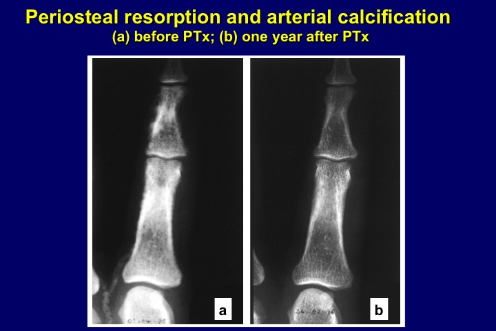
Figure 15. (a) X-ray aspect of periosteal resorption within cortical bone of middle phalanges of the hand, indicative of osteitis fibrosa, and extensive finger artery calcification in a CKD stage 5 patient with severe secondary hyperparathyroidism. (b) Aspect one year after surgical parathyroidectomy (PTX): complete bone lesion healing and disappearance of arterial calcifications.

Figure 15. (a) X-ray aspect of periosteal resorption within cortical bone of middle phalanges of the hand, indicative of osteitis fibrosa, and extensive finger artery calcification in a CKD stage 5 patient with severe secondary hyperparathyroidism. (b) Aspect one year after surgical parathyroidectomy (PTX): complete bone lesion healing and disappearance of arterial calcifications.