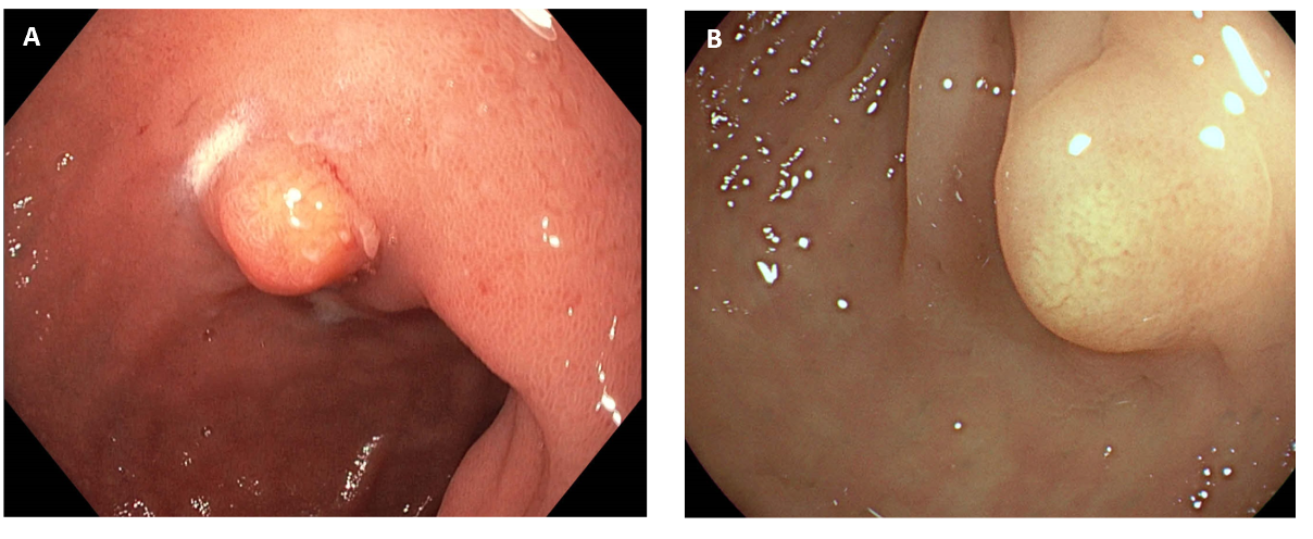
Figure 1. Endoscopy in gastrointestinal NET. (A) Endoscopic image of a 5 mm submucosal lesion in the duodenal bulb. Fine needle aspiration confirmed a grade 1 duodenal NET, which was subsequently removed by endoscopic mucosal resection. (B) Endoscopic view of an 8 mm rectal NET, grade 1, which was successfully resected by endoscopic submucosal dissection.
