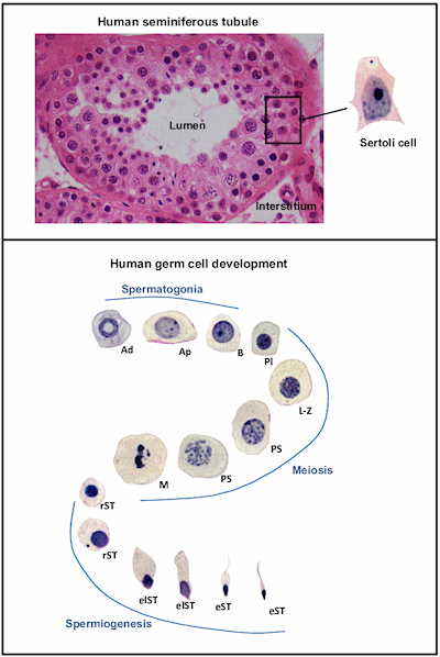
Figure 5. The top panel illustrates the typical structure of the human seminiferous epithelium containing the germ cells and Sertoli cells. The position of Sertoli cell nuclei within the epithelium is indicated, as is the tubule lumen. The tubules are surrounded by thin plate-like contractile cells called peritubular myoid cells. The Leydig cells and blood vessels lie within the interstitium. The bottom panel illustrates the nuclear morphology of the major cell types found within the human seminiferous epithelium, showing the progress of spermatogenesis from immature spermatogonia through meiosis and spermiogenesis to produce mature elongated spermatids. Abbreviations: Ad: A dark spermatogonia, Ap: A pale spermatogonia, B: type B spermatogonia, Pl: preleptotene spermatocyte, L-Z: leptotene to zygotene spermatocyte, PS: pachytene spermatocyte, M: meiotic division, rST: round spermatid, elST: elongating spermatid, eST: elongated spermatid. All germ cell micrographs were taken at the same magnification to indicate relative size. Micrograph of seminiferous epithelium was provided by Dr S. Meachem.
