Archives

Acromegaly

ABSTRACT


Acromegaly is a rare condition with an approximate incidence of 3-11 new cases per million of population per year and a prevalence of approximately 60 per million (1). There are approximately 3000 identified individuals in the UK and 15000 in the USA, although it is possible that more cases exist but do not come to clinical attention. More recent studies suggest a higher incidence of acromegaly, up to 6.9 per 100,000 according to Italian data and 7.7 patients per million per year in Iceland (2,3). The condition was named by Pierre Marie in 1886 using the Greek words akron- extremities and megas- large to describe the typical clinical appearance of the condition (4).The disease occurs as a result of excessive secretion of growth hormone. In more than 99% of cases this is due to a benign pituitary growth hormone secreting adenoma. Pituitary carcinomas are exceedingly rare. Extremely infrequently acromegaly occurs as a result of ectopic secretion of growth hormone releasing hormone (GHRH) from a peripheral neuroendocrine tumor (5,6), excessive hypothalamic GHRH secretion (7), or can result after long term exogenous GH abuse (8). Approximately 5% of cases are associated with familial syndromes, most commonly multiple endocrine neoplasia type 1 (MEN1) syndrome, but also McCune Albright syndrome, familial acromegaly, Carney syndrome, and Familial Isolated Pituitary Adenoma (FIPA). Both genders are equally affected and the diagnosis is typically made in adults aged 40-60 years of age. Younger patients often have more aggressive disease due to more rapidly growing adenomas. Acromegaly is associated with multiple systemic complications and a higher risk of mortality if untreated. Very often a multi-modal treatment approach is required to manage the condition, including surgery, radiotherapy, somatostatin analogues, GH receptor antagonist, and dopamine agonist. The management should be individualized to the patient using best practice guidelines, clinical experience, and individual patient circumstances and guided by biomarkers and clinical predictors.


PHYSIOLOGY: GROWTH HORMONE- STRUCTURE AND PHYSIOLOGY


Growth hormone is a 191 amino acid single chain protein containing two disulphide bonds. It has considerable structural homology with prolactin. Approximately 70% circulates as a 22 kD protein, 10% as a 20 kD isoform and the remainder as dimers or sulphated and glycosylated isoforms (Figure 1). Growth hormone secretion occurs in pulsatile bursts, numbering between 4 and 11 in 24 hours, especially at night, with extremely low or undetectable levels occurring in the nadir between pulses. Thus, a random single serum measurement is very limited as a means of assessing the overall level of secretion. Secretion of growth hormone is governed by both secretory and inhibitory hypothalamic factors. GHRH (growth hormone releasing hormone), ghrelin, and klotho act to stimulate release (9), whereas hypothalamic somatostatin (a 14 amino acid peptide) exerts marked inhibitory effects on GH release. Cortistatin has been found to exert dual, stimulatory and inhibitory effects on GH secretion (10). These stimulatory and inhibitory factors are subject not only to higher influences within the brain but also to peripheral signals such that the overall secretion of growth hormone can vary widely under different physiological and pathological conditions (11) . These are summarized in Table 1.


Table 1. Factors Affecting Growth Hormone Secretion (12)

PHYSIOLOGICAL


PATHOLOGICAL

Factors which increase GH secretion

Factors which reduce GH levels

Factors which increase GH secretion

Factors which reduce GH levels

Sleep

Overeating

Acute glucocorticoid excess

Chronic excess Cortisol/ glucocorticoids

Fasting

Obesity

Type 1 DM

Hyperthyroidism

Stress

Aging

Renal failure

Type 2 DM

Exercise

Increased IGF-1

Acute use of opiods


Hypoglycemia


Anorexia


Dopamine


Depression


Increased Amino acids i.e., high protein meal


Cirrhosis


Reduced Free Fatty acids


Glucagon


Testosterone and estradiol


Figure 1. The 2-D structure of human growth hormone.

Growth hormone circulates in blood bound to a specific binding protein, called GH binding-protein (GHBP). This protein comprises the extracellular portion of the growth hormone receptor (GHR), which is widely distributed and present in most tissues. Activation of the growth hormone receptor occurs when the growth hormone molecule binds two adjacent receptors resulting in dimerization of the growth hormone receptors. Dimerization of the growth hormone receptor results in its activation and binding of the intracellular Janus kinase (Jak 2) tyrosine kinase. The activated JAK2-GHR complex induces multiple signaling pathways responsible for the diverse actions of GH (13,14). These include phosphorylation of a) signal transduction and activators of transcription (STAT) proteins STAT1, STAT3 and most importantly STAT 5, b) SRC family kinases which trigger the MAP kinase pathway, c) insulin receptor substrate (IRS) proteins which activate phosphatidylinositol-3-kinase (PI3K) and Akt pathway, and d) SH2B1, a scaffold protein that upregulates GH action in the actin cytoskeleton (13). Intracellular growth hormone signaling is suppressed by several proteins, especially the suppressors of cytokine signaling (SOCS) 1-3 and protein tyrosine phosphatases SHP1, SHP2 (14,15). While there have been suggestions that GHR polymorphism could play a role in variable responses to the GHR antagonist Pegvisomant therapy, so far studies have not convincingly demonstrated this relationship (16).

Figure 2. The growth hormone molecule binding to the membrane surface growth hormone receptor. Signaling and transduction only occur when adjacent receptors bind the two specific binding sites on the growth hormone moiety to form a dimer.

One of the major proteins induced by growth hormone is insulin-like growth factor-1 (IGF-1)   Although classical endocrinology states that it is hepatic derived IGF-1 acting in an endocrine manner that is responsible for most, if not all, of the effects of growth hormone, it is becoming increasingly clear that local production of IGF-1 acting either in a paracrine (nearby cells) or autocrine (on the same cell) manner also has important biological effects, predominant of which is stimulating cell proliferation and inhibiting apoptosis (17). Elegant gene 'knock-out' experiments have demonstrated that animals with selective hepatic IGF-1 loss have a normal phenotype and growth, despite marked reduction in serum IGF-1 levels (18). Furthermore, patients with severe GH deficiency, perhaps as a result of pituitary surgery, usually have serum IGF-1 levels just below or at the lower end of the normal range. Thus, rather than being the sole effector of growth hormone, serum IGF-1 should perhaps be more accurately regarded as a marker of serum growth hormone concentrations. Circulating IGF-1 does however have important effects in regulating pulsatile growth hormone secretion with IGF-1 acting in a negative feedback fashion suppressing growth hormone release.


Central Regulation of GH Secretion


GHRH consists of 44 amino acids, the first 27 from the N-terminus being essential for physiological activity (19). GHRH containing neurons are located in the arcuate nucleus and surrounding the venteromedial nucleus which is considered the major site of GHRH activity. Somatostatin producing neurons are predominantly present in the dorsolateral arcuate nucleus and share close synaptic connections with the GHRH neurons (20). While these two hormones form the key components of local autocrine short feedback regulation of GH secretion; it is further modulated by interactions with central neuropeptides. Dopamine, serotonin, norepinephrine, neuropeptide Y regulate GH output through their interactions with GHRH and Somatostatin neurons (20). GH exerts its own local negative feedback along with local IGF-1 feedback. The Neuropeptide Y system contributes to the rise in circulating GH seen in the fasting state (21).


A long negative feedback also exists and growth hormone induces hepatic secretion of IGF-1, which in turn inhibits growth hormone release through various mechanisms including modulating GH gene transcription, reducing GH mRNA expression through POUF1 /CREB protein interactions and altering somatostatin release through paracrine activity (20,22,23).


Ghrelin and Growth Hormone Secretion


GH pulsatility is primarily driven by nutritional status. The potent growth hormone stimulating action of ghrelin is now well established.  Ghrelin was identified as a natural ligand responsible for regulating GH secretion in the acylated form (24). It is a 28 amino acid peptide, which is modified by Ghrelin-O-Acyl-transferase (GOAT) enzyme to mediate its GH secretory action through IP3 signaling pathway (25). Expression of ghrelin is found in many tissues including both the gastrointestinal tract and the CNS, with the strongest concentrations located in the stomach. Ghrelin mediates its orexigenic actions through the vagal nerve stimulation and eventually acting on the hypothalamic appetite center though the noradrenergic pathway (26). Gastric expression of ghrelin is reduced following feeding and increased by fasting, hypoglycemia and after leptin administration. On other hand, leptin seems to variably effect GH secretion depending on the nutritional status. The exact mechanisms of leptin driven GH changes are not well understood (27).

Figure 3. The growth hormone/ insulin-like growth factor-I axis.

Peripheral Regulation of GH Secretion


Systemic energy homeostasis is a potent influence on GH secretion, with interactions by glucose, free fatty acids, adipokines, leptin, ghrelin, and insulin (28-30).  Several hormone systems have regulatory effects on growth hormone secretion. Hypothyroidism is associated with low levels of both growth hormone and IGF-1, and in children leads to short stature (31). Thyroxine replacement has been shown to reverse these deficits. Further evidence from studies in rodents indicates that growth hormone gene expression is regulated by thyroid hormone acting through a thyroid hormone responsive element in the promoter region of the growth hormone gene (32,33). GH replacement has been shown to reduce the Free T4 levels and increase Free T3 levels, by the effect on type 2 iodothyronine deiodinase (34). Glucocorticoids are inhibitors of somatic growth both in humans and experimental animals and individuals with either Cushing’s syndrome or taking exogenous corticosteroids have been shown to have reduced growth hormone secretion.


Gonadal hormones also play a role in the neuroregulation of growth hormone secretion. In both sexes spontaneous growth hormone secretion is increased during puberty, and reduced in those with delayed puberty (35), suggesting that both estrogen and testosterone influence growth hormone secretion. The estrogen related reduction of IGF-1 seems to be responsible for relatively higher GH levels in females compared to men, with similar IGF-1 levels (36,37). Hypoglycemia is a potent inducer of growth hormone secretion, and insulin induced hypoglycemia remains the most employed provocative test of growth hormone reserve in humans. Hypoglycemia reduces hypothalamic somatostatin secretion facilitating growth hormone release. A delayed rise in GH levels is noted after acute hyperglycemia and is likely a result of GHRH rise (38) In contrast, hyperglycemia suppresses growth hormone secretion from the healthy pituitary. The availability of amino acids as in the post-prandial state stimulates growth hormone secretion whilst elevated non-esterified fatty acid levels suppress growth hormone release.


IGF-1: Structure and Function


IGF-1 is a single chain polypeptide of 70 amino acids with three intrachain disulphide bridges, coded by a gene situated on the long arm of chromosome (39). It has 48% amino acid sequence homology to pro-insulin, the A and B domains of IGF-1 have 60-70% homology but there is no homology with the C domain. IGF-1 has a specific receptor, which is structurally and functionally very similar to the insulin receptor. It consists of two extracellular α-subunits which are the hormone binding sites and two transmembrane β-subunits which are involved in initiating intracellular signaling. Post-receptor signaling mechanisms are also similar for IGF-1 and insulin receptors, both activating the tyrosine kinase and IRS-1 cascades. IGF-1 can bind to the insulin receptor but with only 1-5% affinity compared to insulin. Under normal physiological conditions it is thought that IGF-1 acts via the specific IGF-1 receptor, but in the presence of high concentrations of IGF-1 there is likely to be cross activation with the insulin receptor. IGF-1 receptors are found on most tissues with the notable exceptions of liver and adipose tissue. Hybrid IGF-1/insulin receptors have now been well documented and sequenced but their role is unclear (40).


The majority of circulating IGF-1 is produced by the liver with bone, adipose tissue, kidney, muscle and many other tissues producing a smaller quantity. Plasma concentrations of IGF-1 in the human are regulated by growth hormone, insulin, age, and nutritional state. Bioavailability of IGF-1 is determined by its binding proteins (see below). Growth hormone and insulin are the main regulators of hepatic IGF-1 production. The precise regulation of local IGF-1 synthesis is uncertain, but it is influenced by many other trophic hormones such as ACTH, fibroblast growth factor, and TSH (41).


Figure 4. The regulation of growth hormone secretion.

IGF Binding Proteins (IGFBPs)


Unlike insulin the majority of IGF-1 circulates in plasma bound to a variety of binding proteins which determine its bioavailability and modulate its biological action (42). The majority of IGF-1 is bound in a 150 KDa complex with IGFBP-3 and form an acid labile subunit ALS (42). This large molecule (termed the ternary complex) is unable to pass through endothelium and acts as an intravascular reservoir of inactive IGF-1. The half-life of IGF-1 in the complex with IGFBP-3 and ALS is 12-15 hours compared with 10-12 minutes for free IGF-1. The exact mechanisms by which IGF-1 is released from the ternary complex to allow access into the tissues is not known; however, IGFBP degrading protease activity has been well documented in many biological fluids and clinical states.


Current knowledge suggests that IGFBP-1 and IGFBP-3/ALS are the binding proteins which have the major effects on the bioavailability of IGF-1(40). IGFBP-1 is inversely related to insulin levels, has a circadian variation with the highest levels being found overnight when insulin levels are lowest, and inhibits the hypoglycemic action of IGF-1 (43). Growth hormone secretory status is the main regulator of plasma levels of ALS (42).


PATHOLOGY OF ACROMEGALY


Acromegaly is most commonly the result of pituitary adenoma and rarely due to non-pituitary neuroendocrine tumor/ neoplasia (NET/ NEN).  Pituitary tumors are commonly monohormonal or plurihormonal in nature with further distinct subtypes. These subtypes of tumors are responsible for their characteristic behavior and can sometimes guide management (44). The common subtypes are discussed below.


1) Monohormonal densely granulated somatotroph adenomas are the most common type of GH-secreting pituitary tumors. They have predominance of the large, dense secretory granules which contain GH and appear deeply eosinophilic on staining.  They account for 30-40% tumors and while tend to lead to higher GH levels, they are usually slow growing tumors, in older patients with mild disease, and retain a predictable response to SSA therapy. Somatic mutation in Gs-α subunit of GNAS has found to be the most common abnormality in GH secreting tumors leading to increased cAMP activity (45).

2) Sparsely granulated somatotroph adenomas form the second most common type of tumor. As the name suggests, they are lightly eosinophilic on HE staining due to the increased presence of keratin aggregates and reduced GH containing granules. While they present with lower GH levels, the tumor behavior is relatively aggressive. They tend to be larger, more invasive with higher ki67 proliferation indices and are less responsive to SSA therapy. Further studies suggest reduced expression of E cadherin and SSTR2 in sparsely granulated tumors as factors likely to be responsible for poor response to SSA (44).  

3) Mammosomatotroph adenoma from a monohormonal Pit-1 lineage cells is a common pathology found in younger individuals with acromegaly. They are densely granulated and co-stain for GH and PRL. They behave in a more benign fashion with high GH and PRL levels leading to early presentation, when the tumor is smaller at diagnosis. They respond to SSA therapy similarly to densely granulated tumors (46).

4) Mixed somatotroph and lactotroph tumors are formed of bihormonal cells with variable combination of somatrotroph and lactotrophs. In variable combinations they comprise of sparsely granulated and densely granulated cells all of which express Pit 1. They tend to be less amenable to treatment and are reported to have frequent disease recurrence (47).

5) Mature Plurihormomal Pit1-Lineage tumors frequently immunostain for TSH along with GH, PRL and are found to express GATA3 (48). Clinical presentation includes features of thyroid overactivity with thyrotoxicosis with non-suppressed TSH.

6) Acidophil stem cell adenomata are tumors comprising of immature GH and PRL secreting tumor cells of a single precursor. The histology shows chromophobic or slightly acidophilic cells, with abundant granular cytoplasm of oncocytic distribution (44). Patients present with hyperprolactinemia which is disproportionate to the size of the tumor. They are frequently invasive and less responsive to dopamine agonist therapy.

7) Poorly differentiated Pit-1 lineage tumors comprise of tumor cells with strong expression of Pit-1 and variable expression of estrogen and GATA3. They are polyglonal and spindle shaped poorly differentiated cells which stain variably for GH, PRL, TSH, and alpha subunit. They are usually macroadenomas, with invasion of surrounding structures and high risk of recurrence (46).

8) Pituitary carcinomas are very rare and form less than 1% of cases. They are difficult to distinguish clinically, and diagnosis is confirmed with evidence of distant metastasis and high ki67 index (e.g., >10%) on histology. These tumors frequently require multi-modal therapy, including chemotherapy, temozolamide and radiation (44).

9) Pituitary hyperplasia is suspected radiologically when there is a uniformly enlarged pituitary gland with no distinct focus of gadolinium enhancement. Hyperplasia is confirmed on histology when the pathology shows expanded pituitary acini containing all of the adenohypophysial cell types, but with increased numbers of somatotrophs and/or mammosomatotrophs (46). The histological diagnosis should prompt the clinician to explore for a GHRH secreting tumor elsewhere or consider investigations for specific genetic conditions associated with acromegaly (highlighted in table 4) such as MEN1, Carney Complex, and McCune Albright Syndrome (49).


Non-pituitary sources of disease include GHRH or GH secreting central and peripheral tumors. Hypothalamic tumors such as hamartomas, choristomas, gliomas, and gangliocytomas producing GHRH result in pituitary hyperplasia and very often the diagnosis remains elusive until patient has undergone pituitary surgery. Carcinoid tumors secreting GHRH are recognized as rare peripheral cause of acromegaly and usually are bronchial in origin (50). An ectopic location of pituitary adenomas has been reported in the tract of dorsal migration of the adenohypophysial cells. While a significant number of peripheral tissues have been found to secrete GH (51), reports of GH secreting peripheral tumors include lung, pancreatic and adrenal tumors.


Table 2. Pathology Associated with Acromegaly (44,52-54)

Pituitary adenoma

Densely granulated somatotroph adenoma

Sparsely granulated somatotroph adenoma

Mixed cell somatotroph and lactrotroph adenoma

Mammosomatrotroph (monohormonal Pit-1 lineage) adenoma

Acidophil stem cell adenoma

Plurihormonal adenoma

Poorly differentiated Pit-1lineage tumor

Pituitary hyperplasia

Pituitary carcinoma


Rare: ectopic pituitary adenomas identified in sphenoid sinus or parapharyngeal tissue

Ectopic hormone secretion

Central:

Hypothalamic tumors

Ganglioneuroma


Peripheral: Bronchial carcinoid, small cell lung cancer, adrenal tumor, pancreatic neuroendocrine tumor

Exogenous GH replacement or abuse


Pseudoacromegaly

Pachydermoperiostosis

IGF signaling pathway diseases

Severe insulin resistance


CLINICAL FEATURES OF ACROMEGALY


The clinical manifestations of acromegaly evolve gradually over a long time and as a consequence there is a lag time of about 5-10 years, from symptom onset to diagnosis (55,56). In the recent decades, there seems to be some early recognition of the condition, particular in individuals investigated for pituitary ‘incidentaloma’, for example with hypogonadism as a presenting feature (57). One third of the cases, have co-existent symptoms of hyperprolactinemia, which aids in early diagnosis (58).


Growth hormone secreting pituitary adenomas are frequently (more than 70%) large tumors (macroadenoma, ≥ 10 mm in diameter) which may present with local mass effects such as headache (often severe and out of proportion to the size of the pituitary tumor), hydrocephalus, visual field defects, ophthalmoplegia, or other cranial nerve palsies (59). As the lesion increases in size deficiencies of other anterior pituitary hormones may also occur. Microadenomas (< 10 mm in diameter) are conventionally thought to be less common, but tend to represent one third of the cases (60,61). However patients presenting with pituitary tumors, without clear features of acromegaly may have elevated IGF-1, and GH positive immunohistochemistry on the resected tumor specimen (62) and such silent growth hormone tumors seem to be more common in females with a higher risk of recurrence (63). The term micromegaly is used to describe such clinical presentations (64,65). Recognition of such presentations should prompt the endocrine specialist to consider GH secreting tumors in all presentations, but especially in the younger patient with pituitary tumor. 


Hypopituitarism has been found to occur in about 40% cases with variable frequency of hypogonadism, adrenal insufficiency, and secondary hypothyroidism (66,67). Hypogonadism, presenting as decreased libido, infertility or oligo/amenorrhea is a common finding at presentation; it may be due to both gonadotrophin deficiency as well as hyperprolactinemia, either from coexistent excessive secretion of prolactin or from stalk compression. Hypogonadism has been reported even in patients with microadenomas with normal prolactin, thereby suggesting an independent effect of GH hypersecretion (68). Menstrual irregularities, PCOS, subfertility and erectile dysfunction can occur as a consequence of GH excess (69,70). The occurrence of diabetes insipidus in relation to a pituitary adenoma is extremely rare and should raise the possibility of an invasive pathology (71,72).


Soft Tissue and Skeletal Changes


The most characteristic feature and one that usually precipitates the diagnosis is a change in appearance as a consequence of soft tissue and bony changes. The common changes include coarsening of the facial features, broadening of the nose, thickening of the lips, macroglossia, and prominence of the supraorbital ridges. There is enlargement of the hands resulting in their characteristic 'spade-like' appearance and soft dough-like consistency of the palms. Ring size increases; a sensitive objective assessment of disease activity and response to treatment. Similar changes occur in the feet which become wider with increase in shoe size. Elongation of the jaw results in prognathism which contributes to dental malocclusion, interdental separation, and temporomandibular joint pain (73,74).


Greasiness of the skin is a frequent finding with excessive sweating, one of the most sensitive signs of growth hormone excess. Skin tags are a frequent finding, likely related to epithelial cell hyperproliferation in response to IGF-1 (74). Additional dermatological manifestations include hypertrichosis, psoriasis, acanthosis nigricans, and cutis verticis gyrata, with the latter two seen more commonly in severe cases (75). Skin changes are a result of deposition of glycosaminoglycans in the subcutaneous tissue, along with increased proliferation of dermal fibroblasts as a consequence of GH and IGF-1 action (74). These changes tend to reverse after treatment, at least partially if not completely. The lean body mass is higher in individuals with acromegaly, while there is increase in adipose tissue post therapeutic intervention (76). This reversal of body composition tends to stabilize by three months of the surgery (77). In addition to the negative impact on body fat, reversal of GH excess has been found to increase intrahepatic lipid accumulation (78).


Generalized organomegaly is not well reported with acromegaly, in contrast some earlier assumptions of the disease process but enlargement of thyroid, prostate, salivary glands, heart, liver and spleen has been recognized (79). Macroglossia, increased thickness of laryngeal structures and vocal cord enlargement increases the risk of anesthesia and makes intubation difficult (80). Ultrasound evidence suggests the presence of increased organ stiffness and commonly reported features include renal cysts, thyroid nodules, multinodular goiter, gallbladder polyps, and polycystic ovaries (79). Mucosal edema and hypertrophy of vocal cord can result in voice changes, but true existence of voice abnormalities is debated (81,82). Patient reported questionnaire and evaluation of voice parameters seem to demonstrate presence of micro perturbations, lower amplitude and poor quality of voice in individuals with active disease (82,83).


The skeletal manifestations of acromegaly result from multiple factors which include direct effect of GH, IGF-1, altered calcium phosphate metabolism, hypogonadism, diabetes, and over replacement of steroids (84,85).  Acromegaly results in increased bone resorption and altered bone formation according to various cross-sectional studies, but the effect of GH, IGF-1 on bone health is complex. GH mediates its effect on bone through systemic IGF-1 with action on cortical bone, whereas bone IGF-1 seems to be responsible for cancellous bone health. There is emerging evidence of correlation of sclerostin levels with acromegaly related bone disease (86). The high prevalence of vertebral fractures (VF) in active acromegaly has been known for a long time with some studies reporting incidence as high as 60%, and dependent on the duration of disease and gender (males>females) (84,87).  In a French study, authors have suggested that the skeletal abnormalities are more likely vertebral deformities than true fractures (88). Screening for VF is recommended in all patients with active acromegaly as it has a significant impact on quality of life, morbidity, and development of cardiac and pulmonary complications (85,89). Standard DXA seems to be less reliable in predicting the risk and Volumetric DXA with quantitative assessment of the trabecular bone density seems to provide more reliable information of acromegaly related bone disease (87,90). Unfortunately it is difficult to undertake this test routinely and therefore alternative use of newer methods such as non-invasive 3D-SHAPER and TBS Trabecular bone score assessments may be considered (90). While vertebral changes are most discussed, consequences of acromegaly include development degenerative diseases in all weight bearing and non- weight bearing joints, predominantly shoulder, hip and knees (84,91). The prevalence of radiographic evidence of at least one joint involvement has been found to be as high as 99% (92).  Patients with active disease seem to be have higher prevalence of reduced cortical density at hip compared to patients with non-functioning pituitary adenomas (93). Degenerative joint changes in early acromegaly related arthropathy appear different from usual osteoarthritis, and are noted as widened joint spaces contributed by cartilage hypertrophy and marked osteophytosis, in contrast to standard OA (94). Acromegaly related arthropathy is associated with a significant impact on quality of life and tends to progress despite improvement in disease status (89). Routine use of anti-resorptive therapy is currently not well established, but considering the wider implications of the disease on bone health in active disease, it seems reasonable to consider offering bone protective therapy in patients with early evidence of bone disease (90).


Figure 5. The typical facial appearance of acromegaly. Evolution of the appearances over 2 decades

Sleep Disordered Breathing


Sleep apnea syndrome (SAS) is a well-recognized manifestation of GH excess and seems to have been variably reported with incidences of 40-80% and about 11.7-20 times higher prevalence than in general population (55,95). Evaluation of the bony changes in the facial skeleton showed significant differences in patients with acromegaly and SAS, compared to patients without SAS. But soft tissue enlargement of upper airways contribute more to the narrowing of the pharyngeal airway space than the craniofacial skeleton changes (95). CT and MRI assessment of upper airways demonstrate pharyngeal hypertrophy and upper airway stenosis correlating with the severity of obstructive sleep apnea (96). Patients with SAS have been found to have more features consistent with metabolic syndrome, such as hypertension and DM compared to patients without SAS (97). Improvement in SAS noted after intervention seems to correlate with positive changes in tongue volume and pharyngeal soft tissue (98). Unfortunately, complete reversal of SAS does not occur and persistent SAS is seen in about 40% of treated cases. It is likely due to effect of additional factors on SAS such as age, male gender, smoking, and obesity (84). A direct inhibitory effect of GH on central respiratory center can result in a non-obstructive central pattern of sleep apnea (99). This phenomenon seems to be much less prevalent than obstructive disease, while a mixed pattern has also been reported (100).


Muscular Changes


Musculoskeletal pain is typically progressively evident during the disease course, with close to 90% patients reporting pain as a dominant symptom (101). Irrespective of the severity of the acromegaly, pain contributes significantly to reduced quality of life (101). Studies demonstrate that GH excess leads to hypertrophy of the type 1 muscle fibers with variable findings for type 2 fibers (102-104). The predominant abnormality of type 1 muscle fibers strength seems to be responsible for evident muscle weakness noted in high velocity activities (105). While earlier studies consistently reported muscle weakness in acromegaly, a recent study used quantitative measures suggest patients with active acromegaly may have higher proximal muscle strength, but reduced hand grip which normalizes after treatment (103). It is postulated that the difference in findings could be related to pain interfering with true assessment of muscle function (103). Ultrasound evidence showed increased tendon thickness, enthesisitis, soft tissue enlargement but reduced muscle volume in some lower limb muscles in a cohort of thirty nine patients (106). There is emerging evidence that acromegaly has a complex effect on muscle strength and volume (107). Differential interactions of GH, Muscle Ring Factor -1 (MuRF-1), and myostatin seem to be responsible for chronic effects of GH excess on skeletal muscles (108).


Neurological Abnormalities


Carpal tunnel syndrome is present in approximately 60% of patients at diagnosis but about 80% will have electrophysiological evidence of median nerve neuropathy (109). The pathophysiology is due to swelling of the median nerve itself within the carpal tunnel rather than extrinsic compression from increased volume of the carpal tunnel contents (110). This is well evident on MRI and Ultrasound studies of the median nerve, and seems to correlate with the abnormal nerve conduction studies (110-112). Similar peripheral nerve abnormalities and abnormal nerve conduction studies have been noted in other peripheral nerves including peroneal, tibial, ulnar, sural nerves (112,113)and polyneuropathy is more common in uncontrolled disease (114). Unfortunately it seems some of the changes are not completely reversible, despite normalization of disease markers (115). Patients with active acromegaly have altered cardiac autonomic function, which contributes to the cardiovascular risk (116,117). This seems to respond to active intervention (118). There is not much evidence of central nervous involvement related to the disease (113), with some report of delayed brainstem auditory evoked potentials (119). Restless leg syndrome has been reported to be present in 20% of cases and negatively impacts the quality of life (120).


Cardiac Complications


Cardiovascular diseases continue to remain one of the most common causes of morbidity in patients with acromegaly and account for 60% of the mortality with this condition (121,122). The range of abnormalities detected at the time of diagnosis have been reported to be hypertension, cardiac hypertrophy, arrhythmias, coronary artery disease, and systolic heart failure, in the order of prevalence (123,124).  Hypertension presents with higher diastolic readings than systolic BP measurements (125). Based on ambulatory BP monitoring readings, it seems that the prevalence of hypertension is about 22% at diagnosis, much lower than earlier studies with single office measurements (126). Anti-natriuretic effect of GH is a direct consequence of GH action on the epithelial sodium channel ENaC in cortical collecting ducts of the kidney (127). This sodium retention leads to volume expansion and further compounded by GH effects on cardiac output, impaired endothelial function, increased peripheral resistance and co-existent sleep apnea are some factors that lead to development of hypertension (128).


Acromegaly related cardiomyopathy is a consequence of GH and IGF-1 effect on cardiac myocytes, regulation of cardiac muscle specific gene transcription, and increased fibrosis (129). It has been described to undergo three stages of disease progression (130). In the first phase biventricular concentric hypertrophy is described related to muscle hypertrophy and increased contractility and leads to a hyperkinetic syndrome. As the disease progresses, patients tend to develop diastolic dysfunction with more prominent ventricular hypertrophy. Patients report reduced exercise tolerance and it is common for disease to be diagnosed in this phase. If untreated patients may progress to develop overt diastolic and systolic dysfunction presenting as congestive heart failure in about 3-4% cases and is a poor prognostic marker (123). The presence of left ventricular hypertrophy correlates with disease duration and various studies report prevalence from 11-78% (84,131). Cardiac Magnetic Resonance (CMR) studies have demonstrated variable prevalence of left ventricular hypertrophy compared to echocardiographic studies (132,133). CMR is more reliable in identifying myocardial fibrosis and RV systolic dysfunction then echocardiography (133). Despite the presence of cardiovascular abnormalities associated with ischemic heart disease, recent studies report no increase in prevalence of Ischemic heart disease, in comparison to normal population (55,134).


Cardiac arrhythmias have been reported to occur in about 7-40% cases of acromegaly. A wide range of rhythm disturbances described in patients with acromegaly include paroxysmal atrial tachycardia, supraventricular tachycardia, sick sinus syndrome, ventricular ectopic, and ventricular tachycardia (130). A typical acromegaly related left ventricular rhythm disturbance, results from abnormal and dyssynchronous loss of peak contraction of corresponding cardiac segments (135). Mitral and aortic regurgitation have been commonly associated with acromegaly and seem to correlate with the duration of the disease (136). Unlike cardiomyopathy and arrhythmias which improve or even completely reverse with disease control, valvular disease is irreversible and only tends to stabilize with intervention (130).


Metabolic Complications


Growth hormone is a potent insulin antagonist and acromegaly results in abnormal glucose tolerance in many patients with frank diabetes mellitus in up to 50% cases at diagnosis. Lipid abnormalities, in particular elevation of serum triglycerides, reduced HDL levels, increased small dense LDL particles, and increased lipoprotein-a (Lp(a)) may be an accompanying feature of insulin resistance and is noted in one third of the cases (137,138). Chronic GH excess results development of insulin resistance by several mechanisms. Reduced glucose uptake occurs by increased levels of free fatty acids and reduced expression of GLUT1 and GLUT 4 receptors (139). GH also results in development of a pro inflammatory state in the adipose tissues with alterations of the genes coding visfatin and IL6 (140). The degree of insulin resistance correlates with IGF-1 levels (141) and improves with management of the disease. Development of IGF receptor resistance beyond a threshold for IGF-1 has been reported in states of chronic GH excess leading to further insulin resistance (142). Visfatin and irisin levels have been suggested to correlate with metabolic abnormalities and cardiovascular risk factors (143,144). When choosing treatment, octreotide, and lanreotide have less impact on the glycemic variations, while pasireotide can aggravate hyperglycemia. Pegvisomant has a favorable on the metabolic parameters (145).


Table 3. Clinical Manifestations and Complications Reported with Acromegaly

Tumor related local effects

Headache

Visual field defects

Cranial nerve abnormalities

Hydrocephalus

Temporal lobe epilepsy

Hyperprolactinemia

Hypopituitarism

Systemic effects

Skin changes

Hyperhidrosis

Oily skin

Skin tags

Hypertrichosis

Acanthosis

Cutis verticis gyrata

Cardiac

HT

Cardiomyopathy

Valvular heart disease

Arrhythmia

Heart failure

Soft tissues changes

Acral enlargement

Change in voice quality

Visceromegaly (thyroid, prostate, liver, salivary glands)

Neurological

Peripheral nerve abnormalities

Autonomic dysregulation

Lumbar canal stenosis

Narcolepsy

Restless leg syndrome

Orofacial changes

Prognathism

Frontal prominence

Dental malocclusion

TMJ pain

Gingival enlargement

Macroglossia

Pulmonary

Sleep apnea

Restrictive lung disease

Subclinical hypoxemia


Musculoskeletal

Vertebral deformities

Kyphosis

Arthralgia and arthritis

Myopathy

Degenerative arthropathy

Calcific discopathy

Hypermobility

Neoplastic

Colon polyps

Thyroid cancer

Breast cancer

Endocrine and Metabolic

Hypogonadism

PCOS

DM, insulin resistance

Hypertriglyceridemia

Erectile dysfunction


Renal

Increased GFR

Hypercalciuria

Glomerulosclerosis


Hematological

Increased thrombosis risk

Psychiatric

Depression


Ocular

Increased risk of diabetic retinopathy

Extraocular myopathy

Glaucoma

Epiphora


Thyroid Abnormalities


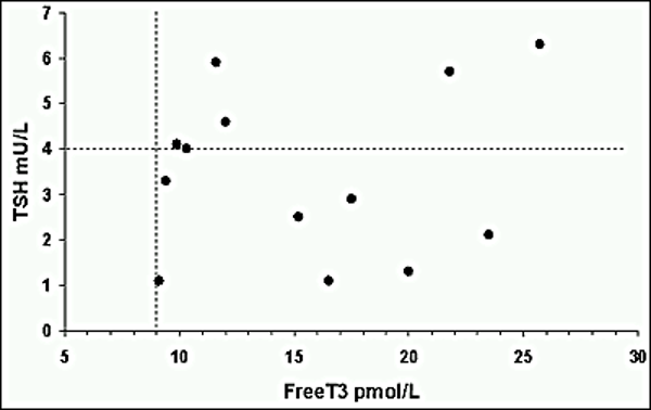
Patients with GH excess have been demonstrated to have a rise in TSH and T3 levels with no significant relation with the FT4 levels (146). There is a direct correlation of IGF-1, GH levels with thyroid volume. Multinodular goiter is one of the most common thyroid abnormality in patients with acromegaly with frequencies of 69.5 to 79.1% being reported by some authors (147). Patients in remission after surgery have been shown to have change in the consistency of the thyroid nodules, reduced vascularity, and volume (148). Over the course of follow up of patients with active acromegaly, thyroid nodule enlargement seemed to correlate with IGF-1 levels with increased prevalence of differentiated thyroid cancer (papillary thyroid carcinoma) in this subgroup (149,150). Despite the increasing understanding, routine screening for thyroid nodules is not yet recommended but assessment should certainly be considered in patients with a palpable nodule (151).


Neoplasia


The true risk of cancer continues to remain debated with concerns of heterogeneity in the study population, selection biases, variable screening strategies, and limitations of using standardized incidence ratios as the reporting indices (84). In the recent years, some large cohort population studies suggest higher cancer risk than general population. Acromegaly has been associated increased risk of cancers, particularly colon, kidney, and thyroid cancer in a large Italian survey (152). A similarly large Danish cohort has reported increased incidence of colon, thyroid, breast, gastric, and urinary bladder cancers (153).


Animal models and in vitro studies suggest anti-apoptotic and tissue proliferative role of GH and IGF-1. IGF-1 deficiency has been found to result in protection from tumor development. GH signaling pathways and autocrine GH action contribute to tumorigenesis and in colonic tissue this mechanism results in reduced action of tumor suppressor proteins (84,154). Colon cancer has been studied in detail in patients with acromegaly. Patients have a higher prevalence of adenomatous and non-adenomatous polyps than general population (155) with reported trend varying between 6-30% (156). Colonic pathology is related to disease activity with patients with elevated serum growth hormone and IGF-1 levels being particularly prone to developing colonic adenomas (157). Although the exact pathogenesis of these tumors remains uncertain it is likely to involve altered homeostasis of cell numbers within the colonic epithelial crypts; increased proliferation and decreased apoptosis within the crypts of patients with acromegaly have both been documented (158). Colorectal neoplasia in acromegaly has different characteristics compared to the general population, in that the adenomas are more likely to be located in the right side of the colon, tend to be bigger and are more often multiple as well as demonstrating increased dysplasia (159).


It is now generally accepted that patients with acromegaly should be regarded as a high-risk group for colorectal cancer and regular colonoscopy screening should be offered to all patients. Current evidence suggests that this should begin at the age of 40 years with the subsequent interval depending both on disease activity and the findings at the original colonoscopy screening (160). In the presence of a polyp (hyperplastic or adenoma) or elevated serum IGF-1 levels screening should be repeated after five years, whilst a normal colonoscopy screening, or serum IGF-1 level within the normal range suggests screening every 10 years may be appropriate. As approximately 30% of lesions occur at the cecum or in the ascending colon, total full-length colonoscopy is required. This should be performed by an experienced colonoscopist, as the cecum is reached in only about 70% of patients in inexperienced hands. Due to their slow bowel transit time and elongated colon, patients with acromegaly require rigorous bowel preparation, often twice that necessary for the patient without acromegaly. Failure to visualize the cecum necessities a repeat colonoscopy or failing this examination using CT virtual colonoscopy (156).


Lung Complications


Pulmonary complications are common in acromegaly. Total lung volume and residual volume are increased, along with narrowing of both large upper airways and more commonly small airways (161,162). In a large study Storrmann et al reported higher prevalence of small airway obstruction in females. They also highlighted presence of subclinical hypoxemia in patients with acromegaly. The findings did not correlate with levels of IGF-1 of duration of disease (163). Tracheal structural abnormalities have been found to be responsible for large airway disease (164).


Ocular and Auditory Complications


A wide variety of ocular complications have been reported in patients with acromegaly, apart from visual field defects. The prevalence of proliferative diabetic retinopathy has been variably reported to be higher in patients with acromegaly (165) in some studies, while others suggest it is likely that there is increased retinal vessel branching, and noted no difference in retinopathy rates (166). Extraocular muscle enlargement has been reported by few authors and rarely has resulted in presentation of diplopia (167-169). Studies have highlighted increased intraocular pressure, increased corneal thickness and increased retinal thickness in patients with acromegaly, with rare reports of epiphora (170-172). The association of acromegaly with hearing disturbances has not been well reported. There have suggestions of abnormal bony changes, changes in middle ear pressures and internal acoustic meatus contributing to variable hearing abnormalities (173,174). But findings have not been widely validated and association of acromegaly with hearing loss is not well established (175).


Other Systemic Complications


Hematological abnormalities are not common with acromegaly. Recent evidence suggests patients with active acromegaly may be at a higher thrombotic risk and this could contribute to cardiovascular risk (176,177). Higher levels of fibrinogen, factor VIII and thrombin tend to result in hypercoagulable state in active untreated disease (178). Rare case reports of polycythemia, myeloma, and Waldenstrom’s macroglobulinemia have been reported (179,180).


GH and IGF-1 receptors have been found in kidneys and suggest local autocrine and endocrine activity of GH at the level of nephrons (51). The effect of GH excess on kidneys has not been well described. Chronic GH exposure leads to renal hypertrophy and structural changes to include glomerulosclerosis (181) . Patients with acromegaly have been reported to have increased GFR, reduced renal excretion of sodium, potassium, hypercalciuria, hyperphosphaturia, greater prevalence of microalbuminuria and micro-nephrolithiasis, irrespective of comorbidities (182-185). 


Morbidity and Mortality in Acromegaly


It is established that uncontrolled acromegaly results in a considerable increase in morbidity with an overall mortality at least two-fold that of the general population (186). In early epidemiological reviews more than 50% of patients had died by the age of 60 years, usually as a result of diabetes, cardiovascular, respiratory or cerebrovascular disease (187). With improved treatment of both the underlying disease and these complications, patients are now surviving longer although may then be susceptible to other complications such as malignancy (188). The determinants of mortality included older age and IGF-1 levels at diagnosis, treatment modality, and malignancy (121). Prolonged diagnostic delay has been found to positively correlate with morbidity and mortality (189). Longitudinal population cohorts indicate a negative impact of female gender on the presence of comorbidities and mortality (37,190).


DIAGNOSIS OF ACROMEGALY


The diagnosis is made using a combination of clinical examination and biochemical assessment. Serum growth hormone concentrations are typically elevated, and although pulsatility may be reduced, levels may fluctuate widely in acromegaly. Due to the pulsatile nature of growth hormone secretion, a single growth hormone measurement is of little use in either monitoring or confirming the diagnosis of acromegaly. GH levels are affected by age, gender, and comorbid disease states and these factors need to be taken into account when interpreting results. These factors have been outlined in table 1. Due to variations between assays, it is recommended that a standardized and similar performing assay is used for monitoring of the disease activity. Most modern assays detect the highly prevalent GH 22kDa isoform and the most common preparation used to calibrate GH assays is the latest recombinant IRP 98/574 (197). It is useful to be aware of the interferences of the local assay with biotin and Pegvisomant. Various tests used in screening, diagnosis, and management of acromegaly are discussed below.


Biochemical Tests at Diagnosis


  1. IGF-1 levels: A single serum IGF-1 level has been advocated as being a useful first line test for the diagnosis of acromegaly as it is elevated in the majority of subjects. It is an indirect assessment of growth hormone secretion with approximately 25% of patients having a discrepancy between the mean value of a growth hormone day curve and an IGF-1 level. IGF-1 secretion is subject to several influences including liver and renal dysfunction, nutrition, diabetes mellitus, physiological factors such as age, gender, and the presence of a statistical correlation between its levels and those of growth hormone should not be used as proof that they are interchangeable (198). However, despite these limitations, from a practical point of view, an elevated serum IGF-1 measurement may be useful as confirmatory evidence, assuming that age and sex matched normal ranges are used, and for monitoring treatment (199). Standardized IGF-1 (IS 02/254) is recommended for manufacturers of IGF-1 assays (52,200).
  2. GH day curve: The assessment of growth hormone hypersecretion requires the mean value of serial samples taken throughout the day (e.g., 5 samples over a 12-hour period). The samples should be taken through an indwelling venous cannula to avoid the stress effects of repeated venipuncture. In normal subjects, the majority of values throughout the day are undetectable, but in acromegaly typically each value is measurable, often with a fixed rate of secretion (201).
  3. Oral glucose tolerance test: Failure of normal suppression of serum growth hormone following administration of oral glucose remains the ‘gold-standard’ biochemical test (202). 75 g of oral glucose is given at 9 am to the fasting patient and plasma glucose and serum growth hormone levels are measured at baseline, 30, 60, 90, 120 (and 150) minutes thereafter. In normal subjects, growth hormone levels suppress to undetectable values (typically <0.1 ng/ml) when ultrasensitive assays are used, whilst in acromegaly serum growth hormone remains detectable, and in approximately 30% of cases there is a paradoxical increase (199). In conventional practice failure to suppress serum growth hormone to a level < 0.4 ng/ml following ingestion of glucose supports the diagnosis of acromegaly. The use of this test also detects those patients with impaired glucose tolerance or diabetes mellitus other than individuals with poor diabetes control (203). False-positive results can be seen in conditions where GH levels are elevated such as stress, type 1 diabetes mellitus, cirrhosis, chronic renal failure, during adolescence, and by drug use (L -dopa, heroin, estrogen 201,204).
  4. TRH test: In cases of remaining doubt about the diagnosis of acromegaly, a TRH test can be used (200 mg of thyrotrophin releasing hormone given intravenously with serum measurement at 0, 20 and 60 minutes). In normal subjects TRH inhibits growth hormone secretion with a fall in serum concentration, whilst approximately 60% of patients with acromegaly demonstrate a paradoxical rise in growth hormone levels (205). In mild disease with relatively low GH levels, TRH stimulation has been suggested to confirm early diagnosis (206).
  5. Others: IGFBP3 levels correlate with mean 24 hour GH levels and IGF-1 levels, but due to the wide overlap with normal and diseased individuals, is a poor parameter for measuring disease activity (207). In the rare patient in whom a non-pituitary etiology is suspected, measurement of serum GHRH may be performed, typically with very elevated levels occurring in ectopic GHRH syndromes such as neuroendocrine tumors. Basal serum prolactin should also be measured as prolactin may be co-secreted with growth hormone in up to a third of patients with acromegaly, which often indicates therapeutic responsiveness to the use of dopamine agonists. In those with hyperprolactinemia the presence of macroprolactin should be excluded (208).


In patients with atypical clinical symptoms such as GI symptoms, hypoglycemia, renal stones, clinicians should consider exploring possibility of coexisting genetic tumors or extra pituitary source of GH abnormality, such as carcinoid tumors.


Biochemical Tests Used to Define Disease Activity to Guide Outcomes


  1. Octreotide test: Acute challenge with octreotide 100mcg sc dose can help predict response to SSA when hourly values are measured over 6 hours (209,210). Not all clinicians perform this test and many treat with a long-acting SSA independent of a challenge test (211).
  2. IGF-1 levels: IGF-1 levels can take over three months to normalize in the post-operative period and until then cannot be reliably used to guide management. In longer term the aim of treatment is to keep levels within the normal ranges (52). In patients with Pegvisomant therapy, IGF-1 is the variable that guides response to treatment (212).
  3. Random GH levels: Random levels can be used to define surgical cure and values as early as day 1 can be undertaken if no pre-operative GH suppressing therapy was used, bearing in mind the effect of post-operative stress. A serum GH <0.4 µg/L favors disease remission and a level <1 µg/L indicates good control and normalization of the mortality risk (151).
  4. OGTT: OGTT can be undertaken to evaluate post-operative outcome if random GH values >1µg/L. A nadir GH cut off of <1µg/L is associated with better long-term outcomes and would be considered as good control. Endocrine Society guidelines suggest GH nadir of less than 0.4mcg/ L could be used to define disease remission (151) It has been suggested that post-operative OGTT at 3 months is more appropriate than an early test in 3 weeks and is likely to help avoid false positives (213).
  5. Other tests: The GH day curve test is rarely required for long term monitoring. The mean GH value of <2.5µg/L correlates better with disease control but it is unreliable in patients who have undergone radiotherapy due to alterations of GH pulsatility pattern (214). Post-operative TRH test has been suggested to predict long term disease remission, but again is rarely required (211).


Radiological Assessment


Historically the diagnosis of pituitary tumors causing acromegaly was made on the basis of changes to skull bones with demonstration of enlargement of the fossa. With advances in radiology, pituitary MRI with gadolinium enhancement is considered the optimal modality and should be undertaken to determine size of the tumor, define tumor characteristics and assess threat to surrounding structures. At diagnosis, more than 70% of patients with acromegaly have a macroadenoma (≥10 mm in diameter) which often extends laterally to the cavernous sinus or superiorly to the supra-sellar region. Younger patients often present with more aggressive disease, with more invasive tumors which often extend inferiorly. On T1 weighted images the pituitary adenoma tends to be of lower signal intensity than the surrounding normal gland and enhances less briskly than the normal gland after injection with intravenous gadolinium contrast. Tumors >15mm, supra-sellar extension, and cavernous sinus extension of the tumor has been associated with lower rate of surgical cure (215). Radiologic grading of pituitary tumors using KNOSP classification is widely used in predict disease invasiveness and tumor response. Some studies report cavernous sinus invasion as the strongest factor predicting surgical outcome (216,217).


In the last decade there is substantial evidence to suggest that hypointensity on T2 weighted MRI is suggestive of good response to somatostatin analogue SSA therapy (218,219). Based on histological characteristics, hypointense T2 weighted tumors correlate with a densely granulated histological subtype and tend to be less aggressive in behavior (220,221). In the post-operative phase, MRI should be considered approximately 3 months after surgery to allow post-operative changes to settle. Subsequent surveillance scans should be guided by biochemical markers as they guide correlation with tumor recurrence. Unless concerned, unenhanced imaging may be preferred for patients expected to require long term surveillance to reduce exposure to gadolinium (222). There are limitations of MRI when it comes to evaluation of persistent or residual disease. In these cases, use of C11 Methionine Positron Emission technology MET-PET with co-registration of volumetric MRI has been found to provide valuable information to guide repeat surgery or targeted gamma knife surgery (223,224). In patients with previously reported empty sella or residual parasellar tissue where surgeons would struggle to consider intervention, the information provided by MET-PET has been found to be useful with positive outcomes. Other tracers that have been studied and reported to guide management include N13 ammonia PET, DOTATATE PET (225,226).


In rare cases of ectopic GHRH secretion, a pituitary MRI can sometimes help differentiate between a pituitary adenoma and hyperplastic pituitary tissue (53). The use of somatostatin receptor scintigraphy (particularly Gallium Dotatate) is useful to correlate the source of ectopic hormone production to an unexpected finding on a conventional body imaging.


Figure 6. Enlargement of pituitary fossae on lateral skull x-ray.

Figure 7. MRI demonstrating a somatotroph macroadenoma of the pituitary gland.


Neuro-Ophthalmological Testing


Neuro-ophthalmological assessment should be undertaken in all patients with macroadenomas, especially where tumor is visibly contacting the optic chiasm. At the initial consultation visual acuity should be assessed with the use of Snellen charts and fundoscopy performed to exclude optic atrophy, retinal vein engorgement, or papilledema from pressure on the visual pathways. Visual fields may be assessed by confrontation using a red pin. Patients with any clinical symptoms or evidence of optic chiasmal compression from imaging studies require formal assessment of visual fields with formal perimetry or visual evoked responses, stimulating each half field in turn.  Optical coherence tomography should be used to assess retinal nerve fiber layer as a marker of chiasmal damage.


Although permanent loss of vision and/or visual field defects usually result from long standing optic chiasmal compression, the shorter the time of compression the easier and more complete is the reversal of any visual field deficit. Surgical decompression may result in rapid improvement in visual fields within hours or days, although the presence of optic atrophy reduces the likelihood of this occurring. Because onset is often insidious, patients may be unaware of any alteration in their vision, although once documented its presence requires them to inform the vehicle licensing authority as driving ability may be impaired. An exception to this usual gradual deterioration is pituitary hemorrhage when visual loss may be sudden with a loss of central vision and development of bitemporal field defects and possible ophthalmoplegia often accompanied by changes in higher mental function.


Assessment of Pituitary Function


Assessment of the integrity of the other pituitary hormones needs to be performed by a combination of the appropriate basal and dynamic tests. These are mentioned in other Endotext chapters. Prior to and following pituitary surgery, both residual pituitary function and the growth hormone secretory status should be evaluated. Basal endocrine testing for early morning cortisol, thyroxine, thyroid stimulating hormone, follicle stimulating hormone, luteinizing hormone, testosterone/estrogen, prolactin, and serum & urinary osmolality, should be performed. Where there is doubt, a provocative test should be made of ACTH reserve.


Histological Assessment


Histology provides valuable information to determine tumor characteristics. Keratin staining is essential to distinguish between densely granulated and sparsely granulated tumors. Further use of immunohistochemical IHC staining modalities is increasing found to help understand tumor behavior. Details of various histological subtypes are highlighted in table 2. IHC staining for different transcription factors Pit-1, SF-1, or Tpit along with hormonal staining is used to understand the tissue of origin. Ki67 labelling index is widely used to determine proliferation rates, though it does not always predict tumor behavior. Evaluation of somatostatin receptor SSTR expression is used to guide response to SSA therapy. Additional immunostaining modalities used in recognition of familial tumors are menin, p27, AIP and SDH expression (227).


Genetic Testing


Over the last decade, we have seen an increasing understanding of the role of specific genes and mechanisms responsible for development of pituitary tumors. The genetic disorders associated with acromegaly are summarized in table 4.


The most exciting developments relate to recognition that aryl hydrocarbon receptor interacting protein (AIP) mutations can account for familial pituitary adenomas. Such cases may present in younger patients with more aggressive tumors, including somatotroph adenomas. Testing for AIP mutations (FIPA) should be considered for any patient with a family history of pituitary tumor, especially GH and/or prolactin secreting tumors (228).  This is particularly so, in those presenting at younger age (<30 years) with aggressive pituitary adenoma. There is some evidence that the response to somatostatin analogue treatment is reduced in acromegaly associated with AIP mutation (229).  Data from the German Pituitary Registry suggest that even in younger patients with acromegaly that the prevalence of AIP mutations is low (<5%) (230). X-LAG X-linked acrogigantism mutation is reported to lead to about 80% cases of pre-pubertal acromegaly (231). GH secreting adenoma is reported to occur in approximately 6% of patients with MEN1 (232).  In practice patients presenting at a young age with acromegaly, co-existent hypercalcemia, and certainly those with a family history of pituitary tumors should be considered for MEN1 and AIP gene sequencing, respectively.


Table 4. Genetic Disorders Associated with Acromegaly (233,234)

Genetic condition

Gene implicated

Inheritance

Clinical features

X-LAG

GPR101

X-linked dominant or sporadic

Mostly females

Age <5 years

Hyperprolactinemia at diagnosis

Can be de-novo and family history may be absent

Familial isolated pituitary adenoma

AIP

Autosomal dominant or sporadic

Younger males

Higher GH levels

Poor response to SSA therapy

MEN1

MEN1

Autosomal dominant or sporadic

Presence of primary hyperparathyroidism, pancreatic disease along with pituitary adenoma

MEN4

CDKN1B

Autosomal dominant

Presence of pituitary and

parathyroid neoplasms with pheochromocytomas, thyroid and other tumors

Carney complex

PRKAR1A

Autosomal dominant

Skin pigmentation, Atrial myxoma, GH or PRL excess, Cushing’s due to primary pigmented nodular adrenocortical disease

Somatrotroph hyperplasia or multifocal adenoma

Mc Cune Albright

GNAS

Autosomal dominant or sporadic

Average age of diagnosis 23 years

Acromegaly seen in 20-30% cases

Hyperprolactinemia present at diagnosis

Café-au-lait spots

Peripheral Precocious puberty

Somatrotroph hyperplasia

SDH mutation

SDHx

Autosomal dominant or sporadic

Phaeochromocytoma, paraganglioma and primary hyperparathyroidism present


Neurofibromatosis

NF1

Autosomal dominant

Few case reports of children and adolescents with optic glioma, rarely adenoma reported

Mechanisms unclear


Investigations for Complication Screening


Acromegaly is associated with a significant number of co-morbidities most of which are present at time of diagnosis. Once the diagnosis is established it is essential to evaluate for complications such as DM, hypertension, dyslipidemia, osteoporosis, sleep apnea, carpal tunnel syndrome, quality of life, and neoplasia.  A suggested screening approach is outlined in table 5.


Table 5. Suggested Strategy for Screening for Complications (52,145,222)

Co-morbidity to evaluate

Screening test

Frequency

Hypertension

Measurement of BP

Ambulatory BP monitoring in selected cases

6 monthly

DM

Hba1c, FBG

OGTT in selected cases

At diagnosis and 6 monthly if normal

OSA

Clinical evaluation and Epworth scale Polysomnography for confirmation

At diagnosis and annually

Cardiomyopathy

ECG

Echocardiography

At diagnosis and 3-5 yearly if normal

Dyslipidemia

Lipid profile

At diagnosis and 6 monthly

Colon polyps

Colonoscopy

At diagnosis in patients >40 years age, and 10 yearly if normal, 3-5 yearly if polyp noted and IGF-1 elevated

Thyroid nodules

Clinical evaluation

Thyroid US guided by examination

Annually

Vertebral disease

Bone morphometric study using thoracic x-ray, thoracic and lumbar spine x-ray

DEXA


At Diagnosis and yearly, guided by symptoms

Cerebral aneurysm

Cerebral MR angiography

Infrequently

QOL

ACROQOL

Annually


DIFFERENTIAL DIAGNOSIS


Patients with tall stature are frequently suspected to have acromegaly, but absence of soft tissue changes and normal biochemistry should prompt investigations for alternative causes such as Marfans syndrome, homocystinuria, or a familial trait. Pseudoacromegaly or acromegaly mimics are rare disorders with clinical manifestations strongly suggestive of GH excess, but without any biochemical evidence favoring the diagnosis. Pachydermoperiostosis is reported more commonly in males and has an autosomal recessive inheritance. It has been reported secondary to HPGD mutations or SLCO2A1 mutation, which lead to an increase in prostaglandin E2 (235).  


MANAGEMENT OF ACROMEGALY


Given the chronic nature and associated significant increased morbidity and mortality of acromegaly, treatment is required for almost all patients. Three modalities of treatment are available: surgery, pituitary irradiation, and medical therapy. All of these have advantages and disadvantages and more than one modality is frequently needed, sometimes all three. The decision as to whether to treat and the modality employed must be based on a number of factors, including patient age and general health, wish for fertility, severity of disease and any associated complications, and the risk/benefit ratio of the proposed treatment modality. The goals of treatment are summarized in Table 6.


Consensus guidelines define goals for treatment of acromegaly.  These include achieving an age-matched normal range IGF-1 and GH <0.4 mcg/L (236). 


Table 6. Acromegaly- Aims of Treatment

1. Removal of the pituitary tumor and resolution of mass effects

2. Relief of the symptoms and signs of acromegaly

3. Restoration of normal rates of secretion of growth hormone and IGF-1

4. Maintenance of normal anterior pituitary function

5. Prevention of recurrence

6. Assessment and treatment of chronic complications


Whilst the general principles of these aims are accepted by all endocrinologists, there remains considerable controversy as to the degree of growth hormone reduction that should be the target and what level should be regarded as normal. The use of sensitive growth hormone assays has demonstrated that abnormal patterns of growth hormone secretion can remain despite reduction in mean circulating concentrations to extremely low levels, and thus complete restoration to normality is often not achieved. Early epidemiological reviews, particularly those documenting the results of surgery, tended to regard a mean level of less than 5 ng/ml as being satisfactory. It has become clear in recent years that the excess mortality associated with acromegaly can be significantly reduced and indeed restored to that of the normal population by aggressive treatment and reduction of serum growth hormone concentrations to a mean level of less than 1 ng/ml and/or a serum IGF-1 within the aged-matched reference range. Thus, rather than using the word cure, it is may be more appropriate to consider an average growth hormone concentration of ≤ 1 ng/ml as representing a "safe" level, whereas current consensus is to aim for <0.4 ng/ml (236). 


Surgery for Acromegaly


Trans-sphenoidal surgery is the initial treatment of choice for most patients. Originally performed by Harvey Cushing in 1910, the lack of adequate visualization prevented its reintroduction for routine use until the mid-1970's. With modern equipment and in experienced hands, it is a safe procedure with a low complication rate and mortality of less than 0.5%. The most commonly used approach is with the patient in a semi-reclining position via a mid-line nasal route. Using a sub-labial or direct nasal approach, the mucosa is cleaved off the nasal septum providing access to the sphenoid sinus and subsequent removal of the fossa floor. A less satisfactory alternative approach is via the ethmoidal sinus. Pituitary adenomas are usually soft and easily removed with curettes although firmer and larger tumors may require piecemeal removal. Using this technique, even tumors with a significant suprasellar extension can be removed via the trans-sphenoidal route, although massive tumors may require a craniotomy. Such transcranial surgery is however associated with increased morbidity and mortality and is rarely required. More recent surgical techniques include the use of intra-operative MRI (237) and intra-operative growth hormone measurement (238). The development of endoscopic trans-sphenoidal surgery offers several advantages over the conventional technique, and is the now the method of choice. Reported advantages over the microscopic technique include superior tumor clearance, especially suprasellar extension, less surgical morbidity, fewer complications, and reduced post-operative discomfort (239), though evidence suggests endoscopic surgery and microsurgery yield similar outcomes in the most experienced hands (240).  Endonasal endoscopic surgery, with ability to resect cavernous sinus located adenoma will increasingly be standard of care in pituitary surgery.


A key recent development in the management of acromegaly internationally is the formalization of a team approach, with endocrinologist, neurosurgeon, specialist nurses, oncologists, radiologists, and histopathologists increasingly working as a single-team, making consensus decisions in a timely and coordinated fashion. There is general acknowledgement that functioning pituitary tumors are best managed in centers with larger volume and experience of rarer conditions (236). Certainly, this practice is increasingly the case in Europe and the USA.


The success rate of trans-sphenoidal surgery depends on several factors: (i) the size of the tumor, (ii) pre-operative growth hormone values and (iii) the skill and experience of the surgeon and (iv) most importantly cavernous sinus invasion. A predictive model using age, KNOSP classification, and pre-operative GH levels has been proposed to predict surgical remission and guide pre-operative medical management and long term management (241). Although different series have often used different criteria to determine success rates, in experienced hands post-operative mean growth hormone levels of less than 1 ng/ml should be achieved in 70%-90% of microadenomas and 30%-50% of macroadenomas (52). Pre-treatment of patients with somatostatin analogues before trans-sphenoidal surgery is increasingly becoming standard practice, even if early surgery is being planned, as it results in significant shrinkage (approximately 50%) of the adenoma and may improve the subsequent surgical cure rates (242).


Complications of trans-sphenoidal surgery include diabetes insipidus, CSF rhinorrhea, meningitis, and hypopituitarism. Diabetes Insipidus is usually transient but may be permanent in approximately 5% of cases depending on the criteria for its diagnosis. A serum osmolality of greater than 295 mosmols/l with a simultaneous urine osmolality of less than 150 mosm/l is confirmatory. It responds well to desmopressin (DDAVP, subcutaneous, oral, or intranasal).


Radiotherapy in Acromegaly


Radiotherapy in the management of pituitary disorders including acromegaly is discussed in detail in another Endotext chapter. Pituitary irradiation is usually used as an adjunct to pituitary surgery when growth hormone levels remain elevated. In elderly patients or those unfit for surgery, it may rarely be used as first-line therapy (243). There are several techniques that have been used: conventional mega-voltage external irradiation, stereotactic single high dose irradiation, interstitial implantation of yttrium 90 seeds, and whole particle proton beam therapy. Only the first two will be discussed here.   


Conventional mega-voltage irradiation has been in routine use for over 40 years and consequently there is a wealth of experience principally relating to it being both a safe and effective technique. A linear accelerator is used as the source; less satisfactory is a cobalt source. Irradiation is focused onto the pituitary fossa using modern CT/MRI imaging and planning, which allows for accurate dosimetry and minimal variation in the daily dosage to surrounding structures, using IMRT. This is particularly so for the optic chiasm, damage to which is avoided by the use of daily fractions of less than 200 cGy. The majority of centers advocate a total dose of 4500 cGy given in 25 fractions of 180 cGy over 5-6 weeks via a minimum of three fields (one frontal and two temporal). Numerous studies have confirmed the efficacy of such mega-voltage irradiation with a 50% fall in growth hormone values occurring in the first two years, regardless of basal levels, followed by a continuing exponential decline thereafter (244). The majority of patients therefore do eventually achieve a level of less than 2 ng/ml, although the interval to reach this depends on the baseline levels. A similar response is seen with IGF-1 with approximately 60% of patients eventually achieving a normal serum level after 10 years. Although it is recognized that pituitary irradiation is associated with several potential adverse consequences, these are rare when irradiation is delivered properly, other than an increased prevalence of hypopituitarism. At ten years after irradiation, approximately 60% of patients are hypogonadal, 50% ACTH deficient, and 40% requiring thyroxine replacement. However, the prevalence prior to irradiation, either due to the pituitary tumor itself or previous surgery should be taken into account, with baseline figures being 40% hypogonadal, 35% ACTH, and 15% TSH deficient (244). Other concerns include development of secondary tumors in up to 2% of cases, radiation induced optic neuropathy in up to 5% cases, cerebrovascular events in up to 20% of cases over 20 years duration, brain necrosis, and psychocognitive impairment (245-247).


Stereotactic single high dose pituitary irradiation using either the gamma knife (radiosurgery) or stereotactic multiple arc radiotherapy (SMART) has received increasing attention in recent years as an alternative to conventional irradiation. These techniques permit the delivery of a single high dose of irradiation to a previously mapped area whilst also ensuring a rapid reduction in radiation exposure to surrounding structures. Median dose delivered is 15-35Gy and invariably achieves good tumor control at 5 years follow up and about 50% biochemical remission at 5 years (247)Care needs to be taken with tumors close to the optic chiasm. Initial impressions suggest that growth hormone levels fall to normal earlier than after conventional radiotherapy, but that hypopituitarism occurs just as often (248). Side effects of secondary brain tumors and cerebral vasculopathy seem to be lower but long term studies are awaited (247). Although the stereotactic technique has clear advantages over conventional external irradiation in terms of precise mapping to a specified tumor volume, it may not encompass tumor tissue that is not visualized radiologically. This is in contrast to conventional irradiation which is usually configured to encompass the whole of the pre-operative tumor volume, and thus will treat tumor beyond the resolution of imaging techniques. It is for this reason that stereotactic irradiation should be seen as complementary to conventional irradiation.


Medical Therapy of Acromegaly


Three different types of medical therapy are currently used in the treatment of acromegaly, dopamine agonists, somatostatin analogs and growth hormone antagonists.


DOPAMINE AGONISTS IN THE TREATMENT OF ACROMEGALY


From their discovery and synthesis in 1971 until the introduction of somatostatin analogs in the mid-1980’s, dopamine agonists, such as bromocriptine, were the sole medical therapy for acromegaly. However, they are relatively ineffective and whilst approximately 80% of patients will show a reduction in growth hormone levels, only about 10-15% achieved a mean level of less than 2 ng/ml (249). Furthermore, the doses required, often 20 - 30 mg of bromocriptine per day, are much higher than those needed for prolactin-secreting pituitary adenoma. Consequently, the side effects of nausea, headache, dizziness, postural hypotension, and nasal stuffiness tend to be worse, although can be minimized by taking the drug in the middle of a main meal to slow absorption and most patients will demonstrate tachyphylaxis. Unlike in patients with prolactinomas (where an excellent treatment response is expected), there may be only a modest reduction in tumor size but this is usually insignificant. Cessation of treatment results in rebound growth hormone hypersecretion. The use of bromocriptine in acromegaly is limited. The development of the long-acting dopamine agonists such as cabergoline offered greater convenience and reduced side effects, although again high doses of up to 4 mg per day may be needed (250). A meta-analysis has demonstrated that its use can achieve normalization of IGF-1 levels in 34% of patients (251). There are no accurate predictive tests as to which patients will respond to dopamine agonists, but it has a place in the management of mixed growth hormone and prolactin secreting tumors and patients with mild IGF-1 elevation. Combination therapy with SSA is frequently used in patients with co-secreting tumors with good effect (252).


Fig 8. Plasma IGF-1 and GH responses to dopamine agonist suppressive therapy in patients with pure GH secreting tumors. The upper squares indicate the pretreatment levels and lower squares correspond to the concentrations obtained by progressively increasing the weekly dose of cabergoline i.e., 1.0, 1.75, and 3.5 mg, respectively. Note the log scale for GH.


SOMATOSTATIN ANALOG TREATMENT OF ACROMEGALY


The development of octreotide (Sandostatin, Novartis, Basel, Switzerland) a synthetic somatostatin analog, represented a major advance in the treatment of acromegaly. In contrast to the short half-life of native somatostatin (approximately 90-seconds), the 8 amino acid octreotide has a half-life of about two hours. Following a single 100 mcg dose, there is prolonged suppression of growth hormone which lasts for several hours, and indeed this response to a single dose can be used to predict the long-term efficacy of octreotide. It is administered by subcutaneous injection and thus a thrice-daily regimen results in stable drug concentrations and maximal effect. More than 90% of patients show a reduction in growth hormone levels, with approximately 50-60% achieving levels of less than 2 ng/ml and a normal serum IGF-1 level. The usual doses are between 100-200 mg three times daily although occasional patients may require higher doses. This biochemical improvement is matched by rapid clinical improvement. The efficiency of octreotide and other somatostatin analogs (SSAs) such as lanreotide is linked to their preferential binding of the human somatostatin receptor type 2 (SSTR2) with reduced or absent binding of SSTR1, SSTR3, SSTR4 or SSTR5. Somatostatin analogs also have additional and independent, but poorly understood, analgesic properties on the headache associated with acromegaly.


Since the introduction of short-acting octreotide, depot formulations of somatostatin analogs have become available. These consist of the active drug incorporated with microspheres of biodegradable polylactide and polyglycolide polymers which allow the slow release of analog after intramuscular injection. There are currently three such preparations available, octreotide LAR (Sandostatin LAR, Novartis) which is given at a variable dose of 10 mg, 20 mg or 30 mg at recommended four weekly intervals, lanreotide (Somatuline Autogel, Ipsen Biotech, Paris, France), which is given as a single dose of 60-120 mg every 28 days as a sub-cutaneous depot formulation, and the more recently licensed Pasireotide LAR.   Pasireotide has increased affinity for SSTR5, and this has led to a license for the treatment of Cushing’s disease in addition to acromegaly.  The SSA medications are also used in the treatment of neuroendocrine tumors arising outside the pituitary gland, in particular small bowel carcinoid tumors and pancreatic neuroendocrine tumors.  Sandostatin and Lanreotide Autogel are of similar efficacy in suppression of growth hormone and IGF-1 with safe growth hormone levels (<2 ng/ml) occurring in approximately 60-70% of patients (253), although a meta-analysis of patients unselected for somatostatin responsiveness indicated that normalized IGF-1 levels and safe growth hormone levels occurred in a higher proportion of LAR treated than lanreotide treated patients (254).


Regardless, of the comparative effects, there is variability in individual patient’s sensitivity to these analogs and more than 90% of patients who achieve adequate control with 4 weekly octreotide LAR injections will also do so with 6 weekly injections (255). Consequently, careful dose titration needs to be performed on each patient. This is particularly important given the cost of these depot formulations; in the UK, the approximate annual cost of octreotide LAR given 4-weekly is £8000 for 10 mg injections, £11000 for 20 mg and £14000 for 30 mg, whilst the cost for lanreotide Autogel 90 mg is approximately £10000 per annum.  Biosimilar agents will increasingly become available perhaps with reduced cost.


Pasireotide is a novel cyclohexapeptide somatostatin analogue which is selective for SSTR2, 3 and 5, but also shows increased binding to SSTR1 compared to octreotide (256). The extended receptor affinity of pasireotide has led to it being referred to as a “second generation somatostatin analogue” with the original depot formulations being termed “first generation” analogues.  More recent clinical trial data relating to Pasireotide in acromegaly indicates that this agent is modestly more potent than Sandostatin LAR in achieving control of GH and IGF-1, has long term safety, and also has a place in the management of seemingly octreotide resistant disease (257).  A Phase III study showed that in new presentations of acromegaly achievement of control of GH and IGF-1 is superior with pasreotide (over octreotide) with about 20% patients achieving complete remission at 6 months, in a group resistant to first generation SSA therapy, suggesting that pasireotide may replace the earlier SSAs in treatment strategies in the future (258). Recent data from the PAOLA extension study showed that of the patients who achieved biochemical control at some point, 65.6% cases did so after 6 months of treatment. Increasing dose from 40-60mg allowed better remission rates (additional 28%) with reasonable safety profile (257). The drug seems to be particularly useful in the management of severe headaches in patient with acromegaly.


Oral octreotide as an agent coupled to a transient gut absorption enhancer and has been very recently approved by the FDA. Phase 3 studies have shown that it helps achieve about 65% control of IGF-1 and GH when switched from injectable SSA and sustains the benefit in about 90% individuals for at least 13 months of follow up (259). It is prescribed in the dose of 40-80mg per day and studies have demonstrated better absorption in a fasting state (260). The most commonly reported adverse effects include headache, nausea, and arthralgia.


The side effects of somatostatin analogs are related to the widespread distribution of somatostatin and include effects on the gastrointestinal system, comprising colic type abdominal pain, diarrhea, flatulence, and nausea, although these tend to resolve with time. In the long-term gastritis occurs in a significant proportion of patients and perhaps most significantly gallstones form in approximately 50% of patients after two years of use. This is due to both an inhibition of gall bladder contraction and alterations in the composition of bile with cholesterol supersaturation. However, perhaps due to the gall bladder paresis, the majority of these remain asymptomatic. The effects of SSAs on glucose metabolism are multifactorial. While they improve insulin sensitivity by reducing growth hormone levels, they also exert direct inhibitory actions on insulin secretion by the pancreatic cells. The net result is normal glucose tolerance in the majority of patients. With their improved patient convenience, there have been suggestions that these depot formulations should be used as first-line treatment for acromegaly. However, their increased cost and the need for continuing treatment should be borne in mind. At present, there remains general consensus that whilst they may have a role prior to surgery to try and decrease tumor size, their major place is post-operatively as an adjunct to irradiation whilst waiting for growth hormone levels to fall. Provisional evidence suggests that treatment of acromegaly with somatostatin analogs prior to surgery improves the cardiovascular risk and respiratory status and may therefore have a place in larger and invasive tumors (261,262). Patients who remain uncontrolled despite the use of these somatostatin analogs may gain additional benefit with the addition of a dopamine agonist, but this is the exception rather than the rule. The incidence of hyperglycemia or diabetes is higher when patients are treated with pasireotide. This and the cost of the drug have thus far limited its use in the UK, though other health care economies have more readily incorporated pasireotide into the acromegaly treatment algorithm. 


GROWTH HORMONE ANTAGONISTS IN ACROMEGALY


The development of Pegvisomant, the novel growth hormone receptor antagonist, is a major advance in the treatment of acromegaly. The development of this molecule utilizes the knowledge that the growth hormone molecule contains two distinct sites which bind to two corresponding unique sites on the respective growth hormone receptor dimer. Pegvisomant is a modified recombinant growth hormone molecule which has increased affinity to the first growth hormone receptor binding site but with decreased affinity to the second binding site. Thus, receptor dimerization and subsequent signal transduction is prevented. Its conjugation with polyethylene glycol (PEG) increases its molecular size, prolongs its half-life and reduces its antigenicity. Based on long term experience, some authors propose Pegvisomant to be most effective medical therapy to date and suitable as first line intervention in selected cases (263). In a study of 152 patients treated for up to 18 months, normalization of IGF-1 occurred in 90% of patients, although doses of up to 40 mg a day were required (264). Growth hormone levels cannot be measured in routine assays as the drug itself interferes with growth hormone assays and pituitary-derived growth hormone increases modestly. Pegvisomant is currently administered as a daily subcutaneous injection of approximately 1 ml in volume. Theoretical concerns exist regarding the increase in circulating growth hormone levels due to the loss of any negative feedback effects on the tumor, but although experience is still limited there is no evidence to date of risk of pituitary tumor growth (263).


Pegvisomant is generally well tolerated although abnormalities of liver function occur in some patients. Its major use is for patients who are resistant to SSAs, either as a sole agent or as an additive agent. A study observed that the combination of 4-weekly octreotide LAR and weekly Pegvisomant normalized IGF-1 in more than 90% of patients with active disease who were not controlled with octreotide alone (265). Other suggestions for its use have been in patients with diabetes or impaired glucose tolerance, in whom SSAs might worsen glycemic control. Higher doses may be required in patients with severe disease, DM, and obesity. However, the change in dosing frequency and additional cost needs to be weighed against the use, if required, of simple oral hypoglycemic agents. The major drawback of Pegvisomant other than its usual requirement for daily injection, as opposed to the 4-6 weekly administration of SSAs, is its cost of approximately £3000 per per month, which can amount to £36000 per annum for patients resistant to SSAs (266).


A combination study demonstrated improved IGF-1 control with Pegvisomant and cabergoline, an approach which might enable a lower dose of the Pegvisomant to be used with reduced costs (267).  Emerging data suggest that Pegvisomant may be an effective long-term treatment for acromegaly (263,268).  Several European countries have registries providing regular outcome data related to Pegvisomant treatment in acromegaly.  However, cost and approval restrictions mean that Pegvisomant is not yet universally available. Combination treated with SSA and Pegvisomant is likely the most effective medical strategy. Table 7 contains a summary of reported studies assessing effectiveness and safety of this treatment approach.


PEG pegvisomant, SRL long-acting somatostatin receptor ligand, QoL Quality of life, OL open-label, LAN Lanreotide, OCT Octreotide LAR, OGTT 75 g oral glucose tolerance test, NA not available

aInclusion criteria: responders to daily PEG monotherapy (presumed previously uncontrolled on SRL therapy), or partial responders to the highest marketed doses of either PEG at 3 months or SRL at 6 months

bPost hoc analysis: eight patients whose mean IGF-1 levels were similar while on pegvisomant monotherapy and during the co-administration period were able to reduce their weekly pegvisomant dose by 50 %

cDefined as > 2 × ULN in this study

dDifferent study criteria for IGF-1 normalization: defined by either end-of–study IGF-1, or lowest IGF-1 achieved

ePrevious pituitary surgery: 1/4; Primary medical therapy: 3/4; none had radiotherapy

fPrevious pituitary surgery: 2/14; Primary medical therapy: 11/14; one patient had radiotherapy

g12/21 patients who did not achieve normal IGF-1 received PEG < 20 mg/day

h2/3 patients with elevations >10 × ULN received OCT 60 mg/28 days

iNone had radiotherapy


Table 8. Medical Agents for Acromegaly Including Drugs Under Trial (Most Relevant Targets in Bold). (269-272)

Agent

Route of administration

Molecule

Target

Dose

Side effects

Cabergoline

Oral

Dopamine agonist

DR2

1-4mg /day

Nausea, headache, dizziness, postural hypotension, and nasal stuffiness. Rare concerns of mood disorders and valvular fibrosis

Octreotide LAR

IM

Somatostatin analog

SSTR2 - SSTR5

10-40mg 4 weekly

Gastrointestinal side effects, GB sludge, reduced GB contractility, cholelithiasis, hypothyroidism. Variable effect on glucose. Rare sinus bradycardia, alopecia

Lanreotide ATG

Deep SC

Somatostatin analog

SSTR2 - SSTR5

60-120mg 4 weekly

Pasireotide LAR

IM

Somatostatin analog

SSTR1, SSTR2, SSTR3, SSTR5

40-60mg 4 weekly

Same as above. Also, Hyperglycemia

Pegvisomant

SC

GH receptor antagonist

GH receptor

10-40mg / day

Injection site reactions, abnormal liver enzymes, increase tumor size?

Tamoxifen

Oral

Selective estrogen receptor modulator


20-40mg / day

Bone marrow suppression, gynecologic malignancies, hepatotoxicity, ocular effects, thromboembolic events (271)

Octreolin®

Oral

Somatostatin analog

SSTR2 - SSTR5

40-80mg/day

Nausea, bloating, diarrhea, GB stones, dysglycemia

THERAPIES UNDER TRIAL

Glide Octreotide Acetate (GP02)

Needle-free version of regular octreotide acetate

Somatostatin analog

SSTR2 - SSTR5

Immediate release drug, details unclear

Trial data not available

IF-2984®

SC

Somatostatin analog

SSTR1, SSTR2, SSTR3, SSTR5

Immediate release drug, details unclear

Trial data not available

CAM2029

SC

Somatostatin analog


20mg monthly depot

Similar to Other SSA

DG3173 (Somatoprim, now called as Veldoreotide)

SC

Somatostatin analog

SSTR2, SSTR4, SSTR5

100-1800µG TDS

Injection site reactions, GI side effects

ATL1103

SC

Antisense molecule

GH receptor (mRNA)

200mg twice weekly

Injection site reaction

Q-chip Octreotide

SC

Somatostatin analog

SSTR2 - SSTR5

10-30mg weekly

Diarrhea, DM

Botulinum neurotoxin SXN101959

Engineered neurotoxin

GHRH receptor

1mg/kg


Intravail Octreotide ProTek ®

Oral/nasal

Somatostatin analog

SSTR2 - SSTR5


VP-003 hydrogel formulation

SC implant

Somatostatin analog


84mg 6 monthly

Similar to SSA


PERSONALISED MANAGEMENT OF THE PATIENT WITH ACROMEGALY


Acromegaly is a rare condition and is best managed by expert teams with a personalized approach. Patient factors, the presence of co-morbidity, quality of life, and resource availability each influence the approach to management.  The Endocrine Society guidance is commonly considered and increasingly biomarkers of disease status and activity are used to guide decision making.


Surgery with an intention of cure, or debulking remains the first line of management in most cases. Current evidence suggests that in cases of growth hormone secreting microadenoma, surgery alone will result in achievement of ‘safe’ growth hormone levels in approximately 70-90% of patients. As per the Knosp criteria tumors that cross the lateral tangent of the intracavernous and supracavernous internal carotid arteries are classified as grades 3A, 3B or 4 and are considered invasive (273,274). Surgical remission rate falls when an invasive macroadenoma (<50%) or a giant adenoma (<20%) is present, in contrast to non-invasive tumors (76%). Those with the highest pre-operative growth hormone concentrations, large tumors, or ones with invasion of cavernous sinus are least likely to be ‘cured’ by surgery alone (275). Older age and lower GH levels are associated with likelihood of cure (276).  There may circumstances where consideration could be given for the use of cabergoline for IGF-1 values below twice the upper limit of normal. Use of SSA could be considered pre-operatively in patients with an intention to reduce disease burden or tumor volume prior to surgery to facilitate intervention.


Several biomarkers have been suggested to help guide response to first generation SSA. In a pre-operative patient T2 hypointense lesion is more likely to respond to SSA. Most of the other biomarkers are guided by histology outcome such as presence of densely granulated tumor, anti-Cam5.2 staining pattern, SSTR2a expression, and Ki-67 are well-established IHC biomarkers for response to first-generation SSA therapy (277). SSTR2a expression is also as useful marker for Pasireotide response (278). Other markers suggested to predict poor SSA response include low AIP expression, low zinc finger protein ZAC1 (a zinc finger protein expression), poor response during acute octreotide test, the presence of a gsp mutation, low expression of E-cadherin, the expression of sst5 and its truncated isoform (sst5TMD4), higher expression of miR-34a, the expression of β-arrestin, and Raf kinase expression, but these are not well validated (274). While presence of AIP expression is useful to guide use of first generation SSA, it does not seem to correlate with effectiveness of use of Pasireotide (279). There are no IHC markers to guide use of Pegvisomant, but patients with lower IGF-1 respond better (280).


Figure 9. Algorithm for management of acromegaly: Colao 2019.


TREATMENT STRATEGY IN ACROMEGALY


Figure 9 summarizes the initial and subsequent strategies and options used at each stage of patient management.  Practical issues including medication and treatment availability, patient factors, and surgical expertise will each have an influence on treatment.  Following confirmation of the diagnosis of acromegaly surgical treatment should be considered for all patients with a confirmed somatotroph adenoma (80). Current evidence suggests that in cases of growth hormone secreting microadenoma, surgery alone will result in achievement of ‘safe’ growth hormone levels in approximately 70-90% of patients. This figure falls when a macroadenoma (<50%) or a giant adenoma (<20%) is present. Those with the highest pre-operative growth hormone concentrations are least likely to be ‘cured’ by surgery alone. In those post-operative patients with continuing growth hormone excess, further treatment is indicated, and this can be medical or radiotherapy treatment.  A second surgical procedure will result in ‘safe’ growth hormone levels in only 20% of patients. Recognizing that radiotherapy does not result in an instant lowering of growth hormone levels, medical treatment is commonly required, especially in the short-term. On average, two years following external beam irradiation growth hormone levels have decreased by approximately 50% with a further fall resulting in 75% reduction at 5 years. Newer stereotactic radiotherapy techniques, when used appropriately, may affect a more rapid reduction in growth hormone levels. However, since the tumor in such cases is usually a macroadenoma, we would only use radiosurgery as “salvage therapy” in the face of poor control of tumor secretion or regrowth following conventional radiotherapy. Available adjunctive medical options include the use of dopamine agonists, somatostatin analogs (first and second generation), and Pegvisomant. Bromocriptine will normalize growth hormone levels in only 10% of patients, although this may rise to 30% with cabergoline. Octreotide and lanreotide, particularly in their depot formulations which last 4-6 weeks, will normalize mean growth hormone levels in 70-80% of patients, and are therefore highly effective, albeit expensive. Pasireotide results in more potent GH lowering, and in many countries is becoming a key part of the treatment algorithm.  The growth hormone receptor antagonist, Pegvisomant, is now well established and may be used in patients resistant to these agents. Periodic assessment with IGF-1 measurement and growth hormone profile testing should be performed at regular intervals to facilitate titration of doses and determine response to radiotherapy. Following irradiation it is reasonable to assess growth hormone status after appropriate discontinuation of medical therapies at 6-monthly intervals for 2 years and thereafter yearly. In all patients with acromegaly efforts should be made to optimize lung and cardiac function and particular attention be made to the management of cardiovascular risk factors including smoking, dyslipidemia, and abnormalities of carbohydrate metabolism.  ‘Extra-hepatic acromegaly’ describes the concept that elevated GH concentration results in tissue specific pathological effects despite normalization of serum levels of IGF-1 (295). It has been postulated that combined use of SSA (to lower GH) and pegvisomant (to control IGF-1) may be the most appropriate strategy for patients who fall into this category.


Treatment of Refractory Disease


Acromegaly with invasive non-responsive adenoma, with either persistent GH excess or invasive adenoma is a rare and difficult management problem often needing multi-modal therapy. Table 7 summarizes data from studies reporting use of combination medical therapy in cases resistant to first line SSA therapy.  Few studies have reported using higher dose Pegvisomant or Combination of Pegvisomant and Pasireotide in these refractory cases with good effect (281,282). Use of radiotherapy has been utilized for control of the tumor volume in aggressive disease, while others have considered using the alkylating agent temozolamide or even cytotoxic therapy (222,283).


Management of Acromegaly in Pregnancy


Pregnancy in a healthy non-pregnant female is associated with gradual decline in pituitary derived GH levels, as the placental GH levels rise throughout the pregnancy. IGF-1 levels initially tend to decrease due to enhanced estrogen effect on liver, but eventually the levels rise as an action of placental GH.  These physiological changes seem to explain the relatively less aggressive or rather benign course of acromegaly in pregnancy (284).


Infertility is more common in active disease, and patients often patients require treatment to achieve pregnancy.  Evaluation of baseline tumor volume in a planned pregnancy is useful to safely plan monitoring during pregnancy. GH and IGF-1 levels are unreliable due to assay interference and are not routinely used to guide decision making (212). As a consequence of physiological changes, prevalence of gestational diabetes and hypertension is higher in women with acromegaly, but this seems to correlate with the pre-pregnancy control and not the degree of rise of IGF-1 (285). For patients with macroadenoma, serial visual field testing is required during pregnancy. Patients with intractable headaches, cranial nerve deficits or visual manifestations are likely to require intervention. If MRI is required it is best undertaken as unenhanced study (212). As GH does not cross the placenta, no direct effect on the disease on fetus have been reported. Studies have shown that the tumor size does not usually increase during pregnancy (286). The current recommendations suggest all medical therapy should be ceased at diagnosis of pregnancy. If pregnancy is pre-planned it is recommended that long acting SSA are discontinued about two months prior to pregnancy and patient switched to short acting octreotide injections (151,287). Safety data for the use of SSA and Pegvisomant is not substantial, but the available evidence has not shown any significant impact on maternal or fetal outcomes. There are reports of SSA use being associated with small for gestation babies, without malformations and similar concerns with use of dopamine agonists (285,288). While there are concerns for premature delivery the data for use of Pegvisomant in pregnancy is more encouraging (289).


NOVEL AGENTS IN THE TREATMENT OF ACROMEGALY


A number of novel agents are in advanced stages of development for the medical treatment of acromegaly. These include agents that continue to work by the somatostatin mechanism as well as new mechanisms of action. Developments in the understanding of the molecular pathogenesis of growth hormone excess and pituitary tumor development have led to the identification of novel targets for drug development. New treatments need to be safe and well tolerated, as well as effective and importantly cost effective.


An anti-sense oligonucleotide has been developed directed against the growth hormone receptor. Early clinical trial data suggests that this strategy may prove effective in reducing growth hormone signaling and IGF-1 generation in patients with acromegaly (290).   The drug was well tolerated in an early clinical trial with injection site reactions the most common adverse event reported.


Novel compounds with combined affinity for SSTR2, SSTR5 and the dopamine D2 receptor are also being developed and in vitro show enhanced inhibition of growth hormone release (291). The ongoing development of these chimeric analogs may increase the efficiency of currently available analogs (292).


Somatoprim or Veldoreotide is a novel somatostatin analogue. This agent has affinity for the SST2, 4 and 5 receptors. A phase II study to investigate the efficacy of this agent in acromegaly is underway.  STAT3 signaling is an important mechanism in the regulation of growth on dependent gene expression. GH-secreting adenomas overexpress STAT3. Recently a STAT3 inhibitor has been shown to suppress growth action. Thus, there is early evidence that this novel strategy may have a role in the treatment of acromegaly in the future (293). In addition, a new formulation of subcutaneous octreotide depot has been trialed in phase II studies, demonstrating superior efficacy to intramuscular octreotide (294).


In summary, continuing advances in the understanding of the mechanisms responsible for pituitary tumor development and the regulation of GH secretion, are aiding the further development of existing therapeutic agents and enabling the creation of new promising treatment for patients with acromegaly.


REFERENCES


  1. Bengtsson BA, Edén S, Ernest I, Odén A, Sjögren B. Epidemiology and long-term survival in acromegaly. A study of 166 cases diagnosed between 1955 and 1984. Acta Med Scand. 1988;223(4):327-335.
  2. Gatto F, Trifirò G, Lapi F, Cocchiara F, Campana C, Dell'Aquila C, Ferrajolo C, Arvigo M, Cricelli C, Giusti M, Ferone D. Epidemiology of acromegaly in Italy: analysis from a large longitudinal primary care database. Endocrine. 2018;61(3):533-541.
  3. Hoskuldsdottir GT, Fjalldal SB, Sigurjonsdottir HA. The incidence and prevalence of acromegaly, a nationwide study from 1955 through 2013. Pituitary. 2015;18(6):803-807.
  4. Marie P. Sur deuxcas d'acromegalie hypertrophie singuliere, non conge-nitale des extremites supe rieures, inferieures, et cephaligue. Rev de Med. 1886;6:297-333.
  5. Krug S, Boch M, Rexin P, Pfestroff A, Gress T, Michl P, Rinke A. Acromegaly in a patient with a pulmonary neuroendocrine tumor: case report and review of current literature. BMC Res Notes. 2016;9:326.
  6. Glikson M, Gil-Ad I, Galun E, Dresner R, Zilberman S, Halperin Y, Okon E, Laron Z, Rubinow A. Acromegaly due to ectopic growth hormone-releasing hormone secretion by a bronchial carcinoid tumour. Dynamic hormonal responses to various stimuli. Acta Endocrinol (Copenh). 1991;125(4):366-371.
  7. Asa SL, Scheithauer BW, Bilbao JM, Horvath E, Ryan N, Kovacs K, Randall RV, Laws ER, Singer W, Linfoot JA. A case for hypothalamic acromegaly: a clinicopathological study of six patients with hypothalamic gangliocytomas producing growth hormone-releasing factor. J Clin Endocrinol Metab. 1984;58(5):796-803.
  8. Nelson AE, Ho KK. Abuse of growth hormone by athletes. Nat Clin Pract Endocrinol Metab. 2007;3(3):198-199.
  9. Rubinek T, Modan-Moses D. Chapter Four - Klotho and the Growth Hormone/Insulin-Like Growth Factor 1 Axis: Novel Insights into Complex Interactions. In: Litwack G, ed. Vitamins & Hormones. Vol 101: Academic Press; 2016:85-118.
  10. Luque RM, Peinado JR, Gracia-Navarro F, Broglio F, Ghigo E, Kineman RD, Malagón MM, Castaño JP. Cortistatin mimics somatostatin by inducing a dual, dose-dependent stimulatory and inhibitory effect on growth hormone secretion in somatotropes. J Mol Endocrinol. 2006;36(3):547-556.
  11. Cuttler L. The regulation of growth hormone secretion. Endocrinol Metab Clin North Am. 1996;25(3):541-571.
  12. Müller EE, Locatelli V, Cocchi D. Neuroendocrine control of growth hormone secretion. Physiol Rev.1999;79(2):511-607.
  13. Carter-Su C, Schwartz J, Argetsinger LS. Growth hormone signaling pathways. Growth Horm IGF Res.2016;28:11-15.
  14. Dehkhoda F, Lee CMM, Medina J, Brooks AJ. The Growth Hormone Receptor: Mechanism of Receptor Activation, Cell Signaling, and Physiological Aspects. Front Endocrinol (Lausanne). 2018;9:35.
  15. Pilecka I, Patrignani C, Pescini R, Curchod M-L, Perrin D, Xue Y, Yasenchak J, Clark A, Magnone MC, Zaratin P, Valenzuela D, Rommel C, van Huijsduijnen RH. Protein-tyrosine Phosphatase H1 Controls Growth Hormone Receptor Signaling and Systemic Growth. Journal of Biological Chemistry. 2007;282(48):35405-35415.
  16. Boguszewski CL, Barbosa EJL, Svensson PA, Johannsson G, Glad CAM. MECHANISMS IN ENDOCRINOLOGY: Clinical and pharmacogenetic aspects of the growth hormone receptor polymorphism. Eur J Endocrinol. 2017;177(6):R309-R321.
  17. Le Roith D, Bondy C, Yakar S, Liu JL, Butler A. The somatomedin hypothesis: 2001. Endocr Rev.2001;22(1):53-74.
  18. Le Roith D, Scavo L, Butler A. What is the role of circulating IGF-I? Trends Endocrinol Metab. 2001;12(2):48-52.
  19. Frohman LA, Downs TR, Chomczynski P, Frohman MA. Growth hormone-releasing hormone: structure, gene expression and molecular heterogeneity. Acta Paediatr Scand Suppl. 1990;367:81-86.
  20. Steyn FJ, Tolle V, Chen C, Epelbaum J. Neuroendocrine Regulation of Growth Hormone Secretion. Compr Physiol. 2016;6(2):687-735.
  21. Huang L, Tan HY, Fogarty MJ, Andrews ZB, Veldhuis JD, Herzog H, Steyn FJ, Chen C. Actions of NPY, and its Y1 and Y2 receptors on pulsatile growth hormone secretion during the fed and fasted state. J Neurosci.2014;34(49):16309-16319.
  22. Romero CJ, Pine-Twaddell E, Sima DI, Miller RS, He L, Wondisford F, Radovick S. Insulin-like growth factor 1 mediates negative feedback to somatotroph GH expression via POU1F1/CREB binding protein interactions. Molecular and cellular biology. 2012;32(21):4258-4269.
  23. Butler AA, Le Roith D. Control of growth by the somatropic axis: growth hormone and the insulin-like growth factors have related and independent roles. Annu Rev Physiol. 2001;63:141-164.
  24. Kojima M, Hosoda H, Date Y, Nakazato M, Matsuo H, Kangawa K. Ghrelin is a growth-hormone-releasing acylated peptide from stomach. Nature. 1999;402(6762):656-660.
  25. Khatib N, Gaidhane S, Gaidhane AM, Khatib M, Simkhada P, Gode D, Zahiruddin QS. Ghrelin: ghrelin as a regulatory Peptide in growth hormone secretion. J Clin Diagn Res. 2014;8(8):MC13-17.
  26. Kojima M, Kangawa K. Ghrelin: more than endogenous growth hormone secretagogue. Annals of the New York Academy of Sciences. 2010;1200(1):140-148.
  27. Chan JL, Williams CJ, Raciti P, Blakeman J, Kelesidis T, Kelesidis I, Johnson ML, Thorner MO, Mantzoros CS. Leptin does not mediate short-term fasting-induced changes in growth hormone pulsatility but increases IGF-I in leptin deficiency states. Journal of Clinical Endocrinology and Metabolism. 2008;93:2819-2827.
  28. Steyn FJ. Nutrient Sensing Overrides Somatostatin and Growth Hormone-Releasing Hormone to Control Pulsatile Growth Hormone Release. J Neuroendocrinol. 2015;27(7):577-587.
  29. Giovanni T. How treatments with endocrine and metabolic drugs influence pituitary cell function. Endocrine Connections. 2020;9(2):R14-R27.
  30. Kato Y, Murakami Y, Sohmiya M, Nishiki M. Regulation of human growth hormone secretion and its disorders. Intern Med. 2002;41(1):7-13.
  31. Leung AM, Brent GA. The Influence of Thyroid Hormone on Growth Hormone Secretion and Action. In: Cohen LE, ed. Growth Hormone Deficiency: Physiology and Clinical Management. Cham: Springer International Publishing; 2016:29-46.
  32. Root AW, Shulman D, Root J, Diamond F. The interrelationships of thyroid and growth hormones: effect of growth hormone releasing hormone in hypo- and hyperthyroid male rats. Acta Endocrinol Suppl (Copenh).1986;279:367-375.
  33. Behan LA, Monson JP, Agha A. The interaction between growth hormone and the thyroid axis in hypopituitary patients. Clin Endocrinol (Oxf). 2011;74(3):281-288.
  34. Yamauchi I, Sakane Y, Yamashita T, Hirota K, Ueda Y, Kanai Y, Yamashita Y, Kondo E, Fujii T, Taura D, Sone M, Yasoda A, Inagaki N. Effects of growth hormone on thyroid function are mediated by type 2 iodothyronine deiodinase in humans. Endocrine. 2018;59(2):353-363.
  35. Mauras N, Blizzard RM, Link K, Johnson ML, Rogol AD, Veldhuis JD. Augmentation of growth hormone secretion during puberty: evidence for a pulse amplitude-modulated phenomenon. J Clin Endocrinol Metab.1987;64(3):596-601.
  36. Frantz AG, Rabkin MT. Effects of estrogen and sex difference on secretion of human growth hormone. Journal of Clinical Endocrinology and Metabolism. 1965;25:1470-1480.
  37. Lenders NF, McCormack AI, Ho KKY. MANAGEMENT OF ENDOCRINE DISEASE: Does gender matter in the management of acromegaly? Eur J Endocrinol. 2020;182(5):R67-R82.
  38. Hage M, Kamenický P, Chanson P. Growth Hormone Response to Oral Glucose Load: From Normal to Pathological Conditions. Neuroendocrinology. 2019;108(3):244-255.
  39. Daughaday WH, Rotwein P. Insulin-like growth factors I and II. Peptide, messenger ribonucleic acid and gene structures, serum, and tissue concentrations. Endocr Rev. 1989;10(1):68-91.
  40. Martin JL, Baxter RC. Signalling pathways of insulin-like growth factors (IGFs) and IGF binding protein-3. Growth Factors. 2011;29(6):235-244.
  41. Rotwein P. Structure, evolution, expression and regulation of insulin-like growth factors I and II. Growth Factors.1991;5(1):3-18.
  42. Baxter RC. Insulin-like growth factor binding proteins in the human circulation: a review. Horm Res. 1994;42(4-5):140-144.
  43. Ferry RJ, Jr., Katz LE, Grimberg A, Cohen P, Weinzimer SA. Cellular actions of insulin-like growth factor binding proteins. Hormone and metabolic research = Hormon- und Stoffwechselforschung = Hormones et metabolisme.1999;31(2-3):192-202.
  44. Akirov A, Asa SL, Amer L, Shimon I, Ezzat S. The Clinicopathological Spectrum of Acromegaly. J Clin Med.2019;8(11).
  45. Hayward BE, Barlier A, Korbonits M, Grossman AB, Jacquet P, Enjalbert A, Bonthron DT. Imprinting of the G(s)alpha gene GNAS1 in the pathogenesis of acromegaly. J Clin Invest. 2001;107(6):R31-36.
  46. Asa SL, Silverberg SG. Tumors of the Pituitary Gland. ARP Press.
  47. Syro LV, Rotondo F, Serna CA, Ortiz LD, Kovacs K. Pathology of GH-producing pituitary adenomas and GH cell hyperplasia of the pituitary. Pituitary. 2017;20(1):84-92.
  48. Mete O, Kefeli M, Çalışkan S, Asa SL. GATA3 immunoreactivity expands the transcription factor profile of pituitary neuroendocrine tumors. Modern Pathology. 2019;32(4):484-489.
  49. Gomez-Hernandez K, Ezzat S, Asa SL, Mete Ö. Clinical Implications of Accurate Subtyping of Pituitary Adenomas: Perspectives from the Treating Physician. Turk Patoloji Derg. 2015;31 Suppl 1:4-17.
  50. Melmed S. Extrapituitary acromegaly. Endocrinol Metab Clin North Am. 1991;20(3):507-518.
  51. Pérez-Ibave DC, Rodríguez-Sánchez IP, Garza-Rodríguez MeL, Barrera-Saldaña HA. Extrapituitary growth hormone synthesis in humans. Growth Horm IGF Res. 2014;24(2-3):47-53.
  52. Colao A, Grasso LFS, Giustina A, Melmed S, Chanson P, Pereira AM, Pivonello R. Acromegaly. Nat Rev Dis Primers. 2019;5(1):20.
  53. Kyriakakis N, Trouillas J, Dang MN, Lynch J, Belchetz P, Korbonits M, Murray RD. Diagnostic challenges and management of a patient with acromegaly due to ectopic growth hormone-releasing hormone secretion from a bronchial carcinoid tumour. Endocrinol Diabetes Metab Case Rep. 2017;2017.
  54. Weiss DE, Vogel H, Lopes MB, Chang SD, Katznelson L. Ectopic acromegaly due to a pancreatic neuroendocrine tumor producing growth hormone-releasing hormone. Endocr Pract. 2011;17(1):79-84.
  55. Dal J, Feldt-Rasmussen U, Andersen M, Kristensen L, Laurberg P, Pedersen L, Dekkers OM, Sørensen HT, Jørgensen JO. Acromegaly incidence, prevalence, complications and long-term prognosis: a nationwide cohort study. Eur J Endocrinol. 2016;175(3):181-190.
  56. Ben-Shlomo A, Melmed S. Acromegaly. Endocrinol Metab Clin North Am. 2008;37(1):101-122, viii.
  57. Ioachimescu AG, Handa T, Goswami N, Pappy AL, Veledar E, Oyesiku NM. Gender differences and temporal trends over two decades in acromegaly: a single center study in 112 patients. Endocrine. 2020;67(2):423-432.
  58. Nyquist P, Laws ER, Elliott E. Novel features of tumors that secrete both growth hormone and prolactin in acromegaly. Neurosurgery. 1994;35(2):179-183; discussion 183-174.
  59. Mestron A, Webb SM, Astorga R, Benito P, Catala M, Gaztambide S, Gomez JM, Halperin I, Lucas-Morante T, Moreno B, Obiols G, de Pablos P, Paramo C, Pico A, Torres E, Varela C, Vazquez JA, Zamora J, Albareda M, Gilabert M. Epidemiology, clinical characteristics, outcome, morbidity and mortality in acromegaly based on the Spanish Acromegaly Registry (Registro Espanol de Acromegalia, REA). Eur J Endocrinol. 2004;151(4):439-446.
  60. Cuevas-Ramos D, Carmichael JD, Cooper O, Bonert VS, Gertych A, Mamelak AN, Melmed S. A structural and functional acromegaly classification. J Clin Endocrinol Metab. 2015;100(1):122-131.
  61. Anagnostis P, Efstathiadou ZA, Polyzos SA, Adamidou F, Slavakis A, Sapranidis M, Litsas ID, Katergari S, Selalmatzidou D, Kita M. Acromegaly: presentation, morbidity and treatment outcomes at a single centre. Int J Clin Pract. 2011;65(8):896-902.
  62. Wade AN, Baccon J, Grady MS, Judy KD, O'Rourke DM, Snyder PJ. Clinically silent somatotroph adenomas are common. Eur J Endocrinol. 2011;165(1):39-44.
  63. Langlois F, Lim DST, Varlamov E, Yedinak CG, Cetas JS, McCartney S, Dogan A, Fleseriu M. Clinical profile of silent growth hormone pituitary adenomas; higher recurrence rate compared to silent gonadotroph pituitary tumors, a large single center experience. Endocrine. 2017;58(3):528-534.
  64. Butz LB, Sullivan SE, Chandler WF, Barkan AL. "Micromegaly": an update on the prevalence of acromegaly with apparently normal GH secretion in the modern era. Pituitary. 2016;19(6):547-551.
  65. Espinosa de Los Monteros AL, Sosa-Eroza E, Gonzalez B, Mendoza V, Mercado M. Prevalence, Clinical and Biochemical Spectrum, and Treatment Outcome of Acromegaly With Normal Basal GH at Diagnosis. J Clin Endocrinol Metab. 2018;103(10):3919-3924.
  66. Greenman Y, Tordjman K, Kisch E, Razon N, Ouaknine G, Stern N. Relative sparing of anterior pituitary function in patients with growth hormone-secreting macroadenomas: comparison with nonfunctioning macroadenomas. J Clin Endocrinol Metab. 1995;80(5):1577-1583.
  67. Melmed S. Acromegaly pathogenesis and treatment. J Clin Invest. 2009;119(11):3189-3202.
  68. Katznelson L, Kleinberg D, Vance ML, Stavrou S, Pulaski KJ, Schoenfeld DA, Hayden DL, Wright ME, Woodburn CJ, Klibanski A, Stravou S. Hypogonadism in patients with acromegaly: data from the multi-centre acromegaly registry pilot study. Clin Endocrinol (Oxf). 2001;54(2):183-188.
  69. Kaltsas GA, Androulakis II, Tziveriotis K, Papadogias D, Tsikini A, Makras P, Dimitriou K, Stathopoulou A, Piaditis G. Polycystic ovaries and the polycystic ovary syndrome phenotype in women with active acromegaly. Clin Endocrinol (Oxf). 2007;67(6):917-922.
  70. Lotti F, Rochira V, Pivonello R, Santi D, Galdiero M, Maseroli E, Balestrieri A, Faustini-Fustini M, Peri A, Sforza A, Colao A, Maggi M, Corona G. Erectile Dysfunction is Common among Men with Acromegaly and is Associated with Morbidities Related to the Disease. J Sex Med. 2015;12(5):1184-1193.
  71. Castro Cabezas M, Zelissen PM, Jansen GH, Van Gils AP, Koppeschaar HP. Acromegaly: report of two patients with an unusual presentation. Neth J Med. 1999;54(4):163-166.
  72. Genka S, Soeda H, Takahashi M, Katakami H, Sanno N, Osamura Y, Fuchinoue T, Teramoto A. Acromegaly, diabetes insipidus, and visual loss caused by metastatic growth hormone-releasing hormone-producing malignant pancreatic endocrine tumor in the pituitary gland. Case report. J Neurosurg. 1995;83(4):719-723.
  73. Ezzat S, Forster MJ, Berchtold P, Redelmeier DA, Boerlin V, Harris AG. Acromegaly. Clinical and biochemical features in 500 patients. Medicine (Baltimore). 1994;73(5):233-240.
  74. Ben-Shlomo A, Melmed S. Skin manifestations in acromegaly. Clin Dermatol. 2006;24(4):256-259.
  75. Degirmentepe EN, Gungor S, Kocaturk E, Kiziltac U, Adas M, Ozekinci S, Khachemoune A. Dermatologic manifestations of acromegaly: A case in point and a focused review. Dermatol Online J. 2017;23(8).
  76. Katznelson L. Alterations in body composition in acromegaly. Pituitary. 2009;12(2):136-142.
  77. Bengtsson BA, Brummer RJ, Edén S, Bosaeus I, Lindstedt G. Body composition in acromegaly: the effect of treatment. Clin Endocrinol (Oxf). 1989;31(4):481-490.
  78. Bredella MA, Schorr M, Dichtel LE, Gerweck AV, Young BJ, Woodmansee WW, Swearingen B, Miller KK. Body Composition and Ectopic Lipid Changes With Biochemical Control of Acromegaly. J Clin Endocrinol Metab.2017;102(11):4218-4225.
  79. Parolin M, Dassie F, Vettor R, Maffei P. Acromegaly and ultrasound: how, when and why? J Endocrinol Invest.2020;43(3):279-287.
  80. Murrant NJ, Gatland DJ. Respiratory problems in acromegaly. J Laryngol Otol. 1990;104(1):52-55.
  81. Wolters TLC, Roerink SHPP, Drenthen LCA, Wagenmakers MAEM, van den Broek GB, Rutten KIM, Herruer JM, Hermus ARMM, Netea-Maier RT. Voice Characteristics in Patients with Acromegaly during Treatment. J Voice. 2020.
  82. Aydin K, Turkyilmaz D, Ozturk B, Dagdelen S, Ozgen B, Unal F, Erbas T. Voice characteristics of acromegaly. Eur Arch Otorhinolaryngol. 2013;270(4):1391-1396.
  83. Bogazzi F, Nacci A, Campomori A, La Vela R, Rossi G, Lombardi M, Fattori B, Bartalena L, Ursino F, Martino E. Analysis of voice in patients with untreated active acromegaly. J Endocrinol Invest. 2010;33(3):178-185.
  84. Gadelha MR, Kasuki L, Lim DST, Fleseriu M. Systemic Complications of Acromegaly and the Impact of the Current Treatment Landscape: An Update. Endocr Rev. 2019;40(1):268-332.
  85. Anthony JR, Ioachimescu AG. Acromegaly and bone disease. Curr Opin Endocrinol Diabetes Obes.2014;21(6):476-482.
  86. Pekkolay Z, Kılınç F, Gozel N, Önalan E, Tuzcu AK. Increased Serum Sclerostin Levels in Patients With Active Acromegaly. J Clin Endocrinol Metab. 2020;105(3).
  87. Giustina A. Acromegaly and Vertebral Fractures: Facts and Questions. Trends Endocrinol Metab.2020;31(4):274-275.
  88. Plard C, Hochman C, Hadjadj S, Goff BL, Maugars Y, Cariou B, Drui D, Guillot P. Acromegaly is associated with vertebral deformations but not vertebral fractures: results of a cross-sectional monocentric study. Joint Bone Spine. 2020.
  89. Bima C, Chiloiro S, Mormando M, Piacentini S, Bracaccia E, Giampietro A, Tartaglione L, Bianchi A, De Marinis L. Understanding the effect of acromegaly on the human skeleton. Expert Rev Endocrinol Metab.2016;11(3):263-270.
  90. Mazziotti G, Lania AGA, Canalis E. MANAGEMENT OF ENDOCRINE DISEASE: Bone disorders associated with acromegaly: mechanisms and treatment. Eur J Endocrinol. 2019;181(2):R45-R56.
  91. Barkan AL. Acromegalic arthropathy. Pituitary. 2001;4(4):263-264.
  92. Wassenaar MJ, Biermasz NR, van Duinen N, van der Klaauw AA, Pereira AM, Roelfsema F, Smit JW, Kroon HM, Kloppenburg M, Romijn JA. High prevalence of arthropathy, according to the definitions of radiological and clinical osteoarthritis, in patients with long-term cure of acromegaly: a case-control study. Eur J Endocrinol.2009;160(3):357-365.
  93. Godang K, Lekva T, Normann KR, Olarescu NC, Øystese KAB, Kolnes A, Ueland T, Bollerslev J, Heck A. Hip Structure Analyses in Acromegaly: Decrease of Cortical Bone Thickness After Treatment: A Longitudinal Cohort Study. JBMR Plus. 2019;3(12):e10240.
  94. Wassenaar MJ, Biermasz NR, Bijsterbosch J, Pereira AM, Meulenbelt I, Smit JW, Roelfsema F, Kroon HM, Romijn JA, Kloppenburg M. Arthropathy in long-term cured acromegaly is characterised by osteophytes without joint space narrowing: a comparison with generalised osteoarthritis. Ann Rheum Dis. 2011;70(2):320-325.
  95. Chevallier M, Pontier S, Sedkaoui K, Caron P, Didier A. [Characteristics of sleep apnea syndrome in a cohort of patients with acromegaly]. Rev Mal Respir. 2012;29(5):673-679.
  96. Guo X, Gao L, Zhao Y, Wang M, Jiang B, Wang Q, Wang Z, Liu X, Feng M, Wang R, Zhang Z, Xing B. Characteristics of the upper respiratory tract in patients with acromegaly and correlations with obstructive sleep apnoea/hypopnea syndrome. Sleep Med. 2018;48:27-34.
  97. Roemmler J, Gutt B, Fischer R, Vay S, Wiesmeth A, Bidlingmaier M, Schopohl J, Angstwurm M. Elevated incidence of sleep apnoea in acromegaly-correlation to disease activity. Sleep Breath. 2012;16(4):1247-1253.
  98. Herrmann BL, Wessendorf TE, Ajaj W, Kahlke S, Teschler H, Mann K. Effects of octreotide on sleep apnoea and tongue volume (magnetic resonance imaging) in patients with acromegaly. Eur J Endocrinol.2004;151(3):309-315.
  99. Grunstein RR, Ho KY, Berthon-Jones M, Stewart D, Sullivan CE. Central sleep apnea is associated with increased ventilatory response to carbon dioxide and hypersecretion of growth hormone in patients with acromegaly. Am J Respir Crit Care Med. 1994;150(2):496-502.
  100. Davì MV, Giustina A. Sleep apnea in acromegaly: a review on prevalence, pathogenetic aspects and treatment. Expert Rev Endocrinol Metab. 2012;7(1):55-62.
  101. Miller A, Doll H, David J, Wass J. Impact of musculoskeletal disease on quality of life in long-standing acromegaly. Eur J Endocrinol. 2008;158(5):587-593.
  102. Mastaglia FL. Pathological changes in skeletal muscle in acromegaly. Acta Neuropathol. 1973;24(4):273-286.
  103. Füchtbauer L, Olsson DS, Bengtsson B, Norrman LL, Sunnerhagen KS, Johannsson G. Muscle strength in patients with acromegaly at diagnosis and during long-term follow-up. Eur J Endocrinol. 2017;177(2):217-226.
  104. Nagulesparen M, Trickey R, Davies MJ, Jenkins JS. Muscle changes in acromegaly. Br Med J.1976;2(6041):914-915.
  105. Walchan EM, Guimarães FS, Soares MS, Kasuki L, Gadelha MR, Lopes AJ. Parameters of knee isokinetic dynamometry in individuals with acromegaly: association with growth hormone levels and general fatigue. Isokinetics and Exercise Science. 2016;24:331-340.
  106. Ozturk Gokce B, Gogus F, Bolayir B, Tecer D, Gokce O, Eroglu Altinova A, Balos Toruner F, Akturk M. The evaluation of the tendon and muscle changes of lower extremity in patients with acromegaly. Pituitary. 2020.
  107. Freda PU, Shen W, Reyes-Vidal CM, Geer EB, Arias-Mendoza F, Gallagher D, Heymsfield SB. Skeletal muscle mass in acromegaly assessed by magnetic resonance imaging and dual-photon x-ray absorptiometry. J Clin Endocrinol Metab. 2009;94(8):2880-2886.
  108. Consitt LA, Saneda A, Saxena G, List EO, Kopchick JJ. Mice overexpressing growth hormone exhibit increased skeletal muscle myostatin and MuRF1 with attenuation of muscle mass. Skeletal muscle. 2017;7(1):17-17.
  109. Colao A, Auriemma RS, Pivonello R, Galdiero M, Lombardi G. Medical consequences of acromegaly: what are the effects of biochemical control? Rev Endocr Metab Disord. 2008;9(1):21-31.
  110. Jenkins PJ, Sohaib SA, Akker S, Phillips RR, Spillane K, Wass JA, Monson JP, Grossman AB, Besser GM, Reznek RH. The pathology of median neuropathy in acromegaly. Ann Intern Med. 2000;133(3):197-201.
  111. Sasagawa Y, Tachibana O, Doai M, Tonami H, Iizuka H. Median nerve conduction studies and wrist magnetic resonance imaging in acromegalic patients with carpal tunnel syndrome. Pituitary. 2015;18(5):695-700.
  112. Tagliafico A, Resmini E, Nizzo R, Bianchi F, Minuto F, Ferone D, Martinoli C. Ultrasound measurement of median and ulnar nerve cross-sectional area in acromegaly. J Clin Endocrinol Metab. 2008;93(3):905-909.
  113. Ozata M, Ozkardes A, Beyhan Z, Corakci A, Gundogan MA. Central and peripheral neural responses in acromegaly. Endocr Pract. 1997;3(3):118-122.
  114. Alibas H, Gogas Yavuz D, Kahraman Koytak P, Uygur M, Tanridag T, Uluc K. Peripheral nervous system assessment in acromegaly patients under somatostatin analogue therapy. J Endocrinol Invest. 2017;40(1):33-40.
  115. Resmini E, Tagliafico A, Nizzo R, Bianchi F, Minuto F, Derchi L, Martinoli C, Ferone D. Ultrasound of peripheral nerves in acromegaly: changes at 1-year follow-up. Clin Endocrinol (Oxf). 2009;71(2):220-225.
  116. Dural M, Kabakcı G, Cınar N, Erbaş T, Canpolat U, Gürses KM, Tokgözoğlu L, Oto A, Kaya EB, Yorgun H, Sahiner L, Dağdelen S, Aytemir K. Assessment of cardiac autonomic functions by heart rate recovery, heart rate variability and QT dynamicity parameters in patients with acromegaly. Pituitary. 2014;17(2):163-170.
  117. Oz O, Taşlıpınar A, Yücel M, Akgün H, Ulaş UH, Bolu E, Kütükçü Y, Odabaşı Z. Electrophysiological assessment of the autonomic nervous system in male patients with acromegaly. Eur Neurol. 2011;66(1):1-5.
  118. Comunello A, Dassie F, Martini C, De Carlo E, Mioni R, Battocchio M, Paoletta A, Fallo F, Vettor R, Maffei P. Heart rate variability is reduced in acromegaly patients and improved by treatment with somatostatin analogues. Pituitary. 2015;18(4):525-534.
  119. Pilecki W, Bolanowski M, Janocha A, Daroszewski J, Kałuzny M, Sebzda T, Kałka D, Sobieszczańska M. Assessment of brainstem auditory evoked potentials (BAEPs) in patients with acromegaly. Neuro Endocrinol Lett. 2008;29(3):373-378.
  120. Cannavò S, Condurso R, Ragonese M, Ferraù F, Alibrandi A, Aricò I, Romanello G, Squadrito S, Trimarchi F, Silvestri R. Increased prevalence of restless legs syndrome in patients with acromegaly and effects on quality of life assessed by Acro-QoL. Pituitary. 2011;14(4):328-334.
  121. Arosio M, Reimondo G, Malchiodi E, Berchialla P, Borraccino A, De Marinis L, Pivonello R, Grottoli S, Losa M, Cannavò S, Minuto F, Montini M, Bondanelli M, De Menis E, Martini C, Angeletti G, Velardo A, Peri A, Faustini-Fustini M, Tita P, Pigliaru F, Borretta G, Scaroni C, Bazzoni N, Bianchi A, Appetecchia M, Cavagnini F, Lombardi G, Ghigo E, Beck-Peccoz P, Colao A, Terzolo M, Acromegaly ISGo. Predictors of morbidity and mortality in acromegaly: an Italian survey. Eur J Endocrinol. 2012;167(2):189-198.
  122. Vitale G, Pivonello R, Lombardi G, Colao A. Cardiovascular complications in acromegaly. Minerva Endocrinol.2004;29(3):77-88.
  123. Goldberg MD, Vadera N, Yandrapalli S, Frishman WH. Acromegalic Cardiomyopathy: An Overview of Risk Factors, Clinical Manifestations, and Therapeutic Options. Cardiol Rev. 2018;26(6):307-311.
  124. Petrossians P, Daly AF, Natchev E, Maione L, Blijdorp K, Sahnoun-Fathallah M, Auriemma R, Diallo AM, Hulting AL, Ferone D, Hana V, Filipponi S, Sievers C, Nogueira C, Fajardo-Montañana C, Carvalho D, Stalla GK, Jaffrain-Réa ML, Delemer B, Colao A, Brue T, Neggers SJCM, Zacharieva S, Chanson P, Beckers A. Acromegaly at diagnosis in 3173 patients from the Liège Acromegaly Survey (LAS) Database. Endocr Relat Cancer. 2017;24(10):505-518.
  125. Vitale G, Pivonello R, Auriemma RS, Guerra E, Milone F, Savastano S, Lombardi G, Colao A. Hypertension in acromegaly and in the normal population: prevalence and determinants. Clin Endocrinol (Oxf). 2005;63(4):470-476.
  126. Costenaro F, Martin A, Horn RF, Czepielewski MA, Rodrigues TC. Role of ambulatory blood pressure monitoring in patients with acromegaly. J Hypertens. 2016;34(7):1357-1363.
  127. Kamenicky P, Viengchareun S, Blanchard A, Meduri G, Zizzari P, Imbert-Teboul M, Doucet A, Chanson P, Lombès M. Epithelial sodium channel is a key mediator of growth hormone-induced sodium retention in acromegaly. Endocrinology. 2008;149(7):3294-3305.
  128. Puglisi S, Terzolo M. Hypertension and Acromegaly. Endocrinol Metab Clin North Am. 2019;48(4):779-793.
  129. Sharma AN, Tan M, Amsterdam EA, Singh GD. Acromegalic cardiomyopathy: Epidemiology, diagnosis, and management. Clin Cardiol. 2018;41(3):419-425.
  130. Ramos-Leví AM, Marazuela M. Cardiovascular comorbidities in acromegaly: an update on their diagnosis and management. Endocrine. 2017;55(2):346-359.
  131. Colao A, Pivonello R, Grasso LF, Auriemma RS, Galdiero M, Savastano S, Lombardi G. Determinants of cardiac disease in newly diagnosed patients with acromegaly: results of a 10 year survey study. Eur J Endocrinol. 2011;165(5):713-721.
  132. Bogazzi F, Lombardi M, Strata E, Aquaro G, Di Bello V, Cosci C, Sardella C, Talini E, Martino E. High prevalence of cardiac hypertophy without detectable signs of fibrosis in patients with untreated active acromegaly: an in vivo study using magnetic resonance imaging. Clin Endocrinol (Oxf). 2008;68(3):361-368.
  133. Guo X, Cao J, Liu P, Cao Y, Li X, Gao L, Wang Z, Fang L, Jin Z, Wang Y, Xing B. Cardiac Abnormalities in Acromegaly Patients: A Cardiac Magnetic Resonance Study. Int J Endocrinol. 2020;2020:2018464.
  134. Schöfl C, Petroff D, Tönjes A, Grussendorf M, Droste M, Stalla G, Jaursch-Hancke C, Störmann S, Schopohl J. Incidence of myocardial infarction and stroke in acromegaly patients: results from the German Acromegaly Registry. Pituitary. 2017;20(6):635-642.
  135. Kırış A, Erem C, Turan OE, Civan N, Kırış G, Nuhoğlu I, Ilter A, Ersöz HO, Kutlu M. Left ventricular synchronicity is impaired in patients with active acromegaly. Endocrine. 2013;44(1):200-206.
  136. Pereira AM, van Thiel SW, Lindner JR, Roelfsema F, van der Wall EE, Morreau H, Smit JW, Romijn JA, Bax JJ. Increased prevalence of regurgitant valvular heart disease in acromegaly. J Clin Endocrinol Metab.2004;89(1):71-75.
  137. Mercado M, Ramírez-Rentería C. Metabolic Complications of Acromegaly. Front Horm Res. 2018;49:20-28.
  138. Feingold KR, Brinton EA, Grunfeld C. The Effect of Endocrine Disorders on Lipids and Lipoproteins. In: Feingold KR, Anawalt B, Boyce A, Chrousos G, Dungan K, Grossman A, Hershman JM, Kaltsas G, Koch C, Kopp P, Korbonits M, McLachlan R, Morley JE, New M, Perreault L, Purnell J, Rebar R, Singer F, Trence DL, Vinik A, Wilson DP, eds. Endotext. South Dartmouth (MA): MDText.com, Inc.

Copyright © 2000-2020, MDText.com, Inc.; 2000.

  1. Frara S, Maffezzoni F, Mazziotti G, Giustina A. Current and Emerging Aspects of Diabetes Mellitus in Acromegaly. Trends Endocrinol Metab. 2016;27(7):470-483.
  2. Olarescu NC, Bollerslev J. The Impact of Adipose Tissue on Insulin Resistance in Acromegaly. Trends Endocrinol Metab. 2016;27(4):226-237.
  3. Reid TJ, Jin Z, Shen W, Reyes-Vidal CM, Fernandez JC, Bruce JN, Kostadinov J, Post KD, Freda PU. IGF-1 levels across the spectrum of normal to elevated in acromegaly: relationship to insulin sensitivity, markers of cardiovascular risk and body composition. Pituitary. 2015;18(6):808-819.
  4. Janssen JAMJ. Mechanisms of putative IGF-I receptor resistance in active acromegaly. Growth Horm IGF Res.2020;52:101319.
  5. Ciresi A, Amato MC, Pizzolanti G, Giordano C. Serum visfatin levels in acromegaly: Correlation with disease activity and metabolic alterations. Growth Horm IGF Res. 2015;25(5):240-246.
  6. Calan M, Demirpence M. Increased circulating levels of irisin are associated with cardiovascular risk factors in subjects with acromegaly. Hormones (Athens). 2019;18(4):435-442.
  7. Giustina A, Barkan A, Beckers A, Biermasz N, Biller BMK, Boguszewski C, Bolanowski M, Bonert V, Bronstein MD, Casanueva FF, Clemmons D, Colao A, Ferone D, Fleseriu M, Frara S, Gadelha MR, Ghigo E, Gurnell M, Heaney AP, Ho K, Ioachimescu A, Katznelson L, Kelestimur F, Kopchick J, Krsek M, Lamberts S, Losa M, Luger A, Maffei P, Marazuela M, Mazziotti G, Mercado M, Mortini P, Neggers S, Pereira AM, Petersenn S, Puig-Domingo M, Salvatori R, Shimon I, Strasburger C, Tsagarakis S, van der Lely AJ, Wass J, Zatelli MC, Melmed S. A Consensus on the Diagnosis and Treatment of Acromegaly Comorbidities: An Update. J Clin Endocrinol Metab. 2020;105(4).
  8. Glynn N, Kenny H, Salim T, Halsall DJ, Smith D, Tun TK, McDermott JH, Tormey W, Thompson CJ, McAdam B, O' Gorman D, Agha A. ALTERATIONS IN THYROID HORMONE LEVELS FOLLOWING GROWTH HORMONE REPLACEMENT EXERT COMPLEX BIOLOGICAL EFFECTS. Endocr Pract. 2018;24(4):342-350.
  9. Dogan S, Atmaca A, Dagdelen S, Erbas B, Erbas T. Evaluation of thyroid diseases and differentiated thyroid cancer in acromegalic patients. Endocrine. 2014;45(1):114-121.
  10. Xu D, Wu B, Li X, Cheng Y, Chen D, Fang Y, Du Q, Chen Z, Wang X. Evaluation of the thyroid characteristics of patients with growth hormone-secreting adenomas. BMC Endocr Disord. 2019;19(1):94.
  11. Dogansen SC, Salmaslioglu A, Yalin GY, Tanrikulu S, Yarman S. Evaluation of the natural course of thyroid nodules in patients with acromegaly. Pituitary. 2019;22(1):29-36.
  12. Boguszewski CL, Ayuk J. MANAGEMENT OF ENDOCRINE DISEASE: Acromegaly and cancer: an old debate revisited. Eur J Endocrinol. 2016;175(4):R147-156.
  13. Katznelson L, Laws ER, Melmed S, Molitch ME, Murad MH, Utz A, Wass JA, Society E. Acromegaly: an endocrine society clinical practice guideline. J Clin Endocrinol Metab. 2014;99(11):3933-3951.
  14. Terzolo M, Reimondo G, Berchialla P, Ferrante E, Malchiodi E, De Marinis L, Pivonello R, Grottoli S, Losa M, Cannavo S, Ferone D, Montini M, Bondanelli M, De Menis E, Martini C, Puxeddu E, Velardo A, Peri A, Faustini-Fustini M, Tita P, Pigliaru F, Peraga G, Borretta G, Scaroni C, Bazzoni N, Bianchi A, Berton A, Serban AL, Baldelli R, Fatti LM, Colao A, Arosio M, Acromegaly ISGo. Acromegaly is associated with increased cancer risk: a survey in Italy. Endocr Relat Cancer. 2017;24(9):495-504.
  15. Dal J, Leisner MZ, Hermansen K, Farkas DK, Bengtsen M, Kistorp C, Nielsen EH, Andersen M, Feldt-Rasmussen U, Dekkers OM, Sørensen HT, Jørgensen JOL. Cancer Incidence in Patients With Acromegaly: A Cohort Study and Meta-Analysis of the Literature. J Clin Endocrinol Metab. 2018;103(6):2182-2188.
  16. Perry JK, Wu ZS, Mertani HC, Zhu T, Lobie PE. Tumour-Derived Human Growth Hormone As a Therapeutic Target in Oncology. Trends Endocrinol Metab. 2017;28(8):587-596.
  17. Rokkas T, Pistiolas D, Sechopoulos P, Margantinis G, Koukoulis G. Risk of colorectal neoplasm in patients with acromegaly: a meta-analysis. World J Gastroenterol. 2008;14(22):3484-3489.
  18. Dworakowska D, Grossman AB. Colonic Cancer and Acromegaly. Front Endocrinol (Lausanne). 2019;10:390.
  19. Jenkins PJ, Frajese V, Jones AM, Camacho-Hubner C, Lowe DG, Fairclough PD, Chew SL, Grossman AB, Monson JP, Besser GM. Insulin-like growth factor I and the development of colorectal neoplasia in acromegaly. J Clin Endocrinol Metab. 2000;85(9):3218-3221.
  20. Jenkins PJ, Besser GM, Fairclough PD. Colorectal neoplasia in acromegaly. Gut. 1999;44(5):585-587.
  21. Jenkins PJ, Besser M. Clinical perspective: acromegaly and cancer: a problem. J Clin Endocrinol Metab.2001;86(7):2935-2941.
  22. Dworakowska D, Gueorguiev M, Kelly P, Monson JP, Besser GM, Chew SL, Akker SA, Drake WM, Fairclough PD, Grossman AB, Jenkins PJ. Repeated colonoscopic screening of patients with acromegaly: 15-year experience identifies those at risk of new colonic neoplasia and allows for effective screening guidelines. Eur J Endocrinol. 2010;163(1):21-28.
  23. Evans CC, Hipkin LJ, Murray GM. Pulmonary function in acromegaly. Thorax. 1977;32(3):322-327.
  24. Camilo GB, Carvalho AR, Machado DC, Mogami R, Melo PL, Lopes AJ. CT pulmonary densitovolumetry in patients with acromegaly: a comparison between active disease and controlled disease. Br J Radiol.2015;88(1054):20150315.
  25. Störmann S, Gutt B, Roemmler-Zehrer J, Bidlingmaier M, Huber RM, Schopohl J, Angstwurm MW. Assessment of lung function in a large cohort of patients with acromegaly. Eur J Endocrinol. 2017;177(1):15-23.
  26. Camilo GB, Guimarães FS, Mogami R, Faria AC, Melo PL, Lopes AJ. Functional changes are associated with tracheal structural abnormalities in patients with acromegaly. Arch Med Sci. 2016;12(1):78-88.
  27. Wu TE, Chen HS. Increased prevalence of proliferative retinopathy in patients with acromegaly. J Chin Med Assoc. 2018;81(3):230-235.
  28. Füchtbauer L, Olsson DS, Coopmans EC, Bengtsson B, Norrman LL, Neggers SJCM, Hellström A, Johannsson G. Increased number of retinal vessels in acromegaly. Eur J Endocrinol. 2020;182(3):293-302.
  29. Zafar A, Jordan DR. Enlarged extraocular muscles as the presenting feature of acromegaly. Ophthalmic Plast Reconstr Surg. 2004;20(4):334-336.
  30. Heireman S, Delaey C, Claerhout I, Decock CE. Restrictive extraocular myopathy: a presenting feature of acromegaly. Indian J Ophthalmol. 2011;59(6):517-519.
  31. Patrinely JR, Osborn AG, Anderson RL, Whiting AS. Computed tomographic features of nonthyroid extraocular muscle enlargement. Ophthalmology. 1989;96(7):1038-1047.
  32. Mehra M, Mohsin M, Sharma P, Dewan T, Taneja A, Kulshreshtha B. Epiphora and proptosis as a presenting complaint in acromegaly: Report of two cases with review of literature. Indian J Endocrinol Metab.2013;17(Suppl 1):S149-151.
  33. Akay F, Akmaz B, Işik MU, Güven YZ, Örük GG. Evaluation of the retinal layers and microvasculature in patients with acromegaly: a case-control OCT angiography study. Eye (Lond). 2020.
  34. Polat SB, Ugurlu N, Ersoy R, Oguz O, Duru N, Cakir B. Evaluation of central corneal and central retinal thicknesses and intraocular pressure in acromegaly patients. Pituitary. 2014;17(4):327-332.
  35. Tabur S, Korkmaz H, Baysal E, Hatipoglu E, Aytac I, Akarsu E. Auditory changes in acromegaly. J Endocrinol Invest. 2017;40(6):621-626.
  36. Aydin K, Ozturk B, Turkyilmaz MD, Dagdelen S, Ozgen B, Unal F, Erbas T. Functional and structural evaluation of hearing in acromegaly. Clin Endocrinol (Oxf). 2012;76(3):415-419.
  37. Teixeira LS, Silva IBO, Sampaio ALL, Oliveira CAP, Bahamad Júnior F. Hearing Loss in Acromegaly - A Review. Int Arch Otorhinolaryngol. 2018;22(3):313-316.
  38. Kyriakakis N, Pechlivani N, Lynch J, Oxley N, Phoenix F, Seejore K, Orme SM, Ajjan R, Murray RD. Prothrombotic fibrin network characteristics in patients with acromegaly: a novel mechanism for vascular complications. Eur J Endocrinol. 2020;182(5):511-521.
  39. Elarabi AM, Mosleh E, Alamlih LI, Albakri MM, Ibrahim WH. Massive Pulmonary Embolism as the Initial Presentation of Acromegaly: Is Acromegaly a Hypercoagulable Condition? Am J Case Rep. 2018;19:1541-1545.
  40. Campello E, Marobin M, Barbot M, Radu CM, Voltan G, Spiezia L, Gavasso S, Ceccato F, Scaroni C, Simioni P. The haemostatic system in acromegaly: a single-centre case-control study. J Endocrinol Invest. 2020.
  41. Zoppoli G, Bianchi F, Bruzzone A, Calvia A, Oneto C, Passalia C, Balleari E, Bedognetti D, Ponomareva E, Nazzari E, Castelletti L, Castellan L, Minuto F, Ghio R, Ferone D. Polycythemia as rare secondary direct manifestation of acromegaly: management and single-centre epidemiological data. Pituitary. 2012;15(2):209-214.
  42. Barbosa FR, Vieira Neto L, Lima GA, Wildemberg LE, Portugal R, Gadelha MR. Hematologic neoplasias and acromegaly. Pituitary. 2011;14(4):377-381.
  43. Kawaguchi H, Itoh K, Mori H, Hayashi Y, Makino S. Renal pathology in rats bearing tumor-secreting growth hormone. Pediatric Nephrology. 1991;5:533-538.
  44. Fujio S, Takano K, Arimura H, Habu M, Bohara M, Hirano H, Hanaya R, Nishio Y, Koriyama C, Kinoshita Y, Arita K. Treatable glomerular hyperfiltration in patients with active acromegaly. Eur J Endocrinol. 2016;175(4):325-333.
  45. Auriemma RS, Galdiero M, De Martino MC, De Leo M, Grasso LF, Vitale P, Cozzolino A, Lombardi G, Colao A, Pivonello R. The kidney in acromegaly: renal structure and function in patients with acromegaly during active disease and 1 year after disease remission. Eur J Endocrinol. 2010;162(6):1035-1042.
  46. Libório AB, Figueiredo PR, Montenegro Junior RM, Montenegro RM, Martins MR, Silva Junior GB, Porto IA, Mota JI, Daher E. Urinary calcium excretion and insulin resistance in patients with acromegaly. Int Urol Nephrol.2012;44(5):1473-1477.
  47. Sindelka G, Skrha J, Hilgertová J, Justová V. [Early diagnosis of impaired glomerular and renal tubule function in patients with acromegaly]. Cas Lek Cesk. 1996;135(20):657-659.
  48. Orme SM, McNally RJ, Cartwright RA, Belchetz PE. Mortality and cancer incidence in acromegaly: a retrospective cohort study. United Kingdom Acromegaly Study Group. J Clin Endocrinol Metab.1998;83(8):2730-2734.
  49. Alexander L, Appleton D, Hall R, Ross WM, Wilkinson R. Epidemiology of acromegaly in the Newcastle region. Clin Endocrinol (Oxf). 1980;12(1):71-79.
  50. Kasuki L, Rocha PDS, Lamback EB, Gadelha MR. Determinants of morbidities and mortality in acromegaly. Arch Endocrinol Metab. 2019;63(6):630-637.
  51. Esposito D, Ragnarsson O, Johannsson G, Olsson DS. Prolonged diagnostic delay in acromegaly is associated with increased morbidity and mortality. Eur J Endocrinol. 2020;182(6):523-531.
  52. Park KH, Lee EJ, Seo GH, Ku CR. Risk for Acromegaly-related Comorbidities by Sex in Korean Acromegaly. J Clin Endocrinol Metab. 2020;105(4).
  53. Giustina A, Bevan JS, Bronstein MD, Casanueva FF, Chanson P, Petersenn S, Thanh XM, Sert C, Houchard A, Guillemin I, Melmed S, Group SI. SAGIT®: clinician-reported outcome instrument for managing acromegaly in clinical practice--development and results from a pilot study. Pituitary. 2016;19(1):39-49.
  54. van der Lely AJ, Gomez R, Pleil A, Badia X, Brue T, Buchfelder M, Burman P, Clemmons D, Ghigo E, Jørgensen JOL, Luger A, van der Lans-Bussemaker J, Webb SM, Strasburger CJ. Development of ACRODAT. Pituitary. 2017;20(6):692-701.
  55. Paisley AN, Rowles SV, Roberts ME, Webb SM, Badia X, Prieto L, Shalet SM, Trainer PJ. Treatment of acromegaly improves quality of life, measured by AcroQol. Clin Endocrinol (Oxf). 2007;67(3):358-362.
  56. Fleseriu M, Fogelfeld L, Gordon MB, Sisco J, Crosby RD, Ludlam WH, Haviv A, Mathias SD. An evaluation of the Acromegaly Treatment Satisfaction Questionnaire (Acro-TSQ) in adult patients with acromegaly, including correlations with other patient-reported outcome measures: data from two large multicenter international studies. Pituitary. 2020.
  57. Wolters TLC, Roerink SHPP, Sterenborg RBTM, Wagenmakers MAEM, Husson O, Smit JWA, Hermus ARMM, Netea-Maier RT. The effect of treatment on quality of life in patients with acromegaly: a prospective study. Eur J Endocrinol. 2020;182(3):319-331.
  58. Webb SM, Badia X. Quality of Life in Acromegaly. Neuroendocrinology. 2016;103(1):106-111.
  59. Schilbach K, Bidlingmaier M. Laboratory investigations in the diagnosis and follow-up of GH-related disorders. Arch Endocrinol Metab. 2019;63(6):618-629.
  60. Junnila RK, Strasburger CJ, Bidlingmaier M. Pitfalls of insulin-like growth factor-i and growth hormone assays. Endocrinol Metab Clin North Am. 2015;44(1):27-34.
  61. Freda PU. Pitfalls in the biochemical assessment of acromegaly. Pituitary. 2003;6(3):135-140.
  62. Clemmons DR, on behalf of the conference p. Consensus Statement on the Standardization and Evaluation of Growth Hormone and Insulin-like Growth Factor Assays. Clinical Chemistry. 2011;57(4):555-559.
  63. Duncan E, Wass JA. Investigation protocol: acromegaly and its investigation. Clin Endocrinol (Oxf).1999;50(3):285-293.
  64. Camacho-Hübner C. Assessment of growth hormone status in acromegaly: what biochemical markers to measure and how? Growth Horm IGF Res. 2000;10 Suppl B:S125-129.
  65. Dobri G, Niwattisaiwong S, Bena JF, Gupta M, Kirwan J, Kennedy L, Hamrahian AH. Is GH nadir during OGTT a reliable test for diagnosis of acromegaly in patients with abnormal glucose metabolism? Endocrine.2019;64(1):139-146.
  66. Tzanela M. Dynamic tests and basal values for defining active acromegaly. Neuroendocrinology. 2006;83(3-4):200-204.
  67. Hulting AL, Theodorsson E, Werner S. Thyrotropin-releasing hormone increases serum levels of growth hormone-releasing hormone and growth hormone in patients with acromegaly. J Intern Med. 1992;232(3):229-235.
  68. Kageyama K, Moriyama T, Sakihara S, Takayasu S, Nigawara T, Suda T. Usefulness of the thyrotropin-releasing hormone test in pre-clinical acromegaly. Tohoku J Exp Med. 2005;206(4):291-297.
  69. de Herder WW, van der Lely AJ, Janssen JA, Uitterlinden P, Hofland LJ, Lamberts SW. IGFBP-3 is a poor parameter for assessment of clinical activity in acromegaly. Clin Endocrinol (Oxf). 1995;43(4):501-505.
  70. Freda PU. Current concepts in the biochemical assessment of the patient with acromegaly. Growth Hormone & IGF Research. 2003;13(4):171-184.
  71. Wang M, Shen M, He W, Yang Y, Liu W, Lu Y, Ma Z, Ye Z, Zhang Y, Zhao X, Lu B, Hu J, Huang Y, Shou X, Wang Y, Ye H, Li Y, Li S, Zhao Y, Zhang Z. The value of an acute octreotide suppression test in predicting short-term efficacy of somatostatin analogues in acromegaly. Endocr J. 2016;63(9):819-834.
  72. Karavitaki N, Botusan I, Radian S, Coculescu M, Turner HE, Wass JA. The value of an acute octreotide suppression test in predicting long-term responses to depot somatostatin analogues in patients with active acromegaly. Clin Endocrinol (Oxf). 2005;62(3):282-288.
  73. Cazabat L, Souberbielle JC, Chanson P. Dynamic tests for the diagnosis and assessment of treatment efficacy in acromegaly. Pituitary. 2008;11(2):129-139.
  74. Cozzi R, Ambrosio MR, Attanasio R, Bozzao A, De Marinis L, De Menis E, Guastamacchia E, Lania A, Lasio G, Logoluso F, Maffei P, Poggi M, Toscano V, Zini M, Chanson P, Katznelson L. ITALIAN ASSOCIATION OF CLINICAL ENDOCRINOLOGISTS (AME) and ITALIAN AACE CHAPTER POSITION STATEMENT FOR CLINICAL PRACTICE: ACROMEGALY - PART 1: DIAGNOSTIC AND CLINICAL ISSUES. Endocr Metab Immune Disord Drug Targets. 2020.
  75. Kristof RA, Neuloh G, Redel L, Klingmüller D, Schramm J. Reliability of the oral glucose tolerance test in the early postoperative assessment of acromegaly remission. J Neurosurg. 2002;97(6):1282-1286.
  76. Peacey SR, Toogood AA, Veldhuis JD, Thorner MO, Shalet SM. The relationship between 24-hour growth hormone secretion and insulin-like growth factor I in patients with successfully treated acromegaly: impact of surgery or radiotherapy. J Clin Endocrinol Metab. 2001;86(1):259-266.
  77. Bourdelot A, Coste J, Hazebroucq V, Gaillard S, Cazabat L, Bertagna X, Bertherat J. Clinical, hormonal and magnetic resonance imaging (MRI) predictors of transsphenoidal surgery outcome in acromegaly. Eur J Endocrinol. 2004;150(6):763-771.
  78. Antunes X, Ventura N, Camilo GB, Wildemberg LE, Guasti A, Pereira PJM, Camacho AHS, Chimelli L, Niemeyer P, Gadelha MR, Kasuki L. Predictors of surgical outcome and early criteria of remission in acromegaly. Endocrine. 2018;60(3):415-422.
  79. Nishioka H, Fukuhara N, Horiguchi K, Yamada S. Aggressive transsphenoidal resection of tumors invading the cavernous sinus in patients with acromegaly: predictive factors, strategies, and outcomes. J Neurosurg.2014;121(3):505-510.
  80. Puig-Domingo M, Resmini E, Gomez-Anson B, Nicolau J, Mora M, Palomera E, Martí C, Halperin I, Webb SM. Magnetic resonance imaging as a predictor of response to somatostatin analogs in acromegaly after surgical failure. J Clin Endocrinol Metab. 2010;95(11):4973-4978.
  81. Potorac I, Petrossians P, Daly AF, Alexopoulou O, Borot S, Sahnoun-Fathallah M, Castinetti F, Devuyst F, Jaffrain-Rea ML, Briet C, Luca F, Lapoirie M, Zoicas F, Simoneau I, Diallo AM, Muhammad A, Kelestimur F, Nazzari E, Centeno RG, Webb SM, Nunes ML, Hana V, Pascal-Vigneron V, Ilovayskaya I, Nasybullina F, Achir S, Ferone D, Neggers SJ, Delemer B, Petit JM, Schöfl C, Raverot G, Goichot B, Rodien P, Corvilain B, Brue T, Schillo F, Tshibanda L, Maiter D, Bonneville JF, Beckers A. T2-weighted MRI signal predicts hormone and tumor responses to somatostatin analogs in acromegaly. Endocr Relat Cancer. 2016;23(11):871-881.
  82. Hagiwara A, Inoue Y, Wakasa K, Haba T, Tashiro T, Miyamoto T. Comparison of growth hormone-producing and non-growth hormone-producing pituitary adenomas: imaging characteristics and pathologic correlation. Radiology. 2003;228(2):533-538.
  83. Potorac I, Petrossians P, Daly AF, Schillo F, Ben Slama C, Nagi S, Sahnoun M, Brue T, Girard N, Chanson P, Nasser G, Caron P, Bonneville F, Raverot G, Lapras V, Cotton F, Delemer B, Higel B, Boulin A, Gaillard S, Luca F, Goichot B, Dietemann JL, Beckers A, Bonneville JF. Pituitary MRI characteristics in 297 acromegaly patients based on T2-weighted sequences. Endocr Relat Cancer. 2015;22(2):169-177.
  84. Cozzi R, Ambrosio MR, Attanasio R, Bozzao A, De Marinis L, De Menis E, Guastamacchia E, Lania A, Lasio G, Logoluso F, Maffei P, Poggi M, Toscano V, Zini M, Chanson P, Katznelson L. Italian Association Of Clinical Endocrinologists (Ame) And Italian Aace Chapter Position Statement For Clinical Practice: Acromegaly - Part 2: Therapeutic Issues. Endocr Metab Immune Disord Drug Targets. 2020.
  85. Rodriguez-Barcelo S, Gutierrez-Cardo A, Dominguez-Paez M, Medina-Imbroda J, Romero-Moreno L, Arraez-Sanchez M. Clinical usefulness of coregistered 11C-methionine positron emission tomography/3-T magnetic resonance imaging at the follow-up of acromegaly. World Neurosurg. 2014;82(3-4):468-473.
  86. Feng Z, He D, Mao Z, Wang Z, Zhu Y, Zhang X, Wang H. Utility of 11C-Methionine and 18F-FDG PET/CT in Patients With Functioning Pituitary Adenomas. Clin Nucl Med. 2016;41(3):e130-134.
  87. Wang Z, Mao Z, Zhang X, He D, Wang X, Du Q, Xiao Z, Zhu D, Zhu Y, Wang H. Utility of (13)N-Ammonia PET/CT to Detect Pituitary Tissue in Patients with Pituitary Adenomas. Acad Radiol. 2019;26(9):1222-1228.
  88. Waligórska-Stachura J, Gut P, Sawicka-Gutaj N, Liebert W, Gryczyńska M, Baszko-Błaszyk D, Blanco-Gangoo AR, Ruchała M. Growth hormone-secreting macroadenoma of the pituitary gland successfully treated with the radiolabeled somatostatin analog (90)Y-DOTATATE: case report. J Neurosurg. 2016;125(2):346-349.
  89. Asa SL, Mete O. Immunohistochemical Biomarkers in Pituitary Pathology. Endocr Pathol. 2018;29(2):130-136.
  90. Chahal HS, Stals K, Unterländer M, Balding DJ, Thomas MG, Kumar AV, Besser GM, Atkinson AB, Morrison PJ, Howlett TA, Levy MJ, Orme SM, Akker SA, Abel RL, Grossman AB, Burger J, Ellard S, Korbonits M. AIP mutation in pituitary adenomas in the 18th century and today. N Engl J Med. 2011;364(1):43-50.
  91. Daly AF, Tichomirowa MA, Petrossians P, Heliövaara E, Jaffrain-Rea ML, Barlier A, Naves LA, Ebeling T, Karhu A, Raappana A, Cazabat L, De Menis E, Montañana CF, Raverot G, Weil RJ, Sane T, Maiter D, Neggers S, Yaneva M, Tabarin A, Verrua E, Eloranta E, Murat A, Vierimaa O, Salmela PI, Emy P, Toledo RA, Sabaté MI, Villa C, Popelier M, Salvatori R, Jennings J, Longás AF, Labarta Aizpún JI, Georgitsi M, Paschke R, Ronchi C, Valimaki M, Saloranta C, De Herder W, Cozzi R, Guitelman M, Magri F, Lagonigro MS, Halaby G, Corman V, Hagelstein MT, Vanbellinghen JF, Barra GB, Gimenez-Roqueplo AP, Cameron FJ, Borson-Chazot F, Holdaway I, Toledo SP, Stalla GK, Spada A, Zacharieva S, Bertherat J, Brue T, Bours V, Chanson P, Aaltonen LA, Beckers A. Clinical characteristics and therapeutic responses in patients with germ-line AIP mutations and pituitary adenomas: an international collaborative study. J Clin Endocrinol Metab. 2010;95(11):E373-383.
  92. Schöfl C, Honegger J, Droste M, Grussendorf M, Finke R, Plöckinger U, Berg C, Willenberg HS, Lammert A, Klingmüller D, Jaursch-Hancke C, Tönjes A, Schneidewind S, Flitsch J, Bullmann C, Dimopoulou C, Stalla G, Mayr B, Hoeppner W, Schopohl J. Frequency of AIP gene mutations in young patients with acromegaly: a registry-based study. J Clin Endocrinol Metab. 2014;99(12):E2789-2793.
  93. Hannah-Shmouni F, Trivellin G, Stratakis CA. Genetics of gigantism and acromegaly. Growth hormone & IGF research : official journal of the Growth Hormone Research Society and the International IGF Research Society.2016;30-31:37-41.
  94. Thakker RV, Newey PJ, Walls GV, Bilezikian J, Dralle H, Ebeling PR, Melmed S, Sakurai A, Tonelli F, Brandi ML. Clinical Practice Guidelines for Multiple Endocrine Neoplasia Type 1 (MEN1). The Journal of Clinical Endocrinology & Metabolism. 2012;97(9):2990-3011.
  95. Gadelha MR, Kasuki L, Korbonits M. The genetic background of acromegaly. Pituitary. 2017;20(1):10-21.
  96. Hannah-Shmouni F, Trivellin G, Stratakis CA. Genetics of gigantism and acromegaly. Growth Horm IGF Res.2016;30-31:37-41.
  97. Karimova MM, Halimova ZY, Urmanova YM, Korbonits M, Cranston T, Grossman AB. Pachydermoperiostosis Masquerading as Acromegaly. J Endocr Soc. 2017;1(2):109-112.
  98. Melmed S, Bronstein MD, Chanson P, Klibanski A, Casanueva FF, Wass JAH, Strasburger CJ, Luger A, Clemmons DR, Giustina A. A Consensus Statement on acromegaly therapeutic outcomes. Nat Rev Endocrinol.2018;14(9):552-561.
  99. Fahlbusch R, Keller B, Ganslandt O, Kreutzer J, Nimsky C. Transsphenoidal surgery in acromegaly investigated by intraoperative high-field magnetic resonance imaging. Eur J Endocrinol. 2005;153(2):239-248.
  100. Abe T, Lüdecke DK. Recent primary transnasal surgical outcomes associated with intraoperative growth hormone measurement in acromegaly. Clin Endocrinol (Oxf). 1999;50(1):27-35.
  101. Fathalla H, Cusimano MD, Di Ieva A, Lee J, Alsharif O, Goguen J, Zhang S, Smyth H. Endoscopic versus microscopic approach for surgical treatment of acromegaly. Neurosurg Rev. 2015;38(3):541-548; discussion 548-549.
  102. Chen CJ, Ironside N, Pomeraniec IJ, Chivukula S, Buell TJ, Ding D, Taylor DG, Dallapiazza RF, Lee CC, Bergsneider M. Microsurgical versus endoscopic transsphenoidal resection for acromegaly: a systematic review of outcomes and complications. Acta Neurochir (Wien). 2017;159(11):2193-2207.
  103. Araujo-Castro M, Pascual-Corrales E, Martínez-Vaello V, Baonza Saiz G, Quiñones de Silva J, Acitores Cancela A, García Cano AM, Rodríguez Berrocal V. Predictive model of surgical remission in acromegaly: age, presurgical GH levels and Knosp grade as the best predictors of surgical remission. J Endocrinol Invest. 2020.
  104. Jenkins PJ, Emery M, Howling SJ, Evanson J, Besser GM, Monson JP. Predicting therapeutic response and degree of pituitary tumour shrinkage during treatment of acromegaly with octreotide LAR. Horm Res.2004;62(5):227-232.
  105. Sims-Williams HP, Rajapaksa K, Sinha S, Radatz M, Walton L, Yianni J, Newell-Price J. Radiosurgery as primary management for acromegaly. Clin Endocrinol (Oxf). 2019;90(1):114-121.
  106. Jenkins PJ, Bates P, Carson MN, Stewart PM, Wass JA. Conventional pituitary irradiation is effective in lowering serum growth hormone and insulin-like growth factor-I in patients with acromegaly. J Clin Endocrinol Metab.2006;91(4):1239-1245.
  107. Cozzi R, Barausse M, Asnaghi D, Dallabonzana D, Lodrini S, Attanasio R. Failure of radiotherapy in acromegaly. Eur J Endocrinol. 2001;145(6):717-726.
  108. Minniti G, Scaringi C, Enrici RM. Radiation techniques for acromegaly. Radiat Oncol. 2011;6:167.
  109. Gheorghiu ML. Updates in outcomes of stereotactic radiation therapy in acromegaly. Pituitary. 2017;20(1):154-168.
  110. Landolt AM, Haller D, Lomax N, Scheib S, Schubiger O, Siegfried J, Wellis G. Stereotactic radiosurgery for recurrent surgically treated acromegaly: comparison with fractionated radiotherapy. J Neurosurg.1998;88(6):1002-1008.
  111. Besser GM, Wass JA, Thorner MO. Acromegaly--results of long term treatment with bromocriptine. Acta Endocrinol Suppl (Copenh). 1978;216:187-198.
  112. Abs R, Verhelst J, Maiter D, Van Acker K, Nobels F, Coolens JL, Mahler C, Beckers A. Cabergoline in the treatment of acromegaly: a study in 64 patients. J Clin Endocrinol Metab. 1998;83(2):374-378.
  113. Sandret L, Maison P, Chanson P. Place of cabergoline in acromegaly: a meta-analysis. J Clin Endocrinol Metab.2011;96(5):1327-1335.
  114. Valea A, Ghervan C, Carsote M, Morar A, Iacob I, Tomesc F, Pop DD, Georgescu C. Effects of combination therapy: somatostatin analogues and dopamine agonists on GH and IGF1 levels in acromegaly. Clujul Med.2015;88(3):310-313.
  115. Auriemma RS, Pivonello R, Galdiero M, De Martino MC, De Leo M, Vitale G, Lombardi G, Colao A. Octreotide-LAR vs lanreotide-SR as first-line therapy for acromegaly: a retrospective, comparative, head-to-head study. J Endocrinol Invest. 2008;31(11):956-965.
  116. Freda PU, Katznelson L, van der Lely AJ, Reyes CM, Zhao S, Rabinowitz D. Long-acting somatostatin analog therapy of acromegaly: a meta-analysis. J Clin Endocrinol Metab. 2005;90(8):4465-4473.
  117. Biermasz NR, van den Oever NC, Frölich M, Arias AM, Smit JW, Romijn JA, Roelfsema F. Sandostatin LAR in acromegaly: a 6-week injection interval suppresses GH secretion as effectively as a 4-week interval. Clin Endocrinol (Oxf). 2003;58(3):288-295.
  118. Bruns C, Lewis I, Briner U, Meno-Tetang G, Weckbecker G. SOM230: a novel somatostatin peptidomimetic with broad somatotropin release inhibiting factor (SRIF) receptor binding and a unique antisecretory profile. Eur J Endocrinol. 2002;146(5):707-716.
  119. Colao A, Bronstein MD, Brue T, De Marinis L, Fleseriu M, Guitelman M, Raverot G, Shimon I, Fleck J, Gupta P, Pedroncelli AM, Gadelha MR. Pasireotide for acromegaly: long-term outcomes from an extension to the Phase III PAOLA study. Eur J Endocrinol. 2020;182(6):583.
  120. Gadelha MR, Bronstein MD, Brue T, Coculescu M, Fleseriu M, Guitelman M, Pronin V, Raverot G, Shimon I, Lievre KK, Fleck J, Aout M, Pedroncelli AM, Colao A, Group PCS. Pasireotide versus continued treatment with octreotide or lanreotide in patients with inadequately controlled acromegaly (PAOLA): a randomised, phase 3 trial. Lancet Diabetes Endocrinol. 2014;2(11):875-884.
  121. Melmed S, Popovic V, Bidlingmaier M, Mercado M, van der Lely AJ, Biermasz N, Bolanowski M, Coculescu M, Schopohl J, Racz K, Glaser B, Goth M, Greenman Y, Trainer P, Mezosi E, Shimon I, Giustina A, Korbonits M, Bronstein MD, Kleinberg D, Teichman S, Gliko-Kabir I, Mamluk R, Haviv A, Strasburger C. Safety and efficacy of oral octreotide in acromegaly: results of a multicenter phase III trial. J Clin Endocrinol Metab. 2015;100(4):1699-1708.
  122. Biermasz NR. New medical therapies on the horizon: oral octreotide. Pituitary. 2017;20(1):149-153.
  123. Nunes VS, Correa JM, Puga ME, Silva EM, Boguszewski CL. Preoperative somatostatin analogues versus direct transsphenoidal surgery for newly-diagnosed acromegaly patients: a systematic review and meta-analysis using the GRADE system. Pituitary. 2015;18(4):500-508.
  124. Albarel F, Castinetti F, Morange I, Guibert N, Graillon T, Dufour H, Brue T. Pre-surgical medical treatment, a major prognostic factor for long-term remission in acromegaly. Pituitary. 2018;21(6):615-623.
  125. van der Lely AJ, Kuhn E, Muhammad A, Coopmans EC, Neggers SJ, Chanson P. Pegvisomant and not somatostatin receptor ligands (SRLs) is first-line medical therapy for acromegaly. Eur J Endocrinol.2020;182(6):D17-D29.
  126. van der Lely AJ, Hutson RK, Trainer PJ, Besser GM, Barkan AL, Katznelson L, Klibanski A, Herman-Bonert V, Melmed S, Vance ML, Freda PU, Stewart PM, Friend KE, Clemmons DR, Johannsson G, Stavrou S, Cook DM, Phillips LS, Strasburger CJ, Hackett S, Zib KA, Davis RJ, Scarlett JA, Thorner MO. Long-term treatment of acromegaly with pegvisomant, a growth hormone receptor antagonist. Lancet. 2001;358(9295):1754-1759.
  127. Feenstra J, de Herder WW, ten Have SM, van den Beld AW, Feelders RA, Janssen JA, van der Lely AJ. Combined therapy with somatostatin analogues and weekly pegvisomant in active acromegaly. Lancet.2005;365(9471):1644-1646.
  128. Leonart LP, Riveros BS, Krahn MD, Pontarolo R. Pharmacological acromegaly treatment: cost-utility and value of information analysis. Neuroendocrinology. 2020.
  129. Higham CE, Atkinson AB, Aylwin S, Bidlingmaier M, Drake WM, Lewis A, Martin NM, Moyes V, Newell-Price J, Trainer PJ. Effective combination treatment with cabergoline and low-dose pegvisomant in active acromegaly: a prospective clinical trial. J Clin Endocrinol Metab. 2012;97(4):1187-1193.
  130. Ramos-Leví AM, Bernabeu I, Álvarez-Escolá C, Aller J, Lucas T, de Miguel P, Rodríguez-Cañete L, Sampedro-Núñez MA, Halperin I, Puig-Domingo M, Marazuela M. Long-term treatment with pegvisomant for acromegaly: a 10-year experience. Clin Endocrinol (Oxf). 2016;84(4):540-550.
  131. Maffezzoni F, Frara S, Doga M, Mazziotti G, Giustina A. New medical therapies of acromegaly. Growth Horm IGF Res. 2016;30-31:58-63.
  132. Melmed S. New therapeutic agents for acromegaly. Nat Rev Endocrinol. 2016;12(2):90-98.
  133. Stone JC, Clark J, Cuneo R, Russell AW, Doi SA. Estrogen and selective estrogen receptor modulators (SERMs) for the treatment of acromegaly: a meta-analysis of published observational studies. Pituitary.2014;17(3):284-295.
  134. Gadelha MR, Chieffo C, Bai SA, Hu X, Frohman LA. A subcutaneous octreotide hydrogel implant for the treatment of acromegaly. Endocr Pract. 2012;18(6):870-881.
  135. Alexander SGM, Adelheid W, Stefan W, Engelbert K. Invasion of the cavernous sinus space in pituitary adenomas: endoscopic verification and its correlation with an MRI-based classification. Journal of Neurosurgery JNS. 2015;122(4):803-811.
  136. Kasuki L, Wildemberg LE, Gadelha MR. MANAGEMENT OF ENDOCRINE DISEASE: Personalized medicine in the treatment of acromegaly. Eur J Endocrinol. 2018;178(3):R89-R100.
  137. Briceno V, Zaidi HA, Doucette JA, Onomichi KB, Alreshidi A, Mekary RA, Smith TR. Efficacy of transsphenoidal surgery in achieving biochemical cure of growth hormone-secreting pituitary adenomas among patients with cavernous sinus invasion: a systematic review and meta-analysis. Neurological Research. 2017;39(5):387-398.
  138. Buchfelder M, Schlaffer SM. The surgical treatment of acromegaly. Pituitary. 2017;20(1):76-83.
  139. Bollerslev J, Heck A, Olarescu NC. MANAGEMENT OF ENDOCRINE DISEASE: Individualised management of acromegaly. Eur J Endocrinol. 2019;181(2):R57-R71.
  140. Muhammad A, Coopmans EC, Gatto F, Franck SE, Janssen JAMJ, van der Lely AJ, Hofland LJ, Neggers SJCM. Pasireotide Responsiveness in Acromegaly Is Mainly Driven by Somatostatin Receptor Subtype 2 Expression. J Clin Endocrinol Metab. 2019;104(3):915-924.
  141. Donato I, Eivind C, Francesca L, Sabrina C, Serena P, Antonio B, Antonella G, Marilda M, Andrew JC, Francesco D, Carmelo A, Giulio M, Libero L, Guido R, Federico R, Alfredo P, Márta K, Laura De M. Factors predicting pasireotide responsiveness in somatotroph pituitary adenomas resistant to first-generation somatostatin analogues: an immunohistochemical study. European Journal of Endocrinology. 2016;174(2):241-250.
  142. Basavilbaso NXG, Ballarino MC, Bruera D, Bruno OD, Chervin AB, Danilowicz K, Fainstein-Day P, Fidalgo SG, Frigeri A, Glerean M, Guelman R, Isaac G, Katz DA, Knoblovits P, Librandi F, Montes ML, Mallea-Gil MS, Manavela M, Mereshian P, Moncet D, Pignatta A, Rogozinsky A, Sago LR, Servidio M, Spezzi M, Stalldecker G, Tkatch J, Vitale NM, Guitelman M. Pegvisomant in acromegaly: a multicenter real-life study in Argentina. Arch Endocrinol Metab. 2019;63(4):320-327.
  143. Ezzat S, Gaspo R, Serri O, Ur E, Chik CL. A Canadian multi-centre, open-label long-term study of Pegvisomant treatment in refractory acromegaly. Clin Invest Med. 2009;32(6):E265.
  144. Chiloiro S, Bima C, Tartaglione T, Giampietro A, Gessi M, Lauretti L, Anile C, Colosimo C, Rindi G, Pontecorvi A, De Marinis L, Bianchi A. Pasireotide and Pegvisomant Combination Treatment in Acromegaly Resistant to Second-Line Therapies: A Longitudinal Study. J Clin Endocrinol Metab. 2019;104(11):5478-5482.
  145. Dutta P, Reddy KS, Rai A, Madugundu AK, Solanki HS, Bhansali A, Radotra BD, Kumar N, Collier D, Iacovazzo D, Gupta P, Raja R, Gowda H, Pandey A, Devgun JS, Korbonits M. Surgery, Octreotide, Temozolomide, Bevacizumab, Radiotherapy, and Pegvisomant Treatment of an AIP Mutation‒Positive Child. J Clin Endocrinol Metab. 2019;104(8):3539-3544.
  146. Beckers A, Stevenaert A, Foidart J-M, Hennen G, Frankenne F. Placental and Pituitary Growth Hormone Secretion during Pregnancy in Acromegalic Women. The Journal of Clinical Endocrinology & Metabolism.1990;71(3):725-731.
  147. Caron P, Broussaud S, Bertherat J, Borson-Chazot F, Brue T, Cortet-Rudelli C, Chanson P. Acromegaly and pregnancy: a retrospective multicenter study of 59 pregnancies in 46 women. J Clin Endocrinol Metab.2010;95(10):4680-4687.
  148. Cheng S, Grasso L, Martinez-Orozco JA, Al-Agha R, Pivonello R, Colao A, Ezzat S. Pregnancy in acromegaly: experience from two referral centers and systematic review of the literature. Clin Endocrinol (Oxf).2012;76(2):264-271.
  149. Muhammad A, Neggers SJ, van der Lely AJ. Pregnancy and acromegaly. Pituitary. 2017;20(1):179-184.
  150. Abucham J, Bronstein MD, Dias ML. MANAGEMENT OF ENDOCRINE DISEASE: Acromegaly and pregnancy: a contemporary review. Eur J Endocrinol. 2017;177(1):R1-R12.
  151. van der Lely AJ, Gomez R, Heissler JF, Åkerblad AC, Jönsson P, Camacho-Hübner C, Kołtowska-Häggström M. Pregnancy in acromegaly patients treated with pegvisomant. Endocrine. 2015;49(3):769-773.
  152. Trainer PJ, Newell-Price JDC, Ayuk J, Aylwin SJB, Rees A, Drake W, Chanson P, Brue T, Webb SM, Fajardo C, Aller J, McCormack AI, Torpy DJ, Tachas G, Atley L, Ryder D, Bidlingmaier M. A randomised, open-label, parallel group phase 2 study of antisense oligonucleotide therapy in acromegaly. Eur J Endocrinol.2018;179(2):97-108.
  153. Jaquet P, Gunz G, Saveanu A, Barlier A, Dufour H, Taylor J, Dong J, Kim S, Moreau JP, Culler MD. BIM-23A760, a chimeric molecule directed towards somatostatin and dopamine receptors, vs universal somatostatin receptors ligands in GH-secreting pituitary adenomas partial responders to octreotide. J Endocrinol Invest.2005;28(11 Suppl International):21-27.
  154. Kim J, Oh JH, Harlem H, Culler MD, Ku CR, Lee EJ. Therapeutic Effect of a Novel Chimeric Molecule Targeting Both Somatostatin and Dopamine Receptors on Growth Hormone-Secreting Pituitary Adenomas. Endocrinol Metab (Seoul). 2020;35(1):177-187.
  155. Zhou C, Jiao Y, Wang R, Ren SG, Wawrowsky K, Melmed S. STAT3 upregulation in pituitary somatotroph adenomas induces growth hormone hypersecretion. J Clin Invest. 2015;125(4):1692-1702.
  156. Pavel M, Borson-Chazot F, Cailleux A, Hörsch D, Lahner H, Pivonello R, Tauchmanova L, Darstein C, Olsson H, Tiberg F, Ferone D. Octreotide SC depot in patients with acromegaly and functioning neuroendocrine tumors: a phase 2, multicenter study. Cancer Chemother Pharmacol. 2019;83(2):375-385.
  157. Neggers SJ, Kopchick JJ, Jorgensen JO, van der Lely AJ.Hypothesis : Extra-hepatic acromegaly : a new paradigm ? 

           European Journal of Endocrinology.  2011 ; 164 : 11-16.


Endocrine Changes In Obesity

ABSTRACT


Obesity can be associated with several endocrine alterations arising from changes in the hypothalamic-pituitary hormones axes. These include hypothyroidism, Cushing’s disease, hypogonadism, and growth hormone deficiency. Besides its role in energy storage, adipose tissue has many other important functions that can be mediated through hormones or substances synthesized and released by adipocytes, including leptin and adiponectin. Further, obesity is also a common feature of polycystic ovarian syndrome with hyperinsulinemia being the primary etiological factor. Here, we provide an overview of several endocrine syndromes known to result in obesity and discuss the endocrine role of adipose tissue in conjunction to its association with hypothalamic-pituitary-endocrine axes.


INTRODUCTION


This chapter will discuss the endocrine role of adipose tissue and how alterations in each of the hypothalamic-pituitary-endocrine axes can occur in association with obesity. Of particular relevance is the possible bidirectionality of the relationships between endocrine changes and obesity: whether they are secondary to obesity or, in some cases, be a contributive factor to the development and/or perpetuation of obesity.


The endocrine axes of the human body are dynamic systems; they frequently show changes in response to stress, disease, or other pathological states. For example, during acute and chronic illnesses, and low calorie or starvation states, levels of thyroid, gonadal, and growth hormone are altered, returning to normal as the subject recovers. These hormonal changes are, therefore, thought to be secondary to the disease state and their recovery is reflective of homeostatic responses. Often these "adaptive" changes in hormonal dynamics may not necessarily be appropriate. Likewise, therapeutic measures aimed at restoring "normal" serum level of perturbed hormones offered in hopes of hastening recovery and improve patient outcomes have generally not been shown to be beneficial.


The weight gain that leads to obesity is the consequence of a positive energy balance, which can result from an increased energy intake, decreased energy expenditure, or both. This misalignment may be thought of as a failure of the body's homeostatic mechanisms to match energy intake with expenditure. Different obesity phenotypes may have variable health implications. For example, abdominal obesity is considered a more hazardous condition than gluteofemoral, or gynecoid, obesity. In those with abdominal obesity, accumulation of intraperitoneal fat (omental and visceral fat) carries greater health risk than the subcutaneous compartment. Therefore, when discussing complications of and metabolic abnormalities associated with obesity, different obesity phenotypes are recognized to carry different degrees of cardiometabolic risk.


Our understanding of the physiology of adipose tissue has greatly advanced in the last decade and extensive research has been dedicated to the study of the interactions between the adipose tissue and other bodily systems, in particular the central nervous system. New hormones have been discovered with potentially important roles in energy balance and food intake. The roles of many of these newly discovered hormones have not been fully elucidated in humans, but the future holds promise in not only improving our knowledge of the pathophysiology of obesity but also in developing novel therapeutic approaches to complement our currently, rather limited, pharmacological arsenal.


ADIPOSE TISSUE AS AN ENDOCRINE ORGAN 


Adipose Tissue


Adipose tissue has many important functions other than energy storage that are mediated through hormones or substances synthesized and released by adipocytes. These substances, termed "adipocytokines," act on distant targets in an endocrine fashion or locally in paracrine and autocrine fashions. In the following paragraphs, we shall discuss a few of the important adipocytokines secreted from “white" fat. For further characterizations of other types of adipose tissue, including "brown" and “pink” fat, see the Endotext chapter (1).


Leptin


The hormone leptin (from the Greek word ''leptos'' meaning ''thin'') is a 167-amino acid peptide hormone encoded by the ob (obesity) gene and secreted by white adipocytes. Its discovery in 1994, has greatly improved our understanding of how adipose tissue "communicates" with other systems in the body, in particular with the central nervous system (CNS). Following release into the circulation, leptin crosses the blood–brain barrier and binds to presynaptic GABAergic neurons of the hypothalamus of the CNS controlling appetite and energy expenditure (2). One of leptin’s key roles is thought to be as a signal of inadequate food intake or starvation. For example, leptin levels decline during fasting, low-calorie dieting, and uncontrolled type 1 diabetes. In these situations, the reduced leptin levels stimulate hunger while decreasing energy expenditure and engendering other physiologic adaptations that restore fat stores, and in turn leptin levels, to baseline (3,4).


On the other hand, serum concentrations of leptin increase in proportion to increasing adiposity. As a regulatory signal in a homeostatic system, if the leptin receptor is functioning normally, then higher circulating leptin levels should result in decreased energy intake and elevated energy expenditure, but this is not the case when individuals become overweight or obese. Instead, in patients with obesity high leptin levels are associated with low circulating soluble leptin receptors (SLR) consistent with a state of leptin resistance (5). Leptin must cross the blood–brain barrier (BBB) to reach the hypothalamus and exert its anorexigenic functions. Decreased transport across the blood-brain barrier (6)and a decreased ability of leptin to activate hypothalamic signaling in diet-induced obesity (7-9) may be crucial in the pathogenesis of leptin resistance.


In addition, anatomical and physiological changes that obesity can cause to the hypothalamus include expression of leptin signaling inhibitors, hypothalamic inflammatory signaling and gliosis, and endoplasmic reticulum stress.Elevated leptin itself may attenuate downstream leptin action, creating a functional ceiling for leptin action (10). These changes, together with the blood-brain barrier alterations, contribute to a failure of rising leptin levels to adequately compensate for the positive energy balance and thus promote the state of unwanted weight gain and obesity. Taken together, evidence points to leptin’s primary function as a defense against decreased body weight rather than to limit increases in body weight (10)


Data also suggests that leptin resistance can be a pre-conditioning factor contributing to diet induced obesity. Animal studies show that rats with a pre-existing reduction in leptin sensitivity develop excessive diet-induced obesity without eating more calories or altering their leptin sensitivity (11). This postulated leptin resistance is a major target in the search for a better understanding of obesity and the development of pharmacological tools to treat this chronic disease.


Most people with obesity are hyperleptinemic and show little or no weight loss after leptin treatment. However, recent evidence has indicated that a subset of patients with obesity have low endogenous plasma leptin levels and robustly respond to leptin treatment (12). These findings have led to a proposed classification of obesity based in leptin secretion and action. Type 1 obesity is associated with low leptin levels and leptin replacement can be an effective treatment in these forms of diabetes and obesity. Examples of patient populations in which this is more likely to be true include children with early onset and severe obesity (congenital leptin deficiency) (13) and those with generalized non-HIV lipodystrophy in whom recombinant methionyl human leptin has been FDA approved (14,15). Type 2 obesity is associated with leptin resistance, in which case leptin replacement is not optimal and other therapeutic approaches should be pursued (12).


Leptin plays a significant permissive role in the physiological regulation of several neuroendocrine axes, including the hypothalamic-pituitary-gonadal, thyroid, growth hormone, and adrenal axes (16,17). Leptin regulates reproductive function by altering the sensitivity of the pituitary gland to GnRH and acting at the ovary to alter follicular and luteal steroidogenesis, proliferation, and apoptosis (17). Thus, leptin serves as a putative signal that links metabolic status with the reproductive axis.


Leptin receptors are also present in peripheral organs, such as the liver, skeletal muscles, pancreatic beta cells, and even adipose cells, indicating endocrine, autocrine, and paracrine roles of leptin in energy regulation. Leptin signaling in these organs is thought to mediate important metabolic effects. For example, leptin has been implicated in glucose and lipid metabolism as an insulin-sensitizer (18). It has been shown to decrease glucagon synthesis and secretion, decrease hepatic glucose production, increase insulin hepatic extraction, decrease lipogenesis in the adipose tissue, and increase lipolysis among multiple other beneficial effects on insulin and lipids metabolism (19).


Other identified links between leptin and biological systems include expression of leptin by placenta and in fetal tissues. In this context, leptin is thought to be important for placentation,  maternal-fetal nutrition, and stimulating hematogenesis and angiogenesis in the regulation of fetal growth and development (20). On the other hand, the pathological expansion of white adipose tissue during expression of obesity and subsequent increases in cytokines and leptin have been implicated in worsening local and systemic inflammation, sustained proliferative signaling, epithelial-to-mesenchymal transition, angiogenesis, and cellular energetics (21) in association with increased risk of endometrial, kidney, and breast cancers (21,22).


Adiponectin


Adiponectin is another important adipocytokine that influences insulin sensitivity and atherogenesis. Adiponectin mediates its effect through binding to receptors AdipoR1 and AdipoR2, leading to activation of adenosine monophosphate dependent kinase, PPAR-α, and other yet-unidentified signaling pathways (23). Lower levels of adiponectin in obesity have been associated with insulin resistance (24), dyslipidemia (25), and atherosclerosis (26) in humans. With weight loss, plasma adiponectin levels significantly increase in parallel with improvements in insulin sensitivity (27). In a study with 2258 children with overweight or obesity, independent of the degree of obesity, leptin, adiponectin, and the leptin/adiponectin (L/A) ratio were associated with insulin resistance and other cardiometabolic comorbidities (hyperglycemia and dyslipidemia), but the L/A ratio exhibited stronger associations than the respective adipokines (28).


Genetic analysis of single nucleotide polymorphisms (SNP) in the adiponectin locus have identified in humans a haplotype that, in presence of reduced adiponectin and obesity might alter metabolic profile posing risk towards type 2 diabetes. Presence of +10211T/G and +276G/T SNP are associated with increased fasting plasma glucose, body mass index (BMI), and hypertriglyceridemia (29). Recently, adiponectin was found to enhance exosome biogenesis and secretion, leading to a decrease in cellular ceramides, the excess of which is known to cause insulin resistance and cardiovascular disease phenotypes (30). Adiponectin has been shown to reduce the action of inflammatory cytokines such as tumor necrosis factor alpha (TNF-alpha) (31), favorably modulate natural killer cell function (32) and other immune regulatory molecules (33), and improve dyslipidemia (34) and other risk factors of cardiovascular disease (31).


In addition to an anti-atherogenic effect, adiponectin may also have a variety of anti-tumor effects. This is thought to be mediated, in part, through inhibition of leptin-induced tumor proliferation (35). It retards the aggressiveness of tumors and their metastatic potential. By cancer site and type, high adiponectin levels are associated with a decreased risk of breast, colorectal, and endometrial cancer (22), whereas hypoadiponectinemia has been associated with increased risk for breast, gastric, lung, and prostate cancers (36-39).


A recent study also linked maintenance of the balance between adiponectin and leptin levels with cellular changes in human milk that enhances the protection and decreases the indices of neonatal infection in the breastfeeding infants of women with high BMI values (40).


Chemerin


Chemerin, also known as Retinoic Acid Receptor Responder Protein 2, is a newly discovered adipokine secreted from mature adipocytes thought to play an important role in the regulation of adipogenesis as well as macrophage infiltration into adipose tissue (41,42). Overexpression of chemerin in people with obesity correlates with early vascular damage, as chemerin was demonstrated to be a better predictor of intima-media thickening than waist circumference and glycated hemoglobin. Weight loss is associated with a decrease of chemerin level and, like adiponectin, an improvement of all parameters of the metabolic syndrome (43). Also, a decrease of  chemerin is independently associated with the reduction of carotid intima-media thickening and the improvement of insulin sensitivity (44).


Omentin


Omentin is an adipokine preferentially produced by visceral adipose tissue that exerts insulin-sensitizing actions (45). Its expression is reduced in obesity, insulin resistance, and type 2 diabetes. Omentin is also positively related with adiponectin and high-density lipoprotein levels, and negatively associated with body mass index, waist circumference, insulin resistance, triglyceride, and leptin levels (46) (47). Apart from obesity, hyperandrogenism and PCOS per-se seem to have an additional role in omentin levels since omentin-1 was lower in girls with obesity, PCOS, and hyperandrogenism compared to girls with obesity but not PCOS (48).


Omentin has anti-inflammatory, anti-atherogenic, anti-cardiovascular disease, and anti-diabetic properties (46). Regarding its effects in the cardiovascular system, omentin causes vasodilatation of blood vessels and mitigates C-reactive protein-induced angiogenesis. The ability of omentin to reduce insulin resistance in conjunction with its anti-inflammatory and anti-atherogenic properties makes it a promising therapeutic/diagnostic target (49).


Omentin levels are not significantly different during pregnancy in mothers with diabetes compared to controls. However, significantly lower levels were observed in offspring of the mothers with diabetes, suggesting an increased risk for the development of insulin resistance in later life (50).


Retinol Binding Protein-4


Retinol binding protein-4 (RBP-4) belongs to the lipocalin family that transports small hydrophobic molecules and is produced primarily in the liver and mature adipocytes (51). Although the relationship between serum RBP-4 and obesity in humans has not been confirmed yet in population studies, several studies have shown positive correlations between the expression of RBP-4 and BMI and glucose concentration (52). RBP-4 levels can be reduced by weight loss, consuming a balanced diet, and exercise in association with increased insulin sensitivity (53,54).


Visceral Adipose Tissue-Derived Serpin: Serpin A12 (Vaspin)


Vaspin is a serine protease inhibitor produced by subcutaneous and visceral adipose tissue. Vaspin is also expressed in the skin, hypothalamus, pancreatic islets, and stomach. Vaspin is considered as an anti-atherogenic insulin-sensitizing factor (55).


Fatty Acid-Binding Proteins


Fatty acid binding protein A (A-FABP) is an isoform expressed in the adipose tissue and macrophages (56). It binds to hydrophobic ligands such as long chain fatty acids and facilitates their transport to specific cell compartments. Several studies have shown positive correlations between A-FABP and proinflammatory factors, such as CRP, and may also have significant importance in predicting insulin resistance (57).


Acylation Stimulating Protein


Acylation stimulating protein is synthesized and secreted by adipocytes and plays a major role in fatty acid uptake and triglyceride synthesis in these same cells, including postprandial clearance of triglycerides (58). It has been shown to induce glucose-stimulated insulin release from pancreatic beta cells, modulates cytokine synthesis by mononuclear cells, as well as inhibit cytotoxicity of natural killer cells (59).


Renin-Angiotensin-Aldosterone System


Several components of the renin-angiotensin system (renin, angiotensinogen, angiotensin-converting enzyme, and angiotensin 2 receptors) are expressed by adipose tissue (60). Recent studies have shown that adipocyte deficiency of angiotensinogen prevents obesity-induced hypertension in male mice (61). Adipocytes promote obesity-induced increases in systolic blood pressure in male high fat-fed C57BL/6 mice via angiotensin 2 dependent mechanism (62). Adipocyte angiotensinogen deficiency prevents high fat-induced elevations in plasma angiotensin 2 concentrations and therefore in systolic blood pressure (61). These results suggest that adipose tissue serves as a major source of angiotensin 2 in the development of obesity-related hypertension.


Other Factors Secreted by Adipose Tissue


Other proteins secreted by adipose tissue include plasminogen activator inhibitor-1 (PAI-1) (63) as well as complement factors adipsin, apelin, and pten, which may have roles in the pathophysiology or the progression of coronary artery disease and type 2 diabetes (64,65).

Circulating levels of Interleukin-6 (IL-6) are significantly higher in patients with overweight and obesity (66). Interleukin-6 is released by macrophages and T-cells in the adipose tissue (67) and has been implicated in regulating insulin signaling in peripheral tissues by promoting insulin-dependent hepatic glycogen synthesis and glucose uptake in adipocytes (68). Recent studies show that IL-6 deficient mice develop late-onset obesity as well as disturbed glucose metabolism (69). The mechanisms underlying the effect of IL-6 on body fat and metabolism are not completely understood. However, IL-6 may exert central effects to decrease fat mass because of increased energy expenditure. Administration of IL-6 to the CNS has, for instance, been shown to induce energy expenditure and reduce fat mass more effectively than peripheral treatment (69). It has been suggested that IL-6 potentiates the action of leptin providing a possible mechanism for its anti-obesity effect (70). In addition, IL-6 has been postulated to play an etiologic role in the increased risk of thromboembolism observed in patients with obesity (71).


Summary


Adipose tissue is an extremely active organ with multiple roles, including endocrine, paracrine, and autocrine, in human physiology and disease. How these roles are performed and their contribution to the health or risk of disease will likely be elucidated as more discoveries continue to shed light on the mechanism of the complex interaction between adipocytes and other body tissues.


OBESITY AND HYPOTHALAMIC-PITUITARY AXES


Obesity and Sex Hormones


Not only is obesity associated with alterations in sex hormone levels but sex hormones may conversely influence expression of different obesity phenotypes. One of the best examples of this is the relationship between obesity and androgen levels in men and women and the roles played by sex hormone-binding globulin (SHBG) and gonadotropins (72-74).


SEX STEROID AND SHBG


Most circulating testosterone and estrogen are bound to proteins, SHBG and albumin. Although a portion of the bound sex hormones may be available for use by the body target cells, only about 2% of circulating sex steroids are unbound, or free, and constitute the bioactive fraction of these hormones. Total hormone levels, therefore, reflect the bound and unbound hormone and are greatly dependent on the serum concentration of SHBG. For example, SHGB levels increase with age and bioactive testosterone levels decrease (Table 1).


Table 1. Common Conditions and Medications that Affect Serum Concentrations of SHBG

Increased SHBG

Decreased SHBG

Older Age

Cirrhosis

Hyperthyroidism
Estrogens

Obesity Androgens
Hypothyroidism
Glucocorticoids
Growth hormone
Insulin


OBESITY AND ANDROGENS IN MEN     


Testosterone should be measured in the morning when serum concentrations peak and we recommend repeating an abnormal measurement for confirmation. Evidence indicates that testosterone (T) deficiency in men induces adiposity and, at the same time, increased adiposity induces hypogonadism (72). An obesity-associated decline in SHBG might partially explain the observed fall in T levels (74,75). However, an increased BMI is associated with low measured, or calculated, free- and bioavailable-testosterone levels as well. In a metanalysis of sixty-eight studies including a total of 19,996 patients with obesity, prevalence of hypogonadism ranged from 22.9 to 78.8% and from 0 to 51.5% depending on whether low total testosterone or low free testosterone was used to define hypogonadism, respectively. Pooled prevalence of hypogonadism when measuring total testosterone or free testosterone was 42.8% and 32.7%, respectively (76).


While the specific pathogenic mechanisms linking obesity with low testosterone levels are not completely understood, both secondary (hypogonadotropic) and, to a minor degree, primary hypogonadism (testicular failure) have been described. Other potentially contributing factors include development of type 2 diabetes, hypertension, and increased adipokines (77,78). Obstructive sleep apnea predisposes to male obesity and secondary hypogonadism (MOSH) through reductions in luteinizing hormone (LH) pulse amplitude and reduced mean serum levels of LH and T in men. Obstructive sleep apnea may also disrupt the association between a rise in serum T levels and the appearance of first REM sleep (79,80).


At the testicular level, studies by Wagner et al have shown that obesity lowers the number of testosterone producing Leydig cells and promotes destruction of existing ones by increasing levels of proinflammatory cytokines (TNF alpha) and cells (macrophages) (81). In both the short and long term, obesity was shown to lower intra testicular levels of testosterone by way of increasing serum leptin and estradiol levels and inhibiting the expression of the gene for cytochrome p450 of the cholesterol side chain cleavage enzyme (Cyp11a1) (81).


Whether testosterone treatment in (MOSH) is beneficial has long been controversial. Only those with low free T levels and signs or symptoms of hypogonadism should be considered androgen deficient. Considering the limited number of rigorous testosterone therapy trials that have shown beneficial effects, the modest amplitude of these effects, and unresolved safety issues, testosterone therapy is currently not advocated in the prevention or reversal of obesity-associated metabolic disturbances (82).


On the other hand, true hypogonadism in men can promote increased fat mass, which in turn may worsen the hypogonadal state. Low testosterone levels lead to a reduction in muscle mass and an increase in adipose tissue within abdominal depots, especially visceral adipose tissue (VAT) that can be reversed with testosterone therapy (83,84). As adiposity increases, there is a further raise in aromatase activity that is associated with an even greater conversion of T to estradiol (often termed the 'testosterone-estradiol shunt'), which is thought to decreased GnRH secretion (85). This further decreases T levels that in turn further increases the preferential deposition of fat within abdominal depots: a 'hypogonadal-obesity cycle' (86,87). Individuals with obesity retain the capacity to reverse this gonadotrophic response with weight loss, demonstrating that MOSH is a reversible condition. This has been made evident on several studies in which weight loss normalized T levels (88,89).


In summary, obesity is frequently associated with low androgen levels in men and true hypogonadism can worsen adiposity and central fat deposition. The pathogenesis of obesity-related hypogonadism is complex and multifactorial, implicating obesity-related comorbidities and changes in body fat mass itself with its multiple adipokines and inflammatory mediators. Ultimately, these changes are frequently reversible with weight loss and preferred strategies to manage these conditions target lifestyle, anti-obesity medications, and weight-loss surgeries when indicated.


OBESITY AND SEX STEROIDS IN WOMEN


Increases in body weight and fat tissue are associated with abnormalities of sex steroid levels in both premenopausal and postmenopausal women. It has been shown that women with central obesity have higher circulating androgen levels, even in the absence of a clinical diagnosis of polycystic ovarian syndrome (PCOS) (90,91). These women have higher total and free testosterone levels than normal-weight woman and lower androstenedione and SHBG levels (91). Some studies examining the co-relationships between the total testosterone levels and phenotypic features of hyperandrogenism, such as hirsutism, found a strong correlation between them, regardless of the assay used for assessment (92).


The timing of menarche is primarily thought to be affected primarily by genetic factors (93,94), but the average age at menarche in US girls has been declining over the past 30 years (95) in conjunction with changes in nutritional status (96). A Mendelian randomization study from the United Kingdom linked a higher BMI with early menarche, suggesting a causal relationship between increasing prevalence of childhood obesity and similar trends in the prevalence of early menarche (97).


Studies have also shown that the earlier the onset of menarche, the higher the risk of developing obesity (98) and other comorbidities in the adult life, independently of BMI, such as: breast cancer, cardiovascular disease, cerebrovascular disease, type 2 diabetes, and adolescence at-risk behaviors (99-104). Consequently, all-cause mortality has been linked with early menarche (105). Also, there is evidence that menarche at or before 12 years of age is associated with higher androgens levels even during adulthood, suggesting that hyperandrogenemia may explain, at least in part, the higher incidence of comorbidities among these women. A recent study demonstrated that with each one-year advance in menarcheal age, the probability of having obesity decreased by 22%; interestingly, in this study women with obesity had higher androgens levels (106).


Menarche age also appears to affect offspring. Boys whose mothers with menarche onset ≤13 years at menarche had an adjusted relative risk of obesity 3-fold greater than sons of mothers with a later menarche onset. The increased obesity risk was not observed in daughters. However, girls who experienced menarche earlier had a less favorable anthropometric profile consisting in a reduced waist and hip circumferences and waist-to-height ratio (107). Early menarche, therefore, has emerged as a risk of later obesity and related medical problems.


RELATIONSHIP BETWEEN LEPTIN AND SEX HORMONES


Leptin participates in the regulation of hypothalamus-pituitary-gonadal (HPG) axis at multiple levels. Leptin appears to facilitate GnRH secretion indirectly by modulating several interneuron secretory neuropeptides (108,109) and directly by stimulating LH and, to a lesser extent, FSH release.


Leptin has a permissive role in timing puberty but is not essential nor is the only trigger for puberty onset, as has been shown in studies (110) of patients with leptin deficiency and several animal studies (111,112).


Kisspeptin play a central role in the modulation of GnRH pulse generator and, thus, downstream regulation of gonadotropins and testosterone secretion in men (113,114). Kisspeptins are mostly distributed in the hypothalamus, dentate gyrus and adrenal cortex. Inactivating mutations of the kisspeptin receptor have been shown to cause hypogonadotropic hypogonadism in men, while an activating mutation is associated with precocious puberty. Data from studies in animals link kisspeptin expression with hyperglycemia, inflammation, leptin and estrogen, factors known to regulate GnRH secretion. It has been hypothesized that decreased endogenous kisspeptin secretion is the common central pathway that links metabolic and endocrine factors in the pathology of T deficiency observed in MOSH and type 2 diabetes (113).


Serum kisspeptin levels are higher in patients with obesity and tend to decrease after weight loss intervention. (115,116). Also, data suggest a higher concentration of serum kisspeptin in women with PCOS irrespective of their BMI but further data are needed to ascertain the role of kisspeptin in PCOS (116).


Kiss1 neurons appear to transmit the regulatory actions of metabolic cues on pubertal maturation. Recently, it has been documented that AMPK and SIRT1 operate as major molecular effectors for the metabolic control of Kiss1 neurons and, thereby, puberty onset. Alterations of these molecular pathways may contribute to the perturbation of pubertal timing linked to conditions of metabolic stress in humans, such as undernutrition and obesity. As such, it has the potential of becoming a druggable targets for better management of pubertal disorders (117).


Leptin receptors are also widely expressed in the human ovaries (118) and testes (119) indicating a direct gonadal regulatory role. Studies by Ma et al. have shown that high-fat diet fed mice produce fewer oocytes compared with control mice receiving a normal diet. Leptin has been noted to act locally within the mice ovarian granulosa cells to reduce estradiol production (120). These actions are mediated via induction of the neuropeptide cocaine- and amphetamine-regulated transcript (CART) in the granulosa cells (GCs), which in turn detrimentally affects intermediate steps of estradiol synthesis including, intracellular cAMP levels, MAPK signaling, and aromatase mRNA expression (121). In humans undergoing in vitro fertilization, Ma et al. demonstrated that subjects with higher BMI had higher levels of CART mRNA and peptide in follicular fluid (121). Therefore, in women with obesity, evidence supports a role for leptin as a mediator of infertility at the level of the ovary.


As mentioned above, in men with obesity, intra testicular levels of testosterone are lower due to leptin and estradiol inhibition of the expression of the gene for cytochrome p450 of the cholesterol side chain cleavage enzyme (Cyp11a1) (81). Gregoraszczuk et al exposed porcine ovarian follicles obtained from prepubertal and mature animals to progressively increasing doses of super active human leptin antagonist (SHLA) and measured levels of leptin receptor (ObR), leptin, CYP11A1 and 17β-hydroxysteroid dehydrogenase (17β-HSD), progesterone (P4), and testosterone (T) in the follicles (122). These experiments showed that SHLA inhibits CYP11A and 17 beta protein expression, subsequently inhibiting leptin, ObR, and hence leptin-mediated follicular P4 and T secretion. Women with obesity and polycystic ovarian syndrome (PCOS), a condition associated with elevated androgen levels and infertility (see also below), were found to have higher levels of leptin (both bound and free form) and lower levels of s-OBR (soluble Leptin receptors) when compared to lean females with PCOS, after adjusting both groups for age, in studies by Rizk, who hypothesized that lower s-OBR may have been in response to impaired leptin function (123).


Leptin and its soluble receptor are thus implicated in the pathophysiology of PCOS, may act as a mediator of infertility at the level of the ovary and testes, and that leptin antagonists acting peripherally in gonadal tissues may thus be useful in modifying the physiology of reproduction.


OBESITY AND POLYCYSTIC OVARIAN SYNDROME


Polycystic ovarian syndrome is a highly prevalent condition of hyperandrogenism frequently associated with obesity. Hence, this disorder has been studied extensively in the context of interactions between sex hormones and obesity. It affects approximately 6-10% of women in reproductive age (124). About two thirds of women with PCOS are obese and 50-70% of them have insulin resistance (IR) (125).


Adult men have more visceral fat than premenopausal women, in which the body fat is more prominent in the periphery and subcutaneous adipose tissue. This sexual dimorphism is mainly related to the differential effects of androgens and estrogens on adipose tissue (126). Visceral adipose tissue (VAT) excess is strongly associated with metabolic disorders such as insulin resistance and dyslipidemia (127). Women with PCOS manifest what has been called "masculinization of the adipose tissue" characterized by increased VAT and even male pattern adipokine gene expression with its associated metabolic complications (128,129). Even though increased VAT plays a significant role in the development of insulin resistance in PCOS, it has been suggested that insulin resistance may represent an intrinsic characteristic of this syndrome, independent of obesity (124). Interestingly, in PCOS, despite the insulin resistance in other organs, the ovaries remain sensitive to the stimulatory effect of insulin on androgen production (130). A recent study showed that despite women with PCOS and women with the metabolic syndrome sharing many features, these are different entities, mainly due to the excess of androgens seen in PCOS, which seems the be the main culprit of its multiple co-morbidities (131) .


Anovulation and menstrual irregularities are major features of PCOS in part due to ovarian hyperandrogenism, hyperinsulinemia due to IR, and altered paracrine signaling within the ovary, which can disrupt follicle growth (124). Hyperinsulinemia also decreases hepatic SHBG with a subsequent increase in free androgens levels. In addition, insulin increases the androgens synthesis stimulated by LH and IGF-1.


An increased ratio of serum LH to FSH may be seen in about 70% of women with PCOS (132,133). The androgen excess reduces the negative feedback in the hypothalamus causing an enhanced pulsatile release of gonadotropin releasing-hormone (GnRH) which will elevate LH levels and pulse frequency (134).


In summary, obesity is a common feature of PCOS and hyperinsulinemia secondary to insulin resistance of the liver and muscle is believed to be the main etiological factor behind the development of PCOS. Obesity also leads to hyperestrogenism. Weight loss and/or use of insulin sensitizing agents (mainly metformin) improve insulin sensitivity, reduce insulin levels, and improve fertility in women with PCOS but not live births (135,136). Therefore, the role of metformin in improving reproductive outcomes in women with PCOS appears to be limited (137). Letrozole, an aromatase inhibitor, is headed toward replacing clomiphene, a selective estrogen receptor modulator. As the first-choice option for ovulation induction, metabolic treatments such as metformin, troglitazone, or d-chiro-inositol have failed to show promise in improving fertility outcomes. Further studies are needed of the newer agents to treat type 2 diabetes (138) .


A clinical trial in 120 infertile PCOS women showed that when metformin is combined to myoinositol (MI) a significant improvement in live birth rate, menstrual cycle (length and bleeding days), and HOMA index is observed compared to use of metformin alone (139). Treatment with MI has been useful also in in-vitro fertilization (IVF), as it allows a decrease in the amount of recombinant FSH administered, in the duration of the ovulation induction for follicular development (140,141) and an increase in the clinical pregnancy rate (142) [45].


OBESITY AND ESTROGENS


Estrogens play an important role in body weight, fat distribution, energy expenditure, and metabolism. In healthy premenopausal women, estrogens are mainly synthesized in the ovaries under the regulation of gonadotropins releasing hormones from the pituitary gland. They are also produced in the adipocytes via aromatization from androgenic precursors, which is especially important in men and post-menopausal women and increase in proportion to the total body adiposity (143,144).


Most metabolic effects of estrogens are mediated through estrogen receptor (ER) alpha, whereas most gynecologic actions are exerted through ER beta. Mice of both sexes with a targeted deletion of the ER alpha gene manifest obesity-induced insulin resistance with altered plasma adipokines and cytokines levels and increased adiposity, mainly VAT (145,146).


Estrogens have a positive effect in glucose homeostasis, acting as an insulin sensitizer at multiple levels, including skeletal muscle, liver. and adipocytes (147). Estrogen effects the immune system to decrease inflammation, thus favoring insulin sensitivity (148,149). Pancreatic islet-cells also have estrogens receptors, which when activated improve beta cell function and survival (150). Estrogen deficiency promotes metabolic dysfunction predisposing to obesity, metabolic syndrome, and type 2 diabetes.


In rodent models, estrogen has been shown to influence energy intake and energy expenditure via hypothalamic signaling. Estrogen receptor alpha is widely expressed in the ventromedial hypothalamus (VMH), area of the brain that controls food intake, energy and body weight homeostasis. In animal models, the lack of ER alpha in the VMH causes dramatic changes in energy balance leading to increased adiposity (147).


The gynecoid body fat distribution, characterized by increased fat depots into the subcutaneous tissue favoring gluteal/femoral areas and decreased VAT is mediated mainly by estrogens (147). Visceral fat is augmented in hypoestrogenic states, as seen in menopause. These changes in body fat composition can be prevented by estrogens replacement (151). Also, estrogen treatment of male-to-female transsexuals significantly increases fat deposition in all subcutaneous fat depots, while having little effect on the visceral fat compartment (152).


Obesity in both men and women is associated with elevated estrogens levels that result from aromatization of androgens in adipocytes (86). Increased adiposity is a known risk factor for the development and progression of breast cancer and this hyperestrogenic state is associated with increased risk of cancer (153), while weight loss improves prognosis of patients diagnosed with breast cancer and the reduction in estrogens levels may be, at least in part, responsible for this finding (154).


Obesity and Growth Hormone


Growth hormone (GH) is secreted by the pituitary gland. Most of GH-promoting effects are mediated by Insulin- like Growth Factor-1 (IGF-1), but GH also has effects independent of IGF-1. Serum IGF-1 concentrations represent the most accurate reflection of growth hormone biologic activity. The liver is the major, but not exclusive, source of IGF-1. About 50% of circulating growth hormone is bound to binding proteins. These include a high affinity Growth Hormone Binding Protein (GHBP), which represents the extracellular portion of the GH receptor. IGFs are mostly bound to IGF- Binding Proteins (IGFBPs) with IGF-1 is bound to IGFBP3.


Together GH and IGF-1 influence lipids, protein, and glucose metabolism so as to inhibit fat accumulation, promote protein accretion, and alter energy expenditure and body fat/muscle composition. Normally, GH secretion is suppressed as insulin increases in the postprandial period, which permits skeletal muscle glucose uptake promoting glycogenesis and adipogenesis (155). The opposite changes in hormonal concentrations occur during fasting to facilitate lipolysis and hepatic glucose output (156).


GH secretion from the anterior pituitary is modulated by the hypothalamic GH releasing hormone (GHRH) and follows a pulsatile pattern that is influenced by age, sex, sleep, feeding, physical activity and weight (157). Obesity is typically accompanied by a decrease in GH levels and increase in GHBP levels. This is the opposite picture to starvation in which GH levels are increased and GHBP levels decreased. An inverse relation exists between GH levels and BMI and percent fat mass, particularly VAT, independently of age or sex (158,159). The reduction in GH levels in obesity is multifactorial and it involves a decreased pituitary release of GH (decreased frequency of GH secretory bursts proportionate to the decree of obesity) and an accelerated GH metabolic clearance rate (160).


Since GH has lipolytic and anabolic properties, it has been postulated that the decline of GH seen in elderly and individuals with obesity may be partly responsible for the progression of metabolic diseases (161). GH is known to induce insulin resistance (IR). The increased IR seen during puberty and gestational diabetes is, in part, attributed to increased GH action (162). One of the clinical manifestations of acromegaly is glucose intolerance and diabetes mellitus. But interestingly, GH deficiency can also be accompanied by increased IR. A recent general population study in Danish adults revealed that both low and high-normal IGF-1 levels are related to IR (163). There are striking similarities between the metabolic syndrome and untreated adult-onset GH deficiency: increased VAT, IR, non-alcoholic fatty liver disease, dyslipidemia and the associated increased risk of premature atherosclerosis and cardiovascular disease (164,165). All these observations have led to an increasing interest in investigating the mechanisms behind the decline of GH seen in obesity since it may have important clinical and therapeutic implications. Weight loss is associated with improved stimulated GH response. However there is uncertainty on how much weight loss is required to completely normalize GH secretion (166).


Despite the reduced GH levels seen in obesity, IGF-1 serum levels are not significantly different between those with and without obesity. Studies have reveled mostly normal or slightly low IGF-1 serum levels in individuals with obesity (159,167,168). This suggests that lower levels of GH are accompanied by increased peripheral sensitivity to GH accounting for the relatively normal IGF-1 levels. This is supported by data from Maccario et al., who found that the administration of a low dose of rhGH had an enhanced stimulatory effect on IGF-1 secretion in subjects with obesity compared to normal weight subjects (169). In another study, the same authors showed a normal feedback inhibitory response of the somatotroph to IGF-1 (170). In addition, decreased GH levels result in up-regulation of GH receptors and increased sensitivity at the liver, as it was shown by higher IGF-1 response to a single GH bolus in subjects with obesity as compared with normal weight individuals (171).


PROPOSED MECHANISMS FOR LOWER GH SECRETION IN OBESITY


Hyperinsulinemia that accompanies obesity could be one of the stronger inhibitors of GH secretion by peripheral and central actions. Insulin produces increased peripheral sensitivity to GH, reduced IGFBP-1 levels and increased IGF-1 in spite of decreased GH secretion by the somatotroph. High free IGF-1 levels in this case exert a negative feedback mechanism on GH secretion. Central effects of insulin were shown in a study where the peak GH secretion after GHRH stimulation was inversely associated with fasting insulin in premenopausal women with obesity (172).


Sex steroid levels may also govern GH activity. It has been shown that testosterone activates the somatotrophic axis in men (173,174) and augments the GH-dependent stimulatory effect on IGF-I production, enhancing protein and energy metabolism (175). Estrogens, in contrast, cause GH resistance in the liver, leading to a relative reduction of IGF-I production per unit of GH secretion (176).


Other possible mechanisms for the altered GH response in obesity are free fatty acids (FFA) and leptin, both of which are increased in obesity. Lee et al showed that reduction in free fatty acids concentrations in subjects with obesity through use of Acipimox leads to increased GH response to GH-releasing hormone (177). In animals, leptin has an inhibitory role on GH secretion from the pituitary gland through its effects on GHRH and neuropeptide Y (NPY) at the hypothalamus level (178).


RECOMBINANT GROWTH HORMONE THERAPY IN PATIENTS WITH OBESITY         


The use of recombinant human growth hormone (rhGH) in elderly and subjects with visceral obesity results in several mild to moderate anthropometric and metabolic effects such as reduced fat mass, increased lean mass, and improved surrogate markers of cardiovascular disease (179). Recombinant growth hormone has been extensively studied as a treatment for obesity. A meta-analysis found that rhGH therapy reduces visceral adiposity and increases lean body mass as well as having beneficial changes in lipid profile in adults with obesity, but without inducing significant weight loss. In fact, the observed reductions in abdominal fat mass are modest and similar to what can be achieved by life style interventions (180). In addition, administration of rhGH was associated with increases in fasting plasma glucose and insulinemia over shorted periods of time (181). However, the dose of rhGH used in these studies was supraphysiological.


Investigations of rhGH in youth have reported favorable outcomes. A pilot study in young adults (18-29 years old) with obesity and non-alcoholic fatty liver disease suggested that rhGH may have benefits to reduce liver fat content (182). Also, in boys with obesity (8-18 years old) treatment with rhGH for one-year reduced body mass index standard deviation scores and insulin-like growth factor 1 levels increased. GH treatment also reduced low density lipoprotein cholesterol, total cholesterol, triglycerides, and alanine aminotransferase when compared with the baseline. (183). However, further studies of longer duration outcomes, including cardiovascular morbidity and insulin sensitivity, are warranted.


In conclusion, obesity is accompanied by a reduction in basal and stimulated GH secretion by the pituitary gland. The reduction in GH does not appear to translate into similar reduction in IGF-1. While some benefits of GH treatment in obesity are seen in body composition, other than in those individuals with documented GH deficiency, these are probably not enough (or greater than was is seen with lifestyle) to outweigh potential long-term side effects and the role of GH replacement in patients with obesity and normal GH axis testing, remains controversial.


Obesity and Adrenal Glands


Cortisol circulates in the bloodstream mainly bound to Cortisol-Binding Globulin (CGB or transcortin) and less to albumin. About 10% of cortisol is free or unbound and this fraction represents the bioactive portion of the hormone. CBG concentrations can be increased or decreased in several conditions and by some medications (Table 2), thus affecting total cortisol levels in these situations.


Table 2. Medical Conditions and Drugs that Affect Cortisol Binding Globulin (CBG) and Total Cortisol Levels

Increase CBG

Decrease CBG

Estrogens
Pregnancy
Oral contraceptives
Diabetes mellitus
Hyperthyroidism

Obesity
Cirrhosis
Testosterone
Nephrotic syndrome
Hypothyroidism


The dynamics of the hypothalamic-pituitary-adrenal (HPA) axis in obesity have been examined. Patients with Cushing's syndrome display several clinical features that resemble those seen in patients with the metabolic syndrome. These features include redistribution of adipose tissue from peripheral to the truncal region increasing VAT, insulin resistance, impaired glucose homeostasis, hypertension, and lipid abnormalities. These similarities led to the hypothesis that a dysregulation of the HPA axis in the form of "functional hypercortisolism" could potentially be a cause for abdominal obesity and its accompanying metabolic consequences (184).


The serum concentrations of cortisol are generally normal in obesity (185-188). Salivary cortisol and 24-hour urine free cortisol (UFC) excretion are usually high-normal or sometimes mildly elevated in obesity. A cross-sectional study of subjects with obesity showed a trend to increase salivary cortisol as BMI increased, but the same association was not found with UFC (189). Other studies in which UFC has been shown to be increased in obesity are due to enhanced cortisol clearance (188,190), with maintenance of normal cortisol levels and circadian appearance in those with obesity through subsequent increases in cortisol production rates (188,190,191).


It has been demonstrated that high-normal ACTH and cortisol levels in individuals with obesity are associated with cardiovascular risk factors, such as hypertension, insulin resistance and dyslipidemia (192,193). On the other hand, depression and/or alcoholism may slightly increase cortisol levels. These conditions have been described as pseudo-Cushing's syndrome (194). A pseudo-Cushing's state is characterized by clinical and biochemical features that resemble true Cushing's syndrome but with resolution of the signs and symptoms once the underlying primary condition is eliminated. It is thought that these primary conditions may stimulate CRH release with subsequent activation of the entire HPA axis (195,196).


Although serum cortisol is not increased in obesity, it is possible that the local production of cortisol in the fat tissue is increased and this, in turn, could lead to increased local action of cortisol with the subsequent metabolic consequences. Adipose tissue is involved in the metabolism of cortisol through action of the enzyme 11 Beta-hydroxysteroid dehydrogenase-1 (11HSD1), which converts cortisone (inactive corticoid) to cortisol (active corticoid) (197). Whole body 11β-HSD1 reductase activity tends to be higher in obesity (~10%) and is further increased by insulin (198). It appears that in obesity, more cortisol is derived from cortisone due to the increased activity of this hormone, which could simply be due to increased visceral fat mass. (198).


Some authors provide evidence that cortisol affects zinc metabolism and indicate possible repercussions on insulin signaling that might contribute to the development of resistance to the actions of insulin in obesity. Thus, alterations in the biochemical parameters of zinc observed in individuals who are obese contribute to the development of disorders in the synthesis, secretion, and action of insulin  (199).


Visceral adipose tissue has higher numbers of glucocorticoid receptors (GR) and mineralocorticoid receptors (MR) than subcutaneous tissue (200,201). Glucocorticoids have higher affinity to MR than to GR. It has been shown that MR activation mediates inflammation and dysregulation of adipokines causing insulin resistance and acceleration of the development of metabolic disorder (202). Interestingly, blockade of the MR improves these outcomes (203,204). In human adipose tissue, MR mRNA levels increase in direct association with BMI and this augmentation is more significant in VAT, whereas GR mRNA levels had no apparent correlation with BMI or fat distribution (201). Even though evidence for an increased cortisol concentration within the VAT in human obesity is "possible, but unlikely" (205), it is not surprising that inhibition of 11HSD1 and MR has become a major therapeutic target in metabolic syndrome (206,207).


The cortisol response to a variety of stimuli such as ACTH, CRH, or meal ingestion is altered in obesity and by sex. Animal studies showed that estrogens sensitize and androgens diminish corticotropic-response to ACTH (208). In obesity these sex hormone differences are blunted. One study showed decreased ACTH potency with higher BMI in men (208) and other studies demonstrated ACTH secretion rates comparatively higher than the cortisol secretion rate in centrally obese premenopausal women; suggesting decreased responsiveness of the adrenal gland to the ACTH stimulation in these subjects (209,210). The same authors showed in a more recent publication, that premenopausal women exhibit diminished ACTH efficacy (maximal cortisol response) and sensitivity (slope of the dose-response curve) (211). This pattern is similar to what has been described in Cushing's syndrome (212). Of note, it is important to mention that older studies have revealed increased responsiveness of adrenal glands to exogenous ACTH pharmacologic stimulation (213), but this finding should not be extrapolated to the effects of endogenous ACTH stimulation.


A decrease in the mineralocorticoid receptor (MR) response to circulating corticosteroids was suggested by Jessop et al as an explanation for the relative insensitivity to glucocorticoid feedback in obesity (214). A more recent study showed that MR represent an important pro-adipogenic transcription factor that may mediate both aldosterone and glucocorticoid effects on adipose tissue development. Mineralocorticoid receptor thus may be of pathophysiological relevance to the development of obesity and the metabolic syndrome (215).


The HPA axis is also activated in response to stress along with the sympathetic nervous system, and the sympathoadrenal system. Whether stress-related obesity due to excess and/or dysfunction of cortisol activity is a distinct medical entity remains unclear and there are contradicting findings in the literature. This topic is evidently difficult to investigate due to multiple confounding variables and therefore well-defined longitudinal studies are needed (216).


Finally, when screening overweight and individuals with obesity for Cushing's syndrome it is imperative to follow the Endocrine Society guidelines which recommend diagnosing the disorder only if two screening tests are abnormal (196). A study of 369 overweight or subjects with obesity with at least two features of Cushing's syndrome found that 25% of these subjects had an abnormal screening test result, but none of them had two positive tests, hence none was found to have Cushing's syndrome (217).


In conclusion, obesity is associated with alterations in the HPA axis that may be a manifestation of a causative effect, adaptive changes to a new homeostatic state or, most likely, a combination of both. And although signs and symptoms of hypercortisolism commonly are also found in patients with central obesity, the finding of an actual case of Cushings disease is very rare in the obese population.


Obesity and the Thyroid


More than 99% of T4 and T3 circulate bound to transport proteins. Only a very small amount, less than 1%, of thyroid hormone is unbound or free and represents the biologically active fraction of the hormone. Thyroxine Binding Globulin (TBG) is the major transport protein for thyroid hormones and serum TBG concentrations are influenced by several conditions and medications, which result in altered total T4 and T3 concentrations (Table 3). Therefore, when evaluating thyroid function, we measure thyroid stimulating hormone (TSH) and free T4 (FT4). Free T3 (FT3) can also be measured in selected circumstances, such as hyperthyroidism, although it represents only a small fraction of circulation total thyroid hormone activity.


Table 3. Medical Conditions and Drugs that Affect Thyroxine Binding Globulin (TBG) and Total Thyroid Hormone Levels

Increase TBG

Decrease TBG

Estrogens
Pregnancy
Hypothyroidism
Acute hepatitis

Androgens
Corticosteroids
Systemic illness
Nephrotic syndrome
Hyperthyroidism
Cirrhosis


Thyroid dysfunction is frequently associated with changes in body weight and composition, body temperature, energy expenditure, food intake, glucose, and lipids metabolism. Hypothyroidism is linked to weight gain and decreased metabolic rate but there is also a positive association across the normal range between serum levels of TSH and BMI. Some cross-sectional population studies suggest that even a slightly elevated serum TSH might be important in determining an excess of body weight and it can be considered a risk factor for overweight and obesity (218-221). Also, individuals with obesity have an increased incidence of subclinical and overt hypothyroidism. Some studies showed a prevalence of these conditions in morbid obesity as high as almost 20% (222,223). Thyroid-stimulating hormone concentrations has also been associated with the presence of the metabolic syndrome, even when TSH is within normal levels. In a study of 2,760 euthyroid young woman, those with high-normal TSH (2.6-4.5 mIU/L) had higher prevalence of metabolic syndrome than those with low-normal TSH (0.3-2.5 mIU/L) (224) .


However, further investigation is needed to determine whether the relationship between TSH and BMI represents causality (mild thyroid failure leading to obesity) or just adaptive changes (physiologic or pathologic) to a new homeostatic state of increased body weight. Contradicting results from different studies illustrate this controversy. For example, a study published by Marzullo et al. supports the idea that obesity increases susceptibility for thyroid autoimmunity, since in their group of individuals with obesity they found higher rates of positive anti-thyroid peroxidase antibodies than in controls (225). This finding was not observed in other cross-sectional studies that included individuals with severe obesity (BMI > 40 kg/m2). In that study, as compared with controls subjects with severe obesity had higher levels of TSH (but with lower rates of positive thyroid antibodies than control individuals (222,223,226). Data from the NHANES III survey showed no difference in thyroid antibodies positivity among individuals with obesityand the general population (227). However, a recent metanalysis showed that even after stratification, the obese population had increased risks of overt hypothyroidism and subclinical hypothyroidism and was clearly associated with Hashimoto’s thyroiditis but not Graves' disease. In patients with Hashimoto’s thyroiditis, obesity was correlated with positive thyroid peroxidase antibody (TPOAb) levels but not with positive thyroglobulin antibody (TGAb) levels (228).


In a euthyroid population, when comparing metabolically healthy obese (MHO) with metabolically unhealthy obese (MUO) phenotypes, the following findings were reported: FT4 levels were negatively associated with the MUO phenotype, FT3 levels were positively associated with both the MHO and the MUO phenotypes, and TSH levels were positively associated with the metabolically unhealthy, non-obese phenotype (229).


Also, in population studies higher levels of T3, FT3, T4, and TSH are seen in individuals with obesity, probably the result of the reset of their central thyrostat at higher level (223).

The idea that these thyroid function tests (TFTs) changes may reflect a state of thyroid hormone resistance has also been considered. This is supported by the observation of decreased thyroid hormone receptors in circulating mononuclear cells of individuals with obesity (230) and decreased negative feedback between TSH and peripheral T3 levels.

Fat accumulation increases in parallel with TSH and FT3 levels independently of insulin sensitivity and other metabolic parameters. Also, a positive association has been described between FT3 to FT4 ratio and BMI and waist circumference (231). These findings may result from a high conversion of T4 to T3 due to increased deiodinase activity in the adipose tissue as a compensatory mechanism to increase energy expenditure (220). On the other hand, during a hypocaloric diet, serum T3 declines significantly, generating changes in the cardiovascular system like those seen in hypothyroidism, suggesting that the decline in T3 may be an adaptive response for energy preservation (232,233). This adaptive decline in T3 may be mediated, in part, by the fall in leptin levels that accompanies weight loss as it can be reversed with leptin administration (234). Subcutaneous and visceral fat showed reduced thyroid gene expression in subjects with obesity, especially TSH Receptor gene expression. These changes were reversed by major weight loss (235).


After weight loss from bariatric surgery, FT3 and TSH levels were significantly reduced and serum thyroperoxidase antibody (TPOAb) and thyroglobulin antibody (TgAb) levels decreased significantly from 79.3 and 177.1 IU/mL to 57.8 and 66.0 IU/mL, respectively, in participants with positive thyroid antibodies (236). Also in patients starting with a subclinical hypothyroidism state, weight loss leaded to normalization of TSH levels in most patients and none developed overt hypothyroidism (237). Furthermore, in children with obesity and overweight without circulating antithyroid antibodies, BMI reductions uniquely predict reductions in TSH, thyroid volume, and improvement in thyroid structure with an altered parenchymal pattern at thyroid ultrasound (238).


Body mass index is directly associated with thyroid volume and the incidence of thyroid nodules. This association appears to be in positive correlation with the degree of insulin resistance (221,239,240). Not only is the incidence of benign thyroid abnormalities increased in obesity, but a higher rate of malignancy has also been reported (241,242). Pathway analysis has identified 1,036 genes associated with thyroid cancer (TC) and 534 regulated by obesity. Five out of the 358 obesity-specific genes, FABP4, CFD, GHR, TNFRSF11B, and LTF, had significantly decreased expression in TC patients (243). Hyperinsulinemia is a common factor found in most studies linking obesity with increased thyroid cancer incidence (244,245). It is not surprising that particularly high percentage of visceral fat mass has a stronger association with thyroid cancer since VAT is highly metabolically active and associated with increased IR. Even though neck circumference as an index of upper-body adiposity, had a positive correlation with thyroid cancer tumor size and lymph node metastasis (246), other studies do not observe any association between obesity and thyroid cancer aggressive features (247,248). Whether obesity increases the risk of thyroid cancer remains controversial as several authors have concluded that obesity is associated with greater risk of thyroid cancer (249,250), while others do not (251).


Synthetic thyroid hormones, as well as various other thyroid hormone preparations, have been used as adjunctive measures to induce or facilitate weight loss. A systematic review reported by Kaptein el al (252) recognized 14 randomized controlled trials and prospective observational studies describing the effects of T3 and T4 therapy in comparison with placebo in euthyroid subjects with obesity during caloric deprivation. Most of these studies had a small sample size, ranging from 5 to 12 treated patients. Thyroid hormone treatment resulted in subclinical hyperthyroidism in most patients and there was no consistent effect on weight loss across the studies.


Since the action of thyroid hormone varies depending on the activated receptor, selective thyroid receptor agonists have been developed. In brief, thyroid hormones exert their actions through two major receptors: thyroid receptor alpha (TRA), which mainly mediates T4 effects in bone, skeletal muscle, brain and heart, and thyroid receptor beta (TRB) that regulates TRH/TSH secretion and the metabolic effects of T3 in the liver, such as lowering lipids.

Adipose tissue expresses both TRs (253). Selective TRB agonists are promising drugs for treatment of dyslipidemia and obesity without the toxic effects of thyroid hormones analogs on bones or heart in euthyroid patients. This has been tested in animal studies but there are no clinical trials in humans yet (254-256).


CONCLUSIONS AND CLINICAL IMPLICATIONS


As discussed in the previous sections, several endocrine alterations can be identified in association with obesity (Table 4). In most cases, these alterations are reversible with weight loss and, therefore, appear to be a consequence of obesity. Emphasis has been focused on the hypothalamic-pituitary hormones axes and the possibility that some "subclinical" alterations in these axes may be at the origin of increased adiposity. At this time this hypothesis needs further testing. What is true is that the interaction between the adipose tissue and the body is far more complex than once believed, and the future will certainly provide more decisive data on the precise mechanisms of these interactions and their contribution to the development and/or the maintenance of obesity.


Certain endocrine syndromes are known to result in obesity. From the clinical practitioner's perspective it is important to remember these syndromes and to be suspicious should a patient with obesity display one or more of the clinical features seen in these disorders. Hypothyroidism is a common clinical problem and can, of course, occur in patients with obesity and could contribute to the presence of symptoms such as fatigue and inability to concentrate. Hypothyroidism is under-diagnosed in the general population and specifically patients with obesity. Routine screening of patients who present with obesity with a sensitive TSH assay and free T4 is reasonable, although there are no specific guidelines with regards to this. Cushing's syndrome is frequently included in the differential diagnosis of obesity and patients with abdominal obesity have many features in common with patients with authentic Cushing's. However, true Cushing's disease (due to excessive endogenous corticosteroids) is rare. Nevertheless, if there is a reasonable suspicion for this condition, the patient should be screened. Attention should be focused on symptoms and signs that are more specific to Cushing's such as proximal muscle weakness, purple striae, thin and bruised skin, hypokalemia, and osteopenia.


Hypogonadism and growth hormone deficiencies are both associated with abdominal obesity. The former is very common and should be kept in mind in males with other symptoms or signs suggestive of androgen deficiency while the latter is usually suspected in the setting of surgery or disease of the hypothalamus-pituitary axis. The treatment of these two conditions can result directly and indirectly (by improving conditioning, muscle strength, and stamina) in weight loss, improved metabolic profile, and improved bone density but is usually reserved for those with true deficiencies, not with low-normal levels.


Table 4. Hormonal Changes in Obesity

Adipose tissue as an endocrine organ 


Type 2 obesity with leptin resistance  Leptin increases


Type 1 obesity with congenital leptin deficiency  Leptin decreases


Adiponectin decreases


Chemerin increases


Omentin decreases


Retinol Binding Protein increases


Angiotensin 2 increases


Plasminogen activator inhibitor-1 (PAI-1) increases


Interleukin-6 increases

Obesity and the pituitary axes


LH pulsatility decreases


Total testosterone decreases in men


Free testosterone decreases in men


SHBG decreases in women and men


Androgens increase in women


Free testosterone increases in women


Androstenedione decreases in women


Increase in kisspeptin levels


Aromatization of androgens in adipocytes leads to elevated estrogens levels


GH level decreases


GH binding protein increases


IGF-1 normal or slightly low


Cortisol normal


24-hour urine free cortisol (UFC) excretion high-normal


TSH normal or slightly increased


REFERENCES


  1. Schulz TJ, Tseng YH. Brown adipose tissue: development, metabolism and beyond. Biochem J.2013;453(2):167-178.
  2. Vong L, Ye C, Yang Z, Choi B, Chua S, Jr., Lowell BB. Leptin action on GABAergic neurons prevents obesity and reduces inhibitory tone to POMC neurons. Neuron. 2011;71(1):142-154.
  3. Cooper JA, Polonsky KS, Schoeller DA. Serum leptin levels in obese males during over- and underfeeding. Obesity (Silver Spring). 2009;17(12):2149-2154.
  4. Klok MD, Jakobsdottir S, Drent ML. The role of leptin and ghrelin in the regulation of food intake and body weight in humans: a review. Obes Rev. 2007;8(1):21-34.
  5. Holm JC, Gamborg M, Ward LC, Gammeltoft S, Kaas-Ibsen K, Heitmann BL, Sorensen TI. Tracking of leptin, soluble leptin receptor, and the free leptin index during weight loss and regain in children. Obes Facts.2011;4(6):461-468.
  6. Levin BE, Dunn-Meynell AA, Banks WA. Obesity-prone rats have normal blood-brain barrier transport but defective central leptin signaling before obesity onset. Am J Physiol Regul Integr Comp Physiol.2004;286(1):R143-150.
  7. El-Haschimi K, Pierroz DD, Hileman SM, Bjorbaek C, Flier JS. Two defects contribute to hypothalamic leptin resistance in mice with diet-induced obesity. J Clin Invest. 2000;105(12):1827-1832.
  8. Rhee SD, Sung YY, Lee YS, Kim JY, Jung WH, Kim MJ, Lee MS, Lee MK, Yang SD, Cheon HG. Obesity of TallyHO/JngJ mouse is due to increased food intake with early development of leptin resistance. Exp Clin Endocrinol Diabetes. 2011;119(4):243-251.
  9. de Lartigue G, Barbier de la Serre C, Espero E, Lee J, Raybould HE. Diet-induced obesity leads to the development of leptin resistance in vagal afferent neurons. Am J Physiol Endocrinol Metab. 2011;301(1):E187-195.
  10. Pan WW, Myers MG, Jr. Leptin and the maintenance of elevated body weight. Nat Rev Neurosci.2018;19(2):95-105.
  11. de Git KCG, Peterse C, Beerens S, Luijendijk MCM, van der Plasse G, la Fleur SE, Adan RAH. Is leptin resistance the cause or the consequence of diet-induced obesity? Int J Obes (Lond). 2018;42(8):1445-1457.
  12. Friedman JM. Leptin and the endocrine control of energy balance. Nat Metab. 2019;1(8):754-764.
  13. Zhao S, Kusminski CM, Elmquist JK, Scherer PE. Leptin: Less Is More. Diabetes. 2020;69(5):823-829.
  14. Triantafyllou GA, Paschou SA, Mantzoros CS. Leptin and Hormones: Energy Homeostasis. Endocrinol Metab Clin North Am. 2016;45(3):633-645.
  15. Sinha G. Leptin therapy gains FDA approval. Nat Biotechnol. 2014;32(4):300-302.
  16. Ahima RS, Saper CB, Flier JS, Elmquist JK. Leptin regulation of neuroendocrine systems. Front Neuroendocrinol. 2000;21(3):263-307.
  17. Hausman GJ, Barb CR, Lents CA. Leptin and reproductive function. Biochimie. 2012;94(10):2075-2081.
  18. Paz-Filho G, Mastronardi C, Wong ML, Licinio J. Leptin therapy, insulin sensitivity, and glucose homeostasis. Indian J Endocrinol Metab. 2012;16(Suppl 3):S549-555.
  19. Ahima RS, Flier JS. Leptin. Annu Rev Physiol. 2000;62:413-437.
  20. Tessier DR, Ferraro ZM, Gruslin A. Role of leptin in pregnancy: consequences of maternal obesity. Placenta.2013;34(3):205-211.
  21. Andò S, Gelsomino L, Panza S, Giordano C, Bonofiglio D, Barone I, Catalano S. Obesity, Leptin and Breast Cancer: Epidemiological Evidence and Proposed Mechanisms. Cancers (Basel). 2019;11(1).
  22. Yoon YS, Kwon AR, Lee YK, Oh SW. Circulating adipokines and risk of obesity related cancers: A systematic review and meta-analysis. Obes Res Clin Pract. 2019;13(4):329-339.
  23. Kadowaki T, Yamauchi T, Kubota N, Hara K, Ueki K, Tobe K. Adiponectin and adiponectin receptors in insulin resistance, diabetes, and the metabolic syndrome. J Clin Invest. 2006;116(7):1784-1792.
  24. Kantartzis K, Fritsche A, Tschritter O, Thamer C, Haap M, Schafer S, Stumvoll M, Haring HU, Stefan N. The association between plasma adiponectin and insulin sensitivity in humans depends on obesity. Obes Res.2005;13(10):1683-1691.
  25. Cnop M, Havel PJ, Utzschneider KM, Carr DB, Sinha MK, Boyko EJ, Retzlaff BM, Knopp RH, Brunzell JD, Kahn SE. Relationship of adiponectin to body fat distribution, insulin sensitivity and plasma lipoproteins: evidence for independent roles of age and sex. Diabetologia. 2003;46(4):459-469.
  26. Sattar N, Wannamethee G, Sarwar N, Tchernova J, Cherry L, Wallace AM, Danesh J, Whincup PH. Adiponectin and coronary heart disease: a prospective study and meta-analysis. Circulation. 2006;114(7):623-629.
  27. Yang WS, Lee WJ, Funahashi T, Tanaka S, Matsuzawa Y, Chao CL, Chen CL, Tai TY, Chuang LM. Weight reduction increases plasma levels of an adipose-derived anti-inflammatory protein, adiponectin. J Clin Endocrinol Metab. 2001;86(8):3815-3819.
  28. Frithioff-Bøjsøe C, Lund MAV, Lausten-Thomsen U, Hedley PL, Pedersen O, Christiansen M, Baker JL, Hansen T, Holm JC. Leptin, adiponectin, and their ratio as markers of insulin resistance and cardiometabolic risk in childhood obesity. Pediatr Diabetes. 2020;21(2):194-202.
  29. Palit SP, Patel R, Jadeja SD, Rathwa N, Mahajan A, Ramachandran AV, Dhar MK, Sharma S, Begum R. A genetic analysis identifies a haplotype at adiponectin locus: Association with obesity and type 2 diabetes. Sci Rep. 2020;10(1):2904.
  30. Kita S, Maeda N, Shimomura I. Interorgan communication by exosomes, adipose tissue, and adiponectin in metabolic syndrome. J Clin Invest. 2019;129(10):4041-4049.
  31. Goldstein BJ, Scalia R. Adiponectin: A novel adipokine linking adipocytes and vascular function. J Clin Endocrinol Metab. 2004;89(6):2563-2568.
  32. Wilk S, Jenke A, Stehr J, Yang CA, Bauer S, Goldner K, Kotsch K, Volk HD, Poller W, Schultheiss HP, Skurk C, Scheibenbogen C. Adiponectin modulates NK-cell function. Eur J Immunol. 2013;43(4):1024-1033.
  33. Wilk S, Scheibenbogen C, Bauer S, Jenke A, Rother M, Guerreiro M, Kudernatsch R, Goerner N, Poller W, Elligsen-Merkel D, Utku N, Magrane J, Volk HD, Skurk C. Adiponectin is a negative regulator of antigen-activated T cells. Eur J Immunol. 2011;41(8):2323-2332.
  34. Karbowska J, Kochan Z. Role of adiponectin in the regulation of carbohydrate and lipid metabolism. J Physiol Pharmacol. 2006;57 Suppl 6:103-113.
  35. Fenton JI, Birmingham JM, Hursting SD, Hord NG. Adiponectin blocks multiple signaling cascades associated with leptin-induced cell proliferation in Apc Min/+ colon epithelial cells. Int J Cancer. 2008;122(11):2437-2445.
  36. Mantzoros C, Petridou E, Dessypris N, Chavelas C, Dalamaga M, Alexe DM, Papadiamantis Y, Markopoulos C, Spanos E, Chrousos G, Trichopoulos D. Adiponectin and breast cancer risk. J Clin Endocrinol Metab.2004;89(3):1102-1107.
  37. Ishikawa M, Kitayama J, Kazama S, Hiramatsu T, Hatano K, Nagawa H. Plasma adiponectin and gastric cancer. Clin Cancer Res. 2005;11(2 Pt 1):466-472.
  38. Abdul-Ghafar J, Oh SS, Park SM, Wairagu P, Lee SN, Jeong Y, Eom M, Yong SJ, Jung SH. Expression of adiponectin receptor 1 is indicative of favorable prognosis in non-small cell lung carcinoma. Tohoku J Exp Med.2013;229(2):153-162.
  39. Michalakis K, Williams CJ, Mitsiades N, Blakeman J, Balafouta-Tselenis S, Giannopoulos A, Mantzoros CS. Serum adiponectin concentrations and tissue expression of adiponectin receptors are reduced in patients with prostate cancer: a case control study. Cancer Epidemiol Biomarkers Prev. 2007;16(2):308-313.
  40. Morais TC, de Abreu LC, de Quental OB, Pessoa RS, Fujimori M, Daboin BEG, França EL, Honorio-França AC. Obesity as an Inflammatory Agent Can Cause Cellular Changes in Human Milk due to the Actions of the Adipokines Leptin and Adiponectin. Cells. 2019;8(6).
  41. Bozaoglu K, Curran JE, Stocker CJ, Zaibi MS, Segal D, Konstantopoulos N, Morrison S, Carless M, Dyer TD, Cole SA, Goring HH, Moses EK, Walder K, Cawthorne MA, Blangero J, Jowett JB. Chemerin, a novel adipokine in the regulation of angiogenesis. J Clin Endocrinol Metab. 2010;95(5):2476-2485.
  42. Fatima SS, Rehman R, Baig M, Khan TA. New roles of the multidimensional adipokine: chemerin. Peptides.2014;62:15-20.
  43. Niklowitz P, Rothermel J, Lass N, Barth A, Reinehr T. Link between chemerin, central obesity, and parameters of the Metabolic Syndrome: findings from a longitudinal study in obese children participating in a lifestyle intervention. Int J Obes (Lond). 2018;42(10):1743-1752.
  44. Ministrini S, Ricci MA, Nulli Migliola E, De Vuono S, D'Abbondanza M, Paganelli MT, Vaudo G, Siepi D, Lupattelli G. Chemerin predicts carotid intima-media thickening in severe obesity. Eur J Clin Invest.2020:e13256.
  45. Yang RZ, Lee MJ, Hu H, Pray J, Wu HB, Hansen BC, Shuldiner AR, Fried SK, McLenithan JC, Gong DW. Identification of omentin as a novel depot-specific adipokine in human adipose tissue: possible role in modulating insulin action. Am J Physiol Endocrinol Metab. 2006;290(6):E1253-1261.
  46. Watanabe T, Watanabe-Kominato K, Takahashi Y, Kojima M, Watanabe R. Adipose Tissue-Derived Omentin-1 Function and Regulation. Compr Physiol. 2017;7(3):765-781.
  47. Rothermel J, Lass N, Barth A, Reinehr T. Link between omentin-1, obesity and insulin resistance in children: Findings from a longitudinal intervention study. Pediatr Obes. 2020;15(5):e12605.
  48. Özgen İ T, Oruçlu Ş, Selek S, Kutlu E, Guzel G, Cesur Y. Omentin-1 level in adolescents with polycystic ovarian syndrome. Pediatr Int. 2019;61(2):147-151.
  49. Zhou JY, Chan L, Zhou SW. Omentin: linking metabolic syndrome and cardiovascular disease. Curr Vasc Pharmacol. 2014;12(1):136-143.
  50. Franz M, Polterauer M, Springer S, Kuessel L, Haslinger P, Worda C, Worda K. Maternal and neonatal omentin-1 levels in gestational diabetes. Arch Gynecol Obstet. 2018;297(4):885-889.
  51. Okuno M, Caraveo VE, Goodman DS, Blaner WS. Regulation of adipocyte gene expression by retinoic acid and hormones: effects on the gene encoding cellular retinol-binding protein. J Lipid Res. 1995;36(1):137-147.
  52. Janke J, Engeli S, Boschmann M, Adams F, Bohnke J, Luft FC, Sharma AM, Jordan J. Retinol-binding protein 4 in human obesity. Diabetes. 2006;55(10):2805-2810.
  53. Haider DG, Schindler K, Prager G, Bohdjalian A, Luger A, Wolzt M, Ludvik B. Serum retinol-binding protein 4 is reduced after weight loss in morbidly obese subjects. J Clin Endocrinol Metab. 2007;92(3):1168-1171.
  54. Christou GA, Tselepis AD, Kiortsis DN. The metabolic role of retinol binding protein 4: an update. Horm Metab Res. 2012;44(1):6-14.
  55. Bluher M. Vaspin in obesity and diabetes: pathophysiological and clinical significance. Endocrine.2012;41(2):176-182.
  56. Kralisch S, Fasshauer M. Adipocyte fatty acid binding protein: a novel adipokine involved in the pathogenesis of metabolic and vascular disease? Diabetologia. 2013;56(1):10-21.
  57. Horakova D, Pastucha D, Stejskal D, Kollarova H, Azeem K, Janout V. Adipocyte fatty acid binding protein and C-reactive protein levels as indicators of insulin resistance development. Biomed Pap Med Fac Univ Palacky Olomouc Czech Repub. 2011;155(4):355-359.
  58. Cianflone K, Xia Z, Chen LY. Critical review of acylation-stimulating protein physiology in humans and rodents. Biochimica et biophysica acta. 2003;1609(2):127-143.
  59. Munkonda MN, Lapointe M, Miegueu P, Roy C, Gauvreau D, Richard D, Cianflone K. Recombinant acylation stimulating protein administration to C3-/- mice increases insulin resistance via adipocyte inflammatory mechanisms. PloS one. 2012;7(10):e46883.
  60. Karlsson C, Lindell K, Ottosson M, Sjostrom L, Carlsson B, Carlsson LM. Human adipose tissue expresses angiotensinogen and enzymes required for its conversion to angiotensin II. J Clin Endocrinol Metab.1998;83(11):3925-3929.
  61. Gupte M, Thatcher SE, Boustany-Kari CM, Shoemaker R, Yiannikouris F, Zhang X, Karounos M, Cassis LA. Angiotensin converting enzyme 2 contributes to sex differences in the development of obesity hypertension in C57BL/6 mice. Arterioscler Thromb Vasc Biol. 2012;32(6):1392-1399.
  62. Gupte M, Boustany-Kari CM, Bharadwaj K, Police S, Thatcher S, Gong MC, English VL, Cassis LA. ACE2 is expressed in mouse adipocytes and regulated by a high-fat diet. Am J Physiol Regul Integr Comp Physiol.2008;295(3):R781-788.
  63. Shimomura I, Funahashi T, Takahashi M, Maeda K, Kotani K, Nakamura T, Yamashita S, Miura M, Fukuda Y, Takemura K, Tokunaga K, Matsuzawa Y. Enhanced expression of PAI-1 in visceral fat: possible contributor to vascular disease in obesity. Nat Med. 1996;2(7):800-803.
  64. Kalea AZ, Batlle D. Apelin and ACE2 in cardiovascular disease. Curr Opin Investig Drugs. 2010;11(3):273-282.
  65. Ortega-Molina A, Efeyan A, Lopez-Guadamillas E, Munoz-Martin M, Gomez-Lopez G, Canamero M, Mulero F, Pastor J, Martinez S, Romanos E, Mar Gonzalez-Barroso M, Rial E, Valverde AM, Bischoff JR, Serrano M. Pten positively regulates brown adipose function, energy expenditure, and longevity. Cell metabolism.2012;15(3):382-394.
  66. El-Mikkawy DME, El-Sadek MA, El-Badawy MA, Samaha D. Circulating level of interleukin-6 in relation to body mass indices and lipid profile in Egyptian adults with overweight and obesity. Egyptian Rheumatology and Rehabilitation. 2020;47(1):7.
  67. Fried SK, Bunkin DA, Greenberg AS. Omental and subcutaneous adipose tissues of obese subjects release interleukin-6: depot difference and regulation by glucocorticoid. J Clin Endocrinol Metab. 1998;83(3):847-850.
  68. Stith RD, Luo J. Endocrine and carbohydrate responses to interleukin-6 in vivo. Circ Shock. 1994;44(4):210-215.
  69. Wallenius V, Wallenius K, Ahren B, Rudling M, Carlsten H, Dickson SL, Ohlsson C, Jansson JO. Interleukin-6-deficient mice develop mature-onset obesity. Nat Med. 2002;8(1):75-79.
  70. Sadagurski M, Norquay L, Farhang J, D'Aquino K, Copps K, White MF. Human IL6 enhances leptin action in mice. Diabetologia. 2010;53(3):525-535.
  71. Matos MF, Lourenco DM, Orikaza CM, Gouveia CP, Morelli VM. Abdominal obesity and the risk of venous thromboembolism among women: a potential role of interleukin-6. Metab Syndr Relat Disord. 2013;11(1):29-34.
  72. Pasquali R. Obesity and androgens: facts and perspectives. Fertil Steril. 2006;85(5):1319-1340.
  73. Bhasin S, Cunningham GR, Hayes FJ, Matsumoto AM, Snyder PJ, Swerdloff RS, Montori VM, Task Force ES. Testosterone therapy in men with androgen deficiency syndromes: an Endocrine Society clinical practice guideline. J Clin Endocrinol Metab. 2010;95(6):2536-2559.
  74. Corona G, Rastrelli G, Morelli A, Vignozzi L, Mannucci E, Maggi M. Hypogonadism and metabolic syndrome. J Endocrinol Invest. 2011;34(7):557-567.
  75. Hofstra J, Loves S, van Wageningen B, Ruinemans-Koerts J, Jansen I, de Boer H. High prevalence of hypogonadotropic hypogonadism in men referred for obesity treatment. Neth J Med. 2008;66(3):103-109.
  76. van Hulsteijn LT, Pasquali R, Casanueva F, Haluzik M, Ledoux S, Monteiro MP, Salvador J, Santini F, Toplak H, Dekkers OM. Prevalence of endocrine disorders in obese patients: systematic review and meta-analysis. Eur J Endocrinol. 2020;182(1):11-21.
  77. Saboor Aftab SA, Kumar S, Barber TM. The role of obesity and type 2 diabetes mellitus in the development of male obesity-associated secondary hypogonadism. Clin Endocrinol (Oxf). 2013;78(3):330-337.
  78. Dandona P, Aljada A, Bandyopadhyay A. Inflammation: the link between insulin resistance, obesity and diabetes. Trends Immunol. 2004;25(1):4-7.
  79. Luboshitzky R, Lavie L, Shen-Orr Z, Herer P. Altered luteinizing hormone and testosterone secretion in middle-aged obese men with obstructive sleep apnea. Obes Res. 2005;13(4):780-786.
  80. Hammoud AO, Carrell DT, Gibson M, Peterson CM, Meikle AW. Updates on the relation of weight excess and reproductive function in men: sleep apnea as a new area of interest. Asian J Androl. 2012;14(1):77-81.
  81. Wagner IV, Kloting N, Atanassova N, Savchuk I, Sprote C, Kiess W, Soder O, Svechnikov K. Prepubertal onset of obesity negatively impacts on testicular steroidogenesis in rats. Mol Cell Endocrinol. 2016;437:154-162.
  82. Lapauw B, Kaufman JM. MANAGEMENT OF ENDOCRINE DISEASE: Rationale and current evidence for testosterone therapy in the management of obesity and its complications. Eur J Endocrinol. 2020;183(6):R167-r183.
  83. Isidori AM, Giannetta E, Greco EA, Gianfrilli D, Bonifacio V, Isidori A, Lenzi A, Fabbri A. Effects of testosterone on body composition, bone metabolism and serum lipid profile in middle-aged men: a meta-analysis. Clin Endocrinol (Oxf). 2005;63(3):280-293.
  84. Kapoor D, Goodwin E, Channer KS, Jones TH. Testosterone replacement therapy improves insulin resistance, glycaemic control, visceral adiposity and hypercholesterolaemia in hypogonadal men with type 2 diabetes. Eur J Endocrinol. 2006;154(6):899-906.
  85. Strain GW, Zumoff B, Miller LK, Rosner W, Levit C, Kalin M, Hershcopf RJ, Rosenfeld RS. Effect of massive weight loss on hypothalamic-pituitary-gonadal function in obese men. J Clin Endocrinol Metab. 1988;66(5):1019-1023.
  86. Belanger C, Luu-The V, Dupont P, Tchernof A. Adipose tissue intracrinology: potential importance of local androgen/estrogen metabolism in the regulation of adiposity. Horm Metab Res. 2002;34(11-12):737-745.
  87. Mammi C, Calanchini M, Antelmi A, Cinti F, Rosano GM, Lenzi A, Caprio M, Fabbri A. Androgens and adipose tissue in males: a complex and reciprocal interplay. International journal of endocrinology. 2012;2012:789653.
  88. Corona G, Rastrelli G, Monami M, Saad F, Luconi M, Lucchese M, Facchiano E, Sforza A, Forti G, Mannucci E, Maggi M. Body weight loss reverts obesity-associated hypogonadotropic hypogonadism: a systematic review and meta-analysis. Eur J Endocrinol. 2013;168(6):829-843.
  89. Hammoud A, Gibson M, Hunt SC, Adams TD, Carrell DT, Kolotkin RL, Meikle AW. Effect of Roux-en-Y gastric bypass surgery on the sex steroids and quality of life in obese men. J Clin Endocrinol Metab. 2009;94(4):1329-1332.
  90. Tchernof A, Labrie F, Belanger A, Despres JP. Obesity and metabolic complications: contribution of dehydroepiandrosterone and other steroid hormones. J Endocrinol. 1996;150 Suppl:S155-164.
  91. Segall-Gutierrez P, Du J, Niu C, Ge M, Tilley I, Mizraji K, Stanczyk FZ. Effect of subcutaneous depot-medroxyprogesterone acetate (DMPA-SC) on serum androgen markers in normal-weight, obese, and extremely obese women. Contraception. 2012;86(6):739-745.
  92. Legro RS, Schlaff WD, Diamond MP, Coutifaris C, Casson PR, Brzyski RG, Christman GM, Trussell JC, Krawetz SA, Snyder PJ, Ohl D, Carson SA, Steinkampf MP, Carr BR, McGovern PG, Cataldo NA, Gosman GG, Nestler JE, Myers ER, Santoro N, Eisenberg E, Zhang M, Zhang H, Reproductive Medicine N. Total testosterone assays in women with polycystic ovary syndrome: precision and correlation with hirsutism. J Clin Endocrinol Metab. 2010;95(12):5305-5313.
  93. Elks CE, Perry JR, Sulem P, Chasman DI, Franceschini N, He C, Lunetta KL, Visser JA, Byrne EM, Cousminer DL, Gudbjartsson DF, Esko T, Feenstra B, Hottenga JJ, Koller DL, Kutalik Z, Lin P, Mangino M, Marongiu M, McArdle PF, Smith AV, Stolk L, van Wingerden SH, Zhao JH, Albrecht E, Corre T, Ingelsson E, Hayward C, Magnusson PK, Smith EN, Ulivi S, Warrington NM, Zgaga L, Alavere H, Amin N, Aspelund T, Bandinelli S, Barroso I, Berenson GS, Bergmann S, Blackburn H, Boerwinkle E, Buring JE, Busonero F, Campbell H, Chanock SJ, Chen W, Cornelis MC, Couper D, Coviello AD, d'Adamo P, de Faire U, de Geus EJ, Deloukas P, Döring A, Smith GD, Easton DF, Eiriksdottir G, Emilsson V, Eriksson J, Ferrucci L, Folsom AR, Foroud T, Garcia M, Gasparini P, Geller F, Gieger C, Gudnason V, Hall P, Hankinson SE, Ferreli L, Heath AC, Hernandez DG, Hofman A, Hu FB, Illig T, Järvelin MR, Johnson AD, Karasik D, Khaw KT, Kiel DP, Kilpeläinen TO, Kolcic I, Kraft P, Launer LJ, Laven JS, Li S, Liu J, Levy D, Martin NG, McArdle WL, Melbye M, Mooser V, Murray JC, Murray SS, Nalls MA, Navarro P, Nelis M, Ness AR, Northstone K, Oostra BA, Peacock M, Palmer LJ, Palotie A, Paré G, Parker AN, Pedersen NL, Peltonen L, Pennell CE, Pharoah P, Polasek O, Plump AS, Pouta A, Porcu E, Rafnar T, Rice JP, Ring SM, Rivadeneira F, Rudan I, Sala C, Salomaa V, Sanna S, Schlessinger D, Schork NJ, Scuteri A, Segrè AV, Shuldiner AR, Soranzo N, Sovio U, Srinivasan SR, Strachan DP, Tammesoo ML, Tikkanen E, Toniolo D, Tsui K, Tryggvadottir L, Tyrer J, Uda M, van Dam RM, van Meurs JB, Vollenweider P, Waeber G, Wareham NJ, Waterworth DM, Weedon MN, Wichmann HE, Willemsen G, Wilson JF, Wright AF, Young L, Zhai G, Zhuang WV, Bierut LJ, Boomsma DI, Boyd HA, Crisponi L, Demerath EW, van Duijn CM, Econs MJ, Harris TB, Hunter DJ, Loos RJ, Metspalu A, Montgomery GW, Ridker PM, Spector TD, Streeten EA, Stefansson K, Thorsteinsdottir U, Uitterlinden AG, Widen E, Murabito JM, Ong KK, Murray A. Thirty new loci for age at menarche identified by a meta-analysis of genome-wide association studies. Nat Genet. 2010;42(12):1077-1085.
  94. Fernandez-Rhodes L, Demerath EW, Cousminer DL, Tao R, Dreyfus JG, Esko T, Smith AV, Gudnason V, Harris TB, Launer L, McArdle PF, Yerges-Armstrong LM, Elks CE, Strachan DP, Kutalik Z, Vollenweider P, Feenstra B, Boyd HA, Metspalu A, Mihailov E, Broer L, Zillikens MC, Oostra B, van Duijn CM, Lunetta KL, Perry JR, Murray A, Koller DL, Lai D, Corre T, Toniolo D, Albrecht E, Stockl D, Grallert H, Gieger C, Hayward C, Polasek O, Rudan I, Wilson JF, He C, Kraft P, Hu FB, Hunter DJ, Hottenga JJ, Willemsen G, Boomsma DI, Byrne EM, Martin NG, Montgomery GW, Warrington NM, Pennell CE, Stolk L, Visser JA, Hofman A, Uitterlinden AG, Rivadeneira F, Lin P, Fisher SL, Bierut LJ, Crisponi L, Porcu E, Mangino M, Zhai G, Spector TD, Buring JE, Rose LM, Ridker PM, Poole C, Hirschhorn JN, Murabito JM, Chasman DI, Widen E, North KE, Ong KK, Franceschini N. Association of adiposity genetic variants with menarche timing in 92,105 women of European descent. Am J Epidemiol. 2013;178(3):451-460.
  95. Herman-Giddens ME. Recent data on pubertal milestones in United States children: the secular trend toward earlier development. Int J Androl. 2006;29(1):241-246; discussion 286-290.
  96. Parent AS, Teilmann G, Juul A, Skakkebaek NE, Toppari J, Bourguignon JP. The timing of normal puberty and the age limits of sexual precocity: variations around the world, secular trends, and changes after migration. Endocr Rev. 2003;24(5):668-693.
  97. Mumby HS, Elks CE, Li S, Sharp SJ, Khaw KT, Luben RN, Wareham NJ, Loos RJ, Ong KK. Mendelian Randomisation Study of Childhood BMI and Early Menarche. Journal of obesity. 2011;2011:180729.
  98. Pierce MB, Leon DA. Age at menarche and adult BMI in the Aberdeen children of the 1950s cohort study. Am J Clin Nutr. 2005;82(4):733-739.
  99. He C, Zhang C, Hunter DJ, Hankinson SE, Buck Louis GM, Hediger ML, Hu FB. Age at menarche and risk of type 2 diabetes: results from 2 large prospective cohort studies. Am J Epidemiol. 2010;171(3):334-344.
  100. Stockl D, Doring A, Peters A, Thorand B, Heier M, Huth C, Stockl H, Rathmann W, Kowall B, Meisinger C. Age at menarche is associated with prediabetes and diabetes in women (aged 32-81 years) from the general population: the KORA F4 Study. Diabetologia. 2012;55(3):681-688.
  101. Elks CE, Ong KK, Scott RA, van der Schouw YT, Brand JS, Wark PA, Amiano P, Balkau B, Barricarte A, Boeing H, Fonseca-Nunes A, Franks PW, Grioni S, Halkjaer J, Kaaks R, Key TJ, Khaw KT, Mattiello A, Nilsson PM, Overvad K, Palli D, Quirós JR, Rinaldi S, Rolandsson O, Romieu I, Sacerdote C, Sánchez MJ, Spijkerman AM, Tjonneland A, Tormo MJ, Tumino R, van der AD, Forouhi NG, Sharp SJ, Langenberg C, Riboli E, Wareham NJ. Age at menarche and type 2 diabetes risk: the EPIC-InterAct study. Diabetes Care. 2013;36(11):3526-3534.
  102. Prentice P, Viner RM. Pubertal timing and adult obesity and cardiometabolic risk in women and men: a systematic review and meta-analysis. Int J Obes (Lond). 2013;37(8):1036-1043.
  103. Canoy D, Beral V, Balkwill A, Wright FL, Kroll ME, Reeves GK, Green J, Cairns BJ. Age at menarche and risks of coronary heart and other vascular diseases in a large UK cohort. Circulation. 2015;131(3):237-244.
  104. Cancer CGoHFiB. Menarche, menopause, and breast cancer risk: individual participant meta-analysis, including 118 964 women with breast cancer from 117 epidemiological studies. Lancet Oncol. 2012;13(11):1141-1151.
  105. Charalampopoulos D, McLoughlin A, Elks CE, Ong KK. Age at menarche and risks of all-cause and cardiovascular death: a systematic review and meta-analysis. Am J Epidemiol. 2014;180(1):29-40.
  106. Bleil ME, Appelhans BM, Adler NE, Gregorich SE, Sternfeld B, Cedars MI. Pubertal timing, androgens, and obesity phenotypes in women at midlife. J Clin Endocrinol Metab. 2012;97(10):E1948-1952.
  107. Hong Y, Maessen SE, Dong G, Huang K, Wu W, Liang L, Wang CL, Chen X, Gibbins JD, Cutfield WS, Derraik JGB, Fu J. Associations between maternal age at menarche and anthropometric and metabolic parameters in the adolescent offspring. Clin Endocrinol (Oxf). 2019;90(5):702-710.
  108. Terasawa E. Cellular mechanism of pulsatile LHRH release. Gen Comp Endocrinol. 1998;112(3):283-295.
  109. Tartaglia LA. The leptin receptor. J Biol Chem. 1997;272(10):6093-6096.
  110. Reinehr T, Roth CL. Is there a causal relationship between obesity and puberty? Lancet Child Adolesc Health.2019;3(1):44-54.
  111. Farooqi IS. Leptin and the onset of puberty: insights from rodent and human genetics. Semin Reprod Med.2002;20(2):139-144.
  112. Welt CK, Chan JL, Bullen J, Murphy R, Smith P, DePaoli AM, Karalis A, Mantzoros CS. Recombinant human leptin in women with hypothalamic amenorrhea. N Engl J Med. 2004;351(10):987-997.
  113. George JT, Millar RP, Anderson RA. Hypothesis: kisspeptin mediates male hypogonadism in obesity and type 2 diabetes. Neuroendocrinology. 2010;91(4):302-307.
  114. Hameed S, Dhillo WS. Biology of kisspeptins. Front Horm Res. 2010;39:25-36.
  115. Kang MJ, Oh YJ, Shim YS, Baek JW, Yang S, Hwang IT. The usefulness of circulating levels of leptin, kisspeptin, and neurokinin B in obese girls with precocious puberty. Gynecol Endocrinol. 2018;34(7):627-630.
  116. Sitticharoon C, Mutirangura P, Chinachoti T, Iamaroon A, Triyasunant N, Churintaraphan M, Keadkraichaiwat I, Maikaew P, Sririwichitchai R. Associations of serum kisspeptin levels with metabolic and reproductive parameters in men. Peptides. 2021;135:170433.
  117. Vazquez MJ, Velasco I, Tena-Sempere M. Novel mechanisms for the metabolic control of puberty: implications for pubertal alterations in early-onset obesity and malnutrition. J Endocrinol. 2019;242(2):R51-r65.
  118. Spicer LJ, Francisco CC. The adipose obese gene product, leptin: evidence of a direct inhibitory role in ovarian function. Endocrinology. 1997;138(8):3374-3379.
  119. Caprio M, Isidori AM, Carta AR, Moretti C, Dufau ML, Fabbri A. Expression of functional leptin receptors in rodent Leydig cells. Endocrinology. 1999;140(11):4939-4947.
  120. Ghizzoni L, Barreca A, Mastorakos G, Furlini M, Vottero A, Ferrari B, Chrousos GP, Bernasconi S. Leptin inhibits steroid biosynthesis by human granulosa-lutein cells. Horm Metab Res. 2001;33(6):323-328.
  121. Ma X, Hayes E, Prizant H, Srivastava RK, Hammes SR, Sen A. Leptin-Induced CART (Cocaine- and Amphetamine-Regulated Transcript) Is a Novel Intraovarian Mediator of Obesity-Related Infertility in Females. Endocrinology. 2016;157(3):1248-1257.
  122. Gregoraszczuk EL, Rak A. Superactive human leptin antagonist reverses leptin-induced excessive progesterone and testosterone secretion in porcine ovarian follicles by blocking leptin receptors. J Physiol Pharmacol. 2015;66(1):39-46.
  123. Rizk NM, Sharif E. Leptin as well as Free Leptin Receptor Is Associated with Polycystic Ovary Syndrome in Young Women. International journal of endocrinology. 2015;2015:927805.
  124. Goodarzi MO, Dumesic DA, Chazenbalk G, Azziz R. Polycystic ovary syndrome: etiology, pathogenesis and diagnosis. Nat Rev Endocrinol. 2011;7(4):219-231.
  125. DeUgarte CM, Bartolucci AA, Azziz R. Prevalence of insulin resistance in the polycystic ovary syndrome using the homeostasis model assessment. Fertil Steril. 2005;83(5):1454-1460.
  126. Wells JC. Sexual dimorphism of body composition. Best Pract Res Clin Endocrinol Metab. 2007;21(3):415-430.
  127. Goodpaster BH, Krishnaswami S, Harris TB, Katsiaras A, Kritchevsky SB, Simonsick EM, Nevitt M, Holvoet P, Newman AB. Obesity, regional body fat distribution, and the metabolic syndrome in older men and women. Arch Intern Med. 2005;165(7):777-783.
  128. Borruel S, Fernandez-Duran E, Alpanes M, Marti D, Alvarez-Blasco F, Luque-Ramirez M, Escobar-Morreale HF. Global adiposity and thickness of intraperitoneal and mesenteric adipose tissue depots are increased in women with polycystic ovary syndrome (PCOS). J Clin Endocrinol Metab. 2013;98(3):1254-1263.
  129. Martinez-Garcia MA, Montes-Nieto R, Fernandez-Duran E, Insenser M, Luque-Ramirez M, Escobar-Morreale HF. Evidence for masculinization of adipokine gene expression in visceral and subcutaneous adipose tissue of obese women with polycystic ovary syndrome (PCOS). J Clin Endocrinol Metab. 2013;98(2):E388-396.
  130. Rojas J, Chavez M, Olivar L, Rojas M, Morillo J, Mejias J, Calvo M, Bermudez V. Polycystic ovary syndrome, insulin resistance, and obesity: navigating the pathophysiologic labyrinth. Int J Reprod Med. 2014;2014:719050.
  131. Tziomalos K, Katsikis I, Papadakis E, Kandaraki EA, Macut D, Panidis D. Comparison of markers of insulin resistance and circulating androgens between women with polycystic ovary syndrome and women with metabolic syndrome. Hum Reprod. 2013;28(3):785-793.
  132. Dignam WJ, Parlow AF, Daane TA. Serum FSH and LH measurements in the evaluation of menstrual disorders. Am J Obstet Gynecol. 1969;105(5):679-695.
  133. Kopelman PG, Pilkington TR, White N, Jeffcoate SL. Abnormal sex steroid secretion and binding in massively obese women. Clin Endocrinol (Oxf). 1980;12(4):363-369.
  134. Blank SK, McCartney CR, Marshall JC. The origins and sequelae of abnormal neuroendocrine function in polycystic ovary syndrome. Hum Reprod Update. 2006;12(4):351-361.
  135. Al-Nozha O, Habib F, Mojaddidi M, El-Bab MF. Body weight reduction and metformin: Roles in polycystic ovary syndrome. Pathophysiology. 2013;20(2):131-137.
  136. Practice Committee of the American Society for Reproductive Medicine. Electronic address Aao, Practice Committee of the American Society for Reproductive M. Role of metformin for ovulation induction in infertile patients with polycystic ovary syndrome (PCOS): a guideline. Fertil Steril. 2017;108(3):426-441.
  137. Tang T, Lord JM, Norman RJ, Yasmin E, Balen AH. Insulin-sensitising drugs (metformin, rosiglitazone, pioglitazone, D-chiro-inositol) for women with polycystic ovary syndrome, oligo amenorrhoea and subfertility. Cochrane Database Syst Rev. 2012(5):CD003053.
  138. Legro RS. Ovulation induction in polycystic ovary syndrome: Current options. Best Pract Res Clin Obstet Gynaecol. 2016;37:152-159.
  139. Agrawal A, Mahey R, Kachhawa G, Khadgawat R, Vanamail P, Kriplani A. Comparison of metformin plus myoinositol vs metformin alone in PCOS women undergoing ovulation induction cycles: randomized controlled trial. Gynecol Endocrinol. 2019;35(6):511-514.
  140. Laganà AS, Vitagliano A, Noventa M, Ambrosini G, D'Anna R. Myo-inositol supplementation reduces the amount of gonadotropins and length of ovarian stimulation in women undergoing IVF: a systematic review and meta-analysis of randomized controlled trials. Arch Gynecol Obstet. 2018;298(4):675-684.
  141. Facchinetti F, Unfer V, Dewailly D, Kamenov ZA, Diamanti-Kandarakis E, Laganà AS, Nestler JE, Soulage CO. Inositols in Polycystic Ovary Syndrome: An Overview on the Advances. Trends Endocrinol Metab.2020;31(6):435-447.
  142. Emekçi Özay Ö, Özay AC, Çağlıyan E, Okyay RE, Gülekli B. Myo-inositol administration positively effects ovulation induction and intrauterine insemination in patients with polycystic ovary syndrome: a prospective, controlled, randomized trial. Gynecol Endocrinol. 2017;33(7):524-528.
  143. Schneider G, Kirschner MA, Berkowitz R, Ertel NH. Increased estrogen production in obese men. J Clin Endocrinol Metab. 1979;48(4):633-638.
  144. Tchernof A, Despres JP, Dupont A, Belanger A, Nadeau A, Prud'homme D, Moorjani S, Lupien PJ, Labrie F. Relation of steroid hormones to glucose tolerance and plasma insulin levels in men. Importance of visceral adipose tissue. Diabetes Care. 1995;18(3):292-299.
  145. Ribas V, Nguyen MT, Henstridge DC, Nguyen AK, Beaven SW, Watt MJ, Hevener AL. Impaired oxidative metabolism and inflammation are associated with insulin resistance in ERalpha-deficient mice. Am J Physiol Endocrinol Metab. 2010;298(2):E304-319.
  146. Heine PA, Taylor JA, Iwamoto GA, Lubahn DB, Cooke PS. Increased adipose tissue in male and female estrogen receptor-alpha knockout mice. Proc Natl Acad Sci U S A. 2000;97(23):12729-12734.
  147. Mauvais-Jarvis F, Clegg DJ, Hevener AL. The role of estrogens in control of energy balance and glucose homeostasis. Endocr Rev. 2013;34(3):309-338.
  148. McNelis JC, Olefsky JM. Macrophages, immunity, and metabolic disease. Immunity. 2014;41(1):36-48.
  149. Chawla A, Nguyen KD, Goh YP. Macrophage-mediated inflammation in metabolic disease. Nat Rev Immunol.2011;11(11):738-749.
  150. Tiano JP, Mauvais-Jarvis F. Importance of oestrogen receptors to preserve functional beta-cell mass in diabetes. Nat Rev Endocrinol. 2012;8(6):342-351.
  151. Lundholm L, Zang H, Hirschberg AL, Gustafsson JA, Arner P, Dahlman-Wright K. Key lipogenic gene expression can be decreased by estrogen in human adipose tissue. Fertil Steril. 2008;90(1):44-48.
  152. Elbers JM, Giltay EJ, Teerlink T, Scheffer PG, Asscheman H, Seidell JC, Gooren LJ. Effects of sex steroids on components of the insulin resistance syndrome in transsexual subjects. Clin Endocrinol (Oxf). 2003;58(5):562-571.
  153. Nichols HB, Trentham-Dietz A, Egan KM, Titus-Ernstoff L, Holmes MD, Bersch AJ, Holick CN, Hampton JM, Stampfer MJ, Willett WC, Newcomb PA. Body mass index before and after breast cancer diagnosis: associations with all-cause, breast cancer, and cardiovascular disease mortality. Cancer Epidemiol Biomarkers Prev. 2009;18(5):1403-1409.
  154. Patterson RE, Cadmus LA, Emond JA, Pierce JP. Physical activity, diet, adiposity and female breast cancer prognosis: a review of the epidemiologic literature. Maturitas. 2010;66(1):5-15.
  155. Berryman DE, Glad CA, List EO, Johannsson G. The GH/IGF-1 axis in obesity: pathophysiology and therapeutic considerations. Nat Rev Endocrinol. 2013;9(6):346-356.
  156. Moller N, Jorgensen JO. Effects of growth hormone on glucose, lipid, and protein metabolism in human subjects. Endocr Rev. 2009;30(2):152-177.
  157. Giustina A, Veldhuis JD. Pathophysiology of the neuroregulation of growth hormone secretion in experimental animals and the human. Endocr Rev. 1998;19(6):717-797.
  158. Vahl N, Jorgensen JO, Skjaerbaek C, Veldhuis JD, Orskov H, Christiansen JS. Abdominal adiposity rather than age and sex predicts mass and regularity of GH secretion in healthy adults. Am J Physiol. 1997;272(6 Pt 1):E1108-1116.
  159. Clasey JL, Weltman A, Patrie J, Weltman JY, Pezzoli S, Bouchard C, Thorner MO, Hartman ML. Abdominal visceral fat and fasting insulin are important predictors of 24-hour GH release independent of age, gender, and other physiological factors. J Clin Endocrinol Metab. 2001;86(8):3845-3852.
  160. Veldhuis JD, Iranmanesh A, Ho KK, Waters MJ, Johnson ML, Lizarralde G. Dual defects in pulsatile growth hormone secretion and clearance subserve the hyposomatotropism of obesity in man. J Clin Endocrinol Metab.1991;72(1):51-59.
  161. Vijayakumar A, Novosyadlyy R, Wu Y, Yakar S, LeRoith D. Biological effects of growth hormone on carbohydrate and lipid metabolism. Growth Horm IGF Res. 2010;20(1):1-7.
  162. Newbern D, Freemark M. Placental hormones and the control of maternal metabolism and fetal growth. Current opinion in endocrinology, diabetes, and obesity. 2011;18(6):409-416.
  163. Friedrich N, Thuesen B, Jorgensen T, Juul A, Spielhagen C, Wallaschofksi H, Linneberg A. The association between IGF-I and insulin resistance: a general population study in Danish adults. Diabetes Care.2012;35(4):768-773.
  164. Johannsson G, Bengtsson BA. Growth hormone and the metabolic syndrome. J Endocrinol Invest. 1999;22(5 Suppl):41-46.
  165. Xu L, Xu C, Yu C, Miao M, Zhang X, Zhu Z, Ding X, Li Y. Association between serum growth hormone levels and nonalcoholic fatty liver disease: a cross-sectional study. PloS one. 2012;7(8):e44136.
  166. Kelijman M, Frohman LA. Enhanced growth hormone (GH) responsiveness to GH-releasing hormone after dietary manipulation in obese and nonobese subjects. J Clin Endocrinol Metab. 1988;66(3):489-494.
  167. Gram IT, Norat T, Rinaldi S, Dossus L, Lukanova A, Tehard B, Clavel-Chapelon F, van Gils CH, van Noord PA, Peeters PH, Bueno-de-Mesquita HB, Nagel G, Linseisen J, Lahmann PH, Boeing H, Palli D, Sacerdote C, Panico S, Tumino R, Sieri S, Dorronsoro M, Quiros JR, Navarro CA, Barricarte A, Tormo MJ, Gonzalez CA, Overvad K, Paaske Johnsen S, Olsen A, Tjonneland A, Travis R, Allen N, Bingham S, Khaw KT, Stattin P, Trichopoulou A, Kalapothaki V, Psaltopoulou T, Casagrande C, Riboli E, Kaaks R. Body mass index, waist circumference and waist-hip ratio and serum levels of IGF-I and IGFBP-3 in European women. Int J Obes (Lond). 2006;30(11):1623-1631.
  168. Lukanova A, Lundin E, Zeleniuch-Jacquotte A, Muti P, Mure A, Rinaldi S, Dossus L, Micheli A, Arslan A, Lenner P, Shore RE, Krogh V, Koenig KL, Riboli E, Berrino F, Hallmans G, Stattin P, Toniolo P, Kaaks R. Body mass index, circulating levels of sex-steroid hormones, IGF-I and IGF-binding protein-3: a cross-sectional study in healthy women. Eur J Endocrinol. 2004;150(2):161-171.
  169. Maccario M, Tassone F, Gauna C, Oleandri SE, Aimaretti G, Procopio M, Grottoli S, Pflaum CD, Strasburger CJ, Ghigo E. Effects of short-term administration of low-dose rhGH on IGF-I levels in obesity and Cushing's syndrome: indirect evaluation of sensitivity to GH. Eur J Endocrinol. 2001;144(3):251-256.
  170. Maccario M, Tassone F, Gianotti L, Lanfranco F, Grottoli S, Arvat E, Muller EE, Ghigo E. Effects of recombinant human insulin-like growth factor I administration on the growth hormone (gh) response to GH-releasing hormone in obesity. J Clin Endocrinol Metab. 2001;86(1):167-171.
  171. Gleeson HK, Lissett CA, Shalet SM. Insulin-like growth factor-I response to a single bolus of growth hormone is increased in obesity. J Clin Endocrinol Metab. 2005;90(2):1061-1067.
  172. Cordido F, Garcia-Buela J, Sangiao-Alvarellos S, Martinez T, Vidal O. The decreased growth hormone response to growth hormone releasing hormone in obesity is associated to cardiometabolic risk factors. Mediators Inflamm. 2010;2010:434562.
  173. Bondanelli M, Ambrosio MR, Margutti A, Franceschetti P, Zatelli MC, degli Uberti EC. Activation of the somatotropic axis by testosterone in adult men: evidence for a role of hypothalamic growth hormone-releasing hormone. Neuroendocrinology. 2003;77(6):380-387.
  174. Veldhuis JD, Keenan DM, Mielke K, Miles JM, Bowers CY. Testosterone supplementation in healthy older men drives GH and IGF-I secretion without potentiating peptidyl secretagogue efficacy. Eur J Endocrinol.2005;153(4):577-586.
  175. Utz AL, Yamamoto A, Sluss P, Breu J, Miller KK. Androgens may mediate a relative preservation of IGF-I levels in overweight and obese women despite reduced growth hormone secretion. J Clin Endocrinol Metab.2008;93(10):4033-4040.
  176. Parkinson C, Ryder WD, Trainer PJ, Sensus Acromegaly Study G. The relationship between serum GH and serum IGF-I in acromegaly is gender-specific. J Clin Endocrinol Metab. 2001;86(11):5240-5244.
  177. Lee EJ, Kim KR, Lee HC, Cho JH, Nam MS, Nam SY, Song YD, Lim SK, Huh KB. Acipimox potentiates growth hormone response to growth hormone-releasing hormone by decreasing serum free fatty acid levels in hyperthyroidism. Metabolism. 1995;44(11):1509-1512.
  178. Dieguez C, Carro E, Seoane LM, Garcia M, Camina JP, Senaris R, Popovic V, Casanueva FF. Regulation of somatotroph cell function by the adipose tissue. Int J Obes Relat Metab Disord. 2000;24 Suppl 2:S100-103.
  179. Franco C, Brandberg J, Lonn L, Andersson B, Bengtsson BA, Johannsson G. Growth hormone treatment reduces abdominal visceral fat in postmenopausal women with abdominal obesity: a 12-month placebo-controlled trial. J Clin Endocrinol Metab. 2005;90(3):1466-1474.
  180. Mekala KC, Tritos NA. Effects of recombinant human growth hormone therapy in obesity in adults: a meta analysis. J Clin Endocrinol Metab. 2009;94(1):130-137.
  181. Rasmussen MH. Obesity, growth hormone and weight loss. Mol Cell Endocrinol. 2010;316(2):147-153.
  182. Pan CS, Weiss JJ, Fourman LT, Buckless C, Branch KL, Lee H, Torriani M, Misra M, Stanley TL. Effect of recombinant human growth hormone on liver fat content in young adults with nonalcoholic fatty liver disease. Clin Endocrinol (Oxf). 2021;94(2):183-192.
  183. Wu J, Zhao F, Zhang Y, Xue J, Kuang J, Jin Z, Zhang T, Jiang C, Wang D, Liang S. Effect of One-Year Growth Hormone Therapy on Cardiometabolic Risk Factors in Boys with Obesity. Biomed Res Int. 2020;2020:2308124.
  184. Bjorntorp P, Rosmond R. Neuroendocrine abnormalities in visceral obesity. Int J Obes Relat Metab Disord.2000;24 Suppl 2:S80-85.
  185. Aldhahi W, Mun E, Goldfine AB. Portal and peripheral cortisol levels in obese humans. Diabetologia.2004;47(5):833-836.
  186. Stimson RH, Andersson J, Andrew R, Redhead DN, Karpe F, Hayes PC, Olsson T, Walker BR. Cortisol release from adipose tissue by 11beta-hydroxysteroid dehydrogenase type 1 in humans. Diabetes. 2009;58(1):46-53.
  187. Salehi M, Ferenczi A, Zumoff B. Obesity and cortisol status. Horm Metab Res. 2005;37(4):193-197.
  188. Purnell JQ, Brandon DD, Isabelle LM, Loriaux DL, Samuels MH. Association of 24-hour cortisol production rates, cortisol-binding globulin, and plasma-free cortisol levels with body composition, leptin levels, and aging in adult men and women. J Clin Endocrinol Metab. 2004;89(1):281-287.
  189. Abraham SB, Rubino D, Sinaii N, Ramsey S, Nieman LK. Cortisol, obesity, and the metabolic syndrome: a cross-sectional study of obese subjects and review of the literature. Obesity (Silver Spring). 2013;21(1):E105-117.
  190. Strain GW, Zumoff B, Strain JJ, Levin J, Fukushima DK. Cortisol production in obesity. Metabolism.1980;29(10):980-985.
  191. Strain GW, Zumoff B, Kream J, Strain JJ, Levin J, Fukushima D. Sex difference in the influence of obesity on the 24 hr mean plasma concentration of cortisol. Metabolism. 1982;31(3):209-212.
  192. Purnell JQ, Kahn SE, Samuels MH, Brandon D, Loriaux DL, Brunzell JD. Enhanced cortisol production rates, free cortisol, and 11beta-HSD-1 expression correlate with visceral fat and insulin resistance in men: effect of weight loss. Am J Physiol Endocrinol Metab. 2009;296(2):E351-357.
  193. Prodam F, Ricotti R, Agarla V, Parlamento S, Genoni G, Balossini C, Walker GE, Aimaretti G, Bona G, Bellone S. High-end normal adrenocorticotropic hormone and cortisol levels are associated with specific cardiovascular risk factors in pediatric obesity: a cross-sectional study. BMC Med. 2013;11:44.
  194. Besemer F, Pereira AM, Smit JW. Alcohol-induced Cushing syndrome. Hypercortisolism caused by alcohol abuse. Neth J Med. 2011;69(7):318-323.
  195. Newell-Price J, Trainer P, Besser M, Grossman A. The diagnosis and differential diagnosis of Cushing's syndrome and pseudo-Cushing's states. Endocr Rev. 1998;19(5):647-672.
  196. Nieman LK, Biller BM, Findling JW, Newell-Price J, Savage MO, Stewart PM, Montori VM. The diagnosis of Cushing's syndrome: an Endocrine Society Clinical Practice Guideline. J Clin Endocrinol Metab.2008;93(5):1526-1540.
  197. Stewart PM. 11 beta-Hydroxysteroid dehydrogenase: implications for clinical medicine. Clin Endocrinol (Oxf).1996;44(5):493-499.
  198. Anderson AJ, Andrew R, Homer NZM, Hughes KA, Boyle LD, Nixon M, Karpe F, Stimson RH, Walker BR. Effects of Obesity And Insulin on Tissue-Specific Recycling Between Cortisol And Cortisone in Men. J Clin Endocrinol Metab. 2020.
  199. Morais JBS, Severo JS, Beserra JB, de Oiveira ARS, Cruz KJC, de Sousa Melo SR, do Nascimento GVR, de Macedo GFS, do Nascimento Marreiro D. Association Between Cortisol, Insulin Resistance and Zinc in Obesity: a Mini-Review. Biol Trace Elem Res. 2019;191(2):323-330.
  200. Bujalska IJ, Kumar S, Stewart PM. Does central obesity reflect "Cushing's disease of the omentum"? Lancet.1997;349(9060):1210-1213.
  201. Hirata A, Maeda N, Nakatsuji H, Hiuge-Shimizu A, Okada T, Funahashi T, Shimomura I. Contribution of glucocorticoid-mineralocorticoid receptor pathway on the obesity-related adipocyte dysfunction. Biochem Biophys Res Commun. 2012;419(2):182-187.
  202. Furukawa S, Fujita T, Shimabukuro M, Iwaki M, Yamada Y, Nakajima Y, Nakayama O, Makishima M, Matsuda M, Shimomura I. Increased oxidative stress in obesity and its impact on metabolic syndrome. J Clin Invest.2004;114(12):1752-1761.
  203. Hirata A, Maeda N, Hiuge A, Hibuse T, Fujita K, Okada T, Kihara S, Funahashi T, Shimomura I. Blockade of mineralocorticoid receptor reverses adipocyte dysfunction and insulin resistance in obese mice. Cardiovasc Res. 2009;84(1):164-172.
  204. Guo C, Ricchiuti V, Lian BQ, Yao TM, Coutinho P, Romero JR, Li J, Williams GH, Adler GK. Mineralocorticoid receptor blockade reverses obesity-related changes in expression of adiponectin, peroxisome proliferator-activated receptor-gamma, and proinflammatory adipokines. Circulation. 2008;117(17):2253-2261.
  205. Alfonso B, Araki T, Zumoff B. Is there visceral adipose tissue (VAT) intracellular hypercortisolism in human obesity? Horm Metab Res. 2013;45(5):329-331.
  206. Rask E, Walker BR, Soderberg S, Livingstone DE, Eliasson M, Johnson O, Andrew R, Olsson T. Tissue-specific changes in peripheral cortisol metabolism in obese women: increased adipose 11beta-hydroxysteroid dehydrogenase type 1 activity. J Clin Endocrinol Metab. 2002;87(7):3330-3336.
  207. Walker BR, Andrew R. Tissue production of cortisol by 11beta-hydroxysteroid dehydrogenase type 1 and metabolic disease. Ann N Y Acad Sci. 2006;1083:165-184.
  208. Keenan DM, Roelfsema F, Carroll BJ, Iranmanesh A, Veldhuis JD. Sex defines the age dependence of endogenous ACTH-cortisol dose responsiveness. Am J Physiol Regul Integr Comp Physiol. 2009;297(2):R515-523.
  209. Kok P, Kok SW, Buijs MM, Westenberg JJ, Roelfsema F, Frolich M, Stokkel MP, Meinders AE, Pijl H. Enhanced circadian ACTH release in obese premenopausal women: reversal by short-term acipimox treatment. Am J Physiol Endocrinol Metab. 2004;287(5):E848-856.
  210. Roelfsema F, Kok P, Frolich M, Pereira AM, Pijl H. Disordered and increased adrenocorticotropin secretion with diminished adrenocorticotropin potency in obese in premenopausal women. J Clin Endocrinol Metab.2009;94(8):2991-2997.
  211. Roelfsema F, Pijl H, Keenan DM, Veldhuis JD. Diminished adrenal sensitivity and ACTH efficacy in obese premenopausal women. Eur J Endocrinol. 2012;167(5):633-642.
  212. van den Berg G, Frolich M, Veldhuis JD, Roelfsema F. Combined amplification of the pulsatile and basal modes of adrenocorticotropin and cortisol secretion in patients with Cushing's disease: evidence for decreased responsiveness of the adrenal glands. J Clin Endocrinol Metab. 1995;80(12):3750-3757.
  213. Pasquali R, Anconetani B, Chattat R, Biscotti M, Spinucci G, Casimirri F, Vicennati V, Carcello A, Labate AM. Hypothalamic-pituitary-adrenal axis activity and its relationship to the autonomic nervous system in women with visceral and subcutaneous obesity: effects of the corticotropin-releasing factor/arginine-vasopressin test and of stress. Metabolism. 1996;45(3):351-356.
  214. Jessop DS, Dallman MF, Fleming D, Lightman SL. Resistance to glucocorticoid feedback in obesity. J Clin Endocrinol Metab. 2001;86(9):4109-4114.
  215. Caprio M, Feve B, Claes A, Viengchareun S, Lombes M, Zennaro MC. Pivotal role of the mineralocorticoid receptor in corticosteroid-induced adipogenesis. FASEB J. 2007;21(9):2185-2194.
  216. Pasquali R. The hypothalamic-pituitary-adrenal axis and sex hormones in chronic stress and obesity: pathophysiological and clinical aspects. Ann N Y Acad Sci. 2012;1264:20-35.
  217. Baid SK, Rubino D, Sinaii N, Ramsey S, Frank A, Nieman LK. Specificity of screening tests for Cushing's syndrome in an overweight and obese population. J Clin Endocrinol Metab. 2009;94(10):3857-3864.
  218. Knudsen N, Laurberg P, Rasmussen LB, Bulow I, Perrild H, Ovesen L, Jorgensen T. Small differences in thyroid function may be important for body mass index and the occurrence of obesity in the population. J Clin Endocrinol Metab. 2005;90(7):4019-4024.
  219. Asvold BO, Bjoro T, Vatten LJ. Association of serum TSH with high body mass differs between smokers and never-smokers. J Clin Endocrinol Metab. 2009;94(12):5023-5027.
  220. Biondi B. Thyroid and obesity: an intriguing relationship. J Clin Endocrinol Metab. 2010;95(8):3614-3617.
  221. Sousa PA, Vaisman M, Carneiro JR, Guimaraes L, Freitas H, Pinheiro MF, Liechocki S, Monteiro CM, Teixeira Pde F. Prevalence of goiter and thyroid nodular disease in patients with class III obesity. Arq Bras Endocrinol Metabol. 2013;57(2):120-125.
  222. Rotondi M, Leporati P, La Manna A, Pirali B, Mondello T, Fonte R, Magri F, Chiovato L. Raised serum TSH levels in patients with morbid obesity: is it enough to diagnose subclinical hypothyroidism? Eur J Endocrinol.2009;160(3):403-408.
  223. Michalaki MA, Vagenakis AG, Leonardou AS, Argentou MN, Habeos IG, Makri MG, Psyrogiannis AI, Kalfarentzos FE, Kyriazopoulou VE. Thyroid function in humans with morbid obesity. Thyroid. 2006;16(1):73-78.
  224. Oh JY, Sung YA, Lee HJ. Elevated thyroid stimulating hormone levels are associated with metabolic syndrome in euthyroid young women. Korean J Intern Med. 2013;28(2):180-186.
  225. Marzullo P, Minocci A, Tagliaferri MA, Guzzaloni G, Di Blasio A, De Medici C, Aimaretti G, Liuzzi A. Investigations of thyroid hormones and antibodies in obesity: leptin levels are associated with thyroid autoimmunity independent of bioanthropometric, hormonal, and weight-related determinants. J Clin Endocrinol Metab. 2010;95(8):3965-3972.
  226. Rotondi M, Magri F, Chiovato L. Thyroid and obesity: not a one-way interaction. J Clin Endocrinol Metab.2011;96(2):344-346.
  227. Spencer CA, Hollowell JG, Kazarosyan M, Braverman LE. National Health and Nutrition Examination Survey III thyroid-stimulating hormone (TSH)-thyroperoxidase antibody relationships demonstrate that TSH upper reference limits may be skewed by occult thyroid dysfunction. J Clin Endocrinol Metab. 2007;92(11):4236-4240.
  228. Song RH, Wang B, Yao QM, Li Q, Jia X, Zhang JA. The Impact of Obesity on Thyroid Autoimmunity and Dysfunction: A Systematic Review and Meta-Analysis. Front Immunol. 2019;10:2349.
  229. Nie X, Ma X, Xu Y, Shen Y, Wang Y, Bao Y. Characteristics of Serum Thyroid Hormones in Different Metabolic Phenotypes of Obesity. Front Endocrinol (Lausanne). 2020;11:68.
  230. Burman KD, Latham KR, Djuh YY, Smallridge RC, Tseng YC, Lukes YG, Maunder R, Wartofsky L. Solubilized nuclear thyroid hormone receptors in circulating human mononuclear cells. J Clin Endocrinol Metab.1980;51(1):106-116.
  231. De Pergola G, Ciampolillo A, Paolotti S, Trerotoli P, Giorgino R. Free triiodothyronine and thyroid stimulating hormone are directly associated with waist circumference, independently of insulin resistance, metabolic parameters and blood pressure in overweight and obese women. Clin Endocrinol (Oxf). 2007;67(2):265-269.
  232. Osburne RC, Myers EA, Rodbard D, Burman KD, Georges LP, O'Brian JT. Adaptation to hypocaloric feeding: physiologic significance of the fall in serum T3 as measured by the pulse wave arrival time (QKd). Metabolism.1983;32(1):9-13.
  233. O'Brian JT, Bybee DE, Burman KD, Osburne RC, Ksiazek MR, Wartofsky L, Georges LP. Thyroid hormone homeostasis in states of relative caloric deprivation. Metabolism. 1980;29(8):721-727.
  234. Rosenbaum M, Goldsmith R, Bloomfield D, Magnano A, Weimer L, Heymsfield S, Gallagher D, Mayer L, Murphy E, Leibel RL. Low-dose leptin reverses skeletal muscle, autonomic, and neuroendocrine adaptations to maintenance of reduced weight. J Clin Invest. 2005;115(12):3579-3586.
  235. Nannipieri M, Cecchetti F, Anselmino M, Camastra S, Niccolini P, Lamacchia M, Rossi M, Iervasi G, Ferrannini E. Expression of thyrotropin and thyroid hormone receptors in adipose tissue of patients with morbid obesity and/or type 2 diabetes: effects of weight loss. Int J Obes (Lond). 2009;33(9):1001-1006.
  236. Xia MF, Chang XX, Zhu XP, Yan HM, Shi CY, Wu W, Zhong M, Zeng HL, Bian H, Wu HF, Gao X. Preoperative Thyroid Autoimmune Status and Changes in Thyroid Function and Body Weight After Bariatric Surgery. Obes Surg. 2019;29(9):2904-2911.
  237. Granzotto PCD, Mesa Junior CO, Strobel R, Radominski R, Graf H, de Carvalho GA. Thyroid function before and after Roux-en-Y gastric bypass: an observational study. Surg Obes Relat Dis. 2020;16(2):261-269.
  238. Licenziati MR, Valerio G, Vetrani I, De Maria G, Liotta F, Radetti G. Altered Thyroid Function and Structure in Children and Adolescents Who Are Overweight and Obese: Reversal After Weight Loss. J Clin Endocrinol Metab. 2019;104(7):2757-2765.
  239. Cakir E, Sahin M, Topaloglu O, Colak NB, Karbek B, Gungunes A, Arslan MS, Unsal IO, Tutal E, Ucan B, Delibasi T. The relationship between LH and thyroid volume in patients with PCOS. J Ovarian Res.2012;5(1):43.
  240. Yasar HY, Ertugrul O, Ertugrul B, Ertugrul D, Sahin M. Insulin resistance in nodular thyroid disease. Endocr Res.2011;36(4):167-174.
  241. Renehan AG, Tyson M, Egger M, Heller RF, Zwahlen M. Body-mass index and incidence of cancer: a systematic review and meta-analysis of prospective observational studies. Lancet. 2008;371(9612):569-578.
  242. Paes JE, Hua K, Nagy R, Kloos RT, Jarjoura D, Ringel MD. The relationship between body mass index and thyroid cancer pathology features and outcomes: a clinicopathological cohort study. J Clin Endocrinol Metab.2010;95(9):4244-4250.
  243. Chen J, Cao H, Lian M, Fang J. Five genes influenced by obesity may contribute to the development of thyroid cancer through the regulation of insulin levels. PeerJ. 2020;8:e9302.
  244. Chen ST, Hsueh C, Chiou WK, Lin JD. Disease-specific mortality and secondary primary cancer in well-differentiated thyroid cancer with type 2 diabetes mellitus. PloS one. 2013;8(1):e55179.
  245. Malaguarnera R, Morcavallo A, Belfiore A. The insulin and igf-I pathway in endocrine glands carcinogenesis. J Oncol. 2012;2012:635614.
  246. Kim MR, Kim SS, Huh JE, Lee BJ, Lee JC, Jeon YK, Kim BH, Kim SJ, Wang SG, Kim YK, Kim IJ. Neck circumference correlates with tumor size and lateral lymph node metastasis in men with small papillary thyroid carcinoma. Korean J Intern Med. 2013;28(1):62-71.
  247. Grani G, Lamartina L, Montesano T, Ronga G, Maggisano V, Falcone R, Ramundo V, Giacomelli L, Durante C, Russo D, Maranghi M. Lack of association between obesity and aggressiveness of differentiated thyroid cancer. J Endocrinol Invest. 2019;42(1):85-90.
  248. Matrone A, Ceccarini G, Beghini M, Ferrari F, Gambale C, D'Aqui M, Piaggi P, Torregrossa L, Molinaro E, Basolo F, Vitti P, Santini F, Elisei R. Potential Impact of BMI on the Aggressiveness of Presentation and Clinical Outcome of Differentiated Thyroid Cancer. J Clin Endocrinol Metab. 2020;105(4).
  249. Rahman ST, Pandeya N, Neale RE, McLeod DSA, Bain CJ, Baade PD, Youl PH, Allison R, Leonard S, Jordan SJ. Obesity Is Associated with BRAF(V600E)-Mutated Thyroid Cancer. Thyroid. 2020;30(10):1518-1527.
  250. Kwon H, Chang Y, Cho A, Ahn J, Park SE, Park CY, Lee WY, Oh KW, Park SW, Shin H, Ryu S, Rhee EJ. Metabolic Obesity Phenotypes and Thyroid Cancer Risk: A Cohort Study. Thyroid. 2019;29(3):349-358.
  251. Fussey JM, Beaumont RN, Wood AR, Vaidya B, Smith J, Tyrrell J. Does Obesity Cause Thyroid Cancer? A Mendelian Randomization Study. J Clin Endocrinol Metab. 2020;105(7):e2398-2407.
  252. Kaptein EM, Beale E, Chan LS. Thyroid hormone therapy for obesity and nonthyroidal illnesses: a systematic review. J Clin Endocrinol Metab. 2009;94(10):3663-3675.
  253. Pearce EN. Thyroid hormone and obesity. Current opinion in endocrinology, diabetes, and obesity.2012;19(5):408-413.
  254. Venditti P, Chiellini G, Bari A, Di Stefano L, Zucchi R, Columbano A, Scanlan TS, Di Meo S. T3 and the thyroid hormone beta-receptor agonist GC-1 differentially affect metabolic capacity and oxidative damage in rat tissues. J Exp Biol. 2009;212(Pt 7):986-993.
  255. Baxter JD, Webb P. Thyroid hormone mimetics: potential applications in atherosclerosis, obesity and type 2 diabetes. Nat Rev Drug Discov. 2009;8(4):308-320.
  256. Ladenson PW, Kristensen JD, Ridgway EC, Olsson AG, Carlsson B, Klein I, Baxter JD, Angelin B. Use of the thyroid hormone analogue eprotirome in statin-treated dyslipidemia. N Engl J Med. 2010;362(10):906-916.


46,XY Differences of Sexual Development

ABSTRACT


The 46,XY differences of sex development (46,XY DSD) can result either from decreased synthesis of testosterone and/or DHT or from impairment of androgen action. 46,XY DSD are characterized by micropenis, atypical or female external genitalia, caused by incomplete intrauterine masculinization with or without the presence of Müllerian structures. Male gonads are identified in the majority of 46,XY DSD patients, but in some of them no gonadal tissue is found. Complete absence of virilization results in normal female external genitalia and these patients generally seek medical attention at pubertal age, due to the absence of breast development and/or primary amenorrhea. A careful clinical evaluation of the neonate is essential because most DSD patients could be recognized in this period and prompt diagnosis allows a better therapeutic approach. Family and prenatal history, complete physical examination and assessment of genital anatomy are the first steps for a correct diagnosis. The diagnostic evaluation of DSD includes hormone measurements (assessment of Leydig and Sertoli cell function), imaging (ultrasonography is always the first and often the most valuable imaging modality in DSD patients’ investigation), cytogenetic, and molecular studies. Endoscopic and laparoscopic exploitation and/or gonadal biopsy are required in very few cases. Psychological evaluation is of crucial importance to treat DSD patients. Every couple that has a child with atypical genitalia must be assessed and receive counseling by an experienced psychologist, specialized in gender identity, who must act as soon as the diagnosis is suspected, and then follow the family periodically, more frequently during the periods before and after genitoplasty. Parents must be well informed by the physician and psychologist about normal sexual development. A simple, detailed, and comprehensive explanation about what to expect regarding integration in social life, sexual activity, need of hormonal and surgical treatment and the likely possibility or not of fertility according to the sex of rearing, should also be discussed with the parents before the assignment of final social sex. Optimal care of DSD patients begins in the newborn period and sometimes in prenatal life and requires a multidisciplinary team. Most of the well-treated DSD patients present a normal quality of life in adulthood.


INTRODUCTION


Male phenotypic development is a 2-step process: 1) testis formation from the primitive gonad (sexual determination) and 2) internal and external genitalia differentiation by action of factors secreted by the fetal testis (sexual differentiation). The first step is very complex and involves interplay of several transcription factors and signaling cells (1-3). Dosage imbalances in genes involved in DSD (deletions or duplication) have also been identified as a cause of these developmental differences (Fig. 1).

Figure 1. Summary of the molecular events in sex determination indicating the genes in which molecular defects can cause gonadal disorders in animal models. Some of these disorders were confirmed in humans. Nr5a1, Wnt4 and Wt1 are expressed in the urogenital ridge whose development results in formation of the gonads, kidneys, and adrenal cortex. Several genes, Wt1, Nr5a1, M33 (CBX2 mouse homologue), Lhx9, Lim1, Gata4/Fog2, Dmrt1, Emx2 and Cited are expressed in the bipotential gonad. Nr5a1 up-regulates Cbx2 expression that is required for upregulation of the Sry gene. Nr5a1 and Wt1 up-regulate Sry expression in pre-Sertoli cells and Sry initiates male gonad development. Sry strongly up-regulates Sox9 in Sertoli cells. Sox9 up-regulates Fgf9 and Fgf9 maintains Sox9 expression, forming a positive feed-forward loop in XY gonads. The balance between Fgf9 and Rspo1/Wnt4 signals is shifted in favor of Fgf9, establishing the male pathway. If Wnt4/Rspo1 is overexpressed activating the β-catenin pathway, this system blocks Fgf9 and disrupts the feed-forward loop between Sox9 and Fgf9. Pdg2 signaling up-regulates Sox9 and Sox9 activate Ptgds. Sox9 establishes a feed-forward loop with the Pgd2. Sox9 inhibits beta-catenin-mediated Wnt signaling. Overexpression in either Dax1 (locus DSS) or Rspo1/Wnt4 antagonizes testis formation. On the other hand, Dax1 regulates the development of peritubular myoid cells and the formation of testicular cords. Dmrt1 has recently been shown to be required for the maintenance of gonadal sex and to prevent female reprogramming in postnatal testis, Cbx2 directly or indirectly repress ovarian development. Dhx37 has critical roles in early human testis determination and also in the maintenance of testicular tissue.

The second step, male sex differentiation, is a more straightforward process. Mesonephric (Wolffian) and paramesonephric (Mullerian) ducts are present in both, male and female fetuses, and originate from the anterolateral epithelium of the urogenital ridge. Anti Müllerian hormone (AMH) secreted by the testicular Sertoli cells acts on its receptor in the Müllerian ducts to cause their regression. Testosterone secreted by the testicular Leydig cells acts on the androgen receptor in the Wolffian ducts to induce the formation of epididymis, deferent ducts and seminal vesicles (Fig. 2) (4). The external genitalia of the fetus derive from mesenchyme cells from the primitive streak. Under androgen stimuli male fetal urethral folds, genital tubercle and genital swellings give rise to corpus spongiosum and primitive urethra, phallus, and scrotal swellings respectively. This process is mediated by testosterone and its further reduced dihydrotestosterone (DHT), which acts on the androgen receptor of the prostate and external genitalia leading to their masculinization (5,6) (Figs. 3 - 8).

Figure 2. Summary of the molecular events in sex differentiation indicating the genes in which molecular defects cause 46,XY DSD in humans. After testis determination, hormones produced by the male gonad induce the differentiation of internal and external genitalia acting on their specific receptor. The regulation of AMH gene requires cooperative interaction between SOX9 and NR5A1, WT1, GATA4 and HSP70 at the AMH promoter. Combined expression of DHH, MAMLD1 and NR5A1 is required for Leydig cell development. NR5A1 regulates gonadal steroidogenesis. The Leydig cells also produce INSL3, which causes the testes to descend to the scrotum.

Figure 3. The development of male internal genitalia in the human embryo. The 6-wk-end embryo is equipped with both male and female genital ducts derived from the mesonephrons.

Figure 4. The development of male internal genitalia in the human embryo. The regression of the Müllerian ducts is mediated by the action of AMH secreted by the fetal Sertoli cells.

Figure 5. The development of male internal genitalia in the human embryo. The stabilization and differentiation of the Wolffian ducts are mediated by testosterone synthesized by the fetal Leydig cells. The enzyme 5α-reductase 2 converts testosterone to dihydrotestosterone (DHT). The Wolffian ducts differentiate into epididymis, vas deferens and seminal vesicles. DHT contributes to prostate differentiation.

Figure 6. Development of male external genitalia in the human embryo. At the 8-wk-end embryo the external genitalia of both sexes are identical and have the capacity to differentiate in both directions: male or female. DHT stimulates growth of the genital tubercle and induces fusion of urethral folds and labioscrotal swellings. It also inhibits growth of the vesicovaginal septum, preventing development of the vagina.

Figure 7. Development of male external genitalia in the human embryo. At the 12-week-end embryo the male external genitalia are entirely formed.

Figure 8. Development of male internal and external genitalia in the human embryo. At the 12-week-end embryo both internal and external genitalia are completely formed.

The term differences of sex development (DSD) include- congenital conditions in which development of chromosomal, or gonadal or anatomical sex is atypical. This nomenclature has been proposed to replace terms such as intersex, pseudo-hermaphroditism and sex reversal   (6,7). These terms, previously used to describe the differences of sex development, are potentially offensive to the patients and the consensus on the management of intersex disorders recommends a new nomenclature that will be followed in this chapter (6). The proposed changes in terminology aim to integrate upcoming advances in molecular genetics in the most recent DSD classification (8).


The 46,XY DSDs are characterized by micro-penis, atypical or female external genitalia, caused by incomplete intrauterine masculinization with or without the presence of Müllerian structures. Male gonads are identified in the majority of 46,XY DSD patients, but in some of them no gonadal tissue is found. Complete absence of virilization results in normal female external genitalia and these patients generally seek medical attention at pubertal age, due to the absence of breast development and/or primary amenorrhea. 46,XY DSD can result either from decreased synthesis of testosterone or DHT or from impairment of androgen action (9,10). Our proposal classification of 46,XY DSD is displayed in Table 1.


Table 1. Classification of 46,XY DSD

46,XY DSD DUE TO ABNORMALITIES OF GONADAL DEVELOPMENT

   Gonadal agenesis

   Gonadal dysgenesis - complete and partial forms

46,XY DSD ASSOCIATED WITH CHOLESTEROL SYNTHESIS DEFECTS

   Smith-Lemli-Opitz syndrome

46,XY DSD DUE TO TESTOSTERONE PRODUCTION DEFECTS

   Impaired Leydig cell differentiation (LHCGR defects)

       Complete and partial forms

   Enzymatic defects in testosterone synthesis

       Defects in adrenal and testicular steroidogenesis

        STAR deficiency

        P450scc deficiency

        3-β-hydroxysteroid dehydrogenase II deficiency

       17α-hydroxylase and 17,20 lyase deficiency

        P450 oxidoreductase defect (electron transfer disruption)

   Defects in testicular steroidogenesis

       Isolated 17,20-lyase deficiency

       Cytochrome b5 defect (allosteric factor for P450c17 and POR interaction)

       17β-hydroxysteroid dehydrogenase III deficiency

   Alternative pathway to DHT

       3α- hydroxysteroid dehydrogenase deficiency due to AKR1C2 and AKR1C4 defects

46,XY DSD DUE TO DEFECTS IN TESTOSTERONE METABOLISM

     5α-reductase type 2 deficiency

46,XY DSD DUE TO DEFECTS IN ANDROGEN ACTION

   Androgen insensitivity syndrome

        Complete and partial forms

46,XY DSD DUE TO PERSISTENCE OF MÜLLERIAN DUCTS

   Defect in AMH synthesis

   Defect in AMH receptor

CONGENITAL NON-GENETIC 46,XY DSD

   Maternal intake of endocrine disruptors

   Associated with impaired prenatal growth

46,XY OVOTESTICULAR DSD

NON-CLASSIFIED FORMS

   Hypospadias

   46,XY gender dysphoria


INVESTIGATION OF DSD PATIENTS


Optimal care of patients with DSD requires a multidisciplinary team and begins in the newborn period. A careful clinical evaluation of the neonate is essential because most DSD patients could be recognized in this period and prompt diagnosis allows a better therapeutic approach. Family and prenatal history, complete physical examination and assessment of genital anatomy are the first steps for a correct diagnosis. The diagnostic evaluation of DSD includes hormone measurements, imaging, cytogenetic, and molecular studies (11). In very few cases, endoscopic and laparoscopic exploitation and/or gonadal biopsy are required (12).


The endocrinological evaluation of 46,XY DSD infants includes assessment of testicular function by basal measurements of LH, FSH, inhibin B, anti-Mullerian hormone (AMH), and steroids. AMH and inhibin B are useful markers of the presence of Sertoli cells and their assessment could help in the diagnosis of testis determination disorders. In boys with bilateral cryptorchidism serum AMH and inhibin B correlate with the presence of testicular tissue and undetectable values are highly suggestive of absence of testicular tissue (13,14).


In minipuberty and in postpubertal patients with testosterone synthesis defects, the diagnosis is made through basal steroid levels. Testosterone levels are low and steroids upstream from the enzymatic blockage are elevated. This pattern can be confirmed by an hCG stimulation test, which increases the accumulation of steroids before the enzymatic blockage, with a slight elevation of testosterone. In prepubertal individuals, an hCG stimulation test is essential for the diagnosis, since basal levels are not altered.


There are several hCG stimulation protocols and normative data must be established for each of them. We established a normal testosterone response 72 and 96 hours after the last of 4 doses of hCG, 50-100 U/kg body weight, given via intramuscular every 4 days in boys with cryptorchidism but otherwise normal external genitalia: testosterone peak levels reached 391 ± 129 ng/dL and we consider a subnormal response a value <130 ng/dL (equivalent to -2 SD) (15).


Imaging evaluation is indicated in the neonatal period when atypical genitalia are identified. If apparent female genitalia with clitoral hypertrophy, posterior labial fusion, foreshortened vulva with single opening or inguinal/labial mass is present, imaging studies may also be performed. A family history of DSD and later presentations as abnormal puberty or primary amenorrhea, cyclic hematuria in a male, and inguinal hernia in a female also require an imaging evaluation.


Ultrasonography is always the first and often the most valuable imaging modality in DSD patients’ investigation. Ultrasound shows the presence or absence of Müllerian structures at all ages and can locate the gonads and characterize their echo texture. This exam can also identify associated malformations such as kidney abnormalities (16).


Genitography and cystourethrography can display the type of urethra, the presence of vagina, cervix, and urogenital sinus. MRI contributes to accurate morphologic evaluation of Mullerian duct structures, the gonads, and the development of the phallus, all of which are essential for appropriate gender assignment and planning of surgical reconstruction (17).


CYTOGENETIC AND MOLECULAR INVESTIGATION


The routine use of genetic testing for reaching a diagnosis in XY DSD is increasingly playing an important role in the diagnostic process. A wide range of techniques may be used, each one having a different investigative application and genetic resolution (18,19).


More than 75 genes involved in gonadal development and/or sex hormone biosynthesis/action are known causes of DSDs and the molecular methodologies have contributed to identify already known as well as novel causes of DSD. These results have led to the adoption of molecular tests into clinical practice for diagnosis and genetic counseling, reducing the need of hormonal and imaging tests to reach the correct diagnosis (20). Advances in molecular biological techniques for diagnosing DSD are reviewed in recent publications (18,19).


Chromosomal Analysis


Early identification of chromosomal regions and candidate-genes involved in the DSD etiology

were established by finding microscopically visible structural changes in the karyotype, using conventional cytogenetic techniques. Many of them were achieved by positional cloning and linkage analysis, which are not widely used tools.


Although conventional karyotyping is still used frequently in routine clinical diagnosis, faster molecular cytogenetic techniques that do not require cell culture can be employed. Array techniques [array comparative genomic hybridization (aCGH) and single-nucleotide polymorphism (SNP) array] are all capable to identify submicroscopic genome imbalance / copy number variation (CNV), as small as 10 KB (CNVs between 10 kb and 5 Mb in size), and which may affect several genes, in patients with an apparently normal karyotype (21,22).


CNV affecting coding sequences or regulatory elements of critical dosage-sensitive genes are known causes of DSDs (23-26). Novel DSD candidate chromosomic regions and genes, with potential roles in sex determination and DSD, such as SUPT3H, C2ORF80, KANK1, ADCY2, VAMP7 and ZEB2, have been also identified by array studies, many of them waiting for further validation (25,27).


Array techniques can diagnose pathogenic CNV in almost 30% of syndromic DSD patients as a single method (27,28). Thus, a CGH or SNP-array was proposed as the first genomic test to investigate this group of DSD patients.


Sequencing Analysis


Among the genetic tests, many use a candidate-gene approach (Sanger sequencing), while targeted DSD gene panels, wider whole-exome (WES) and whole-genome (WGS) scale are high-throughput screening technologies, in which multiple short DNA target sequences are analyzed to identified the presence of allelic variants (29).


Sanger sequencing is often the method of choice if a specific genetic condition is highly suspected by an established clinical and hormonal diagnosis. AR and SRD5A2, in addition to almost all testosterone synthesis defects, are the most requested genes in 46,XY DSD to be sequenced using this approach (20).


The superiority of targeted DSD gene panel tests, that can evaluate simultaneously several and non-standard sets of genes, over single-gene testing approaches is well established, especially considering time and cost-effectiveness (26,30).


Whole-Exome Sequencing (WES) and Whole-Genome Sequencing (WGS) are also based on short-read sequencing. They present a clear improvement over single-gene testing in providing clinical diagnosis for DSD. The advantage of WES/WGS is the potential to identify new DSD-related genes in the research setting. On the other hand, WGS has more consistent coverage of gene sequences throughout the genome, including the non-coding regions and so it has the potential to provide a much higher diagnostic yield than WES (25,31).


Nevertheless, WES and WGS require significant bioinformatic resources and are expensive strategies; consequently, their application for first-line diagnostic investigation in many clinical settings are still limited (18,32).


The target DNA can also be read in longer fragments (several kb). The main advantage of using long-reads during the process is that repetitive elements and complex structural variants can often be resolved to a greater extent than in assemblies generated from short-read sequencing (18). Long-read sequencing offers a potential solution to genome-wide short tandem repeats analysis, which are highly variable elements, which play a pivotal role in the regulation of gene expression.


Studies in animal models have suggested the involvement of epigenetic regulation in the process of gonadal formation, reinforcing a probable role of epigenetic variation in the etiology of DSD (33).


Careful selection of the genetic test indicated for each condition remains important for a good clinical practice (Figure 9).


Figure 9. Algorithm for 46,XY DSD diagnosis

46,XY DSD DUE TO ABNORMALITIES IN GONADAL DEVELOPMENT


Uncountable allelic variants identified in several genes involved in the process of human gonadal determination have been associated with 46,XY gonadal dysgenesis. They will be described according to the period of gene expression in gonadal determination.


Gonadal Determination and Differentiation


The intermediate mesoderm is the primary embryonic tissue at gastrulation that gives rise to the urogenital ridge. This, in turns, is going to derive the primitive gonad from a condensation of the medioventral region of the urogenital ridge. The primitive gonad separates from the adrenal primordium at about 5 weeks but remains bipotential until the 6thweek after conception. Mammals sex determination is a complex process, which involves many genes acting in networks. Several genes have been involved in the development of the urogenital ridge, including Emx2, Lim1, Lhx9, Wt1, Gata-4/Fog2, Nr5a1/Sf1. Although knockout models of these genes produce abnormal gonads in mice, not all of them have been implicated in the human gonadal dysgenesis etiology.


To date, Emx2 null mice have absent kidneys, ureters, gonads and genital tracts and have developmental abnormalities of the brain (34). In humans, variants in EMX2 have been found in patients with schizencephaly (a rare condition in which a person is born with clefts in the brain that are filled with liquor) but no gonadal phenotypes have been described. WT1, NR5A1 and NR0B1/DAX1 are well known genes that are critical for the formation of the urogenital ridge in humans. The products of WT1 are essential for both gonadal and renal formation (35) whereas NR5A1/SF1 protein is essential for gonadal and adrenal formation (36,37). NR0B1/DAX1 is also essential for gonadal and adrenal differentiation and when mutated, results in congenital adrenal hypoplasia and hypogonadotropic hypogonadism (38).


After the formation of the bipotential gonad, by the 6th week after conception, in 46, XY individuals, the expression of the testis-determining gene Sry, which is transcriptionally regulated by the expression of Wt1 (39) and GATA Binding Protein 4  (Gata4), its cofactor the Friend-of-GATA (Fog2) (40) and chromobox protein homolog 2 (Cbx2) (41) trigger the gonadal masculinizing fate process. In the mammalian male embryo, the first molecular signal of sex determination is the expression of Sry within a subpopulation of somatic cells of the indifferent genital ridge (42). The transient expression of Sry drives the initial differentiation of pre-Sertoli cells that would otherwise follow a female pathway, becoming granulosa cells. Once Sry expression begins, it initiates the cascade of gene interactions and cellular events that direct the formation of a testis from the undifferentiated fetal gonad. So, pre-Sertoli cells proliferate, polarize and aggregate around the germ cells to define the testes cords. Migration of cells into the gonad from the mesonephros or the coelomic epithelium is subsequently induced by signals emanating from the pre-Sertoli cells. Peritubular myoid cells surround the testes cords and cooperate with pre-Sertoli cells to deposit the basal lamina and further define the testis cords. Signaling molecules produced by the pre-Sertoli cells promote the differentiation of somatic cells, found outside the cords, into fetal Leydig cells, thus ultimately allowing the production of testosterone. Endothelial cells are associated to form the coelomic vessel, which promotes efficient export of testosterone into plasma.


The gene Sox9 is up-regulated immediately after Sry expression and is involved in the initiation and maintenance of Sertoli cell differentiation during the early phases of testis differentiation. The mechanism by which NR5A1 and SRY increase endogenous SOX9 expression was clearly demonstrated in human embryonal carcinoma cell line NT2/D1 (43).


Extracellular signaling pathways (Fgf9 and Igf1r/Irr/Ir) play a significant role in Sox9 expression. A model has been suggested in that the fate of the bipotential gonad is controlled by mutually antagonistic signals between Fgf9 andWnt4/Rspo1. In this model Sox9 up-regulates Fgf9-Fgfr2 and Fgf9 maintains Sox9 expression, forming a positive feed-forward loop in XY gonads. The balance between Fgf9 and Wnt4/Rspo1 signals is shifted in favor of Fgf9, establishing the male pathway. In addition, Sry inhibits β-catenin-mediated Wnt signaling (44). In the absence of this feed-forward loop between Sox9 and Fgf9, Wnt4/Rspo1, the activated β-catenin pathway, blocks Fgf9 and promotes the ovarian fate (45,46). Furthermore, Sox9 directly binds to the promoter of the Ptgds gene which encodes prostaglandin D synthase that mediates the production of PGD2 (47) which, in turn, promotes nuclear translocation of Sox9, facilitating Sertoli cell differentiation (48). Antagonism between Dmrt1 and Foxl2 comprises another step for sex-determining decision. Dmrt1 has been described as essential to maintain mammalian testis determination, preventing female reprogramming in the postnatal mammalian testis (49). MAP3K1 has been described to be important to the balance between SOX9/FGF9 to WNT/beta-catenin signaling in functional studies (50,51). However, the role of MAP3K1 in human sex-determination remains unknown as the downstream effectors of MAP3K1 in the human developing testis have not been identified (52). Similarly, the precise mechanism by which DHX37 interferes with testis determination/maintenance remains to be elucidated (53). Abnormalities in the expression (underexpression or overexpression or timing of expression) of genes involved in the cascade of testis determination can cause anomalies of gonadal development and consequently, 46,XY DSD. The absence, regression, or the presence of dysgenetic testes results in abnormal development of the genital ducts and/or external genitalia in thosepatients.


46,XY Gonadal Agenesis


Total absence of gonadal tissue confirmed by laparoscopy has rarely been described in XY subjects with female external and internal genitalia indicating the absence of testicular determination (54). Mendonca et al described a pair of siblings, one XY and the other XX, born to a consanguineous marriage, with normal female external and internal genitalia associated with gonadal agenesis (55). Pathogenic allelic variants  in NR5A1 and LHX9 were later ruled out in these siblings (56). The origin of this disorder remains to be determined, but a defect in another gene essential for bipotential gonad development is the most likely cause of this disorder.


46,XY Gonadal Dysgenesis - Complete and Partial Forms


46,XY gonadal dysgenesis consists of a variety of clinical conditions, in which the development of the fetal gonad is abnormal and encompasses both a complete and a partial form. The complete form of gonadal dysgenesis was first described by Swyer et al. (57) and is characterized by female external and internal genitalia, lack of secondary sexual characteristics, normal or tall stature without somatic stigmata of Turner syndrome, eunuchoid habitus and the presence of bilateral dysgenetic gonads in XY subjects. Mild clitoromegaly is present in some cases.


The partial form of this syndrome is characterized by variable degrees of impaired testicular development and testicular function. These patients present a spectrum of atypical genitalia with or without Müllerian structures. Similar phenotypes can also result from a 45,X/46,XY karyotype.

Serum gonadotropin levels are elevated in both the complete and partial forms, mainly FSH levels, which predominate over LH levels. Testosterone levels are at the prepubertal range in the complete form. Meanwhile, in the partial form, it can range from prepubertal levels to normal adult male levels.


The clinical condition named embryonic testicular regression syndrome (ETRS) has been considered part of the clinical spectrum of partial 46,XY gonadal dysgenesis (58). In this syndrome, most of the patients present atypical genitalia or micropenis associated with complete regression of testicular tissue in one or both sides. Pathogenic/likely pathogenic variants in DHX37 were reported in patients with 46,XY GD at a frequency of 14%. Considering only the ETRS phenotype (micropenis and absence of uni- or bilateral testicular tissue), this frequency increases to 50% (53).The masculinization degrees of internal and external genitalia presented are related to the time and duration of the hormonal secretion, prior to cessation of testicular function. The dysgenetic testes showed disorganized seminiferous tubules and stroma with occasional primitive sex cords without germ cells (59). Familial cases of gonadal dysgenesis with variable degrees of genital atypia have been reported, and the nature of the underlying genetic defect is still unknown in several families, despite new genetic investigation methodologies available (58). Regarding the genetic etiology, 46,XY gonadal dysgenesis is heterogeneous and can result from defects of any gene involved in the process of gonadal formation.


The following review will focus on the main genes causing gonadal dysgenesis in humans, presenting as an isolated or syndromic phenotype.


Dysgenetic 46,XY DSD Due to Under Expression of GATA4 and FOG2/ ZFPM2 Genes


Gata4 (GATA-binding factor 4 gene) cooperatively interacts with several proteins to regulate the expression of genes involved in testis determination and differentiation as SRY, SOX9, NR5A1, AMH, DMRT1, STAR, CYP19A1, and others (60).


In humans, GATA4 variants were first described in patients with congenital heart defects without genital abnormalities (61). However, genitourinary anomalies, such as hypospadias and cryptorchidism, were described in 46,XY patients with deletion of the 8p23.1 region, in which GATA4 is located (62).


The p.G221R GATA4 pathogenic variant was identified in five members of a French family, three 46,XY DSD patients, two of them with cardiac anomalies, and in their apparently unaffected mothers (63).


The role of FOG2 in human testis development was corroborated by the identification of a balanced translocation (8;10) (q23.1;q21.1) in a patient with partial gonadal dysgenesis and congenital heart abnormalities (64). Bashamboo et al. identified missense FOG2 variants, using exome sequencing, in two patients with 46,XY gonadal dysgenesis. One patient carried the non-synonymous p.S402R heterozygous variant. The second patient carried the inherited homozygous p.M544I variant and the de novo heterozygous p.R260Q variant. The p.M544I variant by itself has little effect on the biological activity of FOG2 protein in transactivation of the gonadal promoters, but it shows reduced binding with GATA4. In the in vitro assays, a combination of both the p.R260Q and the p.M544I variants altered the biological activity of the FOG2 protein on specific downstream targets, as well as obliterated its interaction with GATA4. In the patient, the two variants together may result in an imbalance of the delicate equilibrium between antagonistic male and female pathways leading ultimately to gonadal dysgenesis (65). Although several GATA4 and FOG2/ZFPM2 variants have been identified in 46,XY DSD patients, the real role of the majority of them in the etiology of gonadal disease is still unclear. The re-study of seven GATA4 and ten FOG2/ZFPM2 variants previously identified by Eggers et al. (26) in a cohort of 46,XY DSD patients, using updated tools and testing their molecular activity in the context of gonadal signaling by in vitro assays, support that the majority of them are benign in their contribution to 46,XY DSD. Only one variant (p. W228C) located in the conserved N-terminal zinc finger of GATA4, was considered pathogenic, with functional analysis confirming differences in its ability to regulate Sox9 and AMH, and in protein interaction with ZFPM2 (66).


Dysgenetic 46,XY DSD Due to Under Expression of the CBX2 gene 


In humans, variants in both CBX2.1 and CBX2.2 isoforms were associated with 46,XY DSD (67,68).


The compound heterozygous CBX2.1 variants, c.C293T (p.P98L) and c.G1370C (p.R443P), inherited from the father and the mother respectively, was identified in a 46,XY patient, who was born with a completely normal female phenotype. The patient had uterus and histologically normal ovaries (67) and high serum FSH levels. Her phenotype resembles the Cbx2 knock-out XY mice phenotype (41). Cbx2 (M33) knockout mice present hypoplastic gonads in both sexes, but a small or absent ovaries are observed in the XY Cbx2 knockout, consequently to the reduced expression of Sry and Sox9 in the gonadal tissue (41). Functional studies demonstrated that these variants do not bind to, or adequately regulate the expression of target genes important for gonadal development, such as NR5A1 (67).


Mutated CBX2.2 isoforms were also implicated in the etiology of partial 46,XY gonadal dysgenesis in two other patients. Each patient carried a distinct variant, the p. C132R (c.394T>C) and the C154fs (c.460delT). These CBX2.2variants were shown to be related to a defective expression of EMX2 in the developing gonad (68). However, analysis of populational genetics data indicates that p.C154fs is present in general populations at high frequency, inconsistent with causing gonadal dysgenesis (69).


46,XY DSD Due to Under Expression of the WT1 Gene


The Wilms’ tumor suppressor gene (WT1) is located on 11p13, and encodes a zinc-finger transcription factor involved in the development and function of the kidneys and gonads. The WT1 contains 10 exons, of which exons 1–6 encode a proline/glutamine-rich transcriptional-regulation region and exons 7–10 encode the four zinc fingers of the DNA-binding domain. Four major species of RNA with conserved relative amounts, different binding specificities, and different subnuclear localizations are generated by two alternative splicing regions (70). Splicing at the first site results in either inclusion or exclusion of exon 5. The second alternative splicing site is in the 3’ end of exon 9 and allows the inclusion or exclusion of three amino acids lysine, threonine and serine between the third and fourth zinc fingers, resulting in either KTS-positive or negative isoforms. Isoforms that only differ by the presence or absence of the KTS amino acids have different affinities for DNA and, therefore, possibly different regulatory functions (70). A precise balance between WT1 isoforms is necessary for its normal function (71).


WT1 presents a complex network of interaction with several protein systems so that abnormalities in it can determine a wide phenotypic spectrum in XY and XX individuals (72). Several syndromes are associated with WT1 pathogenic variants, including: WAGR, Denys-Drash and Frasier syndrome.


WAGR syndrome is characterized by Wilms’ tumor, aniridia, genitourinary abnormalities and mental retardation. Genitourinary anomalies are frequently observed, such as renal agenesis or horseshoe kidney, urethral atresia, hypospadias, cryptorchidism and more rarely atypical genitalia (73). Heterozygous deletions of WT1 and othercontiguous genes are the cause of this syndrome (74). Deletions of PAX6 gene are related to the presence of aniridia in these patients. Severe obesity is present in a substantial proportion of  subjects with the WAGR syndrome, and the acronym WAGRO has been suggested for this condition (75). The phenotype of obesity and hyperphagia in WAGRO syndrome is attributable to deletions that determine haploinsufficiency of the BDNF gene (Brain-Derived Neurotrophic Factor) (76).


Le Caignec et al. described a 46,XY patient with an interstitial deletion of approximately 10 Mb located on 11p13, encompassing WT1 and PAX6 , who  presented a female external and internal genitalia, an unusual phenotype of WAGR syndrome (77). This report demonstrated an overlap of clinical and molecular features in WAGR, Frasier and Denys-Drash syndromes that confirms these conditions as a spectrum of disease due to WT1 alterations.


Denys-Drash syndrome is characterized by dysgenetic 46,XY DSD associated with early-onset renal failure (steroid-resistant nephrotic syndrome with diffuse mesangial sclerosis and progression to end-stage kidney disease) and Wilms´ tumor development in the first decade of life (78). Müllerian duct differentiation varies according to the Sertoli cells' function. The molecular defect of this syndrome is the presence of heterozygous missense allelic variants in the zinc finger encoding exons (DNA-binding domain) of WT1 gene (79). Gonadal development is impaired to variable degrees, resulting in a spectrum of 46,XY DSD (80).


Frasier syndrome is characterized by a female to atypical external genitalia phenotype in 46,XY patients, streak or dysgenetic gonads, which are at high risk for gonadoblastoma development, and renal failure (early steroid-resistant nephrotic syndrome with focal and segmental glomerular sclerosis). In these patients the progression to end-stage renal disease often occurs in adolescence, although the late-onset nephropathy has also been described in Frasier syndrome (81), reinforcing that patients carrying WT1 pathogenic variants should have the renal function carefully monitored (82).


Constitutional heterozygous variants of the WT1, almost all located at intron 9, are found in patients with Frasier syndrome, leading to a change in splicing that results in reversal of the normal KTS positive/negative ratio from 2:1 to 1:2  (78,83).  Exonic variants have been also associated with Frasier syndrome (84).


The report of atypical external genitalia (84) , the presence of Wilms’ tumor  (85), and the description of exonic variants in the DNA binding domain of WT1 gene (84) in patients with Frasier syndrome indicate an overlap of clinical and molecular features between  Denys Drash and Frasier syndromes.


46,XY DSD Due to Under Expression of the NR5A1/SF1 Gene


NR5A1 was originally identified as a master-regulator of steroidogenic enzymes in the early 1990s following the Keith L. Parker and Kenichirou Morohashi inspiring work  (86-88). NR5A1 has since been shown to control many aspects of adrenal and gonadal function (37,89), NR5A1, together with several signaling molecules, are also involved in adrenal stem cell maintenance, proliferation and differentiation inducing adrenal zonation, probably acting in the progenitor cells (90).


Homozygous 46,XY null mice (−/−) have adrenal agenesis, complete testicular dysgenesis, persistent Müllerian structures, partial hypogonadotropic hypogonadism, and other features such as late-onset obesity (91). Therefore, it was clear demonstrated that Nr5a1 is an essential factor in sexual and adrenal differentiation, and a key regulator of adrenal and gonadal steroidogenesis and also of the hypothalamic-pituitary-gonadal axis.


The first reported human case of NR5A1 pathogenic variant, the heterozygous p.G35E, was a 46,XY patient who presented female external genitalia and Müllerian duct derivatives, indicating the absence of male gonadal development, associated with adrenal insufficiency. This patient presented with salt-losing adrenal failure in early infancy and was thought to have a high block in steroidogenesis (e.g., in CYP11A1, STAR) affecting both adrenal and testicular functions. However, the identification of streak-like gonad and Müllerian structures was consistent with testicular dysgenesis, thereby, a disruption of a common developmental regulator such as NR5A1 was hypothesized. The patient was found to have a de novo heterozygous p.G35E change in the P-bo of NR5A1, which is important in dictating DNA binding specificity through its interaction with DNA response elements in the regulatory regions of target genes (92).


The second report of NR5A1 defects in humans was described by Biason-Lauber and Schoenle, in a 14-month-old 46,XX girl who had presented with primary adrenal insufficiency and seizures  (93) . She had a de novo heterozygous NR5A1 change resulting in the p.R255L variant into the proximal part of the ligand-like binding domain of the protein. The ovaries were detected by MRI scan and Inhibin A levels were normal for her age, suggesting that NR5A1 change had not disrupted ovarian function. The follow up of this girl until 16.5 years old showed a normal puberty and regular menstruation showing that phenotypic variant of NR5A1 allelic variant in a 46, XX affected person includes adrenocortical insufficiency but no ovarian dysfunction at pubertal age (94).


The third report of NR5A1 defects in humans was found in an infant with a similar phenotype of the first case: primary adrenal failure and 46,XY DSD. However, this child had inherited the homozygous p.R92Q alteration in a recessive manner  (95). The change lies within the A-box of NR5A1, which interferes with monomeric DNA binding stability, but in vitro functional activity was in the order of 30–40% of the wild type  (95-97). Carrier parents showed normal adrenal function suggesting that the loss of both alleles is required for the phenotype development when disrupted protein keeps this level of functional activity. In addition, another family has been reported with a homozygous missense variant (p.D293N) in the LBD of NR5A1 (98). This change also showed partial loss-of-function (50%) in gene transcription assays.


In 2004, we reported the fourth NR5A1 deleterious variant in humans, which brought two novel variables to the NR5A1 phenotype: it was the first frameshift variant and it was identified in a 34-year-old 46,XY DSD female with normal adrenal function (99). Another interesting aspect in this patient was the absence of gonadal tissue at laparoscopy. Since she had atypical genitalia and absence of Müllerian derivatives, we assumed that testicular tissue regressed completely late in fetal life.


NR5A1 changes associated with 46,XY DSD are usually frameshift, nonsense or missense changes that affect DNA-binding and gene transcription (96). Most of the point variants identified in NR5A1 are located in the DNA-binding domain of the protein. The p.L437Q variant, the first located in the ligand-binding region, was identified in a patient with a mild phenotype, a penoscrotal hypospadias. This protein retained partial function in several NR5A1-expressing cell lines and its location points to the existence of a ligand for NR5A1, considered an orphan receptor so far (97). NR5A1 is bound to sphingosine (SPH) and lyso-sphingomyelin (lysoSM) under basal conditions (100,101).


Progressive androgen production and virilization in adolescence has been observed in several XY patients with NR5A1 variants, in contrast to the severe under virilized external genitalia found in most patients (101,102). The almost normal testosterone levels after hCG stimulation test or at pubertal age suggest that NR5A1 action might be less implicated in pubertal steroidogenesis than during fetal life.


In contrast, fetal Sertoli cell function seems to be preserved in most patients with heterozygous NR5A1 variants based on the common observation of absent Müllerian derivatives and primitive seminiferous tubules in histology. The reviewed data of seventy-two 46,XY DSD patients with NR5A1 pathogenic variants reported in the literature, for whom information on the presence or absence of Müllerian derivatives was available, suggested that Müllerian derivatives are present in about 24% of the cases. However, persistently elevated FSH levels after puberty found in all patients studied suggest an impairment of Sertoli cells function in post pubertal age.


More than 180 different NR5A1 variants, distributed across the full length of the protein, have been described and the majority are nonsynonymous variants (103-105).


 Most of these variants are in the DNA binding domain and are in heterozygous state or compound heterozygous state with the p.G46A (rs1110061) variant. A clear correlation between the location of a variant, it’s in vitro functional performance and the associated phenotype is not observed. Indeed, family members bearing the same NR5A1variant may present with different phenotypes (106).


The contribution of other genetic modifiers has been suggested to explain phenotypic variability. Exome sequencing analyses of DSD patients have identified pathogenic variants or variants of uncertain significance in several genes involved in sexual development (42). In a 46,XY patient with atypical external genitalia, palpable inguinal gonads, absent uterus in pelvic ultrasonography and poor testosterone response to hCG stimulation, Mazen and colleagues identified, by exome sequencing, the previously described p.Arg313Cys NR5A1 variant in compound heterozygous state with a p.Gln237Arg MAP3K1 variant 27169744 (107). This NR5A1 variant was previously reported in association with mild hypospadias (108), and a possible digenic inheritance was proposed to explain the phenotypic heterogeneity (107).


In several cohort studies, NR5A1 changes have been reported in approximately 10–15% of the individuals with gonadal dysgenesis  (89,96). Although many of the heterozygous changes are de novo, about one-third of these changes have been shown to be inherited from the mother in a sex-limited dominant manner  (96). These women are at potential risk of primary ovarian insufficiency but while fertile they can pass NR5A1 heterozygous changes to their children. This mode of transmission can mimic X-linked inheritance (96). The features in different affected family members can be variable.


A different role of NR5A1 in human reproductive function was described by Bashamboo and co-workers (109). They investigated whether changes in NR5A1 could be found in a cohort of 315 men with normal external genitalia and non-obstructive male factor infertility where the underlying cause was unknown (109). Analysis of NR5A1 in this cohort identified heterozygous changes in seven individuals; all of them were located within the hinge region of the NR5A1 protein. The men who harbored NR5A1 changes had more severe forms of infertility (azoospermia, severe oligozoospermia) and in several cases low testosterone and elevated gonadotropins were found. A serial decrease in sperm count was found in one-studied men, raising the possibility that heterozygous changes in NR5A1 might be transmitted to offspring, especially if fatherhood occurs in young adulthood rather than later in life (110). As progressive gonadal dysgenesis is likely, gonadal function should be monitored in adolescence and adulthood, and early sperm cryopreservation considered in male patients, if possible. In conclusion, this study shows that changes in NR5A1 may be found in a small subset of phenotypically normal men with non- obstructive male factor infertility where the cause is currently unknown. These individuals may be at risk of low testosterone in adult life and may represent part of the adult testicular dysgenesis syndrome (110,111).


A novel heterozygous missense variant (p.V355M) in NR5A1 was identified in one boy with a micropenis and testicular regression syndrome (112). NR5A1 variants have also been identified in familial and sporadic forms of 46,XX primary ovarian insufficiency not associated with adrenal failure (98,113). Most of these women harbored heterozygous alterations in NR5A1 and had been identified in families with histories of 46,XY DSD and 46,XX POI. Heterozygous NR5A1 changes were also found in two girls with sporadic forms of POI (98). In one large kindred, a partial loss-of-function NR5A1 change (p.D293N) was inherited in an autosomal recessive manner. These 46,XX women with p.D293N NR5A1 variant presented with either primary or secondary amenorrhea and with a variable age of features onset. The detection of NR5A1 alterations in 46,XX ovarian failure shows that NR5A1 is also a key factor in ovarian development and function in humans. Thus, some 46,XX women with NR5A1 variants have normal ovarian function and can transmit the variant in a sex-limited dominant pattern. Therefore, the inheritance patterns associated with NR5A1 changes can be autosomal dominant, autosomal recessive or sex-limited dominant.


NR5A1 defects can be found in association with a wide range of human reproductive phenotypes such as 46,XY and 46,XX disorders of sex development (DSD) associated or not with primary adrenal insufficiency, male infertility, primary ovarian insufficiency and finally testicular or ovo-testicular 46,XX DSD (101) (103) (Table 4). Spleen development anomalies have been described in patients with NR5A1 variants (103).


Table 2. Spectrum of Phenotypes Caused by NR5A1 Defects


Karyotype


Phenotypes

Number of reported patients


Reference

46,XY 

DSD and adrenal insufficiency

2

(92,95)

DSD without adrenal insufficiency

69

(63,89,96,98,100,101,103,106) 


Male infertility 

10

(63)

Ovotesticular DSD and genitopatellar syndrome*

1

(114)

46,XX

Adrenal insufficiency

2

(92,93)

Female infertility, POI

14

(98,101,103) 

(Ovo) testicular DSD

without adrenal insufficiency

11

(98,115)


46,XY DSD Due to Under Expression of the SRY Gene


Most of the authors reported pathogenic allelic variants in SRY gene in less than 20% of the patients with complete 46,XY gonadal dysgenesis (116-118). In the partial form, the frequency of SRY variants is even lower than in the complete form. To date, most of the SRY variants are located in the HMG box, showing the critical role of this domain, and are predominantly de novo variants. However, some cases of fertile fathers and their XY affected children, sharing the same altered SRY sequence, have been reported (116,119). In a few of these cases, the father’s somatic mosaicism for the normal and mutant SRY gene has been proven (120) The variable penetrance of SRY variants in familial cases have been described in SRY mutant proteins with relatively well preserved in vitro activity (121).


Dysgenetic 46,XY DSD Associated with Campomelic Dysplasia (Under Expression of the SOX9 Gene)


SRY-related HMG-box gene 9 (SOX9) is a transcription factor involved in chondrogenesis and sex determination. SOX9 gene, located on human chromosome 17, is a highly conserved HMG family member and it is also implicated in the male sex-determining pathway (122,123).


Pathogenic allelic variants in SOX9 have been identified in heterozygous state in patients with Campomelic dysplasia (122). This syndrome is characterized by severe skeletal malformations associated with dysgenetic 46,XY DSD. These patients have variable external genitalia ranging from that of normal male with cryptorchidism to atypical or female genitalia, and the internal genitalia may include vagina, uterus, and fallopian tubes (124).


Intact SOX9 were also reported in patients with Campomelic dysplasia and 46,XY gonadal dysgenesis. The genomic analysis of the SOX9 locus in these patients identified a key regulatory element termed RevSex, located approximately 600 kb upstream from SOX9. RevSex is duplicated in individuals with 46,XX (ovo)testicular DSD and deleted in individuals with 46,XY GD (125,126). Moreover, structural changes involving multiple regions both upstream and downstream of the SOX9 gene have been associated with non-syndromic XY DSD (127,128). These findings indicate that variants located in the regulatory elements of SOX9 should be routinely screened in a DSD diagnostic setting (69).


Dysgenetic 46,XY DSD Due to Under Expression of the FGF9/FGFR2 Genes


The importance of Fgf9/Fgfr2 signaling pathway in mouse testis determination is well known (129,130). In the developing testis occurs a positive feedback loop among Fgf9/Fgfr2/Sox9; Fgf9 is upregulated by Sox9 and signals through Fgfr2 maintain Sox9 expression (129) and this loop represses Wnt4 (131).


Mice homozygous for a null variant in Fgf9 or Fgfr2 exhibit male-to-female sex reversal, with all testis-specific cellular events being disrupted, including cell proliferation, mesonephric cell migration, Sertoli cell differentiation, and testis cord formation (129,130,132). However, in human sex development the role of FGF9 and FGFR2 remains unclear.


In humans, the only reported pathogenic variants in FGF9 are associated with craniosynostosis or multiple synostosis phenotypes, and no FGF9 variants were identified in 46,XY GD patients (133).


Human FGFR2 variants have been related with some syndromes as lacrimo-auriculo-dento-digital, characterized by tear tract, ear, teeth and digit abnormalities (133) and craniosynostosis syndromes including Crouzon, Pfeiffer, Apert and Antley-Bixler syndromes (134-136). FGFR2 variants can lead to loss (LAAD syndrome) or gain (craniosynostosis syndromes) of function in these disorders  (137). No gonadal defects were described in patients with LADD or craniosynostosis syndromes.


A single 46,XY patient with gonadal dysgenesis and craniosynostosis was described by Bagheri-Fam et al (138). This patient had abnormalities which are identified in several craniosynostosis syndromes (short stature, brachycephaly, proptosis, down slanting palpebral fissures, low-set dorsally rotated ears, reduced extension at the elbows but absence of hand and feet anomalies). She also presented female external genitalia, primary amenorrhea and gonadal dysgenesis with dysgerminoma. DNA sequencing revealed a cysteine-to-serine substitution at position 342 in the FGFR2c isoform (p.C342S). Cys342 substitutions by Ser or other amino acids (Arg/Phe/Trp/Tyr) occur frequently in the craniosynostosis syndromes Crouzon and Pfeiffer but these patients do not present gonadal abnormalities. Variants in the 2c isoform of FGFR2 is in agreement with knockout data showing that FGFR2c is the critical isoform during sex determination in the mouse. Taken together, these data suggest that the FGFR2c c.1025G>C (p.C342S) variant might contribute to 46,XY DSD in this patient. The authors proposed that this heterozygous variant leads to gain of function in the skull, but to loss of function in the developing gonads and that she might harbor a unique set of modifier genes, which exacerbate this testicular phenotype (138).


The authors proposed that the p.C342S heterozygous variant in FGFR2c leads to gain of function in the skull, but loss of function in the developing gonads; and that the presence of modifier genes would exacerbate the testicular phenotype in this patient (138). However, the presence of a pathogenic variant involving other DSD genes, cannot be completely excluded in this patient.


Dysgenetic 46,XY DSD Due to Disruption in Hedgehog Signaling


DESERT HEDGEHOG (DHH) GENE


It is a member of the hedgehog family of signaling proteins, is located in chromosome 12-q13.1 and is one of the genes involved in the testis-determining pathway (139). Dhh seems to be necessary for Nr5a1 up-regulation in Leydig cells in mice (140). To date, six homozygous variants have been described in DHH gene in 46,XY patients conferring phenotypes ranging from partial to complete gonadal dysgenesis, associated or not with polyneuropathy. The first one, the homozygous missense variant (p.M1T) is located at the initiation codon of exon 1 and was found in a 46,XY patient with partial gonadal dysgenesis associated with polyneuropathy (141). Two other variants, one the p.L162P located at exon 2 and the other the p.L363CfsX4 located in exon 3 were identified in three patients with complete gonadal dysgenesis without polyneuropathy; two of them harbored gonadal tumors (bilateral gonadoblastoma and dysgerminoma, respectively) (142). Later, the c.1086delG variant was identified in heterozygous state in two patients with partial gonadal dysgenesis (143). In addition, two novel homozygous variants were described in two patients with complete 46,XY gonadal dysgenesis without clinically overt polyneuropathy (144). In both sisters, clinical neurological examination revealed signs of a glove and stocking like polyneuropathy. The first defect, the c.271_273delGAC resulted in deletion of one amino acid (p.D90del) and the second one, a duplication c.57_60dupAGCC resulted in a premature termination of DHH protein (144) . The p.R124Q variant was identified by exome sequencing in two sisters of a consanguineous family with 46, XY gonadal dysgenesis and testicular seminoma (145).


HEDGEHOG ACETYL-TRANSFERASE (HHAT) GENE


The HHAT protein is a member of the MBOAT family of membrane-bound acyl-transferases which catalyzes amino-terminal palmitoylation of Hh proteins. The novel variant (p.G287V) in the HHAT gene was found in a syndromic 46,XY DSD patient with complete gonadal dysgenesis and skeletal malformation by exome sequencing. This variant disrupted the ability of HHAT protein to palmitoylated Hh proteins including DHH and SHH (146) In mice, the absence of Hhat in the XY gonad did not affect testis-determination, but impaired fetal Leydig cells and testis cords development (146). The phenotype of the girl carrying the homozygous p.G287V variant is a rare combination of gonadal dysgenesis and chondrodysplasia. Moreover, a de novo dominant variant in the MBOAT domain of HHAT was reported in association with intellectual disability and apparently normal testis development (147).


46,XY DSD Due to Under Expression of the DMRT1 Gene


Raymond et al identified both DNA-binding Motif (DM) domain genes expressed in testis (DMRT1 and DMRT2) located in chromosome 9p24.3, a region associated with gonadal dysgenesis and 46,XY DSD (148-150). The human 9p monosomy syndrome is characterized by variable degrees of 46,XY DSD, from female genitalia to male external genitalia with cryptorchidism associated to agonadism, streak gonads or hypoplastic testes and internal genitalia disclosing normal Müllerian or Wolffian ducts, mental retardation and craniofacial abnormalities (151). Gonadal function varies from insufficient to near normal testicular production. It is inferred that haploinsufficiency of DMRT1and DMRT2 primarily impairs the formation of the undifferentiated gonad, leading to various degrees of testis or ovary formation defects (151).


Although 9p24 deletions are a relatively common cause of syndromic 46,XY gonadal dysgenesis, the pathogenic variants within DMRT1 are rarely identified (152).


Genomic–wide copy number variation screening revealed that DMRT1 deletions were associated with isolated 46,XY gonadal dysgenesis in addition to inactivation variants (133,148). In vitro studies to analyze the functional activity of the DMRT1 (p.R111G) variant identified by exome sequencing in a patient with 46,XY complete gonadal dysgenesis, indicated that this protein had reduced DNA affinity and altered sequence specificity. This mutant DMRT1, when mixed with the wild-type protein bound as a tetramer complex to an in vitro Sox9 DMRT1-binding site, differs from the wild-type DMRT1 that is usually bound as a trimer. This suggests that a combination of haploinsufficiency and a dominant disruption of the normal DMRT1 target binding site is the cause of the abnormal process of testis-determination seen in this patient (153).


Matson et al. (2011) have shown in mice that Dmrt1 and Foxl2 create another regulatory network necessary for maintenance of the testis during adulthood. Loss of Dmrt1 in mouse Sertoli cells induces the reprogramming of those into granulosa cells, due to Foxl2 upregulation. Consequently, theca cells are formed, estrogens are produced, and germ cells appear feminized (49).


ATR-X Syndrome (X-linked α-Thalassemia and Mental Retardation)


ATR-X syndrome results from variants in the gene that encodes for X-linked helicase-2, implicating ATR-X in the development of the human testis (154). Genital anomalies leading to a female sex of rearing were reported in several affected 46,XY patients with ATR-X syndrome (155).


ATR-X syndrome is characterized by severe mental retardation, alpha thalassemia and a range of genital abnormalities in 80% of cases (154). In addition to these definitive phenotypes, patients also present with typical facial anomalies comprising a carp-like mouth and a small triangular nose, skeletal deformities and a range of lung, kidney, and digestive problems. A variety of phenotypically overlapping conditions (Carpenter-Waziri syndrome, Holmes-Gang syndrome, Jubert-Marsidi syndrome, Smith-Fineman-Myers syndrome, Chudley-Lowry syndrome and X-linked mental retardation with spastic paraplegia without thalassemia) have also been associated with ATRX variants (154).


ATRX lies on the X chromosome (Xq13) and the disease has been confined to males; in female carriers of an ATRX variant, the X-inactivating pattern is skewed against the X chromosome carrying the mutant allele.


Urogenital abnormalities associated with variants in human ATRX range from undescended testes to testicular dysgenesis with female or atypical genitalia. Duplication of Xq12.2-Xq21.31 that encompasses ATRX along with other genes has been described in a male patient with bilateral cryptorchidism and severe mental retardation. The patient entered spontaneous puberty by the age of 12 and developed bilateral gynecomastia (156). There are two major functional domains in ATRX protein: 1- the ATRX-DNMT3-DNMT3L (ADD) domain at the N-terminus and 2- the helicase/ATPase domain at the C-terminal half of the protein, both acting as chromatin remodeling. variants in the ADD domain have been related to severe psychomotor impairment associated with urogenital abnormalities. On the other hand, variants in the C-terminus region have been related with mild psychomotor impairment without severe urogenital abnormalities (157,158).


Although all cases of severe genital abnormality reported in ATRX syndrome have been associated with severe mental retardation, this is not true for alpha-thalassemia. The role of ATRX in the sexual development cascade is poorly understood and it is suggested that it could be involved in the development of the Leydig cells  (159).


Dysgenetic 46,XY DSD Due to Under Expression of the MAP3K1 Gene


MAPK signaling pathway role in mammalian sex-determination is still poorly understood. In mice, it has been shown that the Map3k4 gene is essential for testicular determination, since the lack of activity of this protein leads to failure of testicular cord development and disorganization of gonadal tissue in development (160). In mice, the reduction of the Gadd45/Map3k4/p38 pathway activity is associated with a reduction in the Sry expression in the XY mice gonad at sex-determination causing sex-reversal in these animals (161). Studies with knock-in animals for the Map3k1 gene demonstrated a lower repercussion in the testicular tissue, which present a reduction in the Leydig cells number (162,163). However, in patients with 46, XY gonadal dysgenesis, different non-synonymous allelic variants were identified in the MAP3K1 gene. The first variant described was identified for mapping by linkage analysis of an autosomal sex-determining gene locus at the long arm of chromosome 5 in two families with 46,XY DSD, including patients with complete and partial gonadal dysgenesis. The splice-acceptor variant c.634-8T>A in the MAP3K1 disrupted RNA splicing and was segregated with the phenotype in the first family. Variants in the MAP3K1 were also demonstrated in the second family (p.G616R) and in two of 11 sporadic 46,XY DSD patients (p.L189P, p.L189R) studied (51,164). Subsequently, the two novel variants p.P153L and c.2180- 2A>G in the MAP3K1 were identified in non-syndromic patients with 46,XY gonadal dysgenesis. Functional studies of mutated MAP3K1 proteins identified change in phosphorylation targets in subsequent steps of the cascade of MAP3K1, p38 and ERK1/2 and enhanced the binding of the Ras homolog gene family, member A (RHOA) to the MAP3K1 complex (51). In normal male gonadal development, the binding of MAP3K1 to the RHOA protein promotes a normal phosphorylation of p38 and ERK1/2, and a blockade of the β-catenin pathway is determined by MAP3K4. In the female development, hyperphosphorylation of p38 and ERK1/2 occurs and the presence of p38 and ERK1/2 hyperphosphorylated determine the activation of the β-catenin pathway, that result in a block of the positive feedback pathway of SOX9 and the testicular development  (51) .


Cohorts of patients with 46,XY DSD studied by a targeted gene panel have found several new potentially deleterious variants and uncertain significance variants in the MAP3K1 (26). Although the findings strongly indicate the participation of the MAP3K1 variants in the etiology of testicular development abnormalities, a better understanding of the mechanisms of MAPK pathway in the gene regulatory networks of the human testicular determination process is still necessary (52,107).


46,XY DSD Due to Over Expression of the NR0B1/DAX1 Gene


Male patients with female or atypical external and internal genitalia due to partial duplications of Xp in the presence of an intact SRY gene have been described (28). These patients present with dysgenetic or absent gonads associated or not with mental retardation, cleft palate, and dysmorphic face. Bardoni et al. identified in these patients, a common 160-kb region of Xp2 containing NR0B1/DAX1 gene named dosage sensitive sex  locus which, when duplicated, resulted in 46,XY DSD (164).


The large duplications of Xp21 reported prior to array-CGH and MLPA techniques were identified by conventional karyotyping. Patients carried large genomic rearrangements involving several genes. In these patients, the presence of XY gonadal dysgenesis was part of a more complex phenotype, which also included dysmorphic features and/or mental retardation (165).


Interestingly, in all cases with isolated 46,XY gonadal dysgenesis, the IL1RAPL1 gene located immediately to the duplication containing NR0B1/DAX1, is not disrupted. Deletions or variants of this gene have been identified in patients with mental retardation (166). Disruption of this gene could explain the mental retardation previously described in patients with larger Xp21 duplications (167).


Several patients with isolated 46,XY gonadal dysgenesis due to duplications of Xp21 have been described. The first report identified a 637 kb tandem duplication on Xp21.2 that in addition to NR0B1/DAX1 includes the four MAGEBgenes in two sisters with isolated 46,XY gonadal dysgenesis and gonadoblastoma (168). The second case exhibited a duplication with approximately 800 kb in size and, in addition to NR0B1/DAX1, contains the four MAGEB, Cxorf21 and GK genes. The healthy mother was a carrier of the duplication (169).


Smyk et al. described a 21-years-old 46,XY patient manifesting primary amenorrhea, a small immature uterus, gonadal dysgenesis and absence of adrenal insufficiency with a submicroscopic deletion (257 kb) upstream of NR0B1/DAX1. The authors hypothesized that loss of regulatory sequences may have resulted in up-regulation of DAX1 expression, consistent with phenotypic consequences of NR0B1/DAX1 duplication (170).


By using array-CGH and MLPA techniques, additional NR0B1/DAX1 locus duplications have been identified in patients with isolated 46,XY gonadal dysgenesis (28,169,171).


Barbaro et al. identified a relatively small NR0B1/DAX1 locus duplication responsible for isolated complete 46,XY gonadal dysgenesis in a large English family (28). The duplication extends from the MAGEB genes to part of the MAP3K7IP3 gene, including NR0B1, CXorf21, and GK genes. Unfortunately, the authors were unable to set up the rearrangement mechanism and distinguish between a nonallelic homologous recombination or a nonhomologous end joining mechanism. Therefore, until now, there is not a direct proof that an isolated NR0B1/DAX1 duplication is sufficient to cause 46,XY gonadal dysgenesis in humans, suggesting that other contiguous genes located in the DSS locus, should be involved in dosage-sensitive 46,XY DSD.


X-inactivation patterns in fertile female carriers of each of the three small NR0B1 locus duplications were analyzed (169). They established that female carrier of macroscopic Xp21 duplications are healthy and fertile due to the preferential inactivating of the duplicated chromosome and thereby protecting them from increased NR0B1 expression (169).


46,XY DSD Due to the Over Expression of WNT4 Gene


The WNT4 (wingless-type mouse mammary tumor virus integration site member 4) gene belongs to a family that consists of structurally related genes that encode cysteine-rich secreted glycoproteins that act as extracellular signaling factors (172).


Overexpression of the WNT4 and RSPO1 may be a cause of 46,XY DSD. A 46,XY newborn infant, with multiple congenital anomalies including bilateral cleft lips and palate, intrauterine growth retardation, microcephaly, tetralogy of Fallot, atypical external and internal genitalia, and undescended gonads consisted of rete testes and rudimentary seminiferous tubules, who carried a duplication of 1p31-p35, including both WNT4 and RSPO1 genes, was reported (173). In vitro functional studies showed that Wnt4 up-regulates Nr0b1/Dax1 in Sertoli cells, suggesting that Nr0b1/Dax1 overexpression was the cause of 46,XY DSD in this infant (174).


Table 3. Phenotypic Spectrum of Defects in the Genes Involved in Human Male Sex Determination


Genes

Chromosome position

Molecular

defect

External

genitalia

Müllerian ducts derivatives

Testes

Associated anomalies

Associated Syndrome

ARX

Xp22

Deletion/ Inactivating variants

Atypical/ micropenis with cryptorchidism

-

Dysgenetic

Abnormal psychomotor development, epilepsy, spasticity, and intellectual disability

X-linked lissencephaly, Proud syndrome,

Ohtahara syndrome


ATRX

Xq13

Inactivating variants

Atypical / Male with cryptorchidism

-

Dysgenetic

Severe psycho-motor retardation, dysmorphic face, cardiac and skeletal abnormalities, thalassemia

Alpha thalassemia and mental retardation X-linked

CBX2

17q25

Inactivating variants

Female

+

Normal

Ovary

No

No

DHH

12q12

Inactivating variants

Female/Atypical

+/-

Dysgenetic / Testis

Minifascicular

neuropathy

No

DHX37

12q24.31

Inactivating variants

Male with cryptorchidism and micropenis, Atypical

+/-

Dysgenetic/

Absent

No

No

DMRT1

9p24

Deletion/Inactivating variants

Female/ Atypical/ Male with cryptorchidism

+/-

Dysgenetic/Absent/ Hypoplastic

Craniofacial Abnormalities, microcephaly, mental retardation

No

DSS locus

(DAX-1 /MAGEB)

Xp21

Gene

duplication

Female/ Atypical/ male

+/-

Dysgenetic/

Absent

Mental retardation, cleft palate, dysmorphic face

No

FGFR2

10q26

Inactivating variants

Female

ND

Dysgenetic

Short stature, craniofacial abnormalities, elbow and knee contractures

Craniosynostosis

syndrome

FOG2/ZFPM2

8q23

Balanced translocation, inactivating variants

Male

-

Probable

dysgenetic

Heart defects

No

GATA4

8p23

Inactivating variants

Atypical / male with micropenis

-

Normal/

Dysgenetic

Heart defects

No

HHAT

1q32

Inactivating variants

Female

+

Dysgenetic

Chondrodysplasia

Nivelon-Nivelon-Mabille syndrome

MAP3K1

5q11.2

Inactivating mutation

Female/Atypical

+

Dysgenetic

No

No

MYRF

11q12.2

Inactivating variants

Female/Atypical

-

ND

Congenital heart defects, urogenital anomalies, congenital diaphragmatic hernia, and pulmonary hypoplasia

Cardiac urogenital syndrome

NR5A1

9q33

Inactivating variants


Female/Atypical/ Male with cryptorchidism

Male with spermatogenic failure

+/-

Normal/

Dysgenetic/

Absent

Adrenal

Insufficiency

No

PPP1R12A

12q21.2- q21.31

Inactivating variants

Female/Atypical

+/-

Dysgenetic

Genitourinary and/or brain malformations

No

SOX9

17q24.3-25.1

Inactivating variants,

5’ and 3’ Rearrangements

Female/ Atypical Male

+/-

Dysgenetic

Severe skeletal defects

Campomelic

displasia

SRY


Yp11.3

Inactivating variants

Female/ Atypical

+

Dysgenetic

No

No

WNT4

/RSPO1 locus

1p34.3-p35

Gene duplication

Atypical

+

Dysgenetic

Cleft lips and palate, tetralogy of Fallot, intrauterine growth retardation, microcephaly

No

WT1

11p13

Inactivating variants

Female/ Atypical

+/-

Dysgenetic

Late-onset renal failure Gonadoblastoma

Frasier

Inactivating variants

Atypical

+/-

Dysgenetic

Early-onset renal failure, Wilm's tumor

Denys-Drash

Inactivating variants

Female/ Atypical / Male with cryptorchidism

-

Dysgenetic

Mental retardation, Wilm's tumor, Aniridia, renal agenesis or horseshoe kidney

WAGR

WWOX

16q23

Multi-exons deletion

Atypical

+

Dysgenetic

No


-

ND: data not described


46,XY DSD ASSOCIATED WITH CHOLESTEROL SYNTHESIS DEFECTS


Smith-Lemli-Opitz Syndrome (SLOS)


This syndrome, caused by a deficiency of 7-dehydrocholesterol reductase, is the first true metabolic syndrome leading to multiple congenital malformations (179,180).


This disorder is caused by variants in the sterol delta-7-reductase (DHCR7) gene, which maps to 11q12-q13. Typical facial appearance is characterized by short nose with anteverted nostrils, blepharoptosis, microcephaly, photosensitivity, mental retardation, syndactyly of toes 2 and 3, hypotonia, and atypical genitalia. Adrenal insufficiency may be present or evolve with time. Atypical external genitalia are a frequent feature of males (71%) and ranges from hypospadias to female external genitalia despite normal 46,XY karyotype and SRY sequences. Müllerian derivative ducts can also be present (181-183). The etiology of masculinization failure in SLOS remains unclear. However, the description of patients with SLOS who present with hyponatremia, hyperkalemia, and decreased aldosterone-to-renin ratio suggest that the lack of substrate to produce adrenal and testicular steroids is the cause of adrenal insufficiency and atypical genitalia (184), although, a revision of HPA axis in these patients showed normal HPA axis function (185).


Affected children present elevations of 7-dehydrocholesterol (7DHC) in plasma or tissues. 7DHC is best assayed using Gas Chromatography/Mass Spectroscopy (GC/MS). Considering the relative high frequency of Smith-Lemli-Opitz syndrome, approximately 1 in 20,000 to 60,000 births, we suggest that at least cholesterol levels should be routinely measured in patients with 46,XY DSD. However, although frequently low, plasma cholesterol levels can be within normal limits in affected patients.


DHCR7 variant analysis can confirm a diagnosis of SLOS. The human DHCR7 gene is localized on chromosome 11q13 and contains nine exons encoding a 425 amino-acid protein (64). More than 130 different variants of DHCR7have been identified and the great majority of them are located at the exons 6 to 9 (186,187). However, the genotype-phenotype correlation in SLOS is relatively poor (188).


Currently, most SLOS patients are treated with cholesterol supplementation that can be achieved by including high cholesterol foods and/or suspensions of pharmaceutical grade cholesterol. Data suggests that early intervention may be of benefit to SLOS patients (189). Observational studies report improved growth and muscle tone and strength, increased socialization, decreased irritability and aggression in SLOS patients treated with cholesterol supplementation. However, in a group of SLOS patients’ treatment with a high cholesterol diet did not improve developmental scores (190).


Treatment with simvastatin, an HMG-CoA reductase inhibitor, aiming to block the cholesterol synthesis pathway avoiding the formation of large amounts of 7DHC/8DHC, and in this manner limiting exposure to potentially toxic metabolites in SLOS patients has been proposed. Simvastatin can also cross the blood–brain barrier and may provide a means to treat the biochemical defect present in the CNS of SLOS patients (191). A major effect of statin therapy is the transcriptional upregulation of genes controlled by the transcriptional factor SREBP, including DHCR7. Thus, if any residual activity is present in the mutant DHCR7, its upregulation could increase intracellular cholesterol synthesis. Simvastatin use in SLOS patients resulted in a paradoxical increase in serum and cerebrospinal fluid cholesterol levels (191). Randomized controlled-placebo trials were performed with simvastatin in SLOS showing significant reduction in plasmatic 7DHC associated with improvement in irritability symptoms (192). Determination of residual DHCR7 enzymatic activity may be helpful in selecting SLOS patients to be considered for a beneficial response of statins (187). Recently, promising gene therapy using an adeno-associated virus vector carrying a functional copy of the DHCR7 gene was administered by intrathecal injection in a mouse model with improvement of cholesterol levels in the central nervous system (193).


Table 4. Phenotype of 46,XY Subjects with Smith-Lemli-Optiz Sndrome

Inheritance

Autosomal recessive

External genitalia 

Micropenis and/or hypospadias, hypoplastic or bifid scrotum; female

Müllerian duct derivatives

May be present

Wolffian duct derivatives

Absent to male

Testes

Scrotum, inguinal or intra-abdominal region

Clinical features

Facial and bone abnormalities. Heart and pulmonary defects. Renal agenesis. Mental retardation, Seizures, hypotonia, syndactyly of second and third toes.

Puberty

Apparently normal

Hormonal diagnosis

Low cholesterol, elevated 7-dehydrocholesterol. Decreased aldosterone-to-renin ratio

Gender role

Male

DHCR7 gene location

11q12-q13

Molecular defect

variants in DHCR7 gene

Treatment

Dietary cholesterol supplies accompanied by ursodeoxycholic acid, and statins

Outcome

Severe mental retardation


Dysgenetic 46,XY DSD Due to Under Expression of the DHX37 gene


46,XY gonadal dysgenesis (GD) is a heterogeneous group of disorders with a wide phenotypic spectrum, including embryonic testicular regression syndrome (ETRS) (Table 5). Screening of 87 patients with 46,XY DSD (17 familial cases from 8 unrelated families and 70 sporadic cases) using whole-exome sequencing and target gene-panel sequencing identified a new player  in the complex cascade of male gonadal differentiation and maintenance - the Asp-Glu-Ala-His-box (DHX) helicase 37 (DHX37) gene (53). The variants were especially associated with ETRS (7/14 index cases; 50%). The frequency of rare, predicted-to-be-deleterious DHX37 variants in this cohort (14%) is significantly higher than that observed in the Genome Aggregation Database (0.4%; P < 0.001). Immunohistochemistry analysis in human testis showed that DHX37 is mainly expressed in germ cells at different stages of testis maturation, in Leydig cells, and rarely in Sertoli cells. Other papers confirmed these findings, associating 46,XY gonadal dysgenesis with defects in DHX37 gene (152,175).


Table 5. Phenotype of 46,XY Subjects with Gonadal Dysgenesis Due to DHX37Defects

Inheritance

Autosomal dominant

External genitalia 

Micropenis, atypical genitalia or typical female

Müllerian duct derivatives

Absent uterus, Fallopian tubes may be present

Wolffian duct derivatives

Present

Testes

Abdominal region or absent

Histological analysis

Dysgenetic, no gonadal tissue

Puberty

Hypergonadotropic hypogonadism

Hormonal diagnosis

Elevated serum levels of LH and FSH; very low levels of testosterone and normal testosterone precursors levels

Gender role

Male, female, male to female

DHX37 gene location

12q24.31

Molecular defect

Heterozygous variants in DHX37 gene

Treatment

Repair of atypical genitalia; estrogen or testosterone replacement according to social sex

Outcome

Most patients keep the male social sex; some change to female social sex


Different modes of inheritance have been reported in familial cases of 46,XY gonadal dysgenesis, including autosomal dominant, autosomal recessive, X-linked and multifactorial inheritance (polygenic) (107,176-178). Oligogenic mode of inheritance might explain genotype/phenotype variability observed in 46,XY gonadal formation patients.  Pathogenic allelic variants in NR5A1, DHX37, MAP3K1 and SRY are the most frequent molecular causes of 46,XY gonadal dysgenesis (20).


46,XY DSD DUE TO TESTOSTERONE PRODUCTION DEFECTS


46,XY DSD Due to Impaired Leydig Cell Differentiation (Complete and Partial Forms)


Inactivating variants of human LHCG receptor (LHCGR) have been described in 46,XY individuals with a rare form of disorder of sex development, termed Leydig cell hypoplasia. These inactivating variants in the LHCGR prevent LH and hCG signal transduction and thus testosterone production both pre- and postnatally in genetic males (194).


Both hCG and LH act by stimulating a common transmembrane receptor, the LHCGR  (195) LHCGR is a member of G protein-coupled receptors, which are characterized by the canonical serpentine region, composed of seven transmembrane helices interconnected by three extracellular and three intracellular loops (196,197). The large amino-terminal extracellular domain, rich in leucine-repeats, mediates the high affinity binding of pituitary LH or placental human chorionic gonadotropin (hCG) (197).


LHCGR activates the Gs protein, which determines an increase in intracellular cAMP and a subsequent stimulation of steroidogenesis in gonadal cells such as testicular Leydig cells, ovarian theca cells and differentiated granulosa cells (195,198) A secondary mechanism of LHCGR stimulation is through Gq/11 protein activation and the inositol phosphate signaling pathway (197).


The LHCGR gene is located on the short arm of chromosome 2 (2p21). It spans nearly 80 kb and has been thought to be composed of 11 exons and 10 introns. Exon 11 of the LHCGR gene encodes the entire serpentine domain as well as the carboxy-terminal portion of the hinge region (NCBI GeneID 3973; http://www.ncbi.nlm.nih.gov). The amino-terminal portion of the hinge region is encoded by exon 10 and the signal peptide and remaining portion of the extracellular domain are encoded by exons 1-9 (194,196). A novel primate-specific exon (termed exon 6A) was identified within intron 6 of the LHCGR gene. This exon is not used by the wild-type full-length receptor. It displays composite characteristics of an internal/terminal exon and possesses stop codons triggering nonsense-mediated mRNA decay in LHCGR. When exon 6A is utilized, it results in a truncated LHCGR protein (199).


In 1976, Berthezene et al. (200) described the first patient with Leydig cell hypoplasia and subsequently several cases have been reported (201-203). The clinical features are heterogeneous and result from a failure of intrauterine and pubertal virilization. A review of the literature allowed  delineation of the characteristics of 46,XY DSD due to the complete form of Leydig cell hypoplasia as: 1) female external genitalia leading to female sex assignment 2) no development of sexual characteristics at puberty, 3) undescended testes slightly smaller than normal with relatively preserved seminiferous tubules and absence of mature Leydig cells, 4) presence of rudimentary epididymis and vas deferens and absence of uterus and fallopian tubes, 5) low testosterone levels despite elevated gonadotropin levels, with elevated LH levels predominant over FSH levels, 6) testicular unresponsiveness to hCG stimulation, and 7) no abnormal step up in testosterone biosynthesis precursors (194,204) (table 6).


Several different variants in the LHCGR gene were reported in patients with Leydig cell hypoplasia in both sexes (194,205).


Table 6.  Phenotype of 46,XY Subjects with the Complete Form of Leydig Cell Hypoplasia

Inheritance

Autosomal recessive

External genitalia

Female, occasionally mild clitoromegaly or labial fusion

Müllerian derivatives

Absent

Wolffian ducts derivatives

Absent or vestigial

Testes

Inguinal or intra-abdominal, slightly subnormal size

Puberty

Absence of spontaneous virilization or feminization

Hormonal diagnosis

Elevated serum LH, normal or slightly elevated FSH and very low testosterone levels with normal levels of testosterone precursors

Gender role

Female

LHCGR gene location

2p21

Molecular defect

Pathogenic variants in LHCGR gene (complete inactivation) and in the internal exon 6A LHCGR(increase of nonfunctional isoform); defects in LHCGRwere not identified in several families

Treatment

Estrogen replacement at pubertal age, bilateral orchiectomy and vaginal dilation

Outcome

Female gender role and behavior, infertility


In contrast to the homogenous phenotype of the complete form of Leydig cell hypoplasia, the partial form features a broad spectrum, ranging from incomplete male sexual differentiation characterized by micropenis and/or hypospadias to hypergonadotropic hypogonadism without ambiguity of the male external genitalia (194,195,206,207). Testes are cryptorchidic or in the scrotum and during puberty, partial virilization occurs and testicular size is normal or only slightly reduced, while penile growth is significantly impaired. Spontaneous gynecomastia does not occur. Before puberty, the testosterone response to the hCG test is subnormal without accumulation of testosterone precursors. After puberty, LH levels are elevated as a result of insufficient negative feedback of gonadal steroid hormones on the anterior pituitary and testosterone levels are intermediate between those of children and normal males.


Several mutations in the LHCGR gene have also been identified in patients with the partial form of Leydig cell hypoplasia. Latronico et al. reported the first homozygous mutation in the LHCGR (p.Ser616Tyr) in a boy with micropenis (207). Subsequently, other milder mutations were identified in further patients with the partial form of Leydig cell hypoplasia (194,195,207). In vitro studies showed that cells transfected with LHCGR gene containing these mutations had an impaired hCG-stimulated cAMP production (195,207).


Leydig cell hypoplasia was found to be a genetic heterogenous disorder since Zenteno et al. (197) ruled out, by segregation analysis of a known polymorphism in exon 11 of the LHCG receptor gene, molecular defects in the LHCG receptor as being responsible for Leydig cell hypoplasia in three siblings with 46,XY DSD. Most inactivating mutations of the LHCGR are missense mutations that result in a single amino acid substitution in the LHCGR. In addition, mutations causing amino acid deletions, amino acid insertions, splice acceptor mutation or premature truncations of the receptor have also been reported (208). LHCGR mutations are usually located in the coding sequence, resulting in impairment of either LH/CG binding or signal transduction.


Although it is well known that hCG and LH act by stimulating a common receptor, a differential action of them in the LHCGR has been suggested. The identification of a deletion of exon 10 of the LHCGR in a patient with normal male genitalia at birth, but no pubertal development indicated that the mutant LHCGR was responsive to fetal hCG, but resistant to pituitary LH. The binding affinity of hCG for LHCGR was normal in vitro analysis, suggesting that exon 10 is necessary for LH, but not for hCG action (199).


The identification and characterization of a novel, primate-specific bona fide exon (exon 6A) within the LHCGR determined a new regulatory element within the genomic organization of this receptor and a new potential mechanism of this disorder. Kossack et al analyzing the exon 6A in 16 patients with 46,XY DSD due to Leydig cells hypoplasia without molecular diagnosis, detected mutations (p.A557C or p.G558C) in three patients. Functional studies revealed a dramatic increase in expression of the mutated internal exon 6A transcripts, resulting in the generation of predominantly nonfunctional isoforms of the LHCGR, thereby preventing its proper expression and functioning (209).


A new compound heterozygous mutation of the LHCGR, constituted by a previously described missense mutation (p.Cys13Arg) and a large deletion of the paternal chromosome 2 was identified by array-Comparative Genomic Hybridization (array-CGH) in a 46,XY infant with sexual ambiguity and low hCG-stimulated testosterone levels associated with high LH and FSH levels (200).


In addition, causative mutations in LHCGR were absent in around 50% of the patients strongly suspected to have Leydig cell hypoplasia. These findings supported the idea that other genes must be implicated in the molecular basis of this disorder. 


We observed that 46,XX sisters of the patients with 46,XY DSD due to Leydig cell hypoplasia, carrying the same homozygous mutation in the LHCGR, have primary or secondary amenorrhea, spontaneous breast development, infertility, normal or enlarged cystic ovaries with elevated LH and LH/FSH ratio, normal estradiol and progesterone levels for early to mid-follicular phase, but not for luteal phase levels, confirming lack of ovulation (198,207,210). Our findings were subsequently confirmed by other authors who studied 46,XX sisters of 46,XY DSD patients with Leydig cell hypoplasia (201,202,211).


Subsequently, a novel homozygous missense mutation, p.N400S, has been identified by whole genome sequencing in two sisters with empty follicle syndrome (204).


Table 7. Phenotype of 46,XY Subjects with Partial Leydig Cells Hypoplasia

Inheritance 

Autosomal recessive

External genitalia 

Atypical to male

Müllerian derivatives 

Absent

Wolffian ducts derivatives 

Rudimentary to male

Testes

Scrotum, labial folds, or inguinal regions, normal or only slightly subnormal size

Puberty

Partial virilization without gynecomastia, discrepancy between reduced penis size and normal testicular growth

Hormonal diagnosis

Elevated serum LH levels, normal or slightly elevated FSH and low T levels with normal levels of T precursors in relation to T

Gender role

Male

LHCGR gene location

2p21

Molecular defect

Variants which confer partial inactivation of LHCGR

Treatment

Repair of the hypospadias, testosterone replacement at pubertal age

Outcome

Male gender role and behavior, possible fertility under treatment


46,XY DSD Due to Enzymatic Defects in Testosterone Synthesis  


Six enzymatic defects that alter the normal synthesis of testosterone have been described to date (Figure 10). Three of them are associated with defects in cortisol synthesis leading to congenital adrenal hyperplasia. All of them present an autosomal recessive mode of inheritance and genetic counseling is mandatory since the chance of recurring synthesis defects among siblings is 25%.

Figure 10. Standard steroidogenesis and alternative pathway to DHT synthesis.

DEFECTS IN ADRENAL AND TESTICULAR STEROIDOGENESIS  


Adrenal hyperplasia syndromes are examples of hypoadrenocorticism or mixed hypo- and hyper cortico-adrenal steroid secretion. Synthesis of cortisol or both cortisol and aldosterone are impaired. When cortisol production is impaired, there is a compensatory increase in ACTH secretion. If mineralocorticoid production is impeded, there is a compensatory increase in renin-angiotensin production. These compensatory mechanisms may return cortisol or aldosterone production to normal or near normal levels, but at the expense of excessive production of precursors that can cause undesirable hormonal effects.


Lipoid Congenital Adrenal Hyperplasia due to Deficiency of the Steroidogenic Acute Regulatory Protein (StAR)


StAR is a mitochondrial phosphoprotein which facilitates the influx of cholesterol from the outer to the inner mitochondrial membrane for the subsequent action of the P450scc enzyme  (212).

StAR is encoded by the STAR gene and its deficiency leads to congenital lipoid adrenal hyperplasia (CLAH),  the most severe form of congenital adrenal hyperplasia (213) . Lipoid adrenal hyperplasia is rare in Europe and America, but it is thought to be the second most common form of adrenal hyperplasia in Japan where 1 in 300 individuals carries the p.Q258X variant (214).


Affected subjects are phenotypic females irrespective of gonadal sex or sometimes have slightly virilized external genitalia with or without cryptorchidism, underdeveloped internal male organs and an enlarged adrenal cortex, engorged with cholesterol and cholesterol esters (215). Adrenal steroidogenesis deficiency leads to salt wasting, hyponatremia, hyperkalemia, hypovolemia, acidosis, and death in infancy, although patients can survive to adulthood with appropriate mineralocorticoid- and glucocorticoid-replacement therapy (215).


Hormonal diagnosis is based on high ACTH and renin levels and the presence of low levels of all glucocorticoids, mineralocorticoids, and androgens.


The disease was firstly attributed to P450scc deficiency, but most of the cases studied through molecular analysis showed an intact P45011A gene and its RNA (216). Since StAR is also required for the conversion of cholesterol to pregnenolone, molecular studies were performed in StAR gene and variants were found in most of the affected patients (217) Congenital lipoid adrenal hyperplasia (LCAH) in most Palestinian cases is caused by a founder c.201_202delCT variant causing premature termination of the StAR protein (217) Histopathological findings of excised XY gonads included accumulation of fat in Leydig cells since 1 yr. of age, positive placental alkaline phosphatase and octamer binding transcription factor (OCT4) staining indicating a neoplastic potential (217).


A two-hit model has been proposed by Bose et al. (216) as the pathophysiological explanation for LCAH. In response to a stimulus (e.g., ACTH), the normal steroidogenic cell recruits cholesterol from endogenous synthesis, stored lipid droplets or low-density lipoprotein-receptor mediated endocytosis.


Subsequently StAR promotes the cholesterol transport from the outer to the inner mitochondrial membrane in which cholesterol is further processed to pregnenolone. In cells with mutant StAR (first hit), there is no rapid steroid synthesis, but still some StAR-independent cholesterol flows into the mitochondria, resulting in a low level of steroidogenesis. Due to increased steroidogenic stimuli in response to inadequately low steroid levels, additional cholesterol accumulates. Massive cholesterol storage and resulting biochemical reactions eventually destroy all steroidogenic capacity (second hit) (217). This two-hit model has been confirmed by clinical studies (218) as well as StAR knockout mice research (219).


The human STAR gene is localized on chromosome 8p11.2 and consists of seven exons (220). It is translated as a 285-amino acid protein including a mitochondrial target sequence (N terminal 62 amino acids), which guides StAR to the outer mitochondrial membrane and a cholesterol binding site, which is located at the C-terminal region. In vitro studies revealed that StAR protein lacking the N terminal targeting sequence (N-62 StAR) can still stimulate steroidogenesis in transfected COS-1 cells, whereas variants in the C-terminal region led to severely diminished or absent function (221-223). Most of the STAR gene variants associated with LCAH are located in the C-terminal coding region between exons 5 and 7 StAR related lipid transfer (START) domain (224). Mild phenotype of lipoid CAH is a recognized disorder caused by StAR variants that retain partial activity (225). Affected males can present with adrenal insufficiency resembling autoimmune Addison disease with micropenis or normal development with hypergonadotropic hypogonadism (224,225). More than 40 StAR variants causing classic lipoid CAH have been described  (217,226,227), but very few partial loss-of-function variants have been reported (224-226). Therefore, there is a broad clinical spectrum of StAR variants, however, the StAR activities in vitro correlate well with clinical phenotypes (228).


Three 46,XY patients with the homozygous p.R188C STAR variant causing primary adrenocortical insufficiency without atypical genitalia were reported (229). Patients with nonclassical lipoid CAH may present with male genitalia and preserved testicular function (230).


Table 8. Phenotype of 46,XY Subjects with StAR Deficiency

Inheritance

Autosomal recessive

External genitalia

Female

Micropenis (mild form)

Müllerian duct derivatives

Absent

Wolffian duct derivatives

Absent -> hypoplastic

Testes

Small size

Clinical Features

Early adrenal insufficiency; no pubertal development; hypergonadotropic hypogonadism

Hormonal diagnosis

Elevated ACTH and renin levels; low levels of all glucocorticoids, mineralocorticoids, and androgens

Gender role

Female

Male (mild form)

STAR gene location

8p11.2

Molecular defect

Inactivating variants in STAR

Treatment

Early gluco- and mineralocorticoid replacement; estrogen replacement at pubertal age

Outcome

Infertile, female or male gender role and behavior


Deficiency of P450 Side Chain Cleavage Enzyme (P450scc) Due to Variants in CYP11A1


The first step in the conversion of cholesterol to hormonal steroids is hydroxylation at carbon 20, with subsequent cleavage of the 20-22 side chain to form pregnenolone. In steroidogenic tissues, such as adrenal cortex, testis, ovary, and placenta, this is the initial and rate-limiting step in steroidogenesis. This reaction, known as cholesterol side-chain cleavage, is catalyzed by a specific cytochrome P450 called P450scc or CYP11A1 encoded by the CYP11A1 gene (231).


A number of patients with CYP11A1 variants have now been described (232-235), including late-onset non-classical forms secondary to variants that retain partial enzyme activity (236,237). Clinically, these patients are indistinguishable from those with lipoid CAH, but none of them present enlarged adrenals that characterize lipoid CAH.


Analyzing infants with adrenal failure and disorder of sexual differentiation compound heterozygous variants in CYP11A1 have been identified, recognizing that this disorder may be more frequent than originally thought. The phenotypic spectrum of P450scc deficiency ranges from severe loss-of-function variants associated with prematurity, complete under androgenization, and severe early-onset adrenal failure, to partial deficiencies found in children born at term with mild masculinization and later-onset adrenal failure (236,237).


3β-Hydroxysteroid Dehydrogenase type II Deficiency


3β-hydroxysteroid dehydrogenase 2 (3βHSD2) deficiency is a rare form of congenital adrenal hyperplasia (CAH), with fewer than 200 cases reported in the world literature.


3β-HSD converts 3β-hydroxy 5 steroids to 3-keto 4 steroids and is essential for the biosynthesis of mineralocorticoids, glucocorticoids and sex steroids Two forms of the enzyme have been described in man: the type I enzyme which is expressed in placenta and peripheral tissues such as the liver and skin, and type II that is the major form expressed in the adrenals and gonads (238). The two forms are very closely related in structure and substrate specificity, though the type I enzyme has higher substrate affinities and a 5-fold greater enzymatic activity than type II (238).


Male patients with 3β-HSD type II deficiency present with atypical external genitalia, characterized by microphallus, proximal hypospadias, bifid scrotum and a blind vaginal pouch associated or not with salt loss (239,240). Precocious pubarche and gynecomastia at pubertal stage is a common phenotype in 3β-HSD type II deficiency (241).


Serum levels of Δ-5 steroids such as pregnenolone, 17OHpregnenolone (17OHPreg), DHEA, DHEAS are elevated and basal levels of 17OHPreg and 17OHPreg/17OHP ratio are the best markers of this deficiency in both prepubertal and postpubertal stage. Δ-4 steroids are slightly increased due to the peripheral action of 3β-HSD type I enzyme but the ratio of Δ-5/Δ-4 steroids is elevated. Cortisol secretion is reduced but the response to exogenous ACTH stimulation varies from decreased (more severe deficiency) to normal. At adult age, affected males can reach normal or almost normal levels of testosterone due to the peripheral conversion of elevated Δ-5 steroids by 3β-HSD type I enzyme and also due to testicular stimulation by the high LH levels (242).


The human genome encodes two functional 3βHSD genes on chromosome 1p13.1. The HSD3B2 gene is expressed in adrenal and gonads and consists of four exons coding for a 372 amino acid protein (243). To date, around 40 variants in the HSD3B2 gene have been described. Most of them are base substitutions, and they are located especially at the N-terminal region of the protein. The amino acids A10, A82, P222 and T259 could be considered a hotspot since different variants were reported in these HSD3B2 positions.


Variants abolishing 3β-HSD type II activity lead to congenital adrenal hyperplasia (CAH) with severe salt-loss (244). Variants that reduce, but do not abolish type II activity ( > 5% of wild type 3βHSD2 activity in vitro) lead to CAH with mild or no salt-loss, which in males is associated with 46,XY DSD due to the reduction in androgen synthesis (241,242,245,246). Male subjects with 46,XY DSD due 3β-HSD type II deficiency without salt loss showed clinical features in common with the deficiencies of 17β-HSD3 and 5α-reductase 2.


Most of the patients were raised as males and kept the male social sex at puberty. In one Brazilian family, two cousins with 46,XY DSD due to 3β-HSD type II deficiency were reared as females; one of them was underwent orchiectomy in childhood and kept the female social sex; the other did not undergo orchiectomy at childhood and changed to male social sex at puberty (246).


There is little data on the outcomes of 3β-HSD type II deficiency. A mixed longitudinal and cross-sectional study from a single Algerian center reported 14 affected subjects (8 females) with pathogenic variants in HSD3B2 gene (247). Premature pubarche was observed in four patients (3F:1M). Six patients (5F:1M) entered puberty spontaneously, aged 11 (5-13) years in 5 girls and 11.5 years in one boy. Testicular adrenal rest tumors were found in three boys. Four girls reached menarche at 14.3 (11-14.5) years, with three developing adrenal masses and polycystic ovary syndrome (PCOS), with radiological evidence of ovarian adrenal rest tumor in one. The median IQ was 90 (43-105), >100 in only two patients and <70 in three of them (247).


Table 9. Phenotype of 46,XY Subjects with 3β-HSD Type II Deficiency

Inheritance

Autosomal recessive

External genitalia

Atypical (proximal hypospadias, bifid scrotum, urogenital sinus), precocious pubarche

Müllerian derivatives

Absent

Wolffian duct derivatives

Normal

Testes

Well developed; generally topic

Clinical features

Adrenal insufficiency or not in infancy; virilization at puberty with or without gynecomastia

Hormonal diagnosis

Elevated basal and ACTH-stimulated 17OHPreg and 17OHPreg/17OHP ratio

Gender role

Male; female to male

HSD3B2 gene location

1p13.1

Molecular defect

Inactivating variants in HSD3B2

Treatment

Glucocorticoid replacement along with mineralocorticoids in salt-losing form; at puberty variable necessity for testosterone replacement

Outcome

Variable spermatogenesis; fertility possible by in vitrofertilization


Combined 17-Hydroxylase and C-17-20 lyase deficiency


CYP17 is a steroidogenic enzyme that has dual functions: hydroxylation and lyase. It is located in the fasciculata and reticularis zone of the adrenal cortex and gonadal tissues. The first activity results in hydroxylation of pregnenolone and progesterone at the C(17) position to generate 17α-hydroxypregnenolone and 17α-hydroxyprogesterone, while the second enzyme activity cleaves the C(17)-C(20) bond of 17α-hydroxypregnenolone and 17α-hydroxyprogesterone to form dehydroepiandrosterone and androstenedione, respectively. The modulation of these two activities occurs through cytochrome b5, necessary for lyase activity (248).


Deficiency of adrenal 17-hydroxylation activity was first demonstrated by Biglieri et al. (249). The phenotype of 17-hydroxylase deficiency in most of the male patients described is a female-like or slightly virilized external genitalia with blind vaginal pouch, cryptorchidism and high blood pressure, usually associated with hypokalemia. New in 1970, reported the first affected patient with atypical genitalia which was assigned to the male sex (250). The 17-hydroxylase deficiency is the second most common cause of CAH in Brazil (251).


At puberty, patients usually present sparse axillary and pubic hair. Male internal genitalia are hypoplastic and gynecomastia can appear at puberty. Most of the male patients were reared as females and sought treatment due to primary amenorrhea or lack of breast development. Genetic female patients may also be affected and present normal development of internal and external genitalia at birth and hypergonadotropic hypogonadism and amenorrhea at post pubertal age; enlarged ovaries at adult age and infarction from twisting can occur (252,253). These patients do not present signs of glucocorticoid insufficiency, due to the elevated levels of corticosterone, which has a glucocorticoid effect. The phenotype is similar to 46,XX or 46,XY complete gonadal dysgenesis and the presence of systemic hypertension and absence of pubic hair in post pubertal patients suggests the diagnosis of 17-hydroxylase deficiency (254).


Serum levels of progesterone, corticosterone, and 18-OH-corticosterone are elevated, while aldosterone, 17-OH-progesterone, cortisol, androgens and estrogens are decreased. Martin et al, performed a clinical, hormonal, and molecular study of 11 patients from 6 Brazilian families with the combined 17-alpha-hydroxylase/17,20-lyase deficiency phenotype (255). All patients had elevated basal serum levels of progesterone and suppressed plasma renin activity. The authors concluded that basal progesterone measurement is a useful marker of P450c17 deficiency and suggest that its use should reduce the misdiagnosis of this deficiency in patients presenting with male DSD, primary or secondary amenorrhea, and mineralocorticoid excess syndrome.


Excessive production of deoxycorticosterone and corticosterone results in systemic hypertension, suppression of renin levels and inhibition of aldosterone synthesis. The CYP17A1 gene, which encodes the enzymes 17-hydroxylase and 17-20 lyase, is a member of a gene family within the P450 supergene family and is mapped at 10q24.3 (254) (256). Several variants in the CYP17A1 gene have been identified in patients with both 17-hydroxylase and 17,20 lyase deficiencies (252,253,257). Four homozygous variants, p.A302P, p.K327del, p.E331del and p.R416H, were identified by direct sequencing of the CYP17A1 gene. Both P450c17 activities were abolished in all the mutant proteins but the mutant proteins were normally expressed, suggesting that the loss of enzymatic activity is not due to defects of synthesis, stability, or localization of P450c17 proteins (257).


Glucocorticoid replacement for hypertension management, gonadectomy and estrogen replacement at puberty for patients reared in the female social sex are indicated. In male patients, androgen replacement is usually necessary since they present very low levels of testosterone. These patients are very sensitive to glucocorticoids and low doses of dexamethasone (0.125-0.5 mg at night) are sufficient to control blood pressure. In some patients, however, estrogens might aggravate hypertension. The control of blood pressure can be initially achieved by salt restriction although mineralocorticoid antagonists might be necessary (257).


Table 10. Phenotype of 46,XY Subjects with 17a-Hydroxylase and 17,20-Lyase Deficiency

Inheritance

Autosomal recessive

External genitalia

Female like --> atypical

Müllerian duct derivatives

Absent

Wolffian duct derivatives

Hypoplastic --> normal

Testes

Intra-abdominal or inguinal

Clinical features

Low renin hypertension; absent or slight virilization at puberty; gynecomastia

Hormonal diagnosis

Elevated progesterone, DOC, corticosterone; low plasma renin activity low cortisol not stimulated by ACTH

Gender role

Female in most patients

CYP17 gene location

10q24.3

Molecular defect

Variants in CYP17A1 gene

Treatment

Repair of sexual ambiguity; glucocorticoid and estrogen or testosterone replacement according to social sex

Outcome

Female behavior, infertility


Cytochrome P450 Reductase (POR) Deficiency (Electron Transfer Disruption)


The apparent combined P450C17 and P450C21 deficiency is a rare variant of congenital adrenal hyperplasia, first reported by Peterson et al in 1985 (258). Affected girls and boys are born with atypical genitalia, indicating intrauterine androgen excess in females and androgen deficiency in males. Boys and girls can also present with skeletal malformations, which in some cases resemble a pattern seen in patients with Antley-Bixler syndrome. Findings of biochemical investigations of urinary steroid excretion in affected patients have shown accumulation of steroid metabolites, indicating impaired C17 and C21 hydroxylation, suggesting concurrent partial deficiencies of the 2 steroidogenic enzymes, P450C17 and P450C21. However, sequencing of the genes encoding these enzymes showed no variants, suggesting a defect in a cofactor that interacts with both enzymes. POR is a flavoprotein that donates electrons to all microsomal P450 enzymes, including the steroidogenic enzymes P450c17, P450c21 and P450aro (259). Shephard et al. (1989) isolated and sequenced cDNA clones that encode the rat and human NADPH-dependent cytochrome P-450 reductase and located the human gene at 7q11.2 (260).


The underlying molecular basis of congenital adrenal hyperplasia with apparent combined P450C17 and P450C21 deficiency was defined in 3 patients, who were compound heterozygotes for variants in POR (259,260). Antley-Bixler syndrome is characterized by craniosynostosis, severe midface hypoplasia, proptosis, choanal atresia/stenosis, frontal bossing, dysplastic ears, depressed nasal bridge, radio-humeral synostosis, long bone fractures, femoral bowing, phalangeal malformation (arachno-/campto-/clinodactyly, brachy-tele-phalanges, rocker bottom feet) and urogenital abnormalities (259). The occurrence of genital abnormalities in patients with Antley-Bixler syndrome, especially females was reported in 2000 (261). In a recent large survey of patients with Antley-Bixler syndrome, it was demonstrated that individuals with an Antley-Bixler-like phenotype and normal steroidogenesis have FGFR2 variants, whereas those with atypical genitalia and altered steroidogenesis have POR deficiency (262). The skeletal malformations observed in many, but not all patients with POR deficiency, are thought to be due to disruption of enzymes involved in sterol synthesis, 14α-lanosterol demethylase (CYP51A1) and squalene epoxidase, and disruption of retinoic acid metabolism catalyzed by CYP26 isoenzymes that depend on electron transfer from POR (263).


Pubertal presentations in females with congenital POR deficiency were described. Incomplete pubertal development and large ovarian cysts prone to spontaneous rupture were the predominant findings in females (264).The ovarian cysts may be driven not only by high gonadotropins but possibly also by impaired CYP51A1-mediated production of meiosis-activating sterols due to mutant POR. In the two boys evaluated, pubertal development was more mildly affected, with some spontaneous progression. These findings may suggest that testicular steroidogenesis may be less dependent on POR than adrenal and ovarian steroidogenesis (265).


Table 11. Phenotype of 46,XY Patients with POR Deficiency

Inheritance

Autosomal recessive

External genitalia

Atypical

Müllerian duct derivatives

Normally developed

Wolffian duct derivatives

Normally developed

Testes

Well developed, frequent cryptorchidism

Hormonal diagnosis

Low T and cortisol and elevated basal ACTH, Prog and 17OHP

POR gene location

7q11.2

Molecular defect

Inactivating variants of POR gene

Puberty

Spontaneous pubertal development in males

Gender role

Male

Treatment

Repair of sexual ambiguity; glucocorticoid replacement and estrogen or testosterone replacement according to social sex

Outcome

Puberty development, fertility?


DEFECTS IN TESTICULAR STEROIDOGENESIS   


Three defects in testosterone synthesis that are not associated with adrenal insufficiency have been described: CYP17A1 deficiency, cytochrome B5 deficiency and 17-β-HSD3 deficiency


CYP17A1 Deficiency


Human male sexual differentiation requires production of fetal testicular testosterone, whose biosynthesis requires steroid 17,20-lyase activity. The existence of true isolated 17,20-lyase deficiency has been questioned because 17-α-hydroxylase and 17,20-lyase activities are catalyzed by a single enzyme and because combined deficiencies of both activities were found in functional studies of the variant found in a patient thought to have had isolated 17,20-lyase deficiency (266,267). Later, clear molecular evidence of the existence of isolated 17,20 desmolase deficiency was demonstrated (268).


The patients present atypical genitalia with micropenis, proximal hypospadias and cryptorchidism. Gynecomastia Tanner stage V can occur at puberty (268).


Elevated serum levels of 17-OHP and 17-OHPreg, with low levels of androstenedione, dehydroepiandrosterone and testosterone, are described. The hCG stimulation test results in a slight stimulation in androstenedione and testosterone secretion with an accumulation of 17-OHP and 17-OHPreg.


The CYP17A1 gene of two Brazilian 46,XY DSD patients with clinical and hormonal findings indicative of isolated 17,20-lyase deficiency, since they produce cortisol normally, were studied. Both were homozygous for missense variants in CYP17A1 (268). When expressed in COS-1 cells, the mutants retained 17α-hydroxylase activity and had minimal 17,20-lyase activity. Both variants alter the electrostatic charge distribution in the redox-partner binding site, so that the electron transfer for the 17,20-lyase reaction is selectively lost (268).


Table 12. Phenotype of 46,XY Subjects with 17,20 Lyase Deficiency

Inheritance

Autosomal recessive

External genitalia

Atypical (proximal hypospadias, bifid scrotum, urogenital sinus)

Müllerian derivatives

Absent

Wolffian ducts derivatives

Hypoplastic --> normal

Testes

In the inguinal region, small size

Clinical features

Gynecomastia variable; poor virilization at puberty

Hormonal diagnosis

Elevated 17OHP and 17OHP/A ratio after hCG stimulation and decreased A and T levels;

Gender role

Male or female

CYP17 gene location

10q24.3

Molecular defect

Variants in the redox partner binding site of CYP17A1 enzyme

Treatment

Repair of hypospadias and gynecomastia; testosterone replacement at pubertal age

Outcome

Male or female behavior


Cytochrome B5 deficiency (Allosteric Factor for P450c17 and POR Interaction)


In 1986, Hegesh et al described a 46,XY DSD patient with type IV hereditary methemoglobinemia (269). The patient had a 16-bp deletion in the cytochrome b5 mRNA leading to a new in-frame termination codon and a truncated protein. The etiology of 46,XY DSD in this patient was attributed to the cytochrome b5 defect since cytochrome b5 acts as an allosteric factor, promoting the interaction of. P450c17 and POR favoring 17,20 lyase reactions (270).


Two homozygous variants in CYB5 in 46,XY DSD patients with elevated methemoglobin levels but without clinical phenotype of methemoglobinemia were reported (269).


46,XY DSD due to 17β-HSD 3 Deficiency


This disorder consists of a defect in the last phase of steroidogenesis when androstenedione is converted to testosterone and estrone to estradiol. This disorder was described by Saez and his colleagues (271) and is the most common disorder of androgen synthesis, reported from several parts of the world (272,273).


There are 5 steroid 17β-HSD enzymes that catalyze this reaction (274) and 46,XY DSD results from variants in the gene encoding the 17β-HSD3 isoenzyme (275). Patients present female-like or atypical genitalia at birth, with the presence of a blind vaginal pouch, intra-abdominal or inguinal testes and epididymis, vasa deferentia, seminal vesicles and ejaculatory ducts. Most affected males are raised as females (276,277), but some have less severe defects in virilization and are raised as males (274). Virilization in subjects with 17β-HSD3 deficiency occurs at the time of expected puberty. This late virilization is usually a consequence of the presence of testosterone in the circulation because of the conversion of androstenedione to testosterone by some other 17β-HSD isoenzyme (presumably 17β-HSD 5) in extra-gonadal tissue and, occasionally, of the secretion of testosterone by the testes when levels of LH are elevated in subjects with some residual 17β-HSD3 function (274,277). However, the discrepancy between the failure of intrauterine masculinization and the virilization that occurs at the time of expected puberty is poorly understood. A limited capacity to convert androstenedione into testosterone in the fetal extragonadal tissues may explain the impairment of virilization of the external genitalia in the newborn. Bilateral orchiectomy resulted in a clear reduction of androstenedione levels indicating that the main origin of this androgen is the testis (278).  46,XY DSD phenotype is sufficiently variable in 17β-HSD3 deficiency to cause problems in accurate diagnosis, particularly in distinguishing it from partial androgen insensitivity syndrome  (276,279).


Laboratory diagnosis is based on elevated serum levels of androstenedione and estrone and low levels of testosterone and estradiol resulting in elevated androstenedione/testosterone and estrone/ estradiol ratios or low (or low testosterone/androstenedione and estradiol/estrone ratios) indicating impairment in the conversion of 17-keto into 17-hydroxysteroids. Testosterone/Androstenedione ratio of 0.4±0.2 was found in prepubertal patients with 17β-HSD3 deficiency after hCG stimulation. Based on these data, a T/A ratio below <0.8 is suggestive of 17β-HSD3 deficiency (272). At the time of expected puberty, serum LH and testosterone levels rise in all affected males and testosterone levels may reach the normal adult male range (277,278).


Pitfalls in the hormonal diagnosis of 17β-HSD3 deficiency had been reported in the literature. Two of the fourteen cases of 17β-HSD3 deficiency reported from the UK database had a T/A ratio > 0.8 (276). Both patients were from a consanguineous pedigree, with two affected sisters (both assigned in the female gender) and one nephew. The former patient had atypical genitalia with proximal hypospadias and was assigned as male. The hCG test was performed at 2 years and 2 months of age, respectively, resulting in a T/A ratio of 3.4 and 1.5. Two other patients with atypical genitalia, who were also assigned in the female social sex, were evaluated at 5 months and 9.2 year of age, respectively (280). After the hCG stimulation test, there was a clear elevation of serum testosterone (measured by HPLC tandem mass spectrometry) with a small increase of the androstenedione levels resulting in a high T/A ratio (2.47 and 2.27 respectively). Sequencing of the HSD17B3 gene identified deleterious molecular defects in both alleles in both patients. The possible explanation for the normal T/A ratio in these 4 children is the individual and temporal variability in the HSD17B isoenzymes activity (280).


The disorder is due to homozygous or compound heterozygous variants in the HSD17B3 gene which encodes the 17β-HSD3 isoenzyme. Up to now, almost 40 variants in the HSD17B3 gene have been reported. These include missense, nonsense, exonic deletion, duplication and intronic splice site variants (274,277). Although allelic variants have been described throughout HSD17B3, a variant cluster region was identified in the exon 9. The 17β-HSD3 activity was completely eliminated in the majority of the HSD17B3 variants (276). Outside exon 9, the most frequent site of variant in HSD17B3 gene is the R80 in exon 3, which primarily disrupts the binding of the NADPH cofactor to the protein. The p.R80Q variant has been found in Palestinian, Brazilian, and Turkish families (281).


Most patients are raised as girls during childhood. Change to male gender role behavior at puberty has been frequently described in individuals with this disorder who were reared as females (282-285), including members of a large consanguineous family in the Gaza strip (286). In a review of all adult patients with 46,XY DSD due to 17β-HSD3 deficiency reared as female and not castrated during childhood reported until now, we found that 30 of them (61%) kept the female gender and 19 of them (39%) changed to male gender (277).


After a histological analysis of testicular tissue stained with hematoxylin-eosin from 40 reported cases of 46,XY patients with 17β-HSD3 deficiency, the prevalence of germ cell tumor was 5%, which is lower than the estimated GCT risk for some 46,XY DSD etiologies (287-289). However, the maintenance of the testes in male patients is safe if the testes can be positioned into the scrotum (277,290).


Table 13. Phenotype of 46,XY Patients with 17β-HSD 3 Deficiency

Inheritance

Autosomal recessive

External genitalia

Atypical, frequently female-like at birth

Müllerian duct derivatives

Absent

Wolffian duct derivatives

Normally developed

Testes

Well developed, frequent cryptorchidism

Hormonal diagnosis

Low T and elevated basal and hCG-stimulated A and A/T ratio

HSD17B3 gene location

9q22

Molecular defect

Inactivating variants of HSD17B3

Puberty

Virilization at puberty; variable gynecomastia

Gender role

Most patients keep the female social sex; some change to male social sex

Treatment

Repair of sexual ambiguity; estrogen or testosterone replacement according to social sex

Outcome 

Male or female gender identity; in males’ fertility possible by in vitro fertilization


ALTERNATIVE PATHWAY TO DHT SYNTHESIS


46,XY DSD Due to 3α-Hydroxysteroid Dehydrogenase Deficiency (AKR1C2 and AKR1C4 Defects)


Back in 1972, the molecular analysis of  46,XY DSD due to isolated 17,20-lyase deficiency patients failed to find variants in the CYP17A1 (248). However, the hormonal data was inconsistent with other adrenal enzymatic deficiencies. Therefore, the alternative or backdoor pathway was considered to explain the etiology of the DSD in these patients. The backdoor pathway was firstly described in marsupials and is remarkable for having both reductive and oxidative 3α-HSD steps: the reductive reaction converts 17-OH-dihydroprogesterone (17OH-DHP) into 17OH-allopregnanolone (17OH-Allo), and the oxidative reaction converts androstanediol into DHT (291,292) (Figure 6). Therefore, synthesis of dihydrotestosterone (DHT) occurs without the intermediacy of DHEA, androstenedione or testosterone (291). All the human genes participating in the backdoor pathway have not been identified, however it has been thought that the reductive 3α-HSD activity can be catalyzed by an aldo-keto reductase called AKR1C2 (293), as well as by other enzyme, such as the oxidative 3α-HSD activity by 17β-HSD6, also called as RoDH (294)and possibly by AKR1C4 (295).


The first reported cases with isolated 17,20 lyase deficiency from 1972 (266) were found to carry variants in two aldo-keto reductases, AKR1C2 and AKR1C4 which catalyze 3α-hydroxysteroid dehydrogenase activity. The two affected 46,XY females were compound heterozygotes for AKR1C2 variants, the p.I79V/H90Q and p.I79V/N300T. However, the mutant AKR1C2 enzymes retained 22-82% of wild-type activity in vitro analysis suggesting that another gene might be involved (248). Analysis of AKR1C cDNA found that AKR1C4 was spliced incorrectly and gene sequencing displayed an intronic variant 106 bases upstream from exon 2 that caused this exon skipping. So, in this family, a digenetic inheritance was found to impair testicular synthesis of DHT during prenatal life (296).


AKR1C2 is abundantly expressed in the fetal testis, but minimally expressed in the adult testis; on the other hand, the AKR1C4 was found in fetal and adult testes at lower levels (293). Therefore, it appears that both AKR1C2 and AKR1C4 participate in the backdoor pathway to DHT in the fetal testis, and that molecular defects in these genes appear to cause incomplete male genital development (297). However, the relative roles of these two AKR1C enzymes remain unclear and testosterone levels at adult age are not available in these patients (298).

All findings described above, which substantially advanced our understanding of the underlying mechanisms of male sexual differentiation, illustrate the importance of detailed studies of rare 17,20 lyase deficiency patients.


46,XY DSD DUE TO DEFECTS IN TESTOSTERONE METABOLISM


5α-Reductase Type 2 Deficiency


A condition named pseudo-vaginal perineo-scrotal hypospadias in 46,XY individuals was reported in 1961, in which the phenotype included female-like external genitalia, bilateral testes, and male urogenital tracts with a blind-ending vagina (299). Thereafter, experimental studies showed that the male external genitalia virilization depended on the conversion of testosterone into dihydrotestosterone (DHT), an enzymatic reaction catalyzed by the 5α-reductase enzyme. Further, that enzymatic deficiency was biochemically and clinically reported in 24 individuals from the Dominican Republic and two siblings from North America (300,301). Typically, affected individuals are born with female-like external genitalia but develop clinical and psychological virilization at puberty with no gynecomastia (300). Both studies characterized this syndrome as a genetic condition with an autosomal recessive pattern of inheritance, resulting from the inability to convert testosterone into DHT. Later, two different genes encoding two 5α-reductase isoenzymes were isolated by cloning technology: the 5α-reductase type 1 and 2 (SRD5A1 and SRD5A2) (302). Allelic variants in the SRD5A2 gene were found in two individuals from Papua New Guinea with clinical features of 5α-reductase type 2 deficiency, whereas controls did not have variants in this gene, suggesting that variants in the SRD5A2 were the molecular basis of this condition (303). Further, the SRD5A2 gene was mapped at chromosome 2 (2p23), containing 5 exons and 4 introns, and encoding a 254 amino-acids protein (304).


Since then, several SRD5A2 allelic variants have been reported across the whole gene in individuals presenting this particular 46,XY DSD (305). We recently reviewed all 5α-reductase type 2 deficiency cases reported in the literature. We identified 451 cases of 5α-reductase type 2 deficiency from several countries, harboring 121 different SRD5A2 allelic variants (306). These variants have been reported in all exons of this gene, but mainly are located at exons 1 (33%) and at exon 4 (25%). Among the 254 amino acids that make up the SR5A2 protein, we found allelic variants in the SRD5A2 gene in 76 of them (306).


Regarding the SRD5A2 allelic variants, most are missense variants, but small deletions, variants at splicing sites, stop codons, small indels (n = 20) and large deletions have also been described. We also identified homozygosity in 70% of the SRD5A2 allelic variants causing 5α-reductase type 2 deficiency (306).


Neonatal diagnosis was carried out in 29.7%, whereas the remaining had the 5α-reductase type 2 deficiency diagnosis later in life. Most cases were assigned as female (69.4%), and an association between higher scores of external genitalia virilization (less virilization) and female sex assignment was identified. However, when we divided the cases into those who were diagnosed after and before 1999, the percentage of male sex assignment rose from 26.8% to 42.8%, suggesting a temporal trend pointing toward an increased likelihood of 5α-reductase type 2 deficiency patients being raised as boys (305).


Intriguingly, 5α-reductase type 2 deficiency is a condition with no genotype-phenotype correlation (307-309). This observation is based on several 5α-reductase type 2 deficiency families carrying the same genotype but presenting a broad range of external genitalia virilization. However, some SRD5A2 variants are consistent in the way they affect phenotype. It is the case of the p.Arg246Gln variant, which is associated with more external genitalia virilization (304,310-312), and also the case of both p.Gly183Ser and p.Gln126Arg variants, that are consistently reported with more severe external genitalia under-virilization (304,313-315). 


The diagnosis is usually made at birth, infancy or at puberty. In the newborn, the features of 46,XY DSD due to 5a-reductase type 2 deficiency overlap with other forms of male DSD such as androgen insensitivity syndrome (partial form) and testosterone synthesis defects (304,316).


At puberty or in young adult men, the basal hormonal evaluation demonstrates normal male serum testosterone levels, low or low/normal dihydrotestosterone levels, and elevated or normal serum testosterone to dihydrotestosterone ratio (307). For appropriated use of this ratio, the testosterone levels should be in the post puberal range. Likewise, in prepubertal children, a hCG stimulation to increase serum testosterone levels  is necessary (313). The biggest challenge is the diagnosis in newborns. This difficult largely arises from the fact that even when serum testosterone has undergone a neonatal surge, the ratio of serum testosterone to dihydrotestosterone may be normal, because expression of the 5a-reductase type 1 enzyme can occasionally be higher than expected (317,318).


Measurement of dihydrotestosterone is difficult because this steroid is present in very low concentrations and has a high rate of cross-reactions (319). To obtain an accurate dihydrotestosterone measurement, a precise assay must be utilized since serum testosterone levels are higher than dihydrotestosterone levels (about 10-fold). Consequently, the separation of testosterone from dihydrotestosterone is necessary to provide and accurate dihydrotestosterone measurement. Using such methodologies, the testosterone/dihydrotestosterone ratio for 5a-reductase type 2 deficiency hormonal diagnosis is generally over 18 in most cases (320,321). However, the testosterone/dihydrotestosterone ratio for 5a-reductase type 2 deficiency hormonal diagnosis has been debatable.


Another approach to 5a-reductase type 2 deficiency diagnosis is the measurement of urinary steroids by gas chromatography– mass spectrometry (GC–MS) to determine the ratio of 5a- to 5ß- reduced steroids in urine. This evaluation is very helpful for the diagnosis in subjects at prepubertal age and in orchiectomized adults. In one review, extremely low ratios of 5a- to 5ß-reduced steroid metabolites in urine were pathognomonic for 5a-reductase type 2 deficiency (322). Based on the challenges on the hormonal diagnosis, genetic analysis of the SRD5A2 gene is recommended to confirm the diagnosis [39,40].


The management in subjects with female social sex includes a careful psychological evaluation of gender identity (323). Subsequent management is similar to that in women with other forms of 46,XY DSD (324). Treatment must simulate a normal puberty pattern and low to normal estrogen doses, considering the height, and it should be administered at the age of expected puberty (10 – 12 years old). After complete breast development, adult estrogen doses are maintained continuously. Progesterone replacement is not necessary because these patients do not have a uterus (11). For women with this condition, feminizing genitoplasty is often necessary to provide an adequate vaginal opening, a functional vaginal introitus, full separation between urethral and vaginal orifice and phallic erectile tissue removal (11). Vaginal dilatation to promote vaginal length with acrylic molds is recommended when the patients decide to initiate sexual activity (325). Laparoscopic orchiectomy is recommended for all female patients to avoid virilization and gonadal malignancies. Usually, testosterone replacement is not often necessary for male patients since most retain testes and present adequate testicular function towards puberty (307). However, since the degree of virilization is usually unsatisfactory in male patients, a limited use of intramuscular testosterone or transdermal dihydrotestosterone may be helpful to improve virilization (307,319). Maximum penile length is obtained after 6 months of high dose testosterone therapy (e.g., 500 mg of testosterone cypionate per week) (319). The therapeutic penile response does not result in normal penile length in all individuals, even when initiated during childhood, and the final penile length is still below -2 SD in most patients (326). Surgical treatment consists of orthophalloplasty, scrotoplasty, resection of the vaginal pouch and proximal and distal urethroplasty. Correction of hypospadias is indicated in early childhood (up to two years old) (326).


Gender change (from female to male) is common among 5α-reductase type 2 deficiency individuals (327). It occurs in over 50% among those assigned as girls in some series (328). It may change since there is growing evidence suggesting male sex assignment for 5α-reductase type 2 deficiency newborns to avoid gender incongruence and gender dysphoria (329-331). 


Regarding long-term follow up in the males from the Sao Paulo cohort, most of these subjects were satisfied with the appearance of the external genitalia and sexual life, although a small penile length made sexual intercourse difficult for some of them (326). Most of the adult males patients get married, and those reared as male report a more satisfactory quality of life than the female patients (332). Among female individuals, most describe a satisfactory sexual life, but none are married or have adopted children (333).


Table 14. Phenotype of 46,XY Subjects with 5α-Reductase 2 Deficiency

Inheritance 

Autosomal recessive

External genitalia 

Atypical, small phallus, proximal hypospadias, bifid scrotum, blind vaginal pouch

Müllerian duct derivatives 

Absent

Wolffian duct derivatives 

Normal

Testes

Normal size at inguinal, intra-abdominal region or topic

Puberty

Virilization at puberty, absence of gynecomastia

Hormonal diagnosis 

Increased T/DHT ratio in basal and hCG-stimulation conditions in postpubertal patients and after hCG-stimulation in pre-pubertal subjects. Elevated 5β/5α C21 and C19 steroids in urine in all ages

SRD5A2 gene location

2 p23

Molecular defect 

Inactivating variants in 5RD5A2

Gender role 

Female → male in 60% of the cases

Treatment 

High doses of T and/or DHT for 6 months to increase penis size

Outcome 

Maximum penis size in males after treatment = 9 cm; fertility is possible by in vitro fertilization


46,XY DSD DUE TO DEFECTS IN ANDROGEN ACTION


Androgen Insensitivity Syndrome


Androgen insensitivity syndrome (AIS) is the most frequent etiology of 46,XY DSD individuals (334). The underlying molecular basis of AIS is variants in the androgen receptor gene (AR), which is located on the long arm of the X chromosome (Xq11-12) (335). AIS is an X-linked inherited condition, and up to 30% of AIS cases present de novovariants (334,336). Due to disruptive variants in the AR gene, affected individuals present a broad spectrum of under-virilization, which will depend upon the residual activity of the mutant AR. There are three phenotypes of AIS: complete (typically female external genitalia; CAIS), partial (a wide spectrum of external genitalia under virilization; PAIS) or mild (typically male external genitalia with further gynecomastia and/or infertility; MAIS) (337,338).


The AR contains eight exons and encodes a 920 amino-acids protein (10). The AR is composed of three major functional domains: the N-terminal transactivation domain (NTD), a central DNA-binding domain (DBD), a C-terminal ligand-binding domain (LBD), and a hinge region connecting the DBD and LBD (339,340). The main difference between the AR and other steroid receptors is the presence of a longer NTD (341). Exon 1 encodes for the NTD, while exons 2 and 3 encode for the DBD and exons 4-8 encode for the LDB (342). In the presence of androgens, the AR recruits multiple epigenetic coregulators. These co-regulators can be either co-activators or co-repressors and actupon AR influencing DNA binding, nuclear translocation, chromatin remodeling, AR stability and bridging AR with transcriptional machinery (343,344). AR coding region has two polymorphic trinucleotide repeat regions, located at exon 1, the CAG and GGC repeats (345). The length of these repeats can cause human diseases. In general, longer CAG repeats may lead to AR transactivation impairment whereas shorter CAG repeats may enhance the ARtransactivation (346). A high number of CAG repeats (>38) is the molecular cause of Spinal and Bulbar Muscular Atrophy (Kennedy’s disease) (347). This condition is characterized by severe muscular atrophy and a mild AISphenotype, including gynecomastia. On the other hand, shorter CAG repeats are related with increased risk for prostate cancer (348).


There are more than 800 variants in the AR gene reported in AIS patients (www.androgendb.mcgill.ca/; HGMD). Most of them are missense variants leading to amino acid substitutions (349). However, small indels, variants at splicing sites, premature stop codons and large deletions were also reported, most of them related to CAIS (350). Despite a well-characterized monogenic condition, AR variants are identified in 90-95% of CAIS, but only in 28-50% of PAIS (334,350). Therefore, the molecular diagnosis of PAIS individuals remains challenging. Advances in molecular biology have been helpful to clarify unusual molecular mechanisms or deep DNA alterations related to AIS. Alterations immediately upstream of the AR were identified in AIS patients without variants in the coding region of the AR, either by promoting aberrant AR transcripts, or disrupting AR expression by the insertion of a large portion of a long-interspersed element retrotransposon, which were proven to cause AIS (351,352). Rare synonymous variants within the encoding region of the AR gene were proven to play a role in AIS by disrupting splicing (353). The sequencing of intronic regions of the AR was able to identify a deep intronic variant leading to pseudo-exon activation in AIS (354). Additionally, studies involving AR variant-negative individuals with AIS revealed the deficiency of the androgen-responsive apolipoprotein D, indicating functional AIS, and epigenetic repression of the AR transcription was reported in a group of AIS variant-negative individuals, a condition defined as AIS type II (355). However, a specific role of certain coregulators in the pathophysiology of AIS is not established yet and the contribution of AR-associated coregulators in AIS remains poorly understood.


COMPLETE ANDROGEN INSENSITIVITY SYNDROME


Prenatal diagnosis of CAIS is possible and can be suspected based on the discordance between 46,XY karyotype on prenatal fetal sex determination and the identification of a female genitalia at prenatal ultrasound (356). At birth, CAIS individuals present typically female external genitalia. In childhood, the identification of an inguinal hernia in a girl may be a clinical indication of CAIS, since inguinal hernias in girls are rare (357).


At puberty, CAIS patients present with complete breast development and primary amenorrhea (358). Pubic hair and axillar hair are sparse in most of them, and Mullerian ducts are often absent in CAIS patients (337).


The endocrine evaluation after puberty shows normal or elevated serum testosterone levels and slightly elevated LH levels, whereas FSH levels can be slightly elevated, with normal presence of testosterone precursors (334).


Patients with CAIS are assigned and raised as girls and usually present a female gender identity (328,331). Estrogen replacement is recommended to induce puberty if bilateral gonadectomy has been performed before puberty. There is gonadal tumor risk in CAIS patients, but this risk is very low before puberty (359). Therefore, gonadectomy can be postponed because after puberty is complete in CAIS patients (360,361) . An increasing number of adult women with CAIS prefer to decline or delay gonadectomy for several reasons, such as fear of surgery, to avoid estrogen replacement, and expectations for future fertility (362).


Table 15. Phenotype of 46,XY Subjects with Complete Androgen Insensitivity Syndrome

Inheritance 

X-linked recessive

External genitalia 

Female

Müllerian duct derivatives 

Absent

Wolffian duct derivatives 

Absent or vestigial

Testes

Inguinal or intra-abdominal, slightly subnormal size

Puberty 

Complete breast development

Hormonal diagnosis

High or normal serum LH and T levels, normal or slightly elevated FSH levels

Gender role 

Female

AR gene location

Xq11-12

Molecular defect 

Pathogenic allelic variants in AR gene

Treatment

Psychological support

Estrogen replacement after gonadectomy. Vaginal dilation for sexual intercourse

Outcome

Female identity, infertility


PARTIAL ANDROGEN INSENSITIVITY SYNDROME


Patients with PAIS have a broad spectrum of virilization impairment (337). The external genitalia ranged from predominantly female with clitoromegaly and labial fusion to predominantly male with micropenis and hypospadias. Testes are in the inguinal canal or labioscrotal folds or, less frequently, intraabdominal. At puberty, under-virilization and gynecomastia are observed (334). The final height of PAIS individuals is intermediate between the average height for control males and females. In addition, PAIS individuals presented decreased bone mineral density in the lumbar spine compared to controls (363). In male PAIS, gynecomastia is common at puberty which is helpful in the differential diagnosis from other 46,XY DSD etiologies (364).


In the endocrine analysis, serum LH levels are in the normal upper range or slightly elevated, and testosterone levels are normal or slightly elevated (334). A definitive diagnosis of PAIS is established by identifying a variant in the ARgene, but AR variants are found only in about 40% of PAIS (350).


The sex of rearing is female in half of the cases, and gender change is uncommon in PAIS patients either raised as female or male (328).


Estrogen replacement is necessary for female patients to induce adequate puberty since most female PAIS patients undergo gonadectomy in childhood (334,365). For male patients, androgen replacement, either to induce puberty or to enhance virilization post-puberty, is commonly required (11). High doses of intramuscular testosterone preparations or topical DHT can be tried for six months to improve virilization, but it is unnecessary after that (11).


If the testes are at the scrotum, gonadectomy is unnecessary in male PAIS individuals. However, bilateral gonadectomy is still recommended for female PAIS due to avoid partial virilization and due to the gonadal malignancy risk (288,337).


Table 16. Phenotype of 46,XY Subjects with Partial Androgen Insensitivity Syndrome

Inheritance 

X-linked recessive

External genitalia

Broad spectrum from female with mild clitoromegaly to male with micropenis and/or hypospadias

Müllerian duct derivatives 

Absent

Wolffian duct derivatives 

Broad spectrum from absent or male

Testes

Eutopic, inguinal or intra-abdominal, normal or slightly subnormal size

Puberty 

Gynecomastia

Hormonal diagnosis

High or normal serum LH and T levels, normal or slightly elevated FSH levels

Gender role 

Female or male

AR gene location

Xq11-12

Molecular defect 

Pathogenic variants in AR gene

Treatment

Females: surgical feminization, gonadectomy, replacement with estrogens at the time of puberty, vaginal dilation (if necessary)

Males: hypospadias repair, bifid scrotum; high doses of T or DHT to increase penis size

Outcome 

Infertility, female or male gender role


46,XY DSD DUE TO PERSISTENT MÜLLERIAN DUCT


Defect in AMH Synthesis or AMH Receptor


The development of female internal genitalia in a male individual is due to the incapacity of Sertoli cells to synthesize or secrete anti-Mullerian hormone (AMH) or to alterations in the hormone receptor. Persistent Müllerian duct syndrome (PMDS) phenotype can be produced by a variant in the gene encoding anti-Müllerian hormone or by avariant in the AMH receptor. These two forms result in the same phenotype and are referred to as type I and type II, respectively (366).


AMH is a 145,000 MW glycoprotein homodimer produced by Sertoli cells not only during the period when it is responsible for regression of the Müllerian ducts but also in late pregnancy, after birth, and even, albeit at a reduced rate, in adulthood (13,367,368).


AMH is a small gene containing 5 exons, located in chromosome19p.13.3 (367) and its protein product acts through its specific receptor type 2 (AMHR2) a serine/threonine kinase, member of the family of type II receptors for TGF-β-related proteins (369).


Affected patients present a male phenotype, usually along with bilateral cryptorchidism and inguinal hernia (368). Leydig cell function is preserved, but azoospermia is common due to the malformation of ductus deferens or agenesis of epididymis. When the hernia is surgically corrected, the presence of a uterus, fallopian tubes and the superior part of the vagina can be verified.


PMDS is a heterogeneous disorder that is inherited in a sex-limited autosomal recessive manner. variants in AMHgene or AMH receptor 2 gene in similar proportions are the cause of approximately 85% of the cases of PMDS (370,371). In the remaining cases the cause of the persistent Mullerian duct syndrome is unknown (368).


Normally, AMH levels are measurable during childhood and decrease at puberty. Patients with AMH gene defects have low AMH levels since birth whereas patients with variants in AMH receptor gene have elevated AMH levels (372).


Treatment is directed toward an attempt to assure fertility in males. Early orchiopexy, proximal salpingectomy (preserving the epididymis), and a complete hysterectomy with dissection of the vas deferens from the lateral walls of the uterus are indicated (368,373).


CONGENITAL NON-GENETIC 46,XY DSD


Maternal Intake of Endocrine Disruptors


The use of synthetic progesterone or its analogs during the gestational period has been implicated in the etiology of 46,XY DSD (374). Some hypotheses have been proposed to explain the effect of progesterone in the development of male external genitalia, such as reduction of testosterone synthesis by the fetal testes or a decrease in the conversion of testosterone to DHT due to competition with progesterone (also a substrate for 5α-reductase 2 action). The effect of estrogen use during gestation in the etiology of 46,XY DSD has not been confirmed to date (375). Recently, a study in Japanese subjects supports the hypothesis that homozygosis for the specific estrogen receptor alpha 'AGATA' haplotype may increase the susceptibility to the development of male genital abnormalities in response to estrogenic effects of environmental endocrine disruptors (376).


Environmental chemicals that display anti-androgenic activity via multiple mechanisms of action have been identified. They are pesticides, fungicides, insecticides, plasticizers and herbicides. They can work as androgen receptor antagonists like pesticides, or they can decrease mRNA expression of key steroidogenic enzymes and also the peptide hormone insl3 from the fetal Leydig cells, like plasticizers and fungicides (377).


Daily exposure to residues of a fungicide (vinclozolin), either alone or in association with a phytoestrogen genistein (present in soy products), induce hypospadias in 41% of mice, supporting the idea that exposure to environmental endocrine disruptors during gestation could contribute to the development of hypospadias (378).


Supporting the idea that exposure to a mixture of chemicals can produce greater incidences of genital malformations, Rider et al examined the effects of exposure to a mixture of two chemicals that act as androgen receptor antagonists. They observed that the exposure to vinclozolin (fungicide) alone resulted in a 10% incidence of hypospadias and no vaginal pouch development in male rats, whereas procymidone, another fungicide exposure, failed to generate malformations. However, the combined exposure resulted in a 96% incidence of hypospadias and 54% incidence of vaginal pouch in treated animals. Similar results were observed in phthalate (plasticizer) mixture studies (377).


Given that severe alterations of sexual differentiation can be produced in animal laboratory studies, the question arises of what would be expected in exposed humans given that humans are exposed to mixtures of compounds in their environment.


Congenital Non-Genetic 46,XY DSD Associated With Impaired Prenatal Growth


Despite the multiple genetic causes of 46,XY DSD, around 30-40% of cases remain without diagnosis. Currently, there is a frequent, non-genetic variant of 46,XY DSD characterized by reduced prenatal growth and lack of evidence for any associated malformation or endocrinopathy (379,380). Using the model of monozygotic twins, hypospadias has now been linked to low birth weight (379). We have identified a pair of 46,XY monozygotic twins (identical for 13 informative DNA loci) born at term after an uneventful pregnancy sustained by one placenta who were discordant for genital development (perineal hypospadias versus normal male genitalia) and postnatal growth (low birth weight versus normal birth weight). No evidence for uniparental disomy was found (381). The most plausible cause of incomplete male differentiation associated with early-onset growth failure is a post-zygotic, micro-environmental factor since different DNA methylation patterns associated with silencing of genes important for sex differentiation has been shown (382).


Additionally, three cohorts of undetermined 46,XY DSD report around 30% of cases as associated with low birth weight, indicating that adverse events in early pregnancy are frequent causes of congenital non-genetic 46,XY DSD (383-385).


A genetic defect that clarifies the etiology of hypospadias was not found in 41 non-syndromic SGA children, supporting the hypothesis that multifactorial causes, new genes, and/or unidentified epigenetic defects may have an influence in this condition (385).


46,XY OVOTESTICULAR DSD


There are rare descriptions of 46,XY DSD patients with well characterized ovarian tissue with primordial follicles and testicular tissue, a condition that is histologically characterized 46,XY ovo-testicular DSD (386). The differential diagnosis of 46,XY ovo-testicular DSD with partial 46,XY gonadal dysgenesis should be performed considering that in the latter condition there are descriptions of dysgenetic testes with disorganized seminiferous tubules and ovarian stroma with occasional primordial follicles in the first years of life (46). To our knowledge there are no descriptions of an adult patient with 46,XY ovo-testicular DSD with functioning ovarian tissue, as occurs in all 46,XX ovo-testicular DSD. Therefore, the diagnosis of 46,XY ovo-testicular DSD is debatable.


NON-CLASSIFIED FORMS


Hypospadias


Hypospadias is one of the most frequent genital malformations in the male newborn and 40% of the cases are associated with other defects of the urogenital system (387). Hypospadias results from an abnormal penile and urethral development that appears to be a consequence of various mechanisms including genetic and environmental factors. It is usually a sporadic phenomenon, but familial cases can be observed, with several affected members (388,389).


The presence of hypospadias indicates an intra uterus interference in the correct genetic program of the complex tissue interactions and hormonal action through enzymatic activities or transduction signals. MAMLD1 (mastermind-like domain containing 1) has been reported to be a causative gene for hypospadias (390). It appears to play a supportive role in testosterone production around the critical period for sex development. To date, microdeletions involving MAMLD1 and nonsense and frameshift variants in the gene have been found in 46, XY DSD patients, suggesting that MAMLD1 variants cause 46,XY DSD primarily because of compromised fetal testosterone production, however, its role in the molecular network involved in fetal testosterone production is not known so far (391).


The activating transcription factor 3 (ATF3) expression was identified in the developing male urethra. Apparently ATF3 variants may influence the risk of hypospadias (392).


By definition, hypospadias is a form of 46,XY DSD and although most of the patients maintain fertility and masculinization at puberty, their testicular function should be assessed to rule out causes such as defects in testosterone synthesis and action, which require hormonal treatment and genetic counseling in addition to surgical treatment.


GONADAL TUMOR DEVELOPMENT IN 46,XY DSD PATIENTS


Any disturbance in the gonadal development, including testicular descent, increases the risk of developing gonadal malignancies (288). For inherited disturbances in gonadal development or endocrine alterations, patients with 46,XY DSD are at increased risk of developing type II germ cell tumors (GCT) (289). In testicular tissues, GCTs comprise both premalignant conditions, such as germ cell neoplasia in situ (GCNIS) and malignant invasive germ cell tumors, including seminomas and non-seminomas (393).


The term GCNIS was introduced in the 2016 WHO classification of urological tumors to define precursor lesions of invasive GCTs, since GCNIS has the potential to develop into several types of GCTs (394,395). GCNIS cells are fetal gonocytes present in the seminiferous tubules arrested during gonadal development that failed to mature into spermatogonia (396). GCNIS are often detected in testicular tissues from 46,XY DSD subjects (397). It is estimated that 50% of GCNIS progress to an invasive GCT in five years (396,398,399).


A high risk of GCT is found when sex determination is disrupted at an early stage of Sertoli cell differentiation (due to abnormalities in SRY, WT1, SOX9) (289,397,400). For that reason, specific etiologies of 46,XY DSD (401) have a significant risk factor for GCT development (393). Early Sertoli cell development is also disturbed in patients with 45X/46,XY mosaicism (402). The presence of the well-defined Y chromosome region, known as the gonadoblastoma Y locus (GBY), is a prerequisite for malignant transformation. Among the genes located in the GBY region the testis-specific protein Y (TSPY) seems to be the most significant candidate gene for the tumor-promoting process (288,403). The presence of undifferentiated gonadal tissue containing germ cells that abundantly express TSPY has also been identified as a gonadal differentiation pattern bearing a high risk for GCT development (404). Prolonged expression of OCT3/4 (POU5F1) and the stem cell factor KITL after one year of age are also estimated to play a role in GCNIS/GCT development. Other factors implicated in that risk include MAP3K1 variants in 46,XY patients with gonadal dysgenesis due to MAPK signaling pathway upregulation and loss of androgen receptor function in patients with androgen insensitivity syndrome (289,405). Additionally, gonads at the abdominal region are at higher risk of GCNIS/GCT development than those appropriately positioned (393,406).


Unfortunately,  GCNIS/GCT screening is challenging due to a lack of a predictive factor or a biomarker with adequate sensitivity and specificity (407). As far as imaging is concerned, ultrasound (US) is more sensitive than MRI at identifying dysgenetic gonads, but MRI showed better sensitivity and specificity than US at localizing non-palpable gonads (408). However, both imaging techniques are poor at identifying GCNIS/GCT, since MRI failed to identify GCNIS in patients with CAIS and the US only identified one out of ten malignant lesions in 46,XY DSD people (409). There are serum markers that are associated with GCT in non-DSD people, such as alpha-fetoprotein, beta-hCG, and lactate dehydrogenase, but there is poor evidence about how useful they are for GCT screening in 46,XY DSD individuals (410). An interesting perspective for GCT screening are microRNAs (miRNA), since some miRNA clusters are expressed in the presence of GCT (411). For non-DSD people, microRNAs are more sensitive than serum markers and imaging to detect GCT. Noteworthy, GCNIS also expresses some embryonic-type miRNAs (miR-371-3, miR-302, and miR-367) that are also expressed by GCTs (410,412). Therefore, they have the potential to serve as a biomarker even for GCNIS(407).


Overall, neoplastic transformation of germ cells in dysgenetic gonads (gonadoblastoma and/or an invasive germ cell tumor) occurs in 20-30% of 46,XY DSD individuals, but the risk varies among 46,XY DSD etiologies (413). Individuals with Denys-Drash syndrome (40%), Frasier syndrome (60%), and gonadal dysgenesis (15 - 35%) have the highest risk of GCNIS/GCT among 46,XY DSD etiologies (413). On the other hand, individuals with CAIS (at prepubertal age) and ovotestis DSD have lower risk of GCNIS/GCT (414). The age matters in the estimation of GCNIS/GCT risk. For example, it is as low as 1.3% in CAIS individuals before puberty, but it can reach 33% thereafter (287,415).


For 46,XY DSD subjects, gonadectomy is classically recommended to avoid GCNIS/GCT development, preventing additional therapies and related risks (290). Despite a very effective strategy to avoid GCNIS/GCT, gonadectomy leads to hypogonadism and infertility.


Regarding the time for gonadectomy, bilateral gonadectomy should be performed in early childhood in 46,XY DSD patients with gonadal dysgenesis, females with Y chromosome material, and patients with androgen biosynthesis defect, unless the gonad is functional and easily accessible to palpation and imaging studies, which should be performed yearly (11,289). Although data are limited, in the androgen insensitivity syndrome the risk seems to be markedly lower in the complete form before puberty than in the other 46,XY DSD (416). Therefore, gonadectomy can be postponed until puberty is complete in CAIS individuals (417).  Unfortunately, the GCNIS/GCT risk for other causes of 46,XY DSD patients, such as Leydig Cell Hypoplasia and 5 alpha reductase type 2 deficiency has not been estimated yet. 


Rarely, gonadal tumors can produce sexual steroids (418). In those cases that are able to produce estrogens, spontaneous breast development may be a clinical sign that suggests the presence of an estrogen-secreting gonadal tumor, and bilateral gonadectomy is indicated even at early childhood, regardless of the 46,XY DSD etiology.


Overall, 46,XY DSD patients are at increased risk for gonadal malignancy which seems to be related to 46,XY DSD etiology. While it is clear that prepubertal CAIS patients are at low risk for GCT and 46,XY DSD individuals harboring WT1 variants present a high risk for GCT development, the real GCT risk for other 46,XY DSD etiologies is not that clear (413). In the absence of a reliable predictive factor or biomarker of GCNIS/GCT as well as appropriate recommendations for GCT screening, bilateral gonadectomy will still be recommended for most 46,XY DSD etiologies.


FERTILITY IN PATIENTS WITH 46,XY DSD


Most 46,XY DSD individuals face infertility due to abnormal gonadal development, endocrine disturbances, anatomical issues, or prophylactic gonadectomy for malignancy risk (419). However, there has been growing evidence showing that fertility is relevant for several 46, XY DSD people, in addition to the possibility of delaying gonadectomy in some 46,XY DSD etiologies (420). In parallel, fertility preservation technologies have been improved in recent years along with a better social perception of non-traditional family structures (421,422).


In 46,XY DSD, the fertility potential varies depending on the underlying etiology as well as the severity of the condition (421). In this sense, all options for fertility should be discussed considering the 46,XY DSD etiology or the gonadal structure and internal genitalia in those in whom the 46,XY etiology is unknown (420).


For example, individuals with complete gonadal dysgenesis possess uterus, despite lacking gametes (423). Therefore, pregnancy by oocyte donation is an alternative for these patients. On the other hand, male individuals with partial and mild androgen insensitivity often present oligospermia, but biological fertility is possible (334,338).


Overall, there is limited literature about fertility potential among 46,XY DSD people. Successful biological fertility was obtained in a man with PAIS after prolonged high-dose testosterone therapy followed by intracytoplasmic sperm injection (424). The possibility of fertility seems to be more frequent among MAIS since there are six cases of successful fertility (338). There are few reported cases of successful pregnancies and live births in men with 5RD2 deficiency, both spontaneous and with assisted reproductive technology (319,425-427). Biological fertility has also been documented in individuals with nonclassical congenital lipoid adrenal hyperplasia, 3b-HSD2 deficiency, and LHCG receptor defect (420). Conversely, there are no reported cases of biological fertility in individuals with classic congenital lipoid adrenal hyperplasia, cytochrome p450 oxidoreductase deficiency, complete CYP17A1 deficiency, 17b-HSD3 deficiency, and CAIS (419,428).


To estimate fertility potential, a pilot study evaluated the presence of germ cells and the germ cell density in individuals with several 46,XY DSD etiologies (429). In six patients with CAIS, all presented Sertoli Cell nodules and hyperplasia, but germ cells were detected in areas between nodules. All six patients with mixed gonadal dysgenesis and two with ovo-testicular DSD presented germ cells, and ten out of twelve 46,XY DSD patients with unknown etiology presented germ cells in their gonads. On the other hand, germ cells were not found in any of the patients with either complete or partial gonadal dysgenesis. However, the number of germ cells was inversely correlated with age, suggesting that the gonadectomy delay may decrease fertility potential. It needs to be confirmed by more extensive studies, but it indicates that 46,XY DSD fertility potential may be greater than previously thought.


As far as desire for fertility is concerned, a large follow-up study included 1,040 DSD individuals to investigate their fertility preferences (430). The authors reported that 55% of patients expressed a desire to have had fertility treatments in the past or have it in the future, and 40% mentioned that they would like to try new fertility treatment techniques. Additionally, CAIS women reported the possibility of future fertility as one of the reasons to keep their gonads (362).


Indeed, fertility preservation has been primarily assessed in oncology to preserve patients' fertility under gonadotoxic treatments (431). In this sense, cryopreservation of postpubertal testicular tissue is helpful to keep fertility potential in patients having gonadectomy or those before gonadotoxic treatment. As an alternative, cryopreservation of immature testicular tissue containing spermatogonial cells or spermatogonial stem cells can be offered to prepubertal patients (432). These techniques could also be considered for 46,XY DSD patients. 


In summary, addressing fertility is essential in 46,XY DSD management. The fertility potential must be discussed considering the 46,XY DSD etiology and the patient’s desire. As assisted fertility and preservation techniques improve, these advancements should be offered and accessible to all 46,XY individuals.  


46,XY GENDER IDENTITY DISORDERS


Transgender Women are characterized by the wish to live as members of the female sex with conviction and consistently and progressively efforts to achieve such state. 46,XY gender identity disorders are more frequent among the male sex, although it also occurs in the female sex. Its first manifestations usually start during childhood. If it has a biological basis is still unknown, but some hormonal alterations during intrauterine life and familial factors before and after birth cannot be ruled out (433).


The term used to name men and women who live a relevant incongruence between their gender identity and their inborn physical phenotype has changed over time. The term “trans-sexualism” was coined by Hirschfeld in 1923 and was adopted by the International Classification of Diseases – version 10 (ICD-10). The American Psychiatric Association, in its 4th edition, adopted “gender identity disorder” to define persons who are not satisfied with their biological gender (Association, American Psychiatric. "Diagnostic and statistical manual of mental disorders (2000).


Finally, the current classification system of the American Psychiatric Association (DSM-5) replaced the term “gender identity disorder” with “gender dysphoria” and the upcoming version of International Classification of Diseases – version 11 (ICD-11) has proposed the term “gender incongruence” (434).


In this chapter we will use the current DSM-5 term, “gender dysphoria”. To refer to male to female gender-dysphoric persons we will use the term transgender woman (American Psychiatric Association. Diagnostic and Statistical Manual of Mental Disorders, Fifth Edition - DSM-5; 2013). Therefore, the term “transgender woman” refers to all 46, XY individuals with typical male phenotype who wish to live and be accepted as a female.


Higher prevalence of addictions and suicidal thoughts or suicide attempt than those observed in the general population, revealed the need for early care of these patients by health professionals. Among transgender women, total mortality was 51% higher than in the general population, mainly from increased mortality rates due to suicide, acquired immunodeficiency syndrome, cardiovascular disease, drug abuse, and unknown cause (435). Based on these data, supervised gender-affirming treatment for gender dysphoric persons is crucial because they are at increased risk of committing suicide and self-harm (436).


Management of Adult Transgender Women


As proposed by the Harry Benjamin International Gender Dysphoria Association, now known as World Professional Association for Transgender Health (WPATH), the process of gender-affirming treatment should be given by a multi and interdisciplinary team, in which the endocrinologist has a key role (437).


The interdisciplinary team should consist of a psychologist, a psychiatrist, an endocrinologist, and a surgeon, at least (438). It would be ideal that they all participate in an integrated and consistent way across all the steps of the treatment (439).


The mental health professionals (psychologist and psychiatrist) make a distinction between gender dysphoria and conditions with similar features (body dysmorphic disorder and body identity integrity disorder), decide whether the individuals fulfill ICD-11 and DSM-5 criteria, recommend the appropriate treatment and follow-up before, during and after gender-affirming treatment. The endocrinologist will inform about the possibilities and limitations of all sorts of treatment, initiate and monitor the cross-sex hormonal treatment and participate in the indication of gender-affirming surgery. At the final step, the surgeon performs surgical procedures of the treatment (439).


DIAGNOSTIC ASSESSMENT AND MENTAL HEALTH CARE


Psychological evaluation of persons with gender dysphoria should consider the evolution of the individual as whole, using psychological assessment instruments, such as: freely structured interviews and patterned psychological assessment instruments. For the structured interview, we use a specific questionnaire developed by our mental health professionals that covers childhood, adolescence, and adulthood aspects.


During the psychotherapeutic follow up, besides offering an ideal condition for elaborating conflicts and issues regarding gender identity, other variables should be considered, such as individual general state of mental health, ability and manner of conflict resolution, quality of interpersonal relationships, ability to deal with frustrations and limitations, particularly regarding to surgery’s esthetic and functional results idealization. It is recommended that the relatives and/or spouses are invited for interviews to clear them up upon the offered treatment.


HORMONAL THERAPY FOR ADULT TRANSGENDER WOMEN


Endocrinologists have the responsibility to confirm that persons fulfill criteria for hormonal treatment and clarify the consequences, risks, and benefits of treatment.  


Hormone therapy must follow well-defined criteria. The person with gender dysphoria has to: 1) demonstrate knowledge and understanding of the expected and side effects of cross-sex hormone use; 2) complete a real life experience in the gender identity for at least three months, or psychotherapy for a period determined by the mental health professional to consolidate gender identity; and 3) be likely to take hormones appropriately (439).


There are two major goals of hormonal therapy: 1) to replace endogenous sex hormone levels and, thus, induce the appearance of sexual characteristics compatible with female gender identity; 2) to reduce endogenous sex hormone levels and, thereby, the secondary male sexual characteristics and 3) to establish the ideal hormones dosage which allows physiological hormone serum levels compatible with female gender identity by adopting the principles of hormone replacement treatment of hypogonadal patients (439,440).


Hormone therapy provides a strong relief from the suffering caused by the incongruence of the phenotype with the gender identity.


In our clinical practice, we observe that most transgender women consume very high doses of female sex hormones, guided by their wish to obtain fast breast development, and reduce facial hair. However, high doses of hormones are not necessary to achieve the desired effects and are frequently associated with undesirable side effects.


The chosen hormone to induce female secondary sexual characteristics are the estrogens. Several pharmaceutical estrogen preparations, including oral, injectable, transdermal, and intravaginal forms associated or not with progesterone are available. Due to the higher cost of the transdermal preparations, the oral route is the most widely used. Nevertheless, the transdermal route is recommended for transgender women over 40 years of age due to the lower association of transdermal 17β-estradiol replacement with thromboembolic events (441).


Anti-androgens are used as adjuvants to estrogen, especially in the reduction of male sexual characteristics and the suppression of testosterone to levels compatible with the female sex. Cyproterone acetate blocks testosterone binding to its receptor, and in a dose of 50-100 mg/day associated with estrogen can maintain testosterone in female levels in transgender women (442).


At the time, most of the patients in our clinic used conjugated equine estrogens at a dose of 0.625-1.25 mg/day associated with 50 mg/day of cyproterone acetate for an average period of 11 years. At clinical examination we observed satisfactory breast development, decrease of spontaneous erections, thinning of facial and body hair (especially after association with cyproterone acetate), body fat redistribution, enlargement of the areola and nipple and reduction of testicular volume (440).


Testosterone levels remained at pre- or intra-pubertal female range (< 14-99 ng/dL) in all patients; LH levels were pre-pubertal (<0.6-0.7 U/L) in 72% of the cases, and the FSH levels were suppressed (<1.0 U/L) in 40% of cases. Therefore, daily use of oral conjugated estrogens at low doses in association with cyproterone acetate is effective in suppressing the hypothalamic-pituitary-testicular axis in transgender women (440).


Venous thromboembolism may be a serious complication related to estrogen therapy, particularly during the first year of treatment, when the incidence of this event is 2-6% falling to 0.4% in the second year, significantly higher when compared to the overall young population (0.005 to 0.01%/year). This high incidence of thromboembolic events in transgender women seems to be more associated with ethinyl estradiol than natural oral or transdermal estrogens (441). All patients on estrogen therapy have a mild prolactin level increase. However, a small percentage of these subjects have galactorrhea. In our cohort, two patients had macroprolactinoma, which totally regressed with dopamine agonist treatment. Both had previously used high doses of estrogen (443). Endocrinologists should monitor weight, blood pressure, breast enlargement, body hair involution, body fat redistribution and testicular size every six months. The laboratory evaluation should include measurement of LH, FSH, testosterone, estradiol, prolactin, liver enzymes, complete blood count, coagulation factors, and lipid profile. Bone densitometry and breast ultrasound should be performed yearly.
 

After surgery in patients over 50 years old, the measurement of PSA should be conducted yearly (440).


The current key issues include avoiding supraphysiological doses of estrogen and the use of ethinyl estradiol. The preference should be given to conjugated estrogens or transdermal natural estrogen, especially in patients over 40 years of age (444). Hormone therapy provides a strong relief from the suffering caused by gender dysphoria. (440).


MANAGEMENT OF PATIENTS WITH 46,XY DSD


It is important to stress that the treatment of 46,XY DSD patients requires an appropriately trained multi-disciplinary team. Early diagnosis is important for better outcomes and should start with a careful examination of the newborn’s genitalia at birth (445-447).


Psychological Evaluation


It is of crucial importance to treat DSD patients (448). Every couple that has a child with atypical genitalia must be assessed and receive counseling by an experienced psychologist, specialized in gender identity, who must be act as soon as the diagnosis is suspected, and then follow the family periodically, more frequently during the periods before and after genitoplasty (449,450).


Parents must be well informed by the physician and psychologist about sexual development (451). A simple, detailed, and comprehensive explanation about what to expect regarding integration in social life, sexual activity, need of hormonal replacement and surgical treatment and fertility issues should also be discussed with the parents, before sex assignment (11).


The sex assignment must consider the etiological diagnosis, external genitalia, cultural and social aspects, sexual identity and the acceptance of the assigned gender by the parents (452). In case parents and health care providers disagree over the sex of rearing, the parents’ choice must be respected. The affected child and his/her family must be followed throughout life to ascertain the patient’s adjustment to his/her social sex.


Hormonal Therapy


Sex steroid replacement is an important component of management for some types of 46,XY DSD (453,454). The goals of replacement include induction and maintenance of secondary sex characteristics as well as other aspects of pubertal development including growth.  Bone mineral optimization and promotion of uterine development may also be helped by treatment with sex steroids for some patients. Hormone replacement can also impact psychosocial and psychosexual development, as well as general wellbeing, in positive ways for some people (455,456). Induction and maintenance of pubertal development is necessary in most patients affected by 46,XY DSD regardless of male or female rearing; however, specific indications depend on the underlying etiology of the condition.


FEMALE SOCIAL SEX  


The purpose of the hormonal therapy is the development of female sexual characteristics and menses in the patients with uterus. There are several options available for estrogen replacement as well as different combinations and doses of progestins (457) however, 17β-estradiol (oral or transdermal) is preferred. Estrogen therapy should be initiated at a low dose (1/6 to 1/4 of the adult dose) to avoid excessive bone maturation in short children and increase gradually at intervals of 6 months. Doses can then be adjusted to the response (Tanner stage, bone age), with the aim of completing feminization gradually over a period of 2–3 years. In 46,XY females with tall stature, adult estrogen dosage is recommended to avoid high final stature.  Transdermal delivery avoids hepatic first-pass metabolism resulting in less thrombogenicity and more neutral effects on lipids (458,459). It is also easier to administer small doses of estrogen by cutting up a patch or by using a metered-dose gel dispenser. An initial recommended dose of oral 17-βestradiol is 5 μg/kg daily, titrated every 6–12 months to an additional 5 μg/kg daily until an adult dose of 1–2 mg daily is achieved (459) .
 

In case of transdermal replacement, the initial recommended dose for the 17-β estradiol patch is 3.1–6.2 mg/24h overnight (1/8–1/4 of 25 mg/24h patch). Transdermal doses can increase 3.1–6.2 mg/24h every 6 months until an adult dose of 50-100 mg/24 h twice a week is achieved (460) Once breast development is complete, an adult dose can be maintained continuously (11). For patients who do not have a uterus, estrogen alone is indicated (458,461). Progesterone is needed to induce endometrial cycling and menses in patients with a uterus. For the latter group, medroxyprogesterone acetate (5 to 10 mg/day) or micronized progesterone (200 mg/day from the 1st to the 12th day of each month) are appropriate to maintain uterine health.


Some females with CAIS report decreased psychological wellbeing and sexual dissatisfaction following bilateral gonadectomy and subsequent estrogen replacement (462,463).


Testosterone treatment has been proposed as an alternative to estrogen for hormone replacement in these women and such treatment improves sexual desire (464). However, long-term follow-up studies on the impact of T replacement on additional psychological measures, as well as on bone metabolism and cardiovascular outcomes, are needed (465).


The dilation of the blind vaginal pouch with acrylic molds (325) or exceptionally surgical neovagina promote development of a vagina adequate for sexual intercourse after 6-10 months of treatment when patients desire to initiate sexual activity (466).


MALE SOCIAL SEX   


For those raised male, T replacement should strive to mimic masculine pubertal induction between 10 and 12 years of age, provided the child’s predicted height and growth are normal and he indicates a desire and readiness for puberty (5). Intramuscular, short-acting injections of T esters are the most suitable formulation to induce male puberty, although other options include oral T undecanoate and transdermal preparations (467,468).. The initial dose of short-acting T esters is 25–50 mg/month intramuscularly, with further increments of 50- 100 mg every 6–12 months, thereafter. After reaching a replacement dose of 100–150 mg/month, the delivery interval can decrease to every two weeks. 


An adult dose of 200-250 mg every two weeks (short-acting T esters), 1000 mg every 10-14 weeks (long-acting T esters), or 50-100 mg for T gel or other transdermal preparations applied topically are effective to maintain male secondary sex characteristics (12,468). Monitoring of T levels should be performed on the day preceding the next hormone administration, and serum levels should fall just above the lower limit of the normal range for eugonadal men.


In male patients with androgen insensitivity, higher doses of testosterone esters (250-500 mg twice a week) are used to increase penile length and male secondary characteristics. Maximum penis enlargement is obtained after 6 months of high doses and after that, the normal dosage is re-instituted (272,313). The use of topical DHT gel is also useful to increase penile length with the advantage of not causing gynecomastia.


Glucocorticoid Replacement


It is necessary for 46,XY DSD patients with classical forms of congenital lipoid adrenal hyperplasia, POR, 3β-HSD type II deficiency to receive glucocorticoid replacement for adrenal insufficiency and in 17α-hydroxylase/17,20-lyase deficiency for hypertension management (469) (267).


Mineralocorticoid replacement is also required for 46,XY DSD salt-losing patients (470).


Surgical Treatment


Surgical approach for 46,XY DSD patients includes: gonadal management, removal of internal structures that disagree with the social sex and reconstruction of the atypical external genitalia. Genital reconstruction involves the feminization or masculinization of external genitalia; these procedures are being widely discussed and controversy continues over the ideal age for genital surgery (471,472). There is a lack of data concerning this issue: a survey with 459 individuals (≥ 16 years) with a DSD diagnosis concerning patients desire about timing of genital surgery was published (473). A total of 66% of individuals with CAH and 60% of those with 46,XY DSD thought that infancy or childhood were the appropriate age for genital surgery. This report concluded that case-by-case decision-making is the best approach (473). In our experience, patients submitted to surgery in adulthood, preferred surgery in infancy and none of the patients operated during childhood regretted the surgery at that age (474).


Laparoscopy is the ideal method of surgical treatment of the internal genital organs in patients with 46,XY DSD (475). In these patients, the indications for laparoscopy are the removal of gonads and ductal structures that are contrary to the assigned gender and the removal of dysgenetic gonads, which are nonfunctional and present potential for malignancy. In addition to being a minimally invasive surgery, one of the main advantages of this method is the lack of scars.


Feminizing genitoplasty includes the reduction of enlarged clitoral size, opening the urogenital sinus to separate the urethra from the vaginal introitus, and constructing labioscrotal folds. Feminizing techniques have evolved over time to achieve better cosmetic outcomes (476,477). Many techniques have been proposed to separate the urethra from the vaginal introitus and bring both to the surface of the perineum. Fortunoff and Latimer in 1964 described the most commonly used technique until the present day, using an inverted U-shaped perineal skin flap to enlarge the vaginal introitus allowing adequate menstrual flow and future sexual activity (478). Failure to interposing an adequate flap will result in persistent urogenital sinus or vaginal introitus stenosis, requiring later revision (479). Vaginal dilation with acrylic molds in patients with short vagina or introitus stenosis showed to be a good treatment choice when these patients wished to start sexual intercourse, resulting in better outcomes (325). To reduce clitoral enlargement a number of techniques were proposed during the years   (480). Kogan described preserving the neuro-vascular bundle attached to the dorsal portion of the tunica albuginea to protect the nerves and blood supply (481) and this is the technique of choice. The redundant clitoral skin obtained during clitoroplasty is used to create the labia minora; this skin is divided longitudinally and then sutured along either side of the vagina. When necessary, the reduction of labioscrotal folds is performed to create the labia majora, often using a Y-V plasty technique (482).


The most common surgical complications, in feminizing genitoplasty, includes: clitoral ischemia or necrosis that can rarely occur in patients with high grade of virilization; introitus stenosis or vaginal stenosis particularly when the confluence of the vagina and urethra is far from the perineum surface and urinary infections mostly observed in patients with persistence of urogenital sinus (471,479,480).


In order to minimize surgical complications and dissatisfaction in adulthood, only skilled surgeons with specific training should perform these procedures in the DSD patients (8). In our experience, the single-stage feminizing genitoplasty consisting of clitoroplasty with the preservation of dorsal nerves and vessels and ventral mucosa, vulvoplasty and Y-V perineal flap, followed by vaginal dilation with acrylic molds, allowed good cosmetic and functional results (483).


For the males, masculinizing procedures aims to allow the patient to have micturition standing up without effort with a straight and wide stream and to have a satisfactory sexual life with straight erections. The genital surgery consists in correction of hypospadias and scrotal abnormalities, relocation of the testes to the scrotum or removal when dysgenetic, and resection of Mullerian remnants (326,484). Correction of hypospadias includes correction of phallic curvature (orthophalloplasty) and construction of a urethra to the tip of the glans (urethroplasty). Preoperative administration of testosterone is indicated for patients with a small penis (485). Usually, multistage procedures are preferred for male genital reconstruction in DSD patients, due to the severe under virilization represented by proximal hypospadias with severe curvature. The first stage repair consists in ortho-phaloplasty and scrotoplasty (486) (487). The second stage is performed 6-9 months later and consists in urethroplasty). The two-stage approach typically results in better cosmetic outcomes and fewer postoperative complications for patients with severe hypospadias and significant chordee (326,487-489). The most frequent complication in correction of hypospadias is urethral fistula (23%) followed by urethral strictures (9%) and diverticula (4%)  (490). This frequency is highly variable in the literature (490). Fistula can be observed just after surgery or months later, but urethral stenosis in some cases can occur several years after surgery. Reoperations are necessary to correct fistula, diverticula and particularly to treat severe urethral strictures. The buccal mucosa graft is commonly used to enlarge the urethra in these cases (490). For patients with undescended testes, simultaneous orchidopexy may be performed. The surgical treatment of gonads of 46, XY DSD patients aims to preserve testicular function (production of testosterone and sperm) and prevent malignancy (288,360,491). Finally, gonadectomy is recommended for patients at risk for neoplastic transformation of germ cells (gonadoblastomas and/or an invasive germ cell tumor) in dysgenetic gonads (287).


When gonadectomy is recommended, patients may then choose to have a testicular prosthesis placed in the scrotum (492).


Müllerian structures are rudimentary in some patients and present as a cystic prostatic utricle. These utricles may be left in situ when asymptomatic, but in cases of recurrent urinary tract infection, stones, or significant post-void urethral dribbling due to urinary pooling, they can be removed either laparoscopically or through a sagittal posterior incision of the perineum (475). With either approach, great care must be taken to prevent injury to the vas deferens, seminal vesicles and pelvic nerves so as to avoid subsequent infertility, erectile dysfunction and urinary incontinence (488,493). Late evaluation of 46,XY DSD patients operated in childhood due to proximal hypospadias reveals that many felt that their genitals had an unusual appearance or presented some degree of urinary or sexual dysfunction (494). Objectively, most DSD patients have a penile length below the -2.0 SD (5.2 ± 2.0 cm) (326). Dysfunctional voiding and lower urinary tract symptoms are also more frequent in these patients than in controls (495). However, between 55.6 and 91% of these patients after genitoplasty were satisfied with their overall sexual function after genitoplasty, when considering sexual contacts, libido, erections, orgasm, as well as size of the penis and volume of ejaculation (326),(494),(496),(497),(498). The long-term outcomes were evaluated for a long time concerning functional and cosmetic results that could be analyzed by objective criteria. The subjective long-term evaluation analyzing psychological and sexual implications in quality of life were often neglected in the past, but is being currently explored (332) (499) (500). Jones et al reported that 81% were satisfied with their genital appearance and that 90% were satisfied with their overall body image (500). Most of our patients were satisfied with their genital appearance and present satisfactory sexual performance as long as they present a penis size of at least 6 cm (326).


ACKNOWLEDGMENT


The authors would like to thank the postgraduate students Nathalia Lisboa Gomes and Jose Antonio D Faria Junior for their help in the update of this chapter.


REFERENCES


  1. Donahoe PK, Schnitzer JJ. Evaluation of the infant who has ambiguous genitalia, and principles of operative management. Semin Pediatr Surg. 1996;5(1):30-40.
  2. Gomes NL, Chetty T, Jorgensen A, Mitchell RT. Disorders of Sex Development-Novel Regulators, Impacts on Fertility, and Options for Fertility Preservation. Int J Mol Sci. 2020;21(7).
  3. Sreenivasan R, Gonen N, Sinclair A. SOX Genes and Their Role in Disorders of Sex Development. Sex Dev. 2022:1-12.
  4. Pask A. The Reproductive System. Adv Exp Med Biol. 2016;886:1-12.
  5. Tanaka SS, Nishinakamura R. Regulation of male sex determination: genital ridge formation and Sry activation in mice. Cell Mol Life Sci. 2014;71(24):4781-4802.
  6. Lucas-Herald AK, Bashamboo A. Gonadal development. Endocr Dev. 2014;27:1-16.
  7. Hughes IA, Houk C, Ahmed SF, Lee PA, Group LC, Group EC. Consensus statement on management of intersex disorders. Arch Dis Child. 2006;91(7):554-563.
  8. Lee PA, Houk CP, Ahmed SF, Hughes IA, Endocrinology ICCoIobtLWPESatESfP. Consensus statement on management of intersex disorders. International Consensus Conference on Intersex. Pediatrics. 2006;118(2):e488-500.
  9. Russell DW, Wilson JD. Chapter 4A - Steroid 5α-Reductase 2 Deficiency. In: Hammer MINLPTYWOMD, ed. Genetic Steroid Disorders. San Diego: Academic Press; 2014:199-214.
  10. Bennett NC, Gardiner RA, Hooper JD, Johnson DW, Gobe GC. Molecular cell biology of androgen receptor signalling. Int J Biochem Cell Biol. 2010;42(6):813-827.
  11. Wisniewski AB, Batista RL, Costa EMF, Finlayson C, Sircili MHP, Dénes FT, Domenice S, Mendonca BB. Management of 46,XY Differences/Disorders of Sex Development (DSD) Throughout Life. Endocr Rev. 2019.
  12. Mendonca BB, Domenice S, Arnhold IJ, Costa EM. 46,XY disorders of sex development (DSD). Clin Endocrinol (Oxf). 2009;70(2):173-187.
  13. Josso N, Picard JY, Rey R, di Clemente N. Testicular anti-Mullerian hormone: history, genetics, regulation and clinical applications. Pediatr Endocrinol Rev. 2006;3(4):347-358.
  14. Lahlou N, Roger M. Inhibin B in pubertal development and pubertal disorders. Semin Reprod Med.2004;22(3):165-175.
  15. Arnhold IJ, Mendonça BB, Diaz JA, Nogueira C, Batista MC, Madureira G, Oliveira D, Nicolau W, Bloise W. Prepubertal male pseudohermaphroditism due to 17-ketosteroid reductase deficiency: diagnostic value of a hCG test and lack of HLA association. J Endocrinol Invest. 1988;11(4):319-322.
  16. Guerra-Junior G, Andrade KC, Barcelos IHK, Maciel-Guerra AT. Imaging Techniques in the Diagnostic Journey of Disorders of Sex Development. Sex Dev. 2018;12(1-3):95-99.
  17. Garel L. Abnormal sex differentiation: who, how and when to image. Pediatr Radiol. 2008;38 Suppl 3:S508-511.
  18. Parivesh A, Barseghyan H, Délot E, Vilain E. Translating genomics to the clinical diagnosis of disorders/differences of sex development. Curr Top Dev Biol. 2019;134:317-375.
  19. Ahmed SF, Alimusina M, Batista RL, Domenice S, Lisboa Gomes N, McGowan R, Patjamontri S, Mendonca BB. The Use of Genetics for Reaching a Diagnosis in XY DSD. Sex Dev. 2022:1-18.
  20. Gomes NL, Batista RL, Nishi MY, Lerario AM, Silva TE, de Moraes Narcizo A, Benedetti AFF, de Assis Funari MF, Faria Junior JA, Moraes DR, Quintao LML, Montenegro LR, Ferrari MTM, Jorge AA, Arnhold IJP, Costa EMF, Domenice S, Mendonca BB. Contribution of Clinical and Genetic Approaches for Diagnosing 209 Index Cases With 46,XY Differences of Sex Development. J Clin Endocrinol Metab.2022;107(5):e1797-e1806.
  21. Igarashi M, Dung VC, Suzuki E, Ida S, Nakacho M, Nakabayashi K, Mizuno K, Hayashi Y, Kohri K, Kojima Y, Ogata T, Fukami M. Cryptic genomic rearrangements in three patients with 46,XY disorders of sex development. PLoS One. 2013;8(7):e68194.
  22. Kon M, Fukami M. Submicroscopic copy-number variations associated with 46,XY disorders of sex development. Mol Cell Pediatr. 2015;2(1):7.
  23. Ahmed SF, Hughes IA. The genetics of male undermasculinization. Clin Endocrinol (Oxf). 2002;56(1):1-18.
  24. Buonocore F, Achermann JC. Human sex development: targeted technologies to improve diagnosis. Genome Biol. 2016;17(1):257.
  25. Croft B, Ohnesorg T, Sinclair AH. The Role of Copy Number Variants in Disorders of Sex Development. Sex Dev. 2018;12(1-3):19-29.
  26. Eggers S, Sadedin S, van den Bergen JA, Robevska G, Ohnesorg T, Hewitt J, Lambeth L, Bouty A, Knarston IM, Tan TY, Cameron F, Werther G, Hutson J, O'Connell M, Grover SR, Heloury Y, Zacharin M, Bergman P, Kimber C, Brown J, Webb N, Hunter MF, Srinivasan S, Titmuss A, Verge CF, Mowat D, Smith G, Smith J, Ewans L, Shalhoub C, Crock P, Cowell C, Leong GM, Ono M, Lafferty AR, Huynh T, Visser U, Choong CS, McKenzie F, Pachter N, Thompson EM, Couper J, Baxendale A, Gecz J, Wheeler BJ, Jefferies C, MacKenzie K, Hofman P, Carter P, King RI, Krausz C, van Ravenswaaij-Arts CM, Looijenga L, Drop S, Riedl S, Cools M, Dawson A, Juniarto AZ, Khadilkar V, Khadilkar A, Bhatia V, Dũng VC, Atta I, Raza J, Thi Diem Chi N, Hao TK, Harley V, Koopman P, Warne G, Faradz S, Oshlack A, Ayers KL, Sinclair AH. Disorders of sex development: insights from targeted gene sequencing of a large international patient cohort. Genome Biol. 2016;17(1):243.
  27. Norling A, Linden Hirschberg A, Iwarsson E, Persson B, Wedell A, Barbaro M. Novel candidate genes for 46,XY gonadal dysgenesis identified by a customized 1 M array-CGH platform. Eur J Med Genet.2013;56(12):661-668.
  28. Ledig S, Hiort O, Scherer G, Hoffmann M, Wolff G, Morlot S, Kuechler A, Wieacker P. Array-CGH analysis in patients with syndromic and non-syndromic XY gonadal dysgenesis: evaluation of array CGH as diagnostic tool and search for new candidate loci. Hum Reprod. 2010;25(10):2637-2646.
  29. Parivesh A, Barseghyan H, Delot E, Vilain E. Translating genomics to the clinical diagnosis of disorders/differences of sex development. Curr Top Dev Biol. 2019;134:317-375.
  30. Dong Y, Yi Y, Yao H, Yang Z, Hu H, Liu J, Gao C, Zhang M, Zhou L, Asan, Yi X, Liang Z. Targeted next-generation sequencing identification of mutations in patients with disorders of sex development. BMC Med Genet. 2016;17:23.
  31. Bocher O, Genin E. Rare variant association testing in the non-coding genome. Hum Genet.2020;139(11):1345-1362.
  32. Ahmad-Nejad P, Ashavaid T, Vacaflores Salinas A, Huggett J, Harris K, Linder MW, Baluchova K, Steimer W, Payne DA, Diagnostics ICfM. Current and future challenges in quality assurance in molecular diagnostics. Clin Chim Acta. 2021;519:239-246.
  33. Jeong YH, Lu H, Park CH, Li M, Luo H, Kim JJ, Liu S, Ko KH, Huang S, Hwang IS, Kang MN, Gong D, Park KB, Choi EJ, Park JH, Jeong YW, Moon C, Hyun SH, Kim NH, Jeung EB, Yang H, Hwang WS, Gao F. Stochastic anomaly of methylome but persistent SRY hypermethylation in disorder of sex development in canine somatic cell nuclear transfer. Sci Rep. 2016;6:31088.
  34. Miyamoto N, Yoshida M, Kuratani S, Matsuo I, Aizawa S. Defects of urogenital development in mice lacking Emx2. Development. 1997;124(9):1653-1664.
  35. Wilhelm D, Englert C. The Wilms tumor suppressor WT1 regulates early gonad development by activation of Sf1. Genes Dev. 2002;16(14):1839-1851.
  36. Parker KL, Schedl A, Schimmer BP. Gene interactions in gonadal development. Annu Rev Physiol.1999;61:417-433.
  37. Parker KL, Schimmer BP, Schedl A. Genes essential for early events in gonadal development. Cell Mol Life Sci. 1999;55(6-7):831-838.
  38. Ito M, Yu R, Jameson JL. DAX-1 inhibits SF-1-mediated transactivation via a carboxy-terminal domain that is deleted in adrenal hypoplasia congenita. Mol Cell Biol. 1997;17(3):1476-1483.
  39. Hossain A, Saunders GF. The human sex-determining gene SRY is a direct target of WT1. J Biol Chem.2001;276(20):16817-16823.
  40. Tevosian SG, Albrecht KH, Crispino JD, Fujiwara Y, Eicher EM, Orkin SH. Gonadal differentiation, sex determination and normal Sry expression in mice require direct interaction between transcription partners GATA4 and FOG2. Development. 2002;129(19):4627-4634.
  41. Katoh-Fukui Y, Miyabayashi K, Komatsu T, Owaki A, Baba T, Shima Y, Kidokoro T, Kanai Y, Schedl A, Wilhelm D, Koopman P, Okuno Y, Morohashi K. Cbx2, a polycomb group gene, is required for Sry gene expression in mice. Endocrinology. 2012;153(2):913-924.
  42. Bashamboo A, Eozenou C, Rojo S, McElreavey K. Anomalies in human sex determination provide unique insights into the complex genetic interactions of early gonad development. Clin Genet.2017;91(2):143-156.
  43. Knower KC, Sim H, McClive PJ, Bowles J, Koopman P, Sinclair AH, Harley VR. Characterisation of urogenital ridge gene expression in the human embryonal carcinoma cell line NT2/D1. Sex Dev.2007;1(2):114-126.
  44. Bernard P, Sim H, Knower K, Vilain E, Harley V. Human SRY inhibits beta-catenin-mediated transcription. Int J Biochem Cell Biol. 2008;40(12):2889-2900.
  45. Kim Y, Kobayashi A, Sekido R, DiNapoli L, Brennan J, Chaboissier MC, Poulat F, Behringer RR, Lovell-Badge R, Capel B. Fgf9 and Wnt4 act as antagonistic signals to regulate mammalian sex determination. PLoS Biol. 2006;4(6):e187.
  46. Maatouk DM, DiNapoli L, Alvers A, Parker KL, Taketo MM, Capel B. Stabilization of beta-catenin in XY gonads causes male-to-female sex-reversal. Hum Mol Genet. 2008;17(19):2949-2955.
  47. Wilhelm D, Hiramatsu R, Mizusaki H, Widjaja L, Combes AN, Kanai Y, Koopman P. SOX9 regulates prostaglandin D synthase gene transcription in vivo to ensure testis development. J Biol Chem.2007;282(14):10553-10560.
  48. Moniot B, Declosmenil F, Barrionuevo F, Scherer G, Aritake K, Malki S, Marzi L, Cohen-Solal A, Georg I, Klattig J, Englert C, Kim Y, Capel B, Eguchi N, Urade Y, Boizet-Bonhoure B, Poulat F. The PGD2 pathway, independently of FGF9, amplifies SOX9 activity in Sertoli cells during male sexual differentiation. Development. 2009;136(11):1813-1821.
  49. Matson CK, Murphy MW, Sarver AL, Griswold MD, Bardwell VJ, Zarkower D. DMRT1 prevents female reprogramming in the postnatal mammalian testis. Nature. 2011;476(7358):101-104.
  50. Pearlman A, Loke J, Le Caignec C, White S, Chin L, Friedman A, Warr N, Willan J, Brauer D, Farmer C, Brooks E, Oddoux C, Riley B, Shajahan S, Camerino G, Homfray T, Crosby AH, Couper J, David A, Greenfield A, Sinclair A, Ostrer H. Mutations in MAP3K1 cause 46,XY disorders of sex development and implicate a common signal transduction pathway in human testis determination. Am J Hum Genet.2010;87(6):898-904.
  51. Loke J, Pearlman A, Radi O, Zuffardi O, Giussani U, Pallotta R, Camerino G, Ostrer H. Mutations in MAP3K1 tilt the balance from SOX9/FGF9 to WNT/beta-catenin signaling. Hum Mol Genet.2014;23(4):1073-1083.
  52. Bashamboo A, McElreavey K. Human sex-determination and disorders of sex-development (DSD). Semin Cell Dev Biol. 2015;45:77-83.
  53. Evilen da Silva T, Gomes NL, Lerário AM, Keegan CE, Nishi MY, Carvalho FM, Vilain E, Barseghyanm H, Martinez-Aguayo A, Forclaz MV, Papazian R, Pedroso de Paula LC, Costa EC, Carvalho LR, Jorge AA, Elias F, Mitchell R, Frade Costa EM, Mendonca BB, Domenice S. Genetic evidence of the association of DEAH-box helicase 37 defects with 46,XY gonadal dysgenesis spectrum. J Clin Endocrinol Metab. 2019.
  54. De Marchi M, Campagnoli C, Ghiringhello B, Ponzio G, Carbonara A. Gonadal agenesis in a phenotypically normal female with positive H-Y antigen. Hum Genet. 1981;56(3):417-419.
  55. Mendonca BB, Barbosa AS, Arnhold IJ, McElreavey K, Fellous M, Moreira-Filho CA. Gonadal agenesis in XX and XY sisters: evidence for the involvement of an autosomal gene. Am J Med Genet.1994;52(1):39-43.
  56. Ottolenghi C, Moreira-Filho C, Mendonca BB, Barbieri M, Fellous M, Berkovitz GD, McElreavey K. Absence of mutations involving the LIM homeobox domain gene LHX9 in 46,XY gonadal agenesis and dysgenesis. J Clin Endocrinol Metab. 2001;86(6):2465-2469.
  57. Swyer GI. Male pseudohermaphroditism: a hitherto undescribed form. Br Med J. 1955;2(4941):709-712.
  58. Josso N, Briard ML. Embryonic testicular regression syndrome: variable phenotypic expression in siblings. J Pediatr. 1980;97(2):200-204.
  59. Berkovitz GD, Fechner PY, Zacur HW, Rock JA, Snyder HM, 3rd, Migeon CJ, Perlman EJ. Clinical and pathologic spectrum of 46,XY gonadal dysgenesis: its relevance to the understanding of sex differentiation. Medicine (Baltimore). 1991;70(6):375-383.
  60. Viger RS, Guittot SM, Anttonen M, Wilson DB, Heikinheimo M. Role of the GATA family of transcription factors in endocrine development, function, and disease. Mol Endocrinol. 2008;22(4):781-798.
  61. Garg V, Kathiriya IS, Barnes R, Schluterman MK, King IN, Butler CA, Rothrock CR, Eapen RS, Hirayama-Yamada K, Joo K, Matsuoka R, Cohen JC, Srivastava D. GATA4 mutations cause human congenital heart defects and reveal an interaction with TBX5. Nature. 2003;424(6947):443-447.
  62. Wat MJ, Shchelochkov OA, Holder AM, Breman AM, Dagli A, Bacino C, Scaglia F, Zori RT, Cheung SW, Scott DA, Kang SH. Chromosome 8p23.1 deletions as a cause of complex congenital heart defects and diaphragmatic hernia. Am J Med Genet A. 2009;149A(8):1661-1677.
  63. Lourenco D, Brauner R, Rybczynska M, Nihoul-Fekete C, McElreavey K, Bashamboo A. Loss-of-function mutation in GATA4 causes anomalies of human testicular development. Proc Natl Acad Sci U S A. 2011;108(4):1597-1602.
  64. Finelli P, Pincelli AI, Russo S, Bonati MT, Recalcati MP, Masciadri M, Giardino D, Cavagnini F, Larizza L. Disruption of friend of GATA 2 gene (FOG-2) by a de novo t(8;10) chromosomal translocation is associated with heart defects and gonadal dysgenesis. Clin Genet. 2007;71(3):195-204.
  65. Bashamboo A, Brauner R, Bignon-Topalovic J, Lortat-Jacob S, Karageorgou V, Lourenco D, Guffanti A, McElreavey K. Mutations in the FOG2/ZFPM2 gene are associated with anomalies of human testis determination. Hum Mol Genet. 2014;23(14):3657-3665.
  66. van den Bergen JA, Robevska G, Eggers S, Riedl S, Grover SR, Bergman PB, Kimber C, Jiwane A, Khan S, Krausz C, Raza J, Atta I, Davis SR, Ono M, Harley V, Faradz SMH, Sinclair AH, Ayers KL. Analysis of variants in GATA4 and FOG2/ZFPM2 demonstrates benign contribution to 46,XY disorders of sex development. Mol Genet Genomic Med. 2020;8(3):e1095.
  67. Biason-Lauber A, Konrad D, Meyer M, DeBeaufort C, Schoenle EJ. Ovaries and female phenotype in a girl with 46,XY karyotype and mutations in the CBX2 gene. Am J Hum Genet. 2009;84(5):658-663.
  68. Sproll P, Eid W, Gomes CR, Mendonca BB, Gomes NL, Costa EM, Biason-Lauber A. Assembling the jigsaw puzzle: CBX2 isoform 2 and its targets in disorders/differences of sex development. Mol Genet Genomic Med. 2018;6(5):785-795.
  69. Elzaiat M, McElreavey K, Bashamboo A. Genetics of 46,XY gonadal dysgenesis. Best Pract Res Clin Endocrinol Metab. 2022;36(1):101633.
  70. Haber DA, Sohn RL, Buckler AJ, Pelletier J, Call KM, Housman DE. Alternative splicing and genomic structure of the Wilms tumor gene WT1. Proc Natl Acad Sci U S A. 1991;88(21):9618-9622.
  71. Melo KF, Martin RM, Costa EM, Carvalho FM, Jorge AA, Arnhold IJ, Mendonca BB. An unusual phenotype of Frasier syndrome due to IVS9 +4C>T mutation in the WT1 gene: predominantly male ambiguous genitalia and absence of gonadal dysgenesis. J Clin Endocrinol Metab. 2002;87(6):2500-2505.
  72. Ferrari MTM, Watanabe A, da Silva TE, Gomes NL, Batista RL, Nishi MY, de Paula LCP, Costa EC, Costa EMF, Cukier P, Onuchic LF, Mendonca BB, Domenice S. WT1 Pathogenic Variants are Associated with a Broad Spectrum of Differences in Sex Development Phenotypes and Heterogeneous Progression of Renal Disease. Sex Dev. 2022;16(1):46-54.
  73. Riccardi VM, Sujansky E, Smith AC, Francke U. Chromosomal imbalance in the Aniridia-Wilms' tumor association: 11p interstitial deletion. Pediatrics. 1978;61(4):604-610.
  74. van Heyningen V, Bickmore WA, Seawright A, Fletcher JM, Maule J, Fekete G, Gessler M, Bruns GA, Huerre-Jeanpierre C, Junien C, et al. Role for the Wilms tumor gene in genital development? Proc Natl Acad Sci U S A. 1990;87(14):5383-5386.
  75. Tiberio G, Digilio MC, Giannotti A. Obesity and WAGR syndrome. Clin Dysmorphol. 2000;9(1):63-64.
  76. Han JC, Liu QR, Jones M, Levinn RL, Menzie CM, Jefferson-George KS, Adler-Wailes DC, Sanford EL, Lacbawan FL, Uhl GR, Rennert OM, Yanovski JA. Brain-derived neurotrophic factor and obesity in the WAGR syndrome. N Engl J Med. 2008;359(9):918-927.
  77. Le Caignec C, Delnatte C, Vermeesch JR, Boceno M, Joubert M, Lavenant F, David A, Rival JM. Complete sex reversal in a WAGR syndrome patient. Am J Med Genet A. 2007;143A(22):2692-2695.
  78. Mueller RF. The Denys-Drash syndrome. J Med Genet. 1994;31(6):471-477.
  79. Baird PN, Santos A, Groves N, Jadresic L, Cowell JK. Constitutional mutations in the WT1 gene in patients with Denys-Drash syndrome. Hum Mol Genet. 1992;1(5):301-305.
  80. Pelletier J, Bruening W, Kashtan CE, Mauer SM, Manivel JC, Striegel JE, Houghton DC, Junien C, Habib R, Fouser L, et al. Germline mutations in the Wilms' tumor suppressor gene are associated with abnormal urogenital development in Denys-Drash syndrome. Cell. 1991;67(2):437-447.
  81. da Silva TE, Nishi MY, Costa EM, Martin RM, Carvalho FM, Mendonca BB, Domenice S. A novel WT1 heterozygous nonsense mutation (p.K248X) causing a mild and slightly progressive nephropathy in a 46,XY patient with Denys-Drash syndrome. Pediatr Nephrol. 2011;26(8):1311-1315.
  82. Gwin K, Cajaiba MM, Caminoa-Lizarralde A, Picazo ML, Nistal M, Reyes-Mugica M. Expanding the clinical spectrum of Frasier syndrome. Pediatr Dev Pathol. 2008;11(2):122-127.
  83. Drash A, Sherman F, Hartmann WH, Blizzard RM. A syndrome of pseudohermaphroditism, Wilms' tumor, hypertension, and degenerative renal disease. J Pediatr. 1970;76(4):585-593.
  84. Kohsaka T, Tagawa M, Takekoshi Y, Yanagisawa H, Tadokoro K, Yamada M. Exon 9 mutations in the WT1 gene, without influencing KTS splice isoforms, are also responsible for Frasier syndrome. Hum Mutat. 1999;14(6):466-470.
  85. Barbosa AS, Hadjiathanasiou CG, Theodoridis C, Papathanasiou A, Tar A, Merksz M, Gyorvari B, Sultan C, Dumas R, Jaubert F, Niaudet P, Moreira-Filho CA, Cotinot C, Fellous M. The same mutation affecting the splicing of WT1 gene is present on Frasier syndrome patients with or without Wilms' tumor. Hum Mutat. 1999;13(2):146-153.
  86. Morohashi K, Honda S, Inomata Y, Handa H, Omura T. A common trans-acting factor, Ad4-binding protein, to the promoters of steroidogenic P-450s. J Biol Chem. 1992;267(25):17913-17919.
  87. Lala DS, Rice DA, Parker KL. Steroidogenic factor I, a key regulator of steroidogenic enzyme expression, is the mouse homolog of fushi tarazu-factor I. Mol Endocrinol. 1992;6(8):1249-1258.
  88. Rice DA, Mouw AR, Bogerd AM, Parker KL. A shared promoter element regulates the expression of three steroidogenic enzymes. Mol Endocrinol. 1991;5(10):1552-1561.
  89. Lin L, Achermann JC. Steroidogenic factor-1 (SF-1, Ad4BP, NR5A1) and disorders of testis development. Sex Dev. 2008;2(4-5):200-209.
  90. Schimmer BP, White PC. Minireview: steroidogenic factor 1: its roles in differentiation, development, and disease. Mol Endocrinol. 2010;24(7):1322-1337.
  91. Majdic G, Young M, Gomez-Sanchez E, Anderson P, Szczepaniak LS, Dobbins RL, McGarry JD, Parker KL. Knockout mice lacking steroidogenic factor 1 are a novel genetic model of hypothalamic obesity. Endocrinology. 2002;143(2):607-614.
  92. Achermann JC, Ito M, Hindmarsh PC, Jameson JL. A mutation in the gene encoding steroidogenic factor-1 causes XY sex reversal and adrenal failure in humans. Nat Genet. 1999;22(2):125-126.
  93. Biason-Lauber A, Schoenle EJ. Apparently normal ovarian differentiation in a prepubertal girl with transcriptionally inactive steroidogenic factor 1 (NR5A1/SF-1) and adrenocortical insufficiency. Am J Hum Genet. 2000;67(6):1563-1568.
  94. Gerster K, Biason-Lauber A, Schoenle EJ. Clinical follow-up of the first SF-1 insufficient female patient. Ann Endocrinol (Paris). 2017;78(3):156-161.
  95. Achermann JC, Ozisik G, Ito M, Orun UA, Harmanci K, Gurakan B, Jameson JL. Gonadal determination and adrenal development are regulated by the orphan nuclear receptor steroidogenic factor-1, in a dose-dependent manner. J Clin Endocrinol Metab. 2002;87(4):1829-1833.
  96. Ferraz-de-Souza B, Lin L, Achermann JC. Steroidogenic factor-1 (SF-1, NR5A1) and human disease.Mol Cell Endocrinol. 2011;336(1-2):198-205.
  97. Lin L, Philibert P, Ferraz-de-Souza B, Kelberman D, Homfray T, Albanese A, Molini V, Sebire NJ, Einaudi S, Conway GS, Hughes IA, Jameson JL, Sultan C, Dattani MT, Achermann JC. Heterozygous missense mutations in steroidogenic factor 1 (SF1/Ad4BP, NR5A1) are associated with 46,XY disorders of sex development with normal adrenal function. J Clin Endocrinol Metab. 2007;92(3):991-999.
  98. Lourenco D, Brauner R, Lin L, De Perdigo A, Weryha G, Muresan M, Boudjenah R, Guerra-Junior G, Maciel-Guerra AT, Achermann JC, McElreavey K, Bashamboo A. Mutations in NR5A1 associated with ovarian insufficiency. N Engl J Med. 2009;360(12):1200-1210.
  99. Correa RV, Domenice S, Bingham NC, Billerbeck AE, Rainey WE, Parker KL, Mendonca BB. A microdeletion in the ligand binding domain of human steroidogenic factor 1 causes XY sex reversal without adrenal insufficiency. J Clin Endocrinol Metab. 2004;89(4):1767-1772.
  100. Urs AN, Dammer E, Sewer MB. Sphingosine regulates the transcription of CYP17 by binding to steroidogenic factor-1. Endocrinology. 2006;147(11):5249-5258.
  101. Domenice S, Zamboni Machado A, Moraes Ferreira F, Ferraz-de-Souza B, Marcondes Lerario A, Lin L, Yumie Nishi M, Lisboa Gomes N, Evelin da Silva T, Barbosa Silva R, Vieira Correa R, Ribeiro Montenegro L, Narciso A, Maria Frade Costa E, C Achermann J, Bilharinho Mendonca B. Wide spectrum of NR5A1-related phenotypes in 46,XY and 46,XX individuals. Birth Defects Res C Embryo Today. 2016;108(4):309-320.
  102. Fabbri HC, Ribeiro de Andrade JG, Maciel-Guerra AT, Guerra-Júnior G, de Mello MP. NR5A1 Loss-of-Function Mutations Lead to 46,XY Partial Gonadal Dysgenesis Phenotype: Report of Three Novel Mutations. Sex Dev. 2016;10(4):191-199.
  103. Fabbri-Scallet H, de Sousa LM, Maciel-Guerra AT, Guerra-Júnior G, de Mello MP. Mutation update for the NR5A1 gene involved in DSD and infertility. Hum Mutat. 2020;41(1):58-68.
  104. Pedace L, Laino L, Preziosi N, Valentini MS, Scommegna S, Rapone AM, Guarino N, Boscherini B, De Bernardo C, Marrocco G, Majore S, Grammatico P. Longitudinal hormonal evaluation in a patient with disorder of sexual development, 46,XY karyotype and one NR5A1 mutation. Am J Med Genet A.2014;164A(11):2938-2946.
  105. Tantawy S, Mazen I, Soliman H, Anwar G, Atef A, El-Gammal M, El-Kotoury A, Mekkawy M, Torky A, Rudolf A, Schrumpf P, Grüters A, Krude H, Dumargne MC, Astudillo R, Bashamboo A, Biebermann H, Köhler B. Analysis of the gene coding for steroidogenic factor 1 (SF1, NR5A1) in a cohort of 50 Egyptian patients with 46,XY disorders of sex development. Eur J Endocrinol. 2014;170(5):759-767.
  106. Warman DM, Costanzo M, Marino R, Berensztein E, Galeano J, Ramirez PC, Saraco N, Baquedano MS, Ciaccio M, Guercio G, Chaler E, Maceiras M, Lazzatti JM, Bailez M, Rivarola MA, Belgorosky A. Three new SF-1 (NR5A1) gene mutations in two unrelated families with multiple affected members: within-family variability in 46,XY subjects and low ovarian reserve in fertile 46,XX subjects. Horm Res Paediatr. 2011;75(1):70-77.
  107. Mazen I, Abdel-Hamid M, Mekkawy M, Bignon-Topalovic J, Boudjenah R, El Gammal M, Essawi M, Bashamboo A, McElreavey K. Identification of NR5A1 Mutations and Possible Digenic Inheritance in 46,XY Gonadal Dysgenesis. Sex Dev. 2016;10(3):147-151.
  108. Allali S, Muller JB, Brauner R, Lourenco D, Boudjenah R, Karageorgou V, Trivin C, Lottmann H, Lortat-Jacob S, Nihoul-Fekete C, De Dreuzy O, McElreavey K, Bashamboo A. Mutation analysis of NR5A1 encoding steroidogenic factor 1 in 77 patients with 46, XY disorders of sex development (DSD) including hypospadias. PLoS One. 2011;6(10):e24117.
  109. Bashamboo A, Ferraz-de-Souza B, Lourenço D, Lin L, Sebire NJ, Montjean D, Bignon-Topalovic J, Mandelbaum J, Siffroi JP, Christin-Maitre S, Radhakrishna U, Rouba H, Ravel C, Seeler J, Achermann JC, McElreavey K. Human male infertility associated with mutations in NR5A1 encoding steroidogenic factor 1. Am J Hum Genet. 2010;87(4):505-512.
  110. El-Khairi R, Achermann JC. Steroidogenic factor-1 and human disease. Semin Reprod Med.2012;30(5):374-381.
  111. Tantawy S, Lin L, Akkurt I, Borck G, Klingmuller D, Hauffa BP, Krude H, Biebermann H, Achermann JC, Kohler B. Testosterone production during puberty in two 46,XY patients with disorders of sex development and novel NR5A1 (SF-1) mutations. Eur J Endocrinol. 2012;167(1):125-130.
  112. Philibert P, Zenaty D, Lin L, Soskin S, Audran F, Léger J, Achermann JC, Sultan C. Mutational analysis of steroidogenic factor 1 (NR5a1) in 24 boys with bilateral anorchia: a French collaborative study. Hum Reprod. 2007;22(12):3255-3261.
  113. Camats N, Pandey AV, Fernandez-Cancio M, Andaluz P, Janner M, Toran N, Moreno F, Bereket A, Akcay T, Garcia-Garcia E, Munoz MT, Gracia R, Nistal M, Castano L, Mullis PE, Carrascosa A, Audi L, Fluck CE. Ten novel mutations in the NR5A1 gene cause disordered sex development in 46,XY and ovarian insufficiency in 46,XX individuals. J Clin Endocrinol Metab. 2012;97(7):E1294-1306.
  114. Smith AM RN, Robin NH. NR5A1 Pathogenic Variant Identified in Non-Syndromic 46, XY Ovotesticular Disorder of Sexual Development. Archives of Pediatrics. 2019;4(162. Knarston IM, Robevska G, van den Bergen JA, Eggers S, Croft B, Yates J, Hersmus R, Looijenga LHJ, Cameron FJ, Monhike K, Ayers KL, Sinclair AH. NR5A1 gene variants repress the ovarian-specific WNT signaling pathway in 46,XX disorders of sex development patients. Hum Mutat. 2019;40(2):207-216.
  115. Vilain E, Elreavey KM, Richaud F, Fellous M. [Sex genetics]. Presse Med. 1992;21(18):852-856.
  116. Hawkins JR. Mutational analysis of SRY in XY females. Hum Mutat. 1993;2(5):347-350.
  117. McElreavey K, Vilain E, Barbaux S, Fuqua JS, Fechner PY, Souleyreau N, Doco-Fenzy M, Gabriel R, Quereux C, Fellous M, Berkovitz GD. Loss of sequences 3' to the testis-determining gene, SRY, including the Y pseudoautosomal boundary associated with partial testicular determination. Proc Natl Acad Sci U S A. 1996;93(16):8590-8594.
  118. Harley VR, Jackson DI, Hextall PJ, Hawkins JR, Berkovitz GD, Sockanathan S, Lovell-Badge R, Goodfellow PN. DNA binding activity of recombinant SRY from normal males and XY females. Science.1992;255(5043):453-456.
  119. Schmitt-Ney M, Thiele H, Kaltwasser P, Bardoni B, Cisternino M, Scherer G. Two novel SRY missense mutations reducing DNA binding identified in XY females and their mosaic fathers. Am J Hum Genet.1995;56(4):862-869.
  120. Assumpcao JG, Benedetti CE, Maciel-Guerra AT, Guerra G, Jr., Baptista MT, Scolfaro MR, de Mello MP. Novel mutations affecting SRY DNA-binding activity: the HMG box N65H associated with 46,XY pure gonadal dysgenesis and the familial non-HMG box R30I associated with variable phenotypes. J Mol Med (Berl). 2002;80(12):782-790.
  121. Foster JW, Dominguez-Steglich MA, Guioli S, Kwok C, Weller PA, Stevanovic M, Weissenbach J, Mansour S, Young ID, Goodfellow PN, et al. Campomelic dysplasia and autosomal sex reversal caused by mutations in an SRY-related gene. Nature. 1994;372(6506):525-530.
  122. Wagner T, Wirth J, Meyer J, Zabel B, Held M, Zimmer J, Pasantes J, Bricarelli FD, Keutel J, Hustert E, Wolf U, Tommerup N, Schempp W, Scherer G. Autosomal sex reversal and campomelic dysplasia are caused by mutations in and around the SRY-related gene SOX9. Cell. 1994;79(6):1111-1120.
  123. Kwok C, Weller PA, Guioli S, Foster JW, Mansour S, Zuffardi O, Punnett HH, Dominguez-Steglich MA, Brook JD, Young ID, et al. Mutations in SOX9, the gene responsible for Campomelic dysplasia and autosomal sex reversal. Am J Hum Genet. 1995;57(5):1028-1036.
  124. Croft B, Ohnesorg T, Hewitt J, Bowles J, Quinn A, Tan J, Corbin V, Pelosi E, van den Bergen J, Sreenivasan R, Knarston I, Robevska G, Vu DC, Hutson J, Harley V, Ayers K, Koopman P, Sinclair A. Human sex reversal is caused by duplication or deletion of core enhancers upstream of SOX9. Nat Commun. 2018;9(1):5319.
  125. Migale R, Neumann M, Lovell-Badge R. Long-Range Regulation of Key Sex Determination Genes. Sex Dev. 2021;15(5-6):360-380.
  126. Pop R, Conz C, Lindenberg KS, Blesson S, Schmalenberger B, Briault S, Pfeifer D, Scherer G. Screening of the 1 Mb SOX9 5' control region by array CGH identifies a large deletion in a case of campomelic dysplasia with XY sex reversal. J Med Genet. 2004;41(4):e47.
  127. Velagaleti GV, Bien-Willner GA, Northup JK, Lockhart LH, Hawkins JC, Jalal SM, Withers M, Lupski JR, Stankiewicz P. Position effects due to chromosome breakpoints that map approximately 900 Kb upstream and approximately 1.3 Mb downstream of SOX9 in two patients with campomelic dysplasia. Am J Hum Genet. 2005;76(4):652-662.
  128. Bagheri-Fam S, Sim H, Bernard P, Jayakody I, Taketo MM, Scherer G, Harley VR. Loss of Fgfr2 leads to partial XY sex reversal. Dev Biol. 2008;314(1):71-83.
  129. Colvin JS, Green RP, Schmahl J, Capel B, Ornitz DM. Male-to-female sex reversal in mice lacking fibroblast growth factor 9. Cell. 2001;104(6):875-889.
  130. Jameson SA, Lin YT, Capel B. Testis development requires the repression of Wnt4 by Fgf signaling. Dev Biol. 2012;370(1):24-32.
  131. Kim Y, Bingham N, Sekido R, Parker KL, Lovell-Badge R, Capel B. Fibroblast growth factor receptor 2 regulates proliferation and Sertoli differentiation during male sex determination. Proc Natl Acad Sci U S A. 2007;104(42):16558-16563.
  132. Machado AZ, da Silva TE, Frade Costa EM, Dos Santos MG, Nishi MY, Brito VN, Mendonca BB, Domenice S. Absence of inactivating mutations and deletions in the DMRT1 and FGF9 genes in a large cohort of 46,XY patients with gonadal dysgenesis. Eur J Med Genet. 2012;55(12):690-694.
  133. Reardon W, Winter RM, Rutland P, Pulleyn LJ, Jones BM, Malcolm S. Mutations in the fibroblast growth factor receptor 2 gene cause Crouzon syndrome. Nat Genet. 1994;8(1):98-103.
  134. Kan SH, Elanko N, Johnson D, Cornejo-Roldan L, Cook J, Reich EW, Tomkins S, Verloes A, Twigg SR, Rannan-Eliya S, McDonald-McGinn DM, Zackai EH, Wall SA, Muenke M, Wilkie AO. Genomic screening of fibroblast growth-factor receptor 2 reveals a wide spectrum of mutations in patients with syndromic craniosynostosis. Am J Hum Genet. 2002;70(2):472-486.
  135. Roscioli T, Elakis G, Cox TC, Moon DJ, Venselaar H, Turner AM, Le T, Hackett E, Haan E, Colley A, Mowat D, Worgan L, Kirk EP, Sachdev R, Thompson E, Gabbett M, McGaughran J, Gibson K, Gattas M, Freckmann ML, Dixon J, Hoefsloot L, Field M, Hackett A, Kamien B, Edwards M, Ades LC, Collins FA, Wilson MJ, Savarirayan R, Tan TY, Amor DJ, McGillivray G, White SM, Glass IA, David DJ, Anderson PJ, Gianoutsos M, Buckley MF. Genotype and clinical care correlations in craniosynostosis: findings from a cohort of 630 Australian and New Zealand patients. Am J Med Genet C Semin Med Genet. 2013;163C(4):259-270.
  136. Shams I, Rohmann E, Eswarakumar VP, Lew ED, Yuzawa S, Wollnik B, Schlessinger J, Lax I. Lacrimo-auriculo-dento-digital syndrome is caused by reduced activity of the fibroblast growth factor 10 (FGF10)-FGF receptor 2 signaling pathway. Mol Cell Biol. 2007;27(19):6903-6912.
  137. Bagheri-Fam S, Ono M, Li L, Zhao L, Ryan J, Lai R, Katsura Y, Rossello FJ, Koopman P, Scherer G, Bartsch O, Eswarakumar JV, Harley VR. FGFR2 mutation in 46,XY sex reversal with craniosynostosis. Hum Mol Genet. 2015;24(23):6699-6710.
  138. Tate G, Satoh H, Endo Y, Mitsuya T. Assignment of desert hedgehog (DHH) to human chromosome bands 12q12-->q13.1 by in situ hybridization. Cytogenet Cell Genet. 2000;88(1-2):93-94.
  139. Yao HH, Whoriskey W, Capel B. Desert Hedgehog/Patched 1 signaling specifies fetal Leydig cell fate in testis organogenesis. Genes Dev. 2002;16(11):1433-1440.
  140. Umehara F, Tate G, Itoh K, Yamaguchi N, Douchi T, Mitsuya T, Osame M. A novel mutation of desert hedgehog in a patient with 46,XY partial gonadal dysgenesis accompanied by minifascicular neuropathy. Am J Hum Genet. 2000;67(5):1302-1305.
  141. Canto P, Soderlund D, Reyes E, Mendez JP. Mutations in the desert hedgehog (DHH) gene in patients with 46,XY complete pure gonadal dysgenesis. J Clin Endocrinol Metab. 2004;89(9):4480-4483.
  142. Canto P, Vilchis F, Soderlund D, Reyes E, Mendez JP. A heterozygous mutation in the desert hedgehog gene in patients with mixed gonadal dysgenesis. Mol Hum Reprod. 2005;11(11):833-836.
  143. Das DK, Sanghavi D, Gawde H, Idicula-Thomas S, Vasudevan L. Novel homozygous mutations in Desert hedgehog gene in patients with 46,XY complete gonadal dysgenesis and prediction of its structural and functional implications by computational methods. Eur J Med Genet. 2011;54(6):e529-534.
  144. Werner R, Merz H, Birnbaum W, Marshall L, Schroder T, Reiz B, Kavran JM, Baumer T, Capetian P, Hiort O. 46,XY Gonadal Dysgenesis due to a Homozygous Mutation in Desert Hedgehog (DHH) Identified by Exome Sequencing. J Clin Endocrinol Metab. 2015;100(7):E1022-1029.
  145. Callier P, Calvel P, Matevossian A, Makrythanasis P, Bernard P, Kurosaka H, Vannier A, Thauvin-Robinet C, Borel C, Mazaud-Guittot S, Rolland A, Desdoits-Lethimonier C, Guipponi M, Zimmermann C, Stévant I, Kuhne F, Conne B, Santoni F, Lambert S, Huet F, Mugneret F, Jaruzelska J, Faivre L, Wilhelm D, Jégou B, Trainor PA, Resh MD, Antonarakis SE, Nef S. Loss of function mutation in the palmitoyl-transferase HHAT leads to syndromic 46,XY disorder of sex development by impeding Hedgehog protein palmitoylation and signaling. PLoS Genet. 2014;10(5):e1004340.
  146. Agha Z, Iqbal Z, Azam M, Ayub H, Vissers LE, Gilissen C, Ali SH, Riaz M, Veltman JA, Pfundt R, van Bokhoven H, Qamar R. Exome sequencing identifies three novel candidate genes implicated in intellectual disability. PLoS One. 2014;9(11):e112687.
  147. Raymond CS, Parker ED, Kettlewell JR, Brown LG, Page DC, Kusz K, Jaruzelska J, Reinberg Y, Flejter WL, Bardwell VJ, Hirsch B, Zarkower D. A region of human chromosome 9p required for testis development contains two genes related to known sexual regulators. Hum Mol Genet. 1999;8(6):989-996.
  148. Raymond CS, Murphy MW, O'Sullivan MG, Bardwell VJ, Zarkower D. Dmrt1, a gene related to worm and fly sexual regulators, is required for mammalian testis differentiation. Genes Dev.2000;14(20):2587-2595.
  149. Raymond CS, Shamu CE, Shen MM, Seifert KJ, Hirsch B, Hodgkin J, Zarkower D. Evidence for evolutionary conservation of sex-determining genes. Nature. 1998;391(6668):691-695.
  150. Muroya K, Okuyama T, Goishi K, Ogiso Y, Fukuda S, Kameyama J, Sato H, Suzuki Y, Terasaki H, Gomyo H, Wakui K, Fukushima Y, Ogata T. Sex-determining gene(s) on distal 9p: clinical and molecular studies in six cases. J Clin Endocrinol Metab. 2000;85(9):3094-3100.
  151. Buonocore F, Clifford-Mobley O, King TFJ, Striglioni N, Man E, Suntharalingham JP, Del Valle I, Lin L, Lagos CF, Rumsby G, Conway GS, Achermann JC. Next-Generation Sequencing Reveals Novel Genetic Variants (SRY, DMRT1, NR5A1, DHH, DHX37) in Adults With 46,XY DSD. J Endocr Soc.2019;3(12):2341-2360.
  152. Zarkower D, Murphy MW. DMRT1: An Ancient Sexual Regulator Required for Human Gonadogenesis. Sex Dev. 2021:1-14.
  153. Gibbons RJ, Higgs DR. Molecular-clinical spectrum of the ATR-X syndrome. Am J Med Genet.2000;97(3):204-212.
  154. Wilkie AO, Gibbons RJ, Higgs DR, Pembrey ME. X linked alpha thalassaemia/mental retardation: spectrum of clinical features in three related males. J Med Genet. 1991;28(11):738-741.
  155. Linhares ND, Valadares ER, da Costa SS, Arantes RR, de Oliveira LR, Rosenberg C, Vianna-Morgante AM, Svartman M. Inherited Xq13.2-q21.31 duplication in a boy with recurrent seizures and pubertal gynecomastia: Clinical, chromosomal and aCGH characterization. Meta Gene. 2016;9:185-190.
  156. Badens C, Lacoste C, Philip N, Martini N, Courrier S, Giuliano F, Verloes A, Munnich A, Leheup B, Burglen L, Odent S, Van Esch H, Levy N. Mutations in PHD-like domain of the ATRX gene correlate with severe psychomotor impairment and severe urogenital abnormalities in patients with ATRX syndrome. Clin Genet. 2006;70(1):57-62.
  157. Gibbons RJ, Wada T, Fisher CA, Malik N, Mitson MJ, Steensma DP, Fryer A, Goudie DR, Krantz ID, Traeger-Synodinos J. Mutations in the chromatin-associated protein ATRX. Hum Mutat. 2008;29(6):796-802.
  158. Tang P, Park DJ, Marshall Graves JA, Harley VR. ATRX and sex differentiation. Trends Endocrinol Metab. 2004;15(7):339-344.
  159. Bogani D, Siggers P, Brixey R, Warr N, Beddow S, Edwards J, Williams D, Wilhelm D, Koopman P, Flavell RA, Chi H, Ostrer H, Wells S, Cheeseman M, Greenfield A. Loss of mitogen-activated protein kinase kinase kinase 4 (MAP3K4) reveals a requirement for MAPK signalling in mouse sex determination. PLoS Biol. 2009;7(9):e1000196.
  160. Gierl MS, Gruhn WH, von Seggern A, Maltry N, Niehrs C. GADD45G functions in male sex determination by promoting p38 signaling and Sry expression. Dev Cell. 2012;23(5):1032-1042.
  161. Warr N, Bogani D, Siggers P, Brixey R, Tateossian H, Dopplapudi A, Wells S, Cheeseman M, Xia Y, Ostrer H, Greenfield A. Minor abnormalities of testis development in mice lacking the gene encoding the MAPK signalling component, MAP3K1. PLoS One. 2011;6(5):e19572.
  162. Charlaftis N, Suddason T, Wu X, Anwar S, Karin M, Gallagher E. The MEKK1 PHD ubiquitinates TAB1 to activate MAPKs in response to cytokines. EMBO J. 2014;33(21):2581-2596.
  163. Bardoni B, Zanaria E, Guioli S, Floridia G, Worley KC, Tonini G, Ferrante E, Chiumello G, McCabe ER, Fraccaro M, et al. A dosage sensitive locus at chromosome Xp21 is involved in male to female sex reversal. Nat Genet. 1994;7(4):497-501.
  164. Sanlaville D, Vialard F, Thepot F, Vue-Droy L, Ardalan A, Nizard P, Corre A, Devauchelle B, Martin-Denavit T, Nouchy M, Malan V, Taillemite JL, Portnoi MF. Functional disomy of Xp including duplication of DAX1 gene with sex reversal due to t(X;Y)(p21.2;p11.3). Am J Med Genet A. 2004;128A(3):325-330.
  165. Moyses-Oliveira M, Guilherme RS, Meloni VA, Di Battista A, de Mello CB, Bragagnolo S, Moretti-Ferreira D, Kosyakova N, Liehr T, Carvalheira GM, Melaragno MI. X-linked intellectual disability related genes disrupted by balanced X-autosome translocations. Am J Med Genet B Neuropsychiatr Genet.2015;168(8):669-677.
  166. Carrie A, Jun L, Bienvenu T, Vinet MC, McDonell N, Couvert P, Zemni R, Cardona A, Van Buggenhout G, Frints S, Hamel B, Moraine C, Ropers HH, Strom T, Howell GR, Whittaker A, Ross MT, Kahn A, Fryns JP, Beldjord C, Marynen P, Chelly J. A new member of the IL-1 receptor family highly expressed in hippocampus and involved in X-linked mental retardation. Nat Genet. 1999;23(1):25-31.
  167. Barbaro M, Oscarson M, Schoumans J, Staaf J, Ivarsson SA, Wedell A. Isolated 46,XY gonadal dysgenesis in two sisters caused by a Xp21.2 interstitial duplication containing the DAX1 gene. J Clin Endocrinol Metab. 2007;92(8):3305-3313.
  168. Barbaro M, Cicognani A, Balsamo A, Lofgren A, Baldazzi L, Wedell A, Oscarson M. Gene dosage imbalances in patients with 46,XY gonadal DSD detected by an in-house-designed synthetic probe set for multiplex ligation-dependent probe amplification analysis. Clin Genet. 2008;73(5):453-464.
  169. Smyk M, Berg JS, Pursley A, Curtis FK, Fernandez BA, Bien-Willner GA, Lupski JR, Cheung SW, Stankiewicz P. Male-to-female sex reversal associated with an approximately 250 kb deletion upstream of NR0B1 (DAX1). Hum Genet. 2007;122(1):63-70.
  170. White S, Ohnesorg T, Notini A, Roeszler K, Hewitt J, Daggag H, Smith C, Turbitt E, Gustin S, van den Bergen J, Miles D, Western P, Arboleda V, Schumacher V, Gordon L, Bell K, Bengtsson H, Speed T, Hutson J, Warne G, Harley V, Koopman P, Vilain E, Sinclair A. Copy number variation in patients with disorders of sex development due to 46,XY gonadal dysgenesis. PLoS One. 2011;6(3):e17793.
  171. Stark K, Vainio S, Vassileva G, McMahon AP. Epithelial transformation of metanephric mesenchyme in the developing kidney regulated by Wnt-4. Nature. 1994;372(6507):679-683.
  172. Elejalde BR, Opitz JM, de Elejalde MM, Gilbert EF, Abellera M, Meisner L, Lebel RR, Hartigan JM. Tandem dup (1p) within the short arm of chromosome 1 in a child with ambiguous genitalia and multiple congenital anomalies. Am J Med Genet. 1984;17(4):723-730.
  173. Jordan BK, Mohammed M, Ching ST, Delot E, Chen XN, Dewing P, Swain A, Rao PN, Elejalde BR, Vilain E. Up-regulation of WNT-4 signaling and dosage-sensitive sex reversal in humans. Am J Hum Genet. 2001;68(5):1102-1109.
  174. McElreavey K, Jorgensen A, Eozenou C, Merel T, Bignon-Topalovic J, Tan DS, Houzelstein D, Buonocore F, Warr N, Kay RGG, Peycelon M, Siffroi JP, Mazen I, Achermann JC, Shcherbak Y, Leger J, Sallai A, Carel JC, Martinerie L, Le Ru R, Conway GS, Mignot B, Van Maldergem L, Bertalan R, Globa E, Brauner R, Jauch R, Nef S, Greenfield A, Bashamboo A. Pathogenic variants in the DEAH-box RNA helicase DHX37 are a frequent cause of 46,XY gonadal dysgenesis and 46,XY testicular regression syndrome. Genet Med. 2020;22(1):150-159.
  175. Le Caignec C, Baron S, McElreavey K, Joubert M, Rival JM, Mechinaud F, David A. 46,XY gonadal dysgenesis: evidence for autosomal dominant transmission in a large kindred. Am J Med Genet A.2003;116A(1):37-43.
  176. Fechner PY, Marcantonio SM, Ogata T, Rosales TO, Smith KD, Goodfellow PN, Migeon CJ, Berkovitz GD. Report of a kindred with X-linked (or autosomal dominant sex-limited) 46,XY partial gonadal dysgenesis. J Clin Endocrinol Metab. 1993;76(5):1248-1253.
  177. Ostrer H. Pathogenic Variants in MAP3K1 Cause 46,XY Gonadal Dysgenesis: A Review. Sex Dev.2022:1-6.
  178. Opitz JM. RSH/SLO ("Smith-Lemli-Opitz") syndrome: historical, genetic, and developmental considerations. Am J Med Genet. 1994;50(4):344-346.
  179. Tint GS, Irons M, Elias ER, Batta AK, Frieden R, Chen TS, Salen G. Defective cholesterol biosynthesis associated with the Smith-Lemli-Opitz syndrome. N Engl J Med. 1994;330(2):107-113.
  180. Fukazawa R, Nakahori Y, Kogo T, Kawakami T, Akamatsu H, Tanae A, Hibi I, Nagafuchi S, Nakagome Y, Hirayama T. Normal Y sequences in Smith-Lemli-Opitz syndrome with total failure of masculinization. Acta Paediatr. 1992;81(6-7):570-572.
  181. Joseph DB, Uehling DT, Gilbert E, Laxova R. Genitourinary abnormalities associated with the Smith-Lemli-Opitz syndrome. J Urol. 1987;137(4):719-721.
  182. Bianconi SE, Cross JL, Wassif CA, Porter FD. Pathogenesis, Epidemiology, Diagnosis and Clinical Aspects of Smith-Lemli-Opitz Syndrome. Expert Opin Orphan Drugs. 2015;3(3):267-280.
  183. Andersson HC, Frentz J, Martinez JE, Tuck-Muller CM, Bellizaire J. Adrenal insufficiency in Smith-Lemli-Opitz syndrome. Am J Med Genet. 1999;82(5):382-384.
  184. Bianconi SE, Conley SK, Keil MF, Sinaii N, Rother KI, Porter FD, Stratakis CA. Adrenal function in Smith-Lemli-Opitz syndrome. Am J Med Genet A. 2011;155A(11):2732-2738.
  185. Correa-Cerro LS, Porter FD. 3beta-hydroxysterol Delta7-reductase and the Smith-Lemli-Opitz syndrome. Mol Genet Metab. 2005;84(2):112-126.
  186. Porter FD. Smith-Lemli-Opitz syndrome: pathogenesis, diagnosis and management. Eur J Hum Genet.2008;16(5):535-541.
  187. Correa-Cerro LS, Wassif CA, Waye JS, Krakowiak PA, Cozma D, Dobson NR, Levin SW, Anadiotis G, Steiner RD, Krajewska-Walasek M, Nowaczyk MJ, Porter FD. DHCR7 nonsense mutations and characterisation of mRNA nonsense mediated decay in Smith-Lemli-Opitz syndrome. J Med Genet.2005;42(4):350-357.
  188. Tierney E, Nwokoro NA, Porter FD, Freund LS, Ghuman JK, Kelley RI. Behavior phenotype in the RSH/Smith-Lemli-Opitz syndrome. Am J Med Genet. 2001;98(2):191-200.
  189. Sikora DM, Ruggiero M, Petit-Kekel K, Merkens LS, Connor WE, Steiner RD. Cholesterol supplementation does not improve developmental progress in Smith-Lemli-Opitz syndrome. J Pediatr.2004;144(6):783-791.
  190. Jira PE, Wevers RA, de Jong J, Rubio-Gozalbo E, Janssen-Zijlstra FS, van Heyst AF, Sengers RC, Smeitink JA. Simvastatin. A new therapeutic approach for Smith-Lemli-Opitz syndrome. J Lipid Res.2000;41(8):1339-1346.
  191. Wassif CA, Kratz L, Sparks SE, Wheeler C, Bianconi S, Gropman A, Calis KA, Kelley RI, Tierney E, Porter FD. A placebo-controlled trial of simvastatin therapy in Smith-Lemli-Opitz syndrome. Genet Med.2017;19(3):297-305.
  192. Pasta S, Akhile O, Tabron D, Ting F, Shackleton C, Watson G. Delivery of the 7-dehydrocholesterol reductase gene to the central nervous system using adeno-associated virus vector in a mouse model of Smith-Lemli-Opitz Syndrome. Mol Genet Metab Rep. 2015;4:92-98.
  193. Misrahi M, Meduri G, Pissard S, Bouvattier C, Beau I, Loosfelt H, Jolivet A, Rappaport R, Milgrom E, Bougneres P. Comparison of immunocytochemical and molecular features with the phenotype in a case of incomplete male pseudohermaphroditism associated with a mutation of the luteinizing hormone receptor. J Clin Endocrinol Metab. 1997;82(7):2159-2165.
  194. Martens JW, Verhoef-Post M, Abelin N, Ezabella M, Toledo SP, Brunner HG, Themmen AP. A homozygous mutation in the luteinizing hormone receptor causes partial Leydig cell hypoplasia: correlation between receptor activity and phenotype. Mol Endocrinol. 1998;12(6):775-784.
  195. Toledo SP, Arnhold IJ, Luthold W, Russo EM, Saldanha PH. Leydig cell hypoplasia determining familial hypergonadotropic hypogonadism. Prog Clin Biol Res. 1985;200:311-314.
  196. Zenteno JC, Canto P, Kofman-Alfaro S, Mendez JP. Evidence for genetic heterogeneity in male pseudohermaphroditism due to Leydig cell hypoplasia. J Clin Endocrinol Metab. 1999;84(10):3803-3806.
  197. Arnhold IJ, de Mendonca BB, Toledo SP, Madureira G, Nicolau W, Bisi H, Bloise W. Leydig cell hypoplasia causing male pseudohermaphroditism: case report and review of the literature. Rev Hosp Clin Fac Med Sao Paulo. 1987;42(5):227-232.
  198. Gromoll J, Eiholzer U, Nieschlag E, Simoni M. Male hypogonadism caused by homozygous deletion of exon 10 of the luteinizing hormone (LH) receptor: differential action of human chorionic gonadotropin and LH. J Clin Endocrinol Metab. 2000;85(6):2281-2286.
  199. Richard N, Leprince C, Gruchy N, Pigny P, Andrieux J, Mittre H, Manouvrier S, Lahlou N, Weill J, Kottler ML. Identification by array-Comparative Genomic Hybridization (array-CGH) of a large deletion of luteinizing hormone receptor gene combined with a missense mutation in a patient diagnosed with a 46,XY disorder of sex development and application to prenatal diagnosis. Endocr J. 2011;58(9):769-776.
  200. Stavrou SS, Zhu YS, Cai LQ, Katz MD, Herrera C, Defillo-Ricart M, Imperato-McGinley J. A novel mutation of the human luteinizing hormone receptor in 46XY and 46XX sisters. J Clin Endocrinol Metab.1998;83(6):2091-2098.
  201. Qiao J, Han B, Liu BL, Chen X, Ru Y, Cheng KX, Chen FG, Zhao SX, Liang J, Lu YL, Tang JF, Wu YX, Wu WL, Chen JL, Chen MD, Song HD. A splice site mutation combined with a novel missense mutation of LHCGR cause male pseudohermaphroditism. Hum Mutat. 2009;30(9):E855-865.
  202. Zhou HX. Effect of mixed macromolecular crowding agents on protein folding. Proteins.2008;72(4):1109-1113.
  203. Yariz KO, Walsh T, Uzak A, Spiliopoulos M, Duman D, Onalan G, King MC, Tekin M. Inherited mutation of the luteinizing hormone/choriogonadotropin receptor (LHCGR) in empty follicle syndrome. Fertil Steril. 2011;96(2):e125-130.
  204. Aktar Karakaya A, Çayır A, Unal E, Beştaş A, Ece Solmaz A, Kenan Haspolat Y. A rare cause of primary amenorrhea: LHCGR gene mutations. Eur J Obstet Gynecol Reprod Biol. 2022;272:193-197.
  205. Latronico AC, Arnhold IJ. Inactivating mutations of the human luteinizing hormone receptor in both sexes. Semin Reprod Med. 2012;30(5):382-386.
  206. Latronico AC, Anasti J, Arnhold IJ, Rapaport R, Mendonca BB, Bloise W, Castro M, Tsigos C, Chrousos GP. Brief report: testicular and ovarian resistance to luteinizing hormone caused by inactivating mutations of the luteinizing hormone-receptor gene. N Engl J Med. 1996;334(8):507-512.
  207. Segaloff DL. Diseases associated with mutations of the human lutropin receptor. Prog Mol Biol Transl Sci. 2009;89:97-114.
  208. Kossack N, Simoni M, Richter-Unruh A, Themmen AP, Gromoll J. Mutations in a novel, cryptic exon of the luteinizing hormone/chorionic gonadotropin receptor gene cause male pseudohermaphroditism. PLoS Med. 2008;5(4):e88.
  209. Arnhold IJ, Latronico AC, Batista MC, Izzo CR, Mendonca BB. Clinical features of women with resistance to luteinizing hormone. Clin Endocrinol (Oxf). 1999;51(6):701-707.
  210. Bruysters M, Christin-Maitre S, Verhoef-Post M, Sultan C, Auger J, Faugeron I, Larue L, Lumbroso S, Themmen AP, Bouchard P. A new LH receptor splice mutation responsible for male hypogonadism with subnormal sperm production in the propositus, and infertility with regular cycles in an affected sister. Hum Reprod. 2008;23(8):1917-1923.
  211. Miller WL. MECHANISMS IN ENDOCRINOLOGY: Rare defects in adrenal steroidogenesis. Eur J Endocrinol. 2018;179(3):R125-R141.
  212. Prader A, Gurtner HP. [The syndrome of male pseudohermaphrodism in congenital adrenocortical hyperplasia without overproduction of androgens (adrenal male pseudohermaphrodism)]. Helv Paediatr Acta. 1955;10(4):397-412.
  213. Miller WL. Molecular biology of steroid hormone synthesis. Endocr Rev. 1988;9(3):295-318.
  214. Hauffa BP, Miller WL, Grumbach MM, Conte FA, Kaplan SL. Congenital adrenal hyperplasia due to deficient cholesterol side-chain cleavage activity (20, 22-desmolase) in a patient treated for 18 years. Clin Endocrinol (Oxf). 1985;23(5):481-493.
  215. Bose HS, Sugawara T, Strauss JF, 3rd, Miller WL, International Congenital Lipoid Adrenal Hyperplasia C. The pathophysiology and genetics of congenital lipoid adrenal hyperplasia. N Engl J Med.1996;335(25):1870-1878.
  216. Abdulhadi-Atwan M, Jean A, Chung WK, Meir K, Ben Neriah Z, Stratigopoulos G, Oberfield SE, Fennoy I, Hirsch HJ, Bhangoo A, Ten S, Lerer I, Zangen DH. Role of a founder c.201_202delCT mutation and new phenotypic features of congenital lipoid adrenal hyperplasia in Palestinians. J Clin Endocrinol Metab. 2007;92(10):4000-4008.
  217. Fujieda K, Tajima T, Nakae J, Sageshima S, Tachibana K, Suwa S, Sugawara T, Strauss JF, 3rd. Spontaneous puberty in 46,XX subjects with congenital lipoid adrenal hyperplasia. Ovarian steroidogenesis is spared to some extent despite inactivating mutations in the steroidogenic acute regulatory protein (StAR) gene. J Clin Invest. 1997;99(6):1265-1271.
  218. Hasegawa T, Zhao L, Caron KM, Majdic G, Suzuki T, Shizawa S, Sasano H, Parker KL. Developmental roles of the steroidogenic acute regulatory protein (StAR) as revealed by StAR knockout mice. Mol Endocrinol. 2000;14(9):1462-1471.
  219. Sugawara T, Lin D, Holt JA, Martin KO, Javitt NB, Miller WL, Strauss JF, 3rd. Structure of the human steroidogenic acute regulatory protein (StAR) gene: StAR stimulates mitochondrial cholesterol 27-hydroxylase activity. Biochemistry. 1995;34(39):12506-12512.
  220. Miller WL. Congenital lipoid adrenal hyperplasia: the human gene knockout for the steroidogenic acute regulatory protein. J Mol Endocrinol. 1997;19(3):227-240.
  221. Baker BY, Yaworsky DC, Miller WL. A pH-dependent molten globule transition is required for activity of the steroidogenic acute regulatory protein, StAR. J Biol Chem. 2005;280(50):41753-41760.
  222. Baker BY, Epand RF, Epand RM, Miller WL. Cholesterol binding does not predict activity of the steroidogenic acute regulatory protein, StAR. J Biol Chem. 2007;282(14):10223-10232.
  223. Lin D, Sugawara T, Strauss JF, 3rd, Clark BJ, Stocco DM, Saenger P, Rogol A, Miller WL. Role of steroidogenic acute regulatory protein in adrenal and gonadal steroidogenesis. Science.1995;267(5205):1828-1831.
  224. Baker BY, Lin L, Kim CJ, Raza J, Smith CP, Miller WL, Achermann JC. Nonclassic congenital lipoid adrenal hyperplasia: a new disorder of the steroidogenic acute regulatory protein with very late presentation and normal male genitalia. J Clin Endocrinol Metab. 2006;91(12):4781-4785.
  225. Nakae J, Tajima T, Sugawara T, Arakane F, Hanaki K, Hotsubo T, Igarashi N, Igarashi Y, Ishii T, Koda N, Kondo T, Kohno H, Nakagawa Y, Tachibana K, Takeshima Y, Tsubouchi K, Strauss JF, 3rd, Fujieda K. Analysis of the steroidogenic acute regulatory protein (StAR) gene in Japanese patients with congenital lipoid adrenal hyperplasia. Hum Mol Genet. 1997;6(4):571-576.
  226. Fluck CE, Pandey AV, Dick B, Camats N, Fernandez-Cancio M, Clemente M, Gussinye M, Carrascosa A, Mullis PE, Audi L. Characterization of novel StAR (steroidogenic acute regulatory protein) mutations causing non-classic lipoid adrenal hyperplasia. PLoS One. 2011;6(5):e20178.
  227. Sahakitrungruang T, Soccio RE, Lang-Muritano M, Walker JM, Achermann JC, Miller WL. Clinical, genetic, and functional characterization of four patients carrying partial loss-of-function mutations in the steroidogenic acute regulatory protein (StAR). J Clin Endocrinol Metab. 2010;95(7):3352-3359.
  228. Metherell LA, Naville D, Halaby G, Begeot M, Huebner A, Nurnberg G, Nurnberg P, Green J, Tomlinson JW, Krone NP, Lin L, Racine M, Berney DM, Achermann JC, Arlt W, Clark AJ. Nonclassic lipoid congenital adrenal hyperplasia masquerading as familial glucocorticoid deficiency. J Clin Endocrinol Metab. 2009;94(10):3865-3871.
  229. Ishii T, Hori N, Amano N, Aya M, Shibata H, Katsumata N, Hasegawa T. Pubertal and Adult Testicular Functions in Nonclassic Lipoid Congenital Adrenal Hyperplasia: A Case Series and Review. J Endocr Soc. 2019;3(7):1367-1374.
  230. Morohashi K, Fujii-Kuriyama Y, Okada Y, Sogawa K, Hirose T, Inayama S, Omura T. Molecular cloning and nucleotide sequence of cDNA for mRNA of mitochondrial cytochrome P-450(SCC) of bovine adrenal cortex. Proc Natl Acad Sci U S A. 1984;81(15):4647-4651.
  231. Hiort O, Holterhus PM, Werner R, Marschke C, Hoppe U, Partsch CJ, Riepe FG, Achermann JC, Struve D. Homozygous disruption of P450 side-chain cleavage (CYP11A1) is associated with prematurity, complete 46,XY sex reversal, and severe adrenal failure. J Clin Endocrinol Metab. 2005;90(1):538-541.
  232. Tajima T, Fujieda K, Kouda N, Nakae J, Miller WL. Heterozygous mutation in the cholesterol side chain cleavage enzyme (p450scc) gene in a patient with 46,XY sex reversal and adrenal insufficiency. J Clin Endocrinol Metab. 2001;86(8):3820-3825.
  233. Katsumata N, Ohtake M, Hojo T, Ogawa E, Hara T, Sato N, Tanaka T. Compound heterozygous mutations in the cholesterol side-chain cleavage enzyme gene (CYP11A) cause congenital adrenal insufficiency in humans. J Clin Endocrinol Metab. 2002;87(8):3808-3813.
  234. Kim CJ, Lin L, Huang N, Quigley CA, AvRuskin TW, Achermann JC, Miller WL. Severe combined adrenal and gonadal deficiency caused by novel mutations in the cholesterol side chain cleavage enzyme, P450scc. J Clin Endocrinol Metab. 2008;93(3):696-702.
  235. Rubtsov P, Karmanov M, Sverdlova P, Spirin P, Tiulpakov A. A novel homozygous mutation in CYP11A1 gene is associated with late-onset adrenal insufficiency and hypospadias in a 46,XY patient. J Clin Endocrinol Metab. 2009;94(3):936-939.
  236. Sahakitrungruang T, Tee MK, Blackett PR, Miller WL. Partial defect in the cholesterol side-chain cleavage enzyme P450scc (CYP11A1) resembling nonclassic congenital lipoid adrenal hyperplasia. J Clin Endocrinol Metab. 2011;96(3):792-798.
  237. Mason JI, Ushijima K, Doody KM, Nagai K, Naville D, Head JR, Milewich L, Rainey WE, Ralph MM. Regulation of expression of the 3 beta-hydroxysteroid dehydrogenases of human placenta and fetal adrenal. J Steroid Biochem Mol Biol. 1993;47(1-6):151-159.
  238. Bongiovanni AM. The adrenogenital syndrome with deficiency of 3 beta-hydroxysteroid dehydrogenase. J Clin Invest. 1962;41:2086-2092.
  239. Kalfa N, Gaspari L, Ollivier M, Philibert P, Bergougnoux A, Paris F, Sultan C. Molecular genetics of hypospadias and cryptorchidism recent developments. Clin Genet. 2019;95(1):122-131.
  240. Guran T, Kara C, Yildiz M, Bitkin EC, Haklar G, Lin JC, Keskin M, Barnard L, Anik A, Catli G, Guven A, Kirel B, Tutunculer F, Onal H, Turan S, Akcay T, Atay Z, Yilmaz GC, Mamadova J, Akbarzade A, Sirikci O, Storbeck KH, Baris T, Chung BC, Bereket A. Revisiting Classical 3β-hydroxysteroid Dehydrogenase 2 Deficiency: Lessons from 31 Pediatric Cases. J Clin Endocrinol Metab. 2020;105(3).
  241. Mendonça BB, Russell AJ, Vasconcelos-Leite M, Arnhold IJ, Bloise W, Wajchenberg BL, Nicolau W, Sutcliffe RG, Wallace AM. Mutation in 3 beta-hydroxysteroid dehydrogenase type II associated with pseudohermaphroditism in males and premature pubarche or cryptic expression in females. J Mol Endocrinol. 1994;12(1):119-122.
  242. Sutcliffe RG, Russell AJ, Edwards CR, Wallace AM. Human 3 beta-hydroxysteroid dehydrogenase: genes and phenotypes. J Mol Endocrinol. 1996;17(1):1-5.
  243. Simard J, Ricketts ML, Gingras S, Soucy P, Feltus FA, Melner MH. Molecular biology of the 3beta-hydroxysteroid dehydrogenase/delta5-delta4 isomerase gene family. Endocr Rev. 2005;26(4):525-582.
  244. Russell AJ, Wallace AM, Forest MG, Donaldson MD, Edwards CR, Sutcliffe RG. Mutation in the human gene for 3 beta-hydroxysteroid dehydrogenase type II leading to male pseudohermaphroditism without salt loss. J Mol Endocrinol. 1994;12(2):225-237.
  245. Mendonca BB, Bloise W, Arnhold IJ, Batista MC, Toledo SP, Drummond MC, Nicolau W, Mattar E. Male pseudohermaphroditism due to nonsalt-losing 3 beta-hydroxysteroid dehydrogenase deficiency: gender role change and absence of gynecomastia at puberty. J Steroid Biochem. 1987;28(6):669-675.
  246. Ladjouze A, Donaldson M, Plotton I, Djenane N, Mohammedi K, Tardy-Guidollet V, Mallet D, Boulesnane K, Bouzerar Z, Morel Y, Roucher-Boulez F. Genotype, Mortality, Morbidity, and Outcomes of 3β-Hydroxysteroid Dehydrogenase Deficiency in Algeria. Front Endocrinol (Lausanne).2022;13:867073.
  247. Miller WL. The syndrome of 17,20 lyase deficiency. J Clin Endocrinol Metab. 2012;97(1):59-67.
  248. Biglieri EG, Herron MA, Brust N. 17-hydroxylation deficiency in man. J Clin Invest. 1966;45(12):1946-1954.
  249. New MI. Male pseudohermaphroditism due to 17 alpha-hydroxylase deficiency. J Clin Invest.1970;49(10):1930-1941.
  250. Auchus RJ. The uncommon forms of congenital adrenal hyperplasia. Curr Opin Endocrinol Diabetes Obes. 2022;29(3):263-270.
  251. Yanase T, Simpson ER, Waterman MR. 17 alpha-hydroxylase/17,20-lyase deficiency: from clinical investigation to molecular definition. Endocr Rev. 1991;12(1):91-108.
  252. Auchus RJ. The genetics, pathophysiology, and management of human deficiencies of P450c17. Endocrinol Metab Clin North Am. 2001;30(1):101-119, vii.
  253. Zachmann M. Recent aspects of steroid biosynthesis in male sex differentiation. Clinical studies. Horm Res. 1992;38(5-6):211-216.
  254. Martin RM, Lin CJ, Costa EM, de Oliveira ML, Carrilho A, Villar H, Longui CA, Mendonca BB. P450c17 deficiency in Brazilian patients: biochemical diagnosis through progesterone levels confirmed by CYP17 genotyping. J Clin Endocrinol Metab. 2003;88(12):5739-5746.
  255. Matteson KJ, Picado-Leonard J, Chung BC, Mohandas TK, Miller WL. Assignment of the gene for adrenal P450c17 (steroid 17 alpha-hydroxylase/17,20 lyase) to human chromosome 10. J Clin Endocrinol Metab. 1986;63(3):789-791.
  256. Rosa S, Duff C, Meyer M, Lang-Muritano M, Balercia G, Boscaro M, Topaloglu AK, Mioni R, Fallo F, Zuliani L, Mantero F, Schoenle EJ, Biason-Lauber A. P450c17 deficiency: clinical and molecular characterization of six patients. J Clin Endocrinol Metab. 2007;92(3):1000-1007.
  257. Peterson RE, Imperato-McGinley J, Gautier T, Shackleton C. Male pseudohermaphroditism due to multiple defects in steroid-biosynthetic microsomal mixed-function oxidases. A new variant of congenital adrenal hyperplasia. N Engl J Med. 1985;313(19):1182-1191.
  258. Arlt W, Walker EA, Draper N, Ivison HE, Ride JP, Hammer F, Chalder SM, Borucka-Mankiewicz M, Hauffa BP, Malunowicz EM, Stewart PM, Shackleton CH. Congenital adrenal hyperplasia caused by mutant P450 oxidoreductase and human androgen synthesis: analytical study. Lancet.2004;363(9427):2128-2135.
  259. Shephard EA, Phillips IR, Santisteban I, West LF, Palmer CN, Ashworth A, Povey S. Isolation of a human cytochrome P-450 reductase cDNA clone and localization of the corresponding gene to chromosome 7q11.2. Ann Hum Genet. 1989;53(4):291-301.
  260. Reardon W, Smith A, Honour JW, Hindmarsh P, Das D, Rumsby G, Nelson I, Malcolm S, Ades L, Sillence D, Kumar D, DeLozier-Blanchet C, McKee S, Kelly T, McKeehan WL, Baraitser M, Winter RM. Evidence for digenic inheritance in some cases of Antley-Bixler syndrome? J Med Genet.2000;37(1):26-32.
  261. Huang N, Pandey AV, Agrawal V, Reardon W, Lapunzina PD, Mowat D, Jabs EW, Van Vliet G, Sack J, Fluck CE, Miller WL. Diversity and function of mutations in p450 oxidoreductase in patients with Antley-Bixler syndrome and disordered steroidogenesis. Am J Hum Genet. 2005;76(5):729-749.
  262. Schmidt K, Hughes C, Chudek JA, Goodyear SR, Aspden RM, Talbot R, Gundersen TE, Blomhoff R, Henderson C, Wolf CR, Tickle C. Cholesterol metabolism: the main pathway acting downstream of cytochrome P450 oxidoreductase in skeletal development of the limb. Mol Cell Biol. 2009;29(10):2716-2729.
  263. Finkielstain GP, Vieites A, Bergadá I, Rey RA. Disorders of Sex Development of Adrenal Origin. Front Endocrinol (Lausanne). 2021;12:770782.
  264. Idkowiak J, O'Riordan S, Reisch N, Malunowicz EM, Collins F, Kerstens MN, Kohler B, Graul-Neumann LM, Szarras-Czapnik M, Dattani M, Silink M, Shackleton CH, Maiter D, Krone N, Arlt W. Pubertal presentation in seven patients with congenital adrenal hyperplasia due to P450 oxidoreductase deficiency. J Clin Endocrinol Metab. 2011;96(3):E453-462.
  265. Zachmann M, Vollmin JA, Hamilton W, Prader A. Steroid 17,20-desmolase deficiency: a new cause of male pseudohermaphroditism. Clin Endocrinol (Oxf). 1972;1(4):369-385.
  266. Auchus RJ. Steroid 17-hydroxylase and 17,20-lyase deficiencies, genetic and pharmacologic. J Steroid Biochem Mol Biol. 2017;165(Pt A):71-78.
  267. Geller DH, Auchus RJ, Miller WL. P450c17 mutations R347H and R358Q selectively disrupt 17,20-lyase activity by disrupting interactions with P450 oxidoreductase and cytochrome b5. Mol Endocrinol.1999;13(1):167-175.
  268. Hegesh E, Hegesh J, Kaftory A. Congenital methemoglobinemia with a deficiency of cytochrome b5. N Engl J Med. 1986;314(12):757-761.
  269. Idkowiak J, Randell T, Dhir V, Patel P, Shackleton CH, Taylor NF, Krone N, Arlt W. A missense mutation in the human cytochrome b5 gene causes 46,XY disorder of sex development due to true isolated 17,20 lyase deficiency. J Clin Endocrinol Metab. 2012;97(3):E465-475.
  270. Saez JM, De Peretti E, Morera AM, David M, Bertrand J. Familial male pseudohermaphroditism with gynecomastia due to a testicular 17-ketosteroid reductase defect. I. Studies in vivo. J Clin Endocrinol Metab. 1971;32(5):604-610.
  271. Boehmer AL, Brinkmann AO, Sandkuijl LA, Halley DJ, Niermeijer MF, Andersson S, de Jong FH, Kayserili H, de Vroede MA, Otten BJ, Rouwé CW, Mendonça BB, Rodrigues C, Bode HH, de Ruiter PE, Delemarre-van de Waal HA, Drop SL. 17Beta-hydroxysteroid dehydrogenase-3 deficiency: diagnosis, phenotypic variability, population genetics, and worldwide distribution of ancient and de novo mutations. J Clin Endocrinol Metab. 1999;84(12):4713-4721.
  272. George MM, New MI, Ten S, Sultan C, Bhangoo A. The clinical and molecular heterogeneity of 17betaHSD-3 enzyme deficiency. Horm Res Paediatr. 2010;74(4):229-240.
  273. Andersson S, Moghrabi N. Physiology and molecular genetics of 17 beta-hydroxysteroid dehydrogenases. Steroids. 1997;62(1):143-147.
  274. Andersson S, Geissler WM, Wu L, Davis DL, Grumbach MM, New MI, Schwarz HP, Blethen SL, Mendonca BB, Bloise W, Witchel SF, Cutler GB, Griffin JE, Wilson JD, Russel DW. Molecular genetics and pathophysiology of 17 beta-hydroxysteroid dehydrogenase 3 deficiency. J Clin Endocrinol Metab.1996;81(1):130-136.
  275. Lee YS, Kirk JM, Stanhope RG, Johnston DI, Harland S, Auchus RJ, Andersson S, Hughes IA. Phenotypic variability in 17beta-hydroxysteroid dehydrogenase-3 deficiency and diagnostic pitfalls. Clin Endocrinol (Oxf). 2007;67(1):20-28.
  276. Mendonca BB, Gomes NL, Costa EM, Inacio M, Martin RM, Nishi MY, Carvalho FM, Tibor FD, Domenice S. 46,XY disorder of sex development (DSD) due to 17β-hydroxysteroid dehydrogenase type 3 deficiency. J Steroid Biochem Mol Biol. 2017;165(Pt A):79-85.
  277. Mendonca BB, Inacio M, Arnhold IJ, Costa EM, Bloise W, Martin RM, Denes FT, Silva FA, Andersson S, Lindqvist A, Wilson JD. Male pseudohermaphroditism due to 17 beta-hydroxysteroid dehydrogenase 3 deficiency. Diagnosis, psychological evaluation, and management. Medicine (Baltimore).2000;79(5):299-309.
  278. Bertelloni S, Balsamo A, Giordani L, Fischetto R, Russo G, Delvecchio M, Gennari M, Nicoletti A, Maggio MC, Concolino D, Cavallo L, Cicognani A, Chiumello G, Hiort O, Baroncelli GI, Faienza MF. 17beta-Hydroxysteroid dehydrogenase-3 deficiency: from pregnancy to adolescence. J Endocrinol Invest. 2009;32(8):666-670.
  279. Khattab A, Yuen T, Yau M, Domenice S, Frade Costa EM, Diya K, Muhuri D, Pina CE, Nishi MY, Yang AC, de Mendonça BB, New MI. Pitfalls in hormonal diagnosis of 17-beta hydroxysteroid dehydrogenase III deficiency. J Pediatr Endocrinol Metab. 2015;28(5-6):623-628.
  280. McKeever BM, Hawkins BK, Geissler WM, Wu L, Sheridan RP, Mosley RT, Andersson S. Amino acid substitution of arginine 80 in 17beta-hydroxysteroid dehydrogenase type 3 and its effect on NADPH cofactor binding and oxidation/reduction kinetics. Biochim Biophys Acta. 2002;1601(1):29-37.
  281. Cocchetti C, Baldinotti F, Romani A, Ristori J, Mazzoli F, Vignozzi L, Maggi M, Fisher AD. A Novel Compound Heterozygous Mutation of HSD17B3 Gene Identified in a Patient With 46,XY Difference of Sexual Development. Sex Med. 2022;10(4):100522.
  282. von Spreckelsen B, Aksglaede L, Johannsen TH, Nielsen JE, Main KM, Jørgensen A, Jensen RB. Prepubertal and pubertal gonadal morphology, expression of cell lineage markers and hormonal evaluation in two 46,XY siblings with 17β-hydroxysteroid dehydrogenase 3 deficiency. J Pediatr Endocrinol Metab. 2022;35(7):953-961.
  283. Jahagirdar R, Khadilkar V, Deshpande R, Lohiya N. Clinical, Etiological and Laboratory Profile of Children with Disorders of Sexual Development (DSD)-Experience from a Tertiary Pediatric Endocrine Unit in Western India. Indian J Endocrinol Metab. 2021;25(1):48-53.
  284. Manyas H, Eroğlu Filibeli B, Ayrancı İ, Güvenç MS, Dündar BN, Çatlı G. Early and late diagnoses of 17β-Hydroxysteroid dehydrogenase type-3 deficiency in two unrelated patients. Andrologia.2021;53(6):e14017.
  285. Rösler A, Kohn G. Male pseudohermaphroditism due to 17 beta-hydroxysteroid dehydrogenase deficiency: studies on the natural history of the defect and effect of androgens on gender role. J Steroid Biochem. 1983;19(1B):663-674.
  286. Cools M, Drop SL, Wolffenbuttel KP, Oosterhuis JW, Looijenga LH. Germ cell tumors in the intersex gonad: old paths, new directions, moving frontiers. Endocr Rev. 2006;27(5):468-484.
  287. Looijenga LH, Hersmus R, Oosterhuis JW, Cools M, Drop SL, Wolffenbuttel KP. Tumor risk in disorders of sex development (DSD). Best Pract Res Clin Endocrinol Metab. 2007;21(3):480-495.
  288. Kathrins M, Kolon TF. Malignancy in disorders of sex development. Transl Androl Urol. 2016;5(5):794-798.
  289. Abacı A, Çatlı G, Berberoğlu M. Gonadal malignancy risk and prophylactic gonadectomy in disorders of sexual development. J Pediatr Endocrinol Metab. 2015;28(9-10):1019-1027.
  290. Auchus RJ. The backdoor pathway to dihydrotestosterone. Trends Endocrinol Metab. 2004;15(9):432-438.
  291. Wilson JD, Auchus RJ, Leihy MW, Guryev OL, Estabrook RW, Osborn SM, Shaw G, Renfree MB. 5alpha-androstane-3alpha,17beta-diol is formed in tammar wallaby pouch young testes by a pathway involving 5alpha-pregnane-3alpha,17alpha-diol-20-one as a key intermediate. Endocrinology.2003;144(2):575-580.
  292. Lee HG, Kim CJ. Classic and backdoor pathways of androgen biosynthesis in human sexual development. Ann Pediatr Endocrinol Metab. 2022;27(2):83-89.
  293. Biswas MG, Russell DW. Expression cloning and characterization of oxidative 17beta- and 3alpha-hydroxysteroid dehydrogenases from rat and human prostate. J Biol Chem. 1997;272(25):15959-15966.
  294. Dufort I, Soucy P, Labrie F, Luu-The V. Molecular cloning of human type 3 3 alpha-hydroxysteroid dehydrogenase that differs from 20 alpha-hydroxysteroid dehydrogenase by seven amino acids. Biochem Biophys Res Commun. 1996;228(2):474-479.
  295. Fluck CE, Meyer-Boni M, Pandey AV, Kempna P, Miller WL, Schoenle EJ, Biason-Lauber A. Why boys will be boys: two pathways of fetal testicular androgen biosynthesis are needed for male sexual differentiation. Am J Hum Genet. 2011;89(2):201-218.
  296. Mares L, Vilchis F, Chavez B, Ramos L. Molecular genetic analysis of AKR1C2-4 and HSD17B6 genes in subjects 46,XY with hypospadias. J Pediatr Urol. 2020;16(5):689 e681-689 e612.
  297. Penning TM. The aldo-keto reductases (AKRs): Overview. Chem Biol Interact. 2015;234:236-246.
  298. NOWAKOWSKI H, LENZ W. Genetic aspects in male hypogonadism. Recent Prog Horm Res.1961;17:53-95.
  299. Imperato-McGinley J, Guerrero L, Gautier T, Peterson RE. Steroid 5alpha-reductase deficiency in man: an inherited form of male pseudohermaphroditism. Science. 1974;186(4170):1213-1215.
  300. Walsh PC, Madden JD, Harrod MJ, Goldstein JL, MacDonald PC, Wilson JD. Familial incomplete male pseudohermaphroditism, type 2. Decreased dihydrotestosterone formation in pseudovaginal perineoscrotal hypospadias. N Engl J Med. 1974;291(18):944-949.
  301. Andersson S, Russell DW. Structural and biochemical properties of cloned and expressed human and rat steroid 5 alpha-reductases. Proc Natl Acad Sci U S A. 1990;87(10):3640-3644.
  302. Imperato-McGinley J, Miller M, Wilson JD, Peterson RE, Shackleton C, Gajdusek DC. A cluster of male pseudohermaphrodites with 5 alpha-reductase deficiency in Papua New Guinea. Clin Endocrinol (Oxf).1991;34(4):293-298.
  303. Thigpen AE, Davis DL, Milatovich A, Mendonca BB, Imperato-McGinley J, Griffin JE, Francke U, Wilson JD, Russell DW. Molecular genetics of steroid 5 alpha-reductase 2 deficiency. J Clin Invest.1992;90(3):799-809.
  304. Batista RL, Mendonca BB. Integrative and Analytical Review of the 5-Alpha-Reductase Type 2 Deficiency Worldwide. Appl Clin Genet. 2020;13:83-96.
  305. Batista RL, Mendonca BB. The Molecular Basis of 5alpha-Reductase Type 2 Deficiency. Sex Dev.2022:1-13.
  306. Mendonca BB, Batista RL, Domenice S, Costa EM, Arnhold IJ, Russell DW, Wilson JD. Steroid 5α-reductase 2 deficiency. J Steroid Biochem Mol Biol. 2016;163:206-211.
  307. Avendaño A, Paradisi I, Cammarata-Scalisi F, Callea M. 5-α-Reductase type 2 deficiency: is there a genotype-phenotype correlation? A review. Hormones (Athens). 2018;17(2):197-204.
  308. Gui B, Song Y, Su Z, Luo FH, Chen L, Wang X, Chen R, Yang Y, Wang J, Zhao X, Fan L, Liu X, Wang Y, Chen S, Gong C. New insights into 5α-reductase type 2 deficiency based on a multi-centre study: regional distribution and genotype-phenotype profiling of. J Med Genet. 2019;56(10):685-692.
  309. Berra M, Williams EL, Muroni B, Creighton SM, Honour JW, Rumsby G, Conway GS. Recognition of 5α-reductase-2 deficiency in an adult female 46XY DSD clinic. Eur J Endocrinol. 2011;164(6):1019-1025.
  310. Shabir I, Khurana ML, Joseph AA, Eunice M, Mehta M, Ammini AC. Phenotype, genotype and gender identity in a large cohort of patients from India with 5α-reductase 2 deficiency. Andrology.2015;3(6):1132-1139.
  311. Cheng J, Lin R, Zhang W, Liu G, Sheng H, Li X, Zhou Z, Mao X, Liu L. Phenotype and molecular characteristics in 45 Chinese children with 5α-reductase type 2 deficiency from South China. Clin Endocrinol (Oxf). 2015;83(4):518-526.
  312. Mendonca BB, Inacio M, Costa EM, Arnhold IJ, Silva FA, Nicolau W, Bloise W, Russel DW, Wilson JD. Male pseudohermaphroditism due to steroid 5alpha-reductase 2 deficiency. Diagnosis, psychological evaluation, and management. Medicine (Baltimore). 1996;75(2):64-76.
  313. Bertelloni S, Baldinotti F, Russo G, Ghirri P, Dati E, Michelucci A, Moscuzza F, Meroni S, Colombo I, Sessa MR, Baroncelli GI. 5α-Reductase-2 Deficiency: Clinical Findings, Endocrine Pitfalls, and Genetic Features in a Large Italian Cohort. Sex Dev. 2016;10(1):28-36.
  314. Vilchis F, Méndez JP, Canto P, Lieberman E, Chávez B. Identification of missense mutations in the SRD5A2 gene from patients with steroid 5alpha-reductase 2 deficiency. Clin Endocrinol (Oxf).2000;52(3):383-387.
  315. Imperato-McGinley J. 5alpha-reductase-2 deficiency and complete androgen insensitivity: lessons from nature. Adv Exp Med Biol. 2002;511:121-131; discussion 131-124.
  316. Walter KN, Kienzle FB, Frankenschmidt A, Hiort O, Wudy SA, van der Werf-Grohmann N, Superti-Furga A, Schwab KO. Difficulties in diagnosis and treatment of 5alpha-reductase type 2 deficiency in a newborn with 46,XY DSD. Horm Res Paediatr. 2010;74(1):67-71.
  317. Hochberg Z, Chayen R, Reiss N, Falik Z, Makler A, Munichor M, Farkas A, Goldfarb H, Ohana N, Hiort O. Clinical, biochemical, and genetic findings in a large pedigree of male and female patients with 5 alpha-reductase 2 deficiency. J Clin Endocrinol Metab. 1996;81(8):2821-2827.
  318. Costa EM, Domenice S, Sircili MH, Inacio M, Mendonca BB. DSD due to 5α-reductase 2 deficiency - from diagnosis to long term outcome. Semin Reprod Med. 2012;30(5):427-431.
  319. Mendonca BB, Batista RL, Domenice S, Costa EM, Arnhold IJ, Russell DW, Wilson JD. Reprint of "Steroid 5α-reductase 2 deficiency". J Steroid Biochem Mol Biol. 2017;165(Pt A):95-100.
  320. Gomes NL, Batista RL, Nishi MY, Lerario AM, Silva TE, Narcizo AM, Benedetti AFF, Funari MFA, Junior JAF, Moraes DR, Quintao LML, Montenegro LR, Ferrari MTM, Jorge AA, Arnhold IJP, Costa EMF, Domenice S, Mendonca BB. Contribution of clinical and genetic approaches for diagnosing 209 index cases with 46,XY Differences of Sex Development. J Clin Endocrinol Metab. 2022.
  321. Chan AO, But BW, Lee CY, Lam YY, Ng KL, Tung JY, Kwan EY, Chan YK, Tsui TK, Lam AL, Tse WY, Cheung PT, Shek CC. Diagnosis of 5α-reductase 2 deficiency: is measurement of dihydrotestosterone essential? Clin Chem. 2013;59(5):798-806.
  322. Cohen-Kettenis PT. Psychosocial and psychosexual aspects of disorders of sex development. Best Pract Res Clin Endocrinol Metab. 2010;24(2):325-334.
  323. Ahmed SF, Achermann JC, Arlt W, Balen A, Conway G, Edwards Z, Elford S, Hughes IA, Izatt L, Krone N, Miles H, O'Toole S, Perry L, Sanders C, Simmonds M, Watt A, Willis D. Society for Endocrinology UK guidance on the initial evaluation of an infant or an adolescent with a suspected disorder of sex development (Revised 2015). Clin Endocrinol (Oxf). 2016;84(5):771-788.
  324. Costa EM, Mendonca BB, Inácio M, Arnhold IJ, Silva FA, Lodovici O. Management of ambiguous genitalia in pseudohermaphrodites: new perspectives on vaginal dilation. Fertil Steril. 1997;67(2):229-232.
  325. Sircili MH, e Silva FA, Costa EM, Brito VN, Arnhold IJ, Dénes FT, Inacio M, de Mendonca BB. Long-term surgical outcome of masculinizing genitoplasty in large cohort of patients with disorders of sex development. J Urol. 2010;184(3):1122-1127.
  326. Cohen-Kettenis PT. Gender change in 46,XY persons with 5alpha-reductase-2 deficiency and 17beta-hydroxysteroid dehydrogenase-3 deficiency. Arch Sex Behav. 2005;34(4):399-410.
  327. Loch Batista R, Inácio M, Prado Arnhold IJ, Gomes NL, Diniz Faria JA, Rodrigues de Moraes D, Frade Costa EM, Domenice S, Bilharinho Mendonça B. Psychosexual Aspects, Effects of Prenatal Androgen Exposure, and Gender Change in 46,XY Disorders of Sex Development. J Clin Endocrinol Metab.2019;104(4):1160-1170.
  328. Meyer-Bahlburg HF, Baratz Dalke K, Berenbaum SA, Cohen-Kettenis PT, Hines M, Schober JM. Gender Assignment, Reassignment and Outcome in Disorders of Sex Development: Update of the 2005 Consensus Conference. Horm Res Paediatr. 2016;85(2):112-118.
  329. Fisher AD, Ristori J, Fanni E, Castellini G, Forti G, Maggi M. Gender identity, gender assignment and reassignment in individuals with disorders of sex development: a major of dilemma. J Endocrinol Invest.2016;39(11):1207-1224.
  330. Mendonca BB. Gender assignment in patients with disorder of sex development. Curr Opin Endocrinol Diabetes Obes. 2014;21(6):511-514.
  331. Amaral RC, Inacio M, Brito VN, Bachega TA, Domenice S, Arnhold IJ, Madureira G, Gomes L, Costa EM, Mendonca BB. Quality of life of patients with 46,XX and 46,XY disorders of sex development. Clin Endocrinol (Oxf). 2015;82(2):159-164.
  332. Cassia Amaral R, Inacio M, Brito VN, Bachega TA, Oliveira AA, Domenice S, Denes FT, Sircili MH, Arnhold IJ, Madureira G, Gomes L, Costa EM, Mendonca BB. Quality of life in a large cohort of adult Brazilian patients with 46,XX and 46,XY disorders of sex development from a single tertiary centre. Clin Endocrinol (Oxf). 2015;82(2):274-279.
  333. Batista RL, Costa EMF, Rodrigues AS, Gomes NL, Faria JA, Nishi MY, Arnhold IJP, Domenice S, Mendonca BB. Androgen insensitivity syndrome: a review. Arch Endocrinol Metab. 2018;62(2):227-235.
  334. Hiort O. Clinical and molecular aspects of androgen insensitivity. Endocr Dev. 2013;24:33-40.
  335. Mongan NP, Tadokoro-Cuccaro R, Bunch T, Hughes IA. Androgen insensitivity syndrome. Best Pract Res Clin Endocrinol Metab. 2015;29(4):569-580.
  336. Hughes IA, Werner R, Bunch T, Hiort O. Androgen insensitivity syndrome. Semin Reprod Med.2012;30(5):432-442.
  337. Batista RL, Craveiro FL, Ramos RM, Mendonca BB. Mild Androgen Insensitivity Syndrome: The Current Landscape. Endocr Pract. 2022.
  338. Yu X, Yi P, Hamilton RA, Shen H, Chen M, Foulds CE, Mancini MA, Ludtke SJ, Wang Z, O'Malley BW. Structural Insights of Transcriptionally Active, Full-Length Androgen Receptor Coactivator Complexes. Mol Cell. 2020;79(5):812-823.e814.
  339. Clinckemalie L, Vanderschueren D, Boonen S, Claessens F. The hinge region in androgen receptor control. Mol Cell Endocrinol. 2012;358(1):1-8.
  340. Schlanger S, Heemers HV. Functional Studies on Steroid Receptors. Methods Mol Biol. 2018;1786:117-130.
  341. Tan MH, Li J, Xu HE, Melcher K, Yong EL. Androgen receptor: structure, role in prostate cancer and drug discovery. Acta Pharmacol Sin. 2015;36(1):3-23.
  342. Chmelar R, Buchanan G, Need EF, Tilley W, Greenberg NM. Androgen receptor coregulators and their involvement in the development and progression of prostate cancer. Int J Cancer. 2007;120(4):719-733.
  343. Heinlein CA, Chang C. Androgen receptor (AR) coregulators: an overview. Endocr Rev.2002;23(2):175-200.
  344. Tirabassi G, Cignarelli A, Perrini S, Delli Muti N, Furlani G, Gallo M, Pallotti F, Paoli D, Giorgino F, Lombardo F, Gandini L, Lenzi A, Balercia G. Influence of CAG Repeat Polymorphism on the Targets of Testosterone Action. Int J Endocrinol. 2015;2015:298107.
  345. Huang G, Shan W, Zeng L, Huang L. Androgen receptor gene CAG repeat polymorphism and risk of isolated hypospadias: results from a meta-analysis. Genet Mol Res. 2015;14(1):1580-1588.
  346. Malek EG, Salameh JS, Makki A. Kennedy's disease: an under-recognized motor neuron disorder. Acta Neurol Belg. 2020;120(6):1289-1295.
  347. Paz-Y-Miño C, Robles P, Salazar C, Leone PE, García-Cárdenas JM, Naranjo M, López-Cortés A. Positive association of the androgen receptor CAG repeat length polymorphism with the risk of prostate cancer. Mol Med Rep. 2016;14(2):1791-1798.
  348. Gottlieb B, Beitel LK, Nadarajah A, Paliouras M, Trifiro M. The androgen receptor gene mutations database: 2012 update. Hum Mutat. 2012;33(5):887-894.
  349. Hornig NC, Holterhus PM. Molecular basis of androgen insensitivity syndromes. Mol Cell Endocrinol.2021;523:111146.
  350. Batista RL, Yamaguchi K, di Santi Rodrigues A, Nishi MY, Goodier JL, Carvalho LR, Domenice S, Costa EMF, Hazazian H, Mendonca BB. Mobile DNA in Endocrinology: LINE-1 retrotransposon causing Partial Androgen Insensitivity Syndrome. J Clin Endocrinol Metab. 2019.
  351. Hornig NC, de Beaufort C, Denzer F, Cools M, Wabitsch M, Ukat M, Kulle AE, Schweikert HU, Werner R, Hiort O, Audi L, Siebert R, Ammerpohl O, Holterhus PM. A Recurrent Germline Mutation in the 5'UTR of the Androgen Receptor Causes Complete Androgen Insensitivity by Activating Aberrant uORF Translation. PLoS One. 2016;11(4):e0154158.
  352. Batista RL, di Santi Rodrigues A, Nishi MY, Gomes NLRA, Faria JAD, de Moraes DR, Carvalho LR, Frade EMC, Domenice S, de Mendonca BB. A Recurrent Synonymous Mutation in the Human Androgen Receptor Gene Causing Complete Androgen Insensitivity Syndrome. J Steroid Biochem Mol Biol. 2017.
  353. Känsäkoski J, Jääskeläinen J, Jääskeläinen T, Tommiska J, Saarinen L, Lehtonen R, Hautaniemi S, Frilander MJ, Palvimo JJ, Toppari J, Raivio T. Complete androgen insensitivity syndrome caused by a deep intronic pseudoexon-activating mutation in the androgen receptor gene. Sci Rep. 2016;6:32819.
  354. Hornig NC, Ukat M, Schweikert HU, Hiort O, Werner R, Drop SL, Cools M, Hughes IA, Audi L, Ahmed SF, Demiri J, Rodens P, Worch L, Wehner G, Kulle AE, Dunstheimer D, Müller-Roßberg E, Reinehr T, Hadidi AT, Eckstein AK, van der Horst C, Seif C, Siebert R, Ammerpohl O, Holterhus PM. Identification of an AR Mutation-Negative Class of Androgen Insensitivity by Determining Endogenous AR Activity. J Clin Endocrinol Metab. 2016;101(11):4468-4477.
  355. Riskin A, Koren I, Bader D, Grün M, Dar H, Leibovitz Z, Kugelman A, Hiort O. The approach to a neonate with a possible prenatal diagnosis of androgen insensitivity syndrome. J Pediatr Endocrinol Metab. 2006;19(12):1437-1443.
  356. Batista RL. Complete Androgen Insensitivity in Girls with Inguinal Hernias: A Serendipity Opportunity for Early Diagnosis. J Invest Surg. 2019:1-2.
  357. Oakes MB, Eyvazzadeh AD, Quint E, Smith YR. Complete androgen insensitivity syndrome--a review. J Pediatr Adolesc Gynecol. 2008;21(6):305-310.
  358. Döhnert U, Wünsch L, Hiort O. Gonadectomy in Complete Androgen Insensitivity Syndrome: Why and When? Sex Dev. 2017.
  359. Tack LJW, Maris E, Looijenga LHJ, Hannema SE, Audi L, Köhler B, Holterhus PM, Riedl S, Wisniewski A, Flück CE, Davies JH, T Apos Sjoen G, Lucas-Herald AK, Evliyaoglu O, Krone N, Iotova V, Marginean O, Balsamo A, Verkauskas G, Weintrob N, Ellaithi M, Nordenström A, Verrijn Stuart A, Kluivers KB, Wolffenbuttel KP, Ahmed SF, Cools M. Management of Gonads in Adults with Androgen Insensitivity: An International Survey. Horm Res Paediatr. 2018:1-11.
  360. Cools M, Looijenga L. Update on the Pathophysiology and Risk Factors for the Development of Malignant Testicular Germ Cell Tumors in Complete Androgen Insensitivity Syndrome. Sex Dev. 2017.
  361. Deans R, Creighton SM, Liao LM, Conway GS. Timing of gonadectomy in adult women with complete androgen insensitivity syndrome (CAIS): patient preferences and clinical evidence. Clin Endocrinol (Oxf). 2012;76(6):894-898.
  362. Danilovic DL, Correa PH, Costa EM, Melo KF, Mendonca BB, Arnhold IJ. Height and bone mineral density in androgen insensitivity syndrome with mutations in the androgen receptor gene. Osteoporos Int. 2007;18(3):369-374.
  363. Lucas-Herald A, Bertelloni S, Juul A, Bryce J, Jiang J, Rodie M, Sinnott R, Boroujerdi M, Lindhardt-Johansen M, Hiort O, Holterhus PM, Cools M, Guaragna-Filho G, Guerra-Junior G, Weintrob N, Hannema S, Drop S, Guran T, Darendeliler F, Nordenstrom A, Hughes IA, Acerini C, Tadokoro-Cuccaro R, Ahmed SF. The Long Term Outcome Of Boys With Partial Androgen Insensitivity Syndrome And A Mutation In The Androgen Receptor Gene. J Clin Endocrinol Metab. 2016:jc20161372.
  364. Gulía C, Baldassarra S, Zangari A, Briganti V, Gigli S, Gaffi M, Signore F, Vallone C, Nucciotti R, Costantini FM, Pizzuti A, Bernardo S, Porrello A, Piergentili R. Androgen insensitivity syndrome. Eur Rev Med Pharmacol Sci. 2018;22(12):3873-3887.
  365. Josso N, di Clemente N, Gouedard L. Anti-Mullerian hormone and its receptors. Mol Cell Endocrinol.2001;179(1-2):25-32.
  366. Rey R. Anti-Müllerian hormone in disorders of sex determination and differentiation. Arq Bras Endocrinol Metabol. 2005;49(1):26-36.
  367. Loeff DS, Imbeaud S, Reyes HM, Meller JL, Rosenthal IM. Surgical and genetic aspects of persistent mullerian duct syndrome. J Pediatr Surg. 1994;29(1):61-65.
  368. Josso N, di Clemente N. TGF-beta Family Members and Gonadal Development. Trends Endocrinol Metab. 1999;10(6):216-222.
  369. Imbeaud S, Carre-Eusebe D, Rey R, Belville C, Josso N, Picard JY. Molecular genetics of the persistent mullerian duct syndrome: a study of 19 families. Hum Mol Genet. 1994;3(1):125-131.
  370. Imbeaud S, Faure E, Lamarre I, Mattei MG, di Clemente N, Tizard R, Carre-Eusebe D, Belville C, Tragethon L, Tonkin C, Nelson J, McAuliffe M, Bidart JM, Lababidi A, Josso N, Cate RL, Picard JY. Insensitivity to anti-mullerian hormone due to a mutation in the human anti-mullerian hormone receptor. Nat Genet. 1995;11(4):382-388.
  371. Orós-Millán ME, Muñoz-Calvo MT, Nishi MY, Bilharinho Mendonca B, Argente J. [Persistent Müllerian duct syndrome due to a mutation in the anti-Müllerian hormone receptor gene (AMHR2)]. An Pediatr (Barc). 2016.
  372. Saleem M, Ather U, Mirza B, Iqbal S, Sheikh A, Shaukat M, Sheikh MT, Ahmad F, Rehan T. Persistent mullerian duct syndrome: A 24-year experience. J Pediatr Surg. 2016;51(10):1721-1724.
  373. Aarskog D. Maternal progestins as a possible cause of hypospadias. N Engl J Med. 1979;300(2):75-78.
  374. Driscoll SG, Taylor SH. Effects of prenatal maternal estrogen on the male urogenital system. Obstet Gynecol. 1980;56(5):537-542.
  375. Watanabe M, Yoshida R, Ueoka K, Aoki K, Sasagawa I, Hasegawa T, Sueoka K, Kamatani N, Yoshimura Y, Ogata T. Haplotype analysis of the estrogen receptor 1 gene in male genital and reproductive abnormalities. Hum Reprod. 2007;22(5):1279-1284.
  376. Rider CV, Furr J, Wilson VS, Gray LE, Jr. A mixture of seven antiandrogens induces reproductive malformations in rats. Int J Androl. 2008;31(2):249-262.
  377. Vilela ML, Willingham E, Buckley J, Liu BC, Agras K, Shiroyanagi Y, Baskin LS. Endocrine disruptors and hypospadias: role of genistein and the fungicide vinclozolin. Urology. 2007;70(3):618-621.
  378. Fredell L, Lichtenstein P, Pedersen NL, Svensson J, Nordenskjold A. Hypospadias is related to birth weight in discordant monozygotic twins. J Urol. 1998;160(6 Pt 1):2197-2199.
  379. Francois I, van Helvoirt M, de Zegher F. Male pseudohermaphroditism related to complications at conception, in early pregnancy or in prenatal growth. Horm Res. 1999;51(2):91-95.
  380. Mendonca BB, Billerbeck AE, de Zegher F. Nongenetic male pseudohermaphroditism and reduced prenatal growth. N Engl J Med. 2001;345(15):1135.
  381. Rossignol S, Netchine I, Le Bouc Y, Gicquel C. Epigenetics in Silver-Russell syndrome. Best Pract Res Clin Endocrinol Metab. 2008;22(3):403-414.
  382. Morel Y, Rey R, Teinturier C, Nicolino M, Michel-Calemard L, Mowszowicz I, Jaubert F, Fellous M, Chaussain JL, Chatelain P, David M, Nihoul-Fekete C, Forest MG, Josso N. Aetiological diagnosis of male sex ambiguity: a collaborative study. Eur J Pediatr. 2002;161(1):49-59.
  383. Main KM, Jensen RB, Asklund C, Hoi-Hansen CE, Skakkebaek NE. Low birth weight and male reproductive function. Horm Res. 2006;65 Suppl 3:116-122.
  384. Leitao Braga B, Lisboa Gomes N, Nishi MY, Freire BL, Batista RL, JA DFJ, Funari MFA, Figueredo Benedetti AF, de Moraes Narcizo A, Cavalca Cardoso L, Lerario AM, Guerra-Junior G, Frade Costa EM, Domenice S, Jorge AAL, Mendonca BB. Variants in 46,XY DSD-Related Genes in Syndromic and Non-Syndromic Small for Gestational Age Children with Hypospadias. Sex Dev. 2022;16(1):27-33.
  385. Scarpa MG, Grazia MD, Tornese G. 46,XY ovotesticular disorders of sex development: A therapeutic challenge. Pediatr Rep. 2017;9(4):7085.
  386. van der Horst HJ, de Wall LL. Hypospadias, all there is to know. Eur J Pediatr. 2017;176(4):435-441.
  387. Mole RJ, Nash S, MacKenzie DN. Hypospadias. BMJ. 2020;369:m2070.
  388. Hutson JM. Cryptorchidism and Hypospadias. In: Feingold KR, Anawalt B, Boyce A, Chrousos G, de Herder WW, Dhatariya K, Dungan K, Hershman JM, Hofland J, Kalra S, Kaltsas G, Koch C, Kopp P, Korbonits M, Kovacs CS, Kuohung W, Laferrere B, Levy M, McGee EA, McLachlan R, Morley JE, New M, Purnell J, Sahay R, Singer F, Sperling MA, Stratakis CA, Trence DL, Wilson DP, eds. Endotext. South Dartmouth (MA)2000.
  389. Fukami M, Wada Y, Miyabayashi K, Nishino I, Hasegawa T, Nordenskjold A, Camerino G, Kretz C, Buj-Bello A, Laporte J, Yamada G, Morohashi K, Ogata T. CXorf6 is a causative gene for hypospadias. Nat Genet. 2006;38(12):1369-1371.
  390. Ogata T, Sano S, Nagata E, Kato F, Fukami M. MAMLD1 and 46,XY disorders of sex development. Semin Reprod Med. 2012;30(5):410-416.
  391. Beleza-Meireles A, Tohonen V, Soderhall C, Schwentner C, Radmayr C, Kockum I, Nordenskjold A. Activating transcription factor 3: a hormone responsive gene in the etiology of hypospadias. Eur J Endocrinol. 2008;158(5):729-739.
  392. Batool A, Karimi N, Wu XN, Chen SR, Liu YX. Testicular germ cell tumor: a comprehensive review. Cell Mol Life Sci. 2019;76(9):1713-1727.
  393. Moch H, Cubilla AL, Humphrey PA, Reuter VE, Ulbright TM. The 2016 WHO Classification of Tumours of the Urinary System and Male Genital Organs-Part A: Renal, Penile, and Testicular Tumours. Eur Urol. 2016;70(1):93-105.
  394. Berney DM, Looijenga LH, Idrees M, Oosterhuis JW, Rajpert-De Meyts E, Ulbright TM, Skakkebaek NE. Germ cell neoplasia in situ (GCNIS): evolution of the current nomenclature for testicular pre-invasive germ cell malignancy. Histopathology. 2016;69(1):7-10.
  395. Fink C, Baal N, Wilhelm J, Sarode P, Weigel R, Schumacher V, Nettersheim D, Schorle H, Schrock C, Bergmann M, Kliesch S, Kressin M, Savai R. On the origin of germ cell neoplasia in situ: Dedifferentiation of human adult Sertoli cells in cross talk with seminoma cells in vitro. Neoplasia.2021;23(7):731-742.
  396. Morin J, Peard L, Vanadurongvan T, Walker J, Donmez MI, Saltzman AF. Oncologic outcomes of pre-malignant and invasive germ cell tumors in patients with differences in sex development - A systematic review. J Pediatr Urol. 2020;16(5):576-582.
  397. Pierconti F, Martini M, Grande G, Larocca LM, Sacco E, Pugliese D, Gulino G, Bassi PF, Milardi D, Pontecorvi A. Germ Cell Neoplasia in situ (GCNIS) in Testis-Sparing Surgery (TSS) for Small Testicular Masses (STMs). Front Endocrinol (Lausanne). 2019;10:512.
  398. von der Maase H, Rorth M, Walbom-Jorgensen S, Sorensen BL, Christophersen IS, Hald T, Jacobsen GK, Berthelsen JG, Skakkebaek NE. Carcinoma in situ of contralateral testis in patients with testicular germ cell cancer: study of 27 cases in 500 patients. Br Med J (Clin Res Ed). 1986;293(6559):1398-1401.
  399. Jørgensen A, Lindhardt Johansen M, Juul A, Skakkebaek NE, Main KM, Rajpert-De Meyts E. Pathogenesis of germ cell neoplasia in testicular dysgenesis and disorders of sex development. Semin Cell Dev Biol. 2015;45:124-137.
  400. Baxter RM, Arboleda VA, Lee H, Barseghyan H, Adam MP, Fechner PY, Bargman R, Keegan C, Travers S, Schelley S, Hudgins L, Mathew RP, Stalker HJ, Zori R, Gordon OK, Ramos-Platt L, Pawlikowska-Haddal A, Eskin A, Nelson SF, Délot E, Vilain E. Exome sequencing for the diagnosis of 46,XY disorders of sex development. J Clin Endocrinol Metab. 2015;100(2):E333-344.
  401. Cools M, Pleskacova J, Stoop H, Hoebeke P, Van Laecke E, Drop SL, Lebl J, Oosterhuis JW, Looijenga LH, Wolffenbuttel KP, Group MC. Gonadal pathology and tumor risk in relation to clinical characteristics in patients with 45,X/46,XY mosaicism. J Clin Endocrinol Metab. 2011;96(7):E1171-1180.
  402. Spoor JA, Oosterhuis JW, Hersmus R, Biermann K, Wolffenbuttel KP, Cools M, Kazmi Z, Ahmed SF, Looijenga LHJ. Histological Assessment of Gonads in DSD: Relevance for Clinical Management. Sex Dev. 2018;12(1-3):106-122.
  403. Palma I, Garibay N, Pena-Yolanda R, Contreras A, Raya A, Dominguez C, Romero M, Aristi G, Queipo G. Utility of OCT3/4, TSPY and β-catenin as biological markers for gonadoblastoma formation and malignant germ cell tumor development in dysgenetic gonads. Dis Markers. 2013;34(6):419-424.
  404. Granados A, Alaniz VI, Mohnach L, Barseghyan H, Vilain E, Ostrer H, Quint EH, Chen M, Keegan CE. MAP3K1-related gonadal dysgenesis: Six new cases and review of the literature. Am J Med Genet C Semin Med Genet. 2017;175(2):253-259.
  405. Ferguson L, Agoulnik AI. Testicular cancer and cryptorchidism. Front Endocrinol (Lausanne). 2013;4:32.
  406. Leão R, Ahmad AE, Hamilton RJ. Testicular Cancer Biomarkers: A Role for Precision Medicine in Testicular Cancer. Clin Genitourin Cancer. 2019;17(1):e176-e183.
  407. Morin J, Peard L, Saltzman AF. Gonadal malignancy in patients with differences of sex development. Transl Androl Urol. 2020;9(5):2408-2415.
  408. Khan S, Mannel L, Koopman CL, Chimpiri R, Hansen KR, Craig LB. The use of MRI in the pre-surgical evaluation of patients with androgen insensitivity syndrome. J Pediatr Adolesc Gynecol.2014;27(1):e17-20.
  409. Rajpert-De Meyts E, Nielsen JE, Skakkebaek NE, Almstrup K. Diagnostic markers for germ cell neoplasms: from placental-like alkaline phosphatase to micro-RNAs. Folia Histochem Cytobiol.2015;53(3):177-188.
  410. Voorhoeve PM, le Sage C, Schrier M, Gillis AJ, Stoop H, Nagel R, Liu YP, van Duijse J, Drost J, Griekspoor A, Zlotorynski E, Yabuta N, De Vita G, Nojima H, Looijenga LH, Agami R. A genetic screen implicates miRNA-372 and miRNA-373 as oncogenes in testicular germ cell tumors. Cell.2006;124(6):1169-1181.
  411. Leao R, Albersen M, Looijenga LHJ, Tandstad T, Kollmannsberger C, Murray MJ, Culine S, Coleman N, Belge G, Hamilton RJ, Dieckmann KP. Circulating MicroRNAs, the Next-Generation Serum Biomarkers in Testicular Germ Cell Tumours: A Systematic Review. Eur Urol. 2021;80(4):456-466.
  412. Abaci A, Catli G, Berberoglu M. Gonadal malignancy risk and prophylactic gonadectomy in disorders of sexual development. J Pediatr Endocrinol Metab. 2015;28(9-10):1019-1027.
  413. Wünsch L, Holterhus PM, Wessel L, Hiort O. Patients with disorders of sex development (DSD) at risk of gonadal tumour development: management based on laparoscopic biopsy and molecular diagnosis. BJU Int. 2012;110(11 Pt C):E958-965.
  414. Barros BA, Oliveira LR, Surur CRC, Barros-Filho AA, Maciel-Guerra AT, Guerra-Junior G. Complete androgen insensitivity syndrome and risk of gonadal malignancy: systematic review. Ann Pediatr Endocrinol Metab. 2021;26(1):19-23.
  415. Patel V, Casey RK, Gomez-Lobo V. Timing of Gonadectomy in Patients with Complete Androgen Insensitivity Syndrome-Current Recommendations and Future Directions. J Pediatr Adolesc Gynecol.2016;29(4):320-325.
  416. Weidler EM, Linnaus ME, Baratz AB, Goncalves LF, Bailey S, Hernandez SJ, Gomez-Lobo V, van Leeuwen K. A Management Protocol for Gonad Preservation in Patients with Androgen Insensitivity Syndrome. J Pediatr Adolesc Gynecol. 2019;32(6):605-611.
  417. McNeill SA, O'Donnell M, Donat R, Lessells A, Hargreave TB. Estrogen secretion from a malignant sex cord stromal tumor in a patient with complete androgen insensitivity. Am J Obstet Gynecol.1997;177(6):1541-1542.
  418. Guercio G, Rey RA. Fertility issues in the management of patients with disorders of sex development. Endocr Dev. 2014;27:87-98.
  419. Van Batavia JP, Kolon TF. Fertility in disorders of sex development: A review. J Pediatr Urol.2016;12(6):418-425.
  420. Carson SA, Kallen AN. Diagnosis and Management of Infertility: A Review. JAMA. 2021;326(1):65-76.
  421. Foli KJ, VanGraafeiland B, Snethen JA, Greenberg CS. Caring for nontraditional families: Kinship, foster, and adoptive. J Spec Pediatr Nurs. 2022;27(3):e12388.
  422. King TF, Conway GS. Swyer syndrome. Curr Opin Endocrinol Diabetes Obes. 2014;21(6):504-510.
  423. Tordjman KM, Yaron M, Berkovitz A, Botchan A, Sultan C, Lumbroso S. Fertility after high-dose testosterone and intracytoplasmic sperm injection in a patient with androgen insensitivity syndrome with a previously unreported androgen receptor mutation. Andrologia. 2014;46(6):703-706.
  424. Matsubara K, Iwamoto H, Yoshida A, Ogata T. Semen analysis and successful paternity by intracytoplasmic sperm injection in a man with steroid 5α-reductase-2 deficiency. Fertil Steril.2010;94(7):2770.e2777-2710.
  425. Nordenskjöld A, Ivarsson SA. Molecular characterization of 5 alpha-reductase type 2 deficiency and fertility in a Swedish family. J Clin Endocrinol Metab. 1998;83(9):3236-3238.
  426. Bertelloni S, Baldinotti F, Baroncelli GI, Caligo MA, Peroni D. Paternity in 5α-Reductase-2 Deficiency: Report of Two Brothers with Spontaneous or Assisted Fertility and Literature Review. Sex Dev.2019;13(2):55-59.
  427. Guercio G, Costanzo M, Grinspon RP, Rey RA. Fertility Issues in Disorders of Sex Development. Endocrinol Metab Clin North Am. 2015;44(4):867-881.
  428. Finlayson C, Fritsch MK, Johnson EK, Rosoklija I, Gosiengfiao Y, Yerkes E, Madonna MB, Woodruff TK, Cheng E. Presence of Germ Cells in Disorders of Sex Development: Implications for Fertility Potential and Preservation. J Urol. 2017;197(3 Pt 2):937-943.
  429. Słowikowska-Hilczer J, Hirschberg AL, Claahsen-van der Grinten H, Reisch N, Bouvattier C, Thyen U, Cohen Kettenis P, Roehle R, Köhler B, Nordenström A, Group d-L. Fertility outcome and information on fertility issues in individuals with different forms of disorders of sex development: findings from the dsd-LIFE study. Fertil Steril. 2017;108(5):822-831.
  430. Oktay K, Harvey BE, Partridge AH, Quinn GP, Reinecke J, Taylor HS, Wallace WH, Wang ET, Loren AW. Fertility Preservation in Patients With Cancer: ASCO Clinical Practice Guideline Update. J Clin Oncol. 2018;36(19):1994-2001.
  431. Sadri-Ardekani H, Atala A. Testicular tissue cryopreservation and spermatogonial stem cell transplantation to restore fertility: from bench to bedside. Stem Cell Res Ther. 2014;5(3):68.
  432. Michel A, Mormont C, Legros JJ. A psycho-endocrinological overview of transsexualism. Eur J Endocrinol. 2001;145(4):365-376.
  433. Drescher J, Cohen-Kettenis P, Winter S. Minding the body: situating gender identity diagnoses in the ICD-11. Int Rev Psychiatry. 2012;24(6):568-577.
  434. Asscheman H, Giltay EJ, Megens JA, de Ronde WP, van Trotsenburg MA, Gooren LJ. A long-term follow-up study of mortality in transsexuals receiving treatment with cross-sex hormones. Eur J Endocrinol. 2011;164(4):635-642.
  435. Mustanski B, Liu RT. A longitudinal study of predictors of suicide attempts among lesbian, gay, bisexual, and transgender youth. Arch Sex Behav. 2013;42(3):437-448.
  436. Wylie K, Knudson G, Khan SI, Bonierbale M, Watanyusakul S, Baral S. Serving transgender people: clinical care considerations and service delivery models in transgender health. Lancet.2016;388(10042):401-411.
  437. Hembree WC, Cohen-Kettenis PT, Gooren L, Hannema SE, Meyer WJ, Murad MH, Rosenthal SM, Safer JD, Tangpricha V, T'Sjoen GG. Endocrine Treatment of Gender-Dysphoric/Gender-Incongruent Persons: An Endocrine Society Clinical Practice Guideline. J Clin Endocrinol Metab.2017;102(11):3869-3903.
  438. Hembree WC, Cohen-Kettenis P, Delemarre-van de Waal HA, Gooren LJ, Meyer WJ, 3rd, Spack NP, Tangpricha V, Montori VM, Endocrine S. Endocrine treatment of transsexual persons: an Endocrine Society clinical practice guideline. J Clin Endocrinol Metab. 2009;94(9):3132-3154.
  439. Costa EM, Mendonca BB. Clinical management of transsexual subjects. Arq Bras Endocrinol Metabol.2014;58(2):188-196.
  440. Toorians AW, Thomassen MC, Zweegman S, Magdeleyns EJ, Tans G, Gooren LJ, Rosing J. Venous thrombosis and changes of hemostatic variables during cross-sex hormone treatment in transsexual people. J Clin Endocrinol Metab. 2003;88(12):5723-5729.
  441. Jequier AM, Bullimore NJ, Bishop MJ. Cyproterone acetate and a small dose of oestrogen in the pre-operative management of male transsexuals. A report of three cases. Andrologia. 1989;21(5):456-461.
  442. Cunha FS, Domenice S, Camara VL, Sircili MH, Gooren LJ, Mendonca BB, Costa EM. Diagnosis of prolactinoma in two male-to-female transsexual subjects following high-dose cross-sex hormone therapy. Andrologia. 2015;47(6):680-684.
  443. Gooren LJ. Clinical practice. Care of transsexual persons. N Engl J Med. 2011;364(13):1251-1257.
  444. Wilson JD, Rivarola MA, Mendonca BB, Warne GL, Josso N, Drop SL, Grumbach MM. Advice on the management of ambiguous genitalia to a young endocrinologist from experienced clinicians. Semin Reprod Med. 2012;30(5):339-350.
  445. Achermann JC, Domenice S, Bachega TA, Nishi MY, Mendonca BB. Disorders of sex development: effect of molecular diagnostics. Nat Rev Endocrinol. 2015;11(8):478-488.
  446. Hiort O, Birnbaum W, Marshall L, Wünsch L, Werner R, Schröder T, Döhnert U, Holterhus PM. Management of disorders of sex development. Nat Rev Endocrinol. 2014;10(9):520-529.
  447. Bennecke E, Werner-Rosen K, Thyen U, Kleinemeier E, Lux A, Jürgensen M, Grüters A, Köhler B. Subjective need for psychological support (PsySupp) in parents of children and adolescents with disorders of sex development (dsd). Eur J Pediatr. 2015;174(10):1287-1297.
  448. Markosyan R, Ahmed SF. Sex Assignment in Conditions Affecting Sex Development. J Clin Res Pediatr Endocrinol. 2017;9(Suppl 2):106-112.
  449. Sandberg DE, Callens N, Wisniewski AB. Disorders of Sex Development (DSD): Networking and Standardization Considerations. Horm Metab Res. 2015;47(5):387-393.
  450. Streuli JC, Vayena E, Cavicchia-Balmer Y, Huber J. Shaping parents: impact of contrasting professional counseling on parents' decision making for children with disorders of sex development. J Sex Med.2013;10(8):1953-1960.
  451. Ediati A, Maharani N, Utari A. Sociocultural aspects of disorders of sex development. Birth Defects Res C Embryo Today. 2016;108(4):380-383.
  452. Moshiri M, Chapman T, Fechner PY, Dubinsky TJ, Shnorhavorian M, Osman S, Bhargava P, Katz DS. Evaluation and management of disorders of sex development: multidisciplinary approach to a complex diagnosis. Radiographics. 2012;32(6):1599-1618.
  453. Massanyi EZ, Dicarlo HN, Migeon CJ, Gearhart JP. Review and management of 46,XY disorders of sex development. J Pediatr Urol. 2013;9(3):368-379.
  454. Lee PA, Houk CP, Ahmed SF, Hughes IA. Consensus statement on management of intersex disorders. International Consensus Conference on Intersex. Pediatrics. 2006;118(2):e488-500.
  455. Hewitt J, Zacharin M. Hormone replacement in disorders of sex development: Current thinking. Best Pract Res Clin Endocrinol Metab. 2015;29(3):437-447.
  456. Birnbaum W, Bertelloni S. Sex hormone replacement in disorders of sex development. Endocr Dev.2014;27:149-159.
  457. Crandall CJ, Hovey KM, Andrews C, Cauley JA, Stefanick M, Shufelt C, Prentice RL, Kaunitz AM, Eaton C, Wactawski-Wende J, Manson JE. Comparison of clinical outcomes among users of oral and transdermal estrogen therapy in the Women's Health Initiative Observational Study. Menopause.2017;24(10):1145-1153.
  458. Adami S, Rossini M, Zamberlan N, Bertoldo F, Dorizzi R, Lo Cascio V. Long-term effects of transdermal and oral estrogens on serum lipids and lipoproteins in postmenopausal women. Maturitas.1993;17(3):191-196.
  459. Ankarberg-Lindgren C, Kriström B, Norjavaara E. Physiological estrogen replacement therapy for puberty induction in girls: a clinical observational study. Horm Res Paediatr. 2014;81(4):239-244.
  460. Cools M, Nordenström A, Robeva R, Hall J, Westerveld P, Flück C, Köhler B, Berra M, Springer A, Schweizer K, Pasterski V, 1 CABwg. Caring for individuals with a difference of sex development (DSD): a Consensus Statement. Nat Rev Endocrinol. 2018;14(7):415-429.
  461. Schonbucher V, Schweizer K, Richter-Appelt H. Sexual quality of life of individuals with disorders of sex development and a 46,XY karyotype: a review of international research. J Sex Marital Ther.2010;36(3):193-215.
  462. Minto CL, Liao KL, Conway GS, Creighton SM. Sexual function in women with complete androgen insensitivity syndrome. Fertil Steril. 2003;80(1):157-164.
  463. Birnbaum W, Marshall L, Werner R, Kulle A, Holterhus PM, Rall K, Köhler B, Richter-Unruh A, Hartmann MF, Wudy SA, Auer MK, Lux A, Kropf S, Hiort O. Oestrogen versus androgen in hormone-replacement therapy for complete androgen insensitivity syndrome: a multicentre, randomised, double-dummy, double-blind crossover trial. Lancet Diabetes Endocrinol. 2018;6(10):771-780.
  464. Batista RL, Mendonca BB. Testosterone replacement in androgen insensitivity: is there an advantage? Ann Transl Med. 2018;6(Suppl 1):S85.
  465. Khen-Dunlop N, Lortat-Jacob S, Thibaud E, Clement-Ziza M, Lyonnet S, Nihoul-Fekete C. Rokitansky syndrome: clinical experience and results of sigmoid vaginoplasty in 23 young girls. J Urol.2007;177(3):1107-1111.
  466. Werner R, Grötsch H, Hiort O. 46,XY disorders of sex development--the undermasculinised male with disorders of androgen action. Best Pract Res Clin Endocrinol Metab. 2010;24(2):263-277.
  467. McGriff NJ, Csako G, Kabbani M, Diep L, Chrousos GP, Pucino F. Treatment options for a patient experiencing pruritic rash associated with transdermal testosterone: a review of the literature. Pharmacotherapy. 2001;21(11):1425-1435.
  468. El-Maouche D, Arlt W, Merke DP. Congenital adrenal hyperplasia. Lancet. 2017;390(10108):2194-2210.
  469. Fleming L, Van Riper M, Knafl K. Management of Childhood Congenital Adrenal Hyperplasia-An Integrative Review of the Literature. J Pediatr Health Care. 2017;31(5):560-577.
  470. Mouriquand PD, Gorduza DB, Gay CL, Meyer-Bahlburg HF, Baker L, Baskin LS, Bouvattier C, Braga LH, Caldamone AC, Duranteau L, El Ghoneimi A, Hensle TW, Hoebeke P, Kaefer M, Kalfa N, Kolon TF, Manzoni G, Mure PY, Nordenskjöld A, Pippi Salle JL, Poppas DP, Ransley PG, Rink RC, Rodrigo R, Sann L, Schober J, Sibai H, Wisniewski A, Wolffenbuttel KP, Lee P. Surgery in disorders of sex development (DSD) with a gender issue: If (why), when, and how? J Pediatr Urol. 2016;12(3):139-149.
  471. Creighton S, Chernausek SD, Romao R, Ransley P, Salle JP. Timing and nature of reconstructive surgery for disorders of sex development - introduction. J Pediatr Urol. 2012;8(6):602-610.
  472. Bennecke E, Bernstein S, Lee P, van de Grift TC, Nordenskjöld A, Rapp M, Simmonds M, Streuli JC, Thyen U, Wiesemann C, Group d-L. Early Genital Surgery in Disorders/Differences of Sex Development: Patients' Perspectives. Arch Sex Behav. 2021;50(3):913-923.
  473. Sircili MH, de Mendonca BB, Denes FT, Madureira G, Bachega TA, e Silva FA. Anatomical and functional outcomes of feminizing genitoplasty for ambiguous genitalia in patients with virilizing congenital adrenal hyperplasia. Clinics. 2006;61(3):209-214.
  474. Dénes FT, Cocuzza MA, Schneider-Monteiro ED, Silva FA, Costa EM, Mendonca BB, Arap S. The laparoscopic management of intersex patients: the preferred approach. BJU Int. 2005;95(6):863-867.
  475. Bernabé KJ, Nokoff NJ, Galan D, Felsen D, Aston CE, Austin P, Baskin L, Chan YM, Cheng EY, Diamond DA, Ellens R, Fried A, Greenfield S, Kolon T, Kropp B, Lakshmanan Y, Meyer S, Meyer T, Delozier AM, Mullins LL, Palmer B, Paradis A, Reddy P, Reyes KJS, Schulte M, Swartz JM, Yerkes E, Wolfe-Christensen C, Wisniewski AB, Poppas DP. Preliminary report: Surgical outcomes following genitoplasty in children with moderate to severe genital atypia. J Pediatr Urol. 2018;14(2):157.e151-157.e158.
  476. Jesus LE. Feminizing genitoplasties: Where are we now? J Pediatr Urol. 2018;14(5):407-415.
  477. FORTUNOFF S, LATTIMER JK, EDSON M. VAGINOPLASTY TECHNIQUE FOR FEMALE PSEUDOHERMAPHRODITES. Surg Gynecol Obstet. 1964;118:545-548.
  478. Sircili MH, Bachega TS, Madureira G, Gomes L, Mendonca BB, Dénes FT. Surgical Treatment after Failed Primary Correction of Urogenital Sinus in Female Patients with Virilizing Congenital Adrenal Hyperplasia: Are Good Results Possible? Front Pediatr. 2016;4:118.
  479. Baskin LS, Erol A, Li YW, Liu WH, Kurzrock E, Cunha GR. Anatomical studies of the human clitoris. J Urol. 1999;162(3 Pt 2):1015-1020.
  480. Kogan SJ, Smey P, Levitt SB. Subtunical total reduction clitoroplasty: a safe modification of existing techniques. J Urol. 1983;130(4):746-748.
  481. Rink RC, Cain MP. Urogenital mobilization for urogenital sinus repair. BJU Int. 2008;102(9):1182-1197.
  482. Sircili MH, de Mendonca BB, Denes FT, Madureira G, Bachega TA, e Silva FA. Anatomical and functional outcomes of feminizing genitoplasty for ambiguous genitalia in patients with virilizing congenital adrenal hyperplasia. Clinics (Sao Paulo). 2006;61(3):209-214.
  483. Kolon TF, Herndon CD, Baker LA, Baskin LS, Baxter CG, Cheng EY, Diaz M, Lee PA, Seashore CJ, Tasian GE, Barthold JS, Assocation AU. Evaluation and treatment of cryptorchidism: AUA guideline. J Urol. 2014;192(2):337-345.
  484. Sircili MH, Denes FT, Costa EM, Machado MG, Inacio M, Silva RB, Srougi M, Mendonca BB, Domenice S. Long-term followup of a large cohort of patients with ovotesticular disorder of sex development. J Urol. 2014;191(5 Suppl):1532-1536.
  485. Pippi Salle JL, Sayed S, Salle A, Bagli D, Farhat W, Koyle M, Lorenzo AJ. Proximal hypospadias: A persistent challenge. Single institution outcome analysis of three surgical techniques over a 10-year period. J Pediatr Urol. 2016;12(1):28.e21-27.
  486. Snodgrass WT, Granberg C, Bush NC. Urethral strictures following urethral plate and proximal urethral elevation during proximal TIP hypospadias repair. J Pediatr Urol. 2013;9(6 Pt B):990-994.
  487. Romao RLP, Pippi Salle JL. Update on the surgical approach for reconstruction of the male genitalia. Semin Perinatol. 2017;41(4):218-226.
  488. Steven L, Cherian A, Yankovic F, Mathur A, Kulkarni M, Cuckow P. Current practice in paediatric hypospadias surgery; a specialist survey. J Pediatr Urol. 2013;9(6 Pt B):1126-1130.
  489. Hafez AT, Helmy T. Tubularized incised plate repair for penoscrotal hypospadias: role of surgeon's experience. Urology. 2012;79(2):425-427.
  490. Cools M, Looijenga LH, Wolffenbuttel KP, T'Sjoen G. Managing the risk of germ cell tumourigenesis in disorders of sex development patients. Endocr Dev. 2014;27:185-196.
  491. Lee P, Schober J, Nordenström A, Hoebeke P, Houk C, Looijenga L, Manzoni G, Reiner W, Woodhouse C. Review of recent outcome data of disorders of sex development (DSD): emphasis on surgical and sexual outcomes. J Pediatr Urol. 2012;8(6):611-615.
  492. Ark JT, Moses KA. Operative considerations for late-presenting persistent Müllerian duct syndrome. Urol Ann. 2016;8(3):363-365.
  493. Andersson M, Sjöström S, Wängqvist M, Örtqvist L, Nordenskjöld A, Holmdahl G. Psychosocial and Sexual Outcomes in Adolescents following Surgery for Proximal Hypospadias in Childhood. J Urol.2018;200(6):1362-1370.
  494. Rynja SP, de Jong TP, Bosch JL, de Kort LM. Functional, cosmetic and psychosexual results in adult men who underwent hypospadias correction in childhood. J Pediatr Urol. 2011;7(5):504-515.
  495. van der Zwan YG, Callens N, van Kuppenveld J, Kwak K, Drop SL, Kortmann B, Dessens AB, Wolffenbuttel KP, DSD DSGo. Long-term outcomes in males with disorders of sex development. J Urol.2013;190(3):1038-1042.
  496. Köhler B, Kleinemeier E, Lux A, Hiort O, Grüters A, Thyen U, Group DNW. Satisfaction with genital surgery and sexual life of adults with XY disorders of sex development: results from the German clinical evaluation study. J Clin Endocrinol Metab. 2012;97(2):577-588.
  497. Meyer-Bahlburg HF, Migeon CJ, Berkovitz GD, Gearhart JP, Dolezal C, Wisniewski AB. Attitudes of adult 46, XY intersex persons to clinical management policies. J Urol. 2004;171(4):1615-1619; discussion 1619.
  498. Mureau MA, Slijper FM, van der Meulen JC, Verhulst FC, Slob AK. Psychosexual adjustment of men who underwent hypospadias repair: a norm-related study. J Urol. 1995;154(4):1351-1355.
  499. Bubanj TB, Perovic SV, Milicevic RM, Jovcic SB, Marjanovic ZO, Djordjevic MM. Sexual behavior and sexual function of adults after hypospadias surgery: a comparative study. J Urol. 2004;171(5):1876-1879.


Pituitary and Hypothalamic Tumor Syndromes in Childhood

ABSTRACT


Central nervous system (CNS) tumors are the second commonest childhood malignancy, with 10% of these affecting the suprasellar and/or intrasellar regions. Survival has increased significantly over the last decade as a result of improved multimodality cancer therapies and better supportive care. Measurements of serum prolactin, α-fetoprotein, and β-hCG as well as baseline pituitary function tests are essential at diagnosis prior to commencement of any therapy. Craniopharyngiomas and low-grade gliomas account for most of these tumors, whilst other histological subtypes such as pituitary adenomas, germinomas, and hamartomas are rare. Non-neoplastic masses include pituitary hyperplasia and Rathke’s cleft cysts. Neurological syndromes and endocrine dysfunction are often present at diagnosis, and may be missed if not sought for. Post-diagnosis, endocrinopathies can evolve over decades secondary to tumor and/or treatment, necessitating long-term follow-up of such patients. Treatment of endocrine dysfunction is crucial not just to avoid the fatal consequences of untreated secondary adrenal insufficiency and/or diabetes insipidus, but also to improve quality of survival, and should be closely supervised by a pediatric endocrinologist with experience in the management of such patients. Growth hormone therapy in replacement doses in particular has not been shown to increase the risk of tumor recurrence. The “hypothalamic syndrome”, including variable hypothalamic dysfunction (e.g., sleep-wake cycle disturbances, temperature dysregulation, adipsia, and behavioral disorders) and hypothalamic obesity, is a common and as yet untreatable sequela of both tumor and treatment. The latter is caused by dysregulation of a network anorexigenic and orexigenic hormone signals which is only beginning to be elucidated.


INTRODUCTION


Central nervous system (CNS) tumors are the second commonest childhood malignancy after leukemias, accounting for 25% of cancers in children <15 years of age with an annual incidence rate of 35 cases/million/year (1–4). As with all childhood cancers, their incidence is gradually increasing worldwide (1,2,5), an effect largely attributed to improvements in diagnosis and tumor registration (6–8), and more recently campaigns such as the UK HeadSmart project aimed at increasing awareness of pediatric brain tumor symptoms (http://www.headsmart.org.uk/) (9). Concurrently, 5-year survival for CNS tumors has increased much more steeply from 57% to 65% in the last decade (~95% in low-grade gliomas) as a result of improved multimodality cancer therapies and better supportive care (10–12).


However, while survival is high, increasingly intensive treatment strategies aimed at improving cure in a small minority can conversely cause a higher toxicity burden in the larger majority, with a rapidly accruing cohort of survivors faced with reduced quality of life due to late and evolving multi-organ toxicities (13–15). Over 40% of these chronic morbidities (“late effects”) are severe, disabling or life-threatening (16), and more than 80% of CNS tumor survivors develop at least one endocrinopathy, most frequently growth hormone deficiency  (17). Indeed, suprasellar tumors have been found to be the commonest cause of hypothalamo-pituitary dysfunction in adult cohort studies (18,19). However, when compared with adult CNS tumors, pediatric tumors tend to be more curable, and the early presentation of some tumors (e.g., craniopharyngiomas, primitive neuroectodermal tumors (PNET)), and their association with mutations in neural development genes blur the delineation between congenital malformations and neoplasia (20–22).


Tumor location and histology is distinctly age-dependent: 30% of tumors under the age of 14 years are infratentorial (medulloblastomas, posterior fossa juvenile pilocytic astrocytomas, and ependymomas), whilst 26% and 16% of tumors diagnosed in young adulthood (15 to 24 years) are supratentorial or suprasellar respectively (non-pilocytic astrocytomas, other gliomas, pituitary adenomas, and germinomas) (4,23). Supra- and intrasellar tumors constitute 10% of all pediatric CNS tumors (23,24) and their close proximity to the vital hypothalamo-pituitary axis (HPA) increases the risk of important endocrine dysfunction. This may occur secondary to tumor mass effect and/or treatment, and can therefore be manifest at presentation or evolve subsequently during or after completion of oncological therapies. Dissecting the effect of tumor from treatment on endocrinopathies diagnosed after commencement of therapy is particularly complicated. We aim here to (1) outline the epidemiology, clinical features, and management of common pediatric suprasellar tumors not readily addressed in other chapters, (2) examine the common clinical neuroendocrine presenting features and (3) summarize common themes in the neuroendocrine late effects observed at follow-up of these patients.


THE DIFFERENTIAL DIAGNOSIS OF PEDIATRIC SUPRA- AND INTRASELLAR MASSES


The definitive diagnosis of pediatric suprasellar and intrasellar masses is crucial, as therapeutic strategies differ markedly depending on histological subtype. However, a tissue diagnosis may not always be possible due to their location, as even minor procedures such as biopsies can lead to life-threatening endocrinopathies such as diabetes insipidus (DI) (25). Biochemical measurements of serum prolactin (PRL), α-fetoprotein (AFP), and β-human chorionic gonadotrophin (β-hCG) to aid the diagnosis of prolactinomas and secreting germinomas respectively are therefore absolutely essential prior to commencement of any therapy.


Table 1. The Differential Diagnosis of Pediatric Suprasellar Tumors and Other Disorders

Neoplastic

Craniopharyngioma

Low-grade glioma (mainly pilocytic astrocytoma)

Pituitary adenoma

Germ cell tumor (mainly germinoma)

Hamartoma

Meningeal metastases

Non-neoplastic

Pituitary hyperplasia

Pituitary stalk thickening

Langerhans cell histiocytosis*

Tuberculosis

Sarcoidosis

Rathke cleft cyst

Arachnoid cyst

Epidermoid/dermoid cyst

Meningioma

*The classification of Langerhans cell histiocytosis as a non-neoplastic disease is debatable.


Craniopharyngiomas


Figure 1. T1-weighted MRI images of a craniopharyngioma demonstrating the coexistence of solid, cystic and calcified components with the tendency for multiple progressions over seven years. (a) After initial endoscopic cyst fenestration and ventriculoperitoneal shunt insertion, (b) after first transcranial debulking, (c) first cystic progression, (d) after first cyst drainage via reservoir, (e) second cystic progression, (f) after second transcranial debulking, (g) after adjuvant radiotherapy and third cystic progression, (h) after second cyst drainage via reservoir, (a) after fourth cystic & solid progression, (j) after complete resection.


Craniopharyngiomas are by far the commonest suprasellar tumor of childhood, accounting for up to 50-80% of masses in this region (24,26–28) and 1.5-11.6% of all pediatric CNS tumors (3,24,26,29,30).  There is a bimodal age distribution in incidence, with the peak incidence in childhood occurring between the ages of 5-14 years at 1.4 cases/million/year (29,31). They are benign tumors originating from the embryonal epithelium lining Rathke’s pouch and are almost invariably adamantinomatous in childhood, characterized by the presence of intratumoral calcifications(32). Over-activation of the Sonic hedgehog (SHH) and Wnt/β-catenin pathways, both important in both pituitary stem cell development and carcinogenesis, have been shown to be key to their formation (20,21), but they occur typically sporadically, with only one case report of familial adamantinomatous craniopharyngiomas occurring in a consanguineous pedigree reported in the English literature (33). Contrastingly, papillary craniopharyngiomas are found almost exclusively in adults and harbor the BRAF V600E mutation instead (34).


Symptoms related to hypothalamo-pituitary dysfunction, such as weight gain, growth failure, prolonged recovery from infections, and abnormalities of puberty are often under-recognized but in fact constitute the third commonest group of clinical findings at diagnosis, after symptoms related to raised intracranial pressure (e.g., headaches, vomiting) and visual deterioration (22,35–47). Radiologically, 65-93% of these tumors are calcified but a plain X-ray or computerized tomography (CT) scan may be required to demonstrate this. The coexistence of solid, cystic, and calcified structures on neuroimaging, as well as the characteristic cholesterol crystals seen under microscopy of the “engine fluid” aspirated surgically from cystic components are so highly suggestive of the diagnosis that histological confirmation from biopsies of solid components may be unnecessary, particularly as this may further compromise hypothalamo-pituitary function (32,48). Anatomically, 75% of craniopharyngiomas are suprasellar with an intrasellar extension, 20% are exclusively suprasellar, and 5% are exclusively intrasellar, with over 50% involving the hypothalamus and nearly one-third invading the floor of the third ventricle (26,37,44).


Due to their location, a significant proportion of these tumors are not completely resectable, but their relative rarity, high rates of survival, and benign histology have precluded them from pan-European randomized trials, resulting in a lack of agreement on the optimal treatment strategy. Most recently, the first evidence- and consensus-based national UK guideline for the management of craniopharyngiomas in children and young people has been published by the UK Children’s Cancer and Leukemia Group (CCLG), with endorsement from the Royal College of Pediatrics and Child Health (RCPCH) and British Society of Pediatric Endocrinology & Diabetes (BSPED) (49).Importantly, these guidelines advocate a more conservative approach to the degree of surgical resection in the presence of significant hypothalamic involvement in order to minimize further damage to the hypothalamo-pituitary axis (39,50,51), balanced against the need to relieve symptoms of raised intracranial pressure, preserve vision, and provide long-term control and reduced recurrence rates (49,52,53). The use of adjuvant radiotherapy in combination with subtotal tumor resection has been shown to achieve survival rates which are on par with complete tumor resection (5-year progression-free survival 73-100% vs 73-82%), with the potential for less neuroendocrine dysfunction (54–56). More recently, the use of proton beam therapy has increased, with equivalent survival outcomes to conventional radiotherapy, but there remains the issue of insufficient follow-up data to ascertain its long-term toxicity profile (57,58). Experience with systemic or intracystic chemotherapy, intracystic interferon, and radioisotope instillation of 32P or 90Y have been met with conflicting success and cannot therefore be currently recommended as primary treatment approaches in children (59–62). Ultimately, despite high long-term overall survival (80% at 30 years), (37) up to 98% of survivors experience dysfunction in at least one hypothalamo-pituitary axis with high rates of morbid obesity(45,63).


Low-grade Gliomas (LGGs)

Figure 2. T1-weighted MRI image demonstrating appearances of a large, lobulated optic pathway astrocytoma with hydrocephalus and widespread leptomeningeal dissemination affecting the brainstem, cerebellum, and spinal cord.


LGGs account for >40% of all CNS tumors and are thus the commonest pediatric intracranial tumor (3,8). The optic pathway, hypothalamus, and suprasellar midline are the second most frequent location for LGGs (30-50%) after the cerebellum, cerebral hemispheres, and brainstem (12,64). Even in the suprasellar region they are the second commonest pediatric tumor after craniopharyngiomas, and are similarly regarded as benign (grade I or II), the vast majority being juvenile pilocytic astrocytomas (65). The genetic tumor predisposition syndrome neurofibromatosis type 1 (NF-1) is present in 10-16% of cases, whilst 15% of asymptomatic NF-1 children will have LGGs on neuroimaging. NF-1-associated tumors more often originate from the optic nerves (70%) than from the hypothalamochiasmatic area (27-40%) and tend to a more indolent course (11,12,64,66–69). Mutations involving KIAA1549, BRAF and Ras proto-oncogenes are associated with pilocytic astrocytomas and disruptors targeted at these pathways form the basis of current clinical therapeutic trials (70–72). Similar to craniopharyngiomas, the commonest symptoms at diagnosis are related to visual changes or raised intracranial pressure, with disorders of the LH/ FSH axis being the most prevalent endocrinopathy at presentation (25,66,73–75). In infancy, hypothalamic LGGs can present with diencephalic syndrome (see below) (11,76–78), which significantly increases the risk of future neuroendocrine dysfunction (79).


Complete tumor resection has been shown to be a favorable risk factor for survival (12,64) but suprasellar and/or optic pathway tumors cannot be completely resected without causing major visual and neuroendocrine morbidity. Treatment trials have thus focused on medical strategies, with radiotherapy being delayed in favor of chemotherapy in young children due to concerns of cognitive dysfunction (80), subsequent primary cancers (SPCs) (81,82) and radiation-induced vasculopathies (83), despite showing superior 5-year progression-free survival rates (65% vs. 47%) (11). However, to date none of the previous international treatment trials – LGG1 (1997-2004) or LGG2 (2005-2010) – were randomized, these being purely observational studies aimed at improving visual outcomes but with little reported success (11,12,84). At the time of writing, the first randomized interventional study of chemotherapeutic strategies (LGG3) is being designed with careful long-term prospective measurements of visual and neuroendocrine outcomes. More recently, tumors harboring BRAF mutations have been the target of MAPK/ERK kinase (MEK) and BRAF inhibitors such as trametinib and dabrafenib (72,85–87), although these can still lead to various side effects including endocrinopathies(88).


A 30-year survival analysis has revealed the extent of long-term neuroendocrine dysfunction affecting these patients with new endocrine deficits appearing up to 15 years post-diagnosis, and 20-year endocrine event-free survival approaching 20% (25). Hypothalamic tumor location is a more important independent risk factor for long-term anterior hypothalamo-pituitary deficits than radiotherapy exposure; however only surgical intervention (regardless of extent) has been shown to be independently associated with posterior pituitary dysfunction and life-threatening salt and water imbalances (25,64). Similar to craniopharyngiomas, overall survival is high (85% at 25 years), but ~80% of survivors experience at least one endocrinopathy (25,79).


Pituitary Adenomas

Figure 3. T1-weighted MRI image demonstrating appearances of a giant prolactinoma. There is obscuration of normal pituitary morphology due to the tumor arising from the pituitary gland itself.


Pituitary adenomas are rare in childhood, accounting for just 3% of all supratentorial tumors with an estimated annual incidence of 0.1 cases/million/year in children (89). The vast majority are functioning, with prolactinomas alone accounting for 50% of adenomas and 2% of all pediatric and adolescent intracranial tumors. Therefore, the measurement of plasma prolactin (PRL) may be diagnostic and is absolutely mandatory prior to planning surgery for any pituitary mass, as medical treatment alone may be entirely curative (90,91). ACTH- and GH-secreting adenomas are the next commonest, whilst TSH-secreting, gonadotrophin-secreting, and non-functioning adenomas are vanishingly rare (91–93).


A child with a pituitary adenoma may be the index case for a genetic tumor predisposition syndrome (up to 22%), particularly given their rarity, and therefore careful documentation of their family history and testing for multiple endocrine neoplasia type 1 (MEN1) and aryl-hydrocarbon receptor interacting protein (AIP) gene mutations are therefore paramount in all cases (94–96). Other genetic syndromes associated with pituitary adenomas that need to be considered are multiple endocrine neoplasia type 4 (CDKN1B), Carney complex (PRKAR1A), McCune-Albright syndrome (GNAS), SDH-related pituitary adenoma syndrome (SDHB, SDHC, SDHD), and DICER1 syndrome (97).


Investigation and management of pituitary adenomas depends on whether they are functioning or non-functioning, and in the case of the former, which hormones are being secreted in excess. Similar to craniopharyngiomas, an evidence- and consensus-based national UK guideline is being written for the management of pituitary adenomas in children and young people as a collaborative effort between the CCLG, RCPCH and BSPED.


PROLACTINOMA


Pituitary adenomas are classified as microadenomas (<1 cm), macroadenomas (>1 cm), and giant adenomas (>4 cm). In prolactinomas plasma PRL levels generally, but not exclusively, increase with tumor size. Hyperprolactinemia may also result from stalk compression by tumor mass (interrupting hypothalamic dopaminergic inhibition of PRL secretion) and antipsychotic medication but PRL concentrations are usually <2000 mU/l and patients rarely symptomatic (98). Laboratories should always screen for artefactual hyperprolactinemia due to macroprolactin, but levels >5000 mU/l are usually diagnostic and symptomatic. Occasionally, falsely low results can be due to interference by extreme hyperprolactinemia on antibody-antigen sandwich complex formation, a phenomenon known as the hook effect. In cases of large tumors, samples should therefore be diluted 100-fold and repeated for confirmation (99). Clinical presentation varies according to the size of tumor, gender, and pubertal status, with girls usually experiencing galactorrhea, pubertal delay, or amenorrhea and boys presenting later with larger, more aggressive tumors with raised intracranial pressure (90).


Given the paucity of good quality outcome data in children, treatment guidelines follow those for adults (53,91), recommending dopamine agonists (DAs) as first line, ideally cabergoline due to its high efficacy and tolerability (98). Starting doses, dose escalation and duration of therapy in children remain undefined and are critical questions given the potential for more aggressive disease and cardiac valve abnormalities with long-term cumulative exposure (100). Surgery should be reserved for those cases resistant to DAs or for neurosurgical emergencies (e.g., neuro-ophthalmic deficits, pituitary apoplexy) and both trans-sphenoidal and transcranial approaches should be considered by an experienced pediatric neurosurgeon. Radiotherapy has usually been reserved for treatment failures in view of the presumed risk of post-treatment endocrine morbidity and second primary cancers. However, the former may have been overestimated in view of the high incidence of endocrinopathies already present at diagnosis (101), and therefore this treatment modality should be considered earlier and prior to other more experimental treatments such as temozolomide chemotherapy (98). As with other hypothalamo-pituitary tumors, long-term neuroendocrine and secondary cardiovascular morbidity is significant (102).


CORTICOTROPHINOMAS


The age distribution for corticotrophinomas is younger than that of prolactinomas (where the incidence increases in adolescence and young adulthood), with Cushing disease accounting for the vast proportion of Cushing syndrome in children aged >5 years, and >70% of pituitary adenomas in the prepubertal age group (103,104). These tumors are nearly always microadenomas. Common presenting features include weight gain with linear growth arrest or short stature, change in facial appearance, fatigue, striae, hirsutism, emotional lability, hypertension, acne, headaches, or psychosis (104–106). Diagnosis is achieved by firstly screening for Cushing syndrome indicated by a raised urine free cortisol (sensitivity 89%) or midnight cortisol concentration of >50 nmol/l (sensitivity 99%, specificity 20%). This is then followed by a low-dose (sensitivity 100%, specificity 80%) then high-dose (sensitivity 94%, specificity 70%) dexamethasone suppression test (104,107–111). CRH-stimulated bilateral inferior petrosal sinus sampling (BIPSS) may help successfully localize the position of the microadenoma (104,105). Transsphenoidal resection is the first-line treatment of choice, superseding bilateral adrenalectomy which carries a risk of post-operative Nelson syndrome(112). Cure rates are 45-78% with nearly 40% requiring adjuvant radiotherapy (113–115).


SOMATOTROPHINOMAS


8-15% of all pituitary adenomas in patients <20 years of age secrete GH, with a significant proportion co-secreting PRL and TSH (103,116). Genetic syndromes associated with somatotrophinomas include MEN-1 (MEN1), Carney complex (PRKAR1A), McCune-Albright syndrome (GNAS), and familial isolated pituitary adenoma (FIPA, AIP) syndrome (97). Due to the absence of complete epiphyseal fusion, in childhood and adolescence, somatotrophinomas present with pituitary gigantism rather than acromegaly. Tall stature and increased growth velocity however can still be associated with other acromegalic features such as mild obesity, macrocephaly, acral enlargement, frontal bossing, and macrognathia (93,117). Investigations reveal high random GH and IGF-1 concentrations, loss of GH pulsatility, and failure of GH suppression to an oral glucose tolerance test (87). Like corticotrophinomas, transsphendoidal resection is the treatment of choice but a significant proportion of patients require adjuvant medical therapy with somatostatin analogues (octreotide, lanreotide), dopamine agonists (cabergoline, bromocriptine), or the GH receptor antagonist pegvisomant (118). Radiotherapy has been used with limited effect (119).


Germ Cell Tumors

Figure 4. T1-weighted MRI image demonstrating the appearance of a contrast-enhancing suprasellar β-hCG-secreting germinoma in a patient who presented with central diabetes insipidus.


Germ cell tumors (GCTs) are tumors arising from primordial germ cells normally sited in the testes and ovaries and can be subclassified into germinomatous (GGCT, usually non-secreting but can occasionally produce βhCG) and non-germinomatous germ cell tumors (NGGCT). NGGCTs and can be further classified into yolk sac tumors (secreting α-fetoprotein (AFP)), choriocarcinomas (secreting βhCG), and embryonal carcinomas. In contrast to craniopharyngiomas and LGGs, intracranial GCTs account for just 3-4% of all primary pediatric and young adult CNS tumors <24 years (23,120). There is a clear peak in incidence in adolescence and young adulthood, with age-adjusted incidence rates rising from 0.9 cases/million/year in patients <10 years to 1.3-2.1 cases/million/year in patients aged 15-24 years (23,120). Boys are affected nearly three times as often as girls, and this sex distribution is magnified in adolescence (male: female ratio of >8:1) (23). GCTs are also the commonest CNS tumor in Klinefelter and Down syndromes (121). Diabetes Insipidus (DI) and gonadotrophin-independent precocious puberty (due to βhCG acting on the LH receptor) are common findings at diagnosis and present in 30-50% and 11-12% of patients respectively. Unlike NGGCTs, GGCTs can grow indolently (if at all), meaning that both clinical and radiological features can often be subtle at onset, and delays in diagnosis up to 21 years have been reported (122–124).


Histologically, intracranial GCTs resemble their gonadal counterparts (ovarian teratoma or testicular seminoma) and account for 34% of all such tumors (125). They have a particular predilection for the pineal gland (37-66%) and suprasellar region (23-35%), such that synchronous (bifocal) pineal and suprasellar tumors are pathognomonic. Both GGCTs and NGGCTs are extremely chemo- and radiosensitive, and their propensity to metastasize throughout the cerebrospinal fluid (26,121,126) has meant that whole neuraxial (craniospinal) irradiation has been standard therapy for decades, with overall and progression-free survival rates approaching 100% (119). Chemotherapy alone has been shown to result in inferior survival (127), and more recent attempts to reduce the irradiation field with adjuvant chemotherapy in an effort to preserve cognitive function have shown little reduction in overall survival (121,128,129). The latest SIOP CNS GCTII however aims to reduce the radiation dose and field by stratifying treatment strategies between NGGCT and GGCTs, and based on the absence or presence of metastatic disease (https://www.skion.nl/workspace/uploads/2_siop_cns_gct_ii_final_version_2_15062011_unterschrift_hoppenheit.pdf). As for other suprasellar tumors, the rate of post-treatment endocrine morbidity is significant, with 50-60% of patients having at least one endocrinopathy (122).


Hypothalamic Hamartomas

Figure 5. T1-weighted MRI image demonstrating the appearances of a pedunculated hypothalamic hamartoma (arrowheads) arising from the floor of the third ventricle in a patient who presented with central precocious puberty. The pituitary morphology is otherwise normal.


Hypothalamic hamartomas are extremely rare congenital (rather than neoplastic) malformations consisting of grey matter heterotopia in the tuber cinereum and inferior hypothalamus (24,26,130). Their true prevalence is unknown but is estimated to occur in between 1 in 50,000 – 1 million individuals (131–133). Symptom onset occurs in infancy to early childhood, with the mean age of first seizures occurring between 6 weeks – 4.5 years (133–136). The triad of epilepsy (usually gelastic (laughing) or dacrystic (crying) seizures), central precocious puberty, and developmental delay is classic with the seizure semiology eventually evolving into multiple, more severe seizure types (130). Rarely, they are associated with Pallister-Hall syndrome, an autosomal dominant disorder characterized by polydactyly and other midline defects (imperforate anus, bifid epiglottis, panhypopituitarism and dysmorphic facies) (132,137), or with SOX2 mutations (138).


The intrinsic epileptogenicity of these lesions (139,140), the trend towards evolving seizure semiology, the worsening of behavioral and psychiatric co-morbidities, and the general failure of anti-epileptic drug therapy has led clinicians to explore the options of surgical or stereotactic radiosurgical resection despite their deep-seated location, with variably reported success in the remission of seizure activity and behavioral disturbances, but more modest improvements in cognitive function (130,131,141–143). Li et al.'s (144) case series reported successful remission of central precocious puberty (CPP) and little, if any, late-onset endocrinopathy; but a larger cohort study by Freeman et al. (145) suggested that clinically silent endocrine dysfunction (particularly GH and TSH deficiency) is common both at diagnosis and postoperatively. Transient posterior pituitary dysfunction leading to DI and the syndrome of inappropriate antidiuretic hormone secretion (SIADH) has also been described (145,146). One adult cohort study corroborates these findings, showing that >1/3 of these patients had endocrine dysfunction and approximately 2/3 experienced excessive weight gain postoperatively (147). More recently laser induced thermal therapy (LiTT) of these lesions has shown promising results with regards to seizure control, with little late onset additional endocrinopathies (148,149).

Langerhans Cell Histiocytosis (LCH)


Figure 6. T1-weighted MRI image demonstrating the appearances of a contrast-enhancing suprasellar LCH lesion. There is a small anterior pituitary and absent posterior pituitary bright spot in keeping with the known panhypopituitarism (including central DI) present at diagnosis.


LCH (previously “histiocytosis X”) is one of the three major histiocyte disorders, and involves clonal proliferation of bone marrow-derived dendritic antigen-presenting (“Langerhans”) cells which accumulate in various organs (150). It is a rare disease with an incidence of 2.6-8.9 cases/million/year, the majority presenting in infancy (median age at diagnosis 2-3.8 years, incidence at age <1 year 9.0-15.3 cases/million/year vs. age >5 years 0.7-4.5 cases/million/year) with no sex predilection (151–154). The variability in organ involvement causes a spectrum of clinical features ranging from a single self-healing cutaneous lesion to fatal multiorgan disease, particularly if the liver, spleen, lungs, and hemopoietic system (the “risk” organs) are involved (150). Multisystem involvement is present in 27-56% of cases, of which 28-80% have “risk” organ involvement (151–153,155,156). LCH can thus be considered a primary hematological disorder which, in a proportion of cases, infiltrates the CNS, although its etiology, whether neoplastic or reactive, remain poorly understood (155). More than half of biopsied lesions contain BRAF mutations(157).


In the CNS, the hypothalamo-pituitary region is involved in up to 25% of cases, which almost invariably leads to DI (previously known as Hand-Schuller-Christian disease if associated with orbital and bony lesions)(151,152,154,158,159). Commonly associated radiological findings include thickening of the pituitary stalk with progression to space-occupying tumors and an absence of the posterior pituitary bright spot (159). Indeed, LCH is the commonest underlying diagnosis in patients with central DI and an intracranial mass, occurring in 70% of this cohort(160). The presence of multisystem involvement, particularly if involving “risk” organs, craniofacial bones, gastrointestinal tract, skin, or genitalia) is a particular risk factor for DI (159,161).


Treatment is dependent on the number of organs involved and may range from biopsy/curettage, intralesional steroids, indomethacin, and radiotherapy/UV phototherapy for single bone and cutaneous lesions to systemic chemotherapy with steroids and vinblastine for multisystem disease (155,162,163). Refractory cases have been treated with cytarabine, cladribine, clofarabine, hemopoietic stem cell transplantation, or BRAF inhibitors (164–168). Notably, no treatment protocol has been shown to reverse existing or prevent future DI or other endocrinopathies(159), though current therapeutic recommendations are aimed at preventing disease progression and limiting endocrinopathy with prolonged, low-dose systemic chemotherapy (155,169–171). Overall, 5-year survival remains relatively high at 71-95%, but 3-25% of patients experience at least one endocrinopathy (particularly GH deficiency), with no current chemotherapeutic regimens showing superior overall- or endocrine event-free survival (151,156,158,161).


Pituitary Stalk Thickening

Figure 7. T1-weighted MRI image illustrating the appearances of a contrast-enhancing thickened pituitary stalk lesion (arrow) and an absent posterior pituitary bright spot in a patient presenting with central DI. The differential diagnosis included germinoma and LCH. However, approximately one year after diagnosis, the pituitary stalk lesion resolved completely, although the patient has been left with GH deficiency and central DI.


A thickened pituitary stalk (TPS) may be discovered either as part of the evaluation of a patient presenting with central DI, visual impairment, or other endocrine dysfunction or incidentally on neuroimaging performed for other purposes. It is discussed here as it is an important differential for germ cell tumors and Langerhans cell histiocytosis (LCH), resulting frequently in diagnostic and management dilemmas, due to a number of reasons:


  1. There is no clear consensus as to what constitutes abnormality for children; previous adult studies have shown that the 95th centile for the transverse dimensions of the infundibulum at the optic chiasm and pituitary insertion are 4.21-4.35 mm and 2.69-2.89 mm respectively (upper limit 4.21-4.58 mm and 2.93-3.04 mm) (172,173). Raybaud and Barkovich suggest using a pediatric threshold thickness of 3.8 mm at the optic chiasm and 2.7 mm at the pituitary insertion for investigating further pathology, particularly if there are interruptions in the normal smooth tapering of the infundibulum from median eminence to pituitary insertion (174).
  2. The radiological appearances of a TPS, LCH and germinomas cannot be easily differentiated and there is substantial overlap (Table 2). The normal infundibulum lacks a blood-brain barrier and therefore always enhances with contrast, obscuring neoplastic processes. TPS is the commonest initial radiological finding in both LCH and germinomas, and concurrent absence of the posterior pituitary bright spot is inconsistent (123,175,176). Similarly, the two commonest causes of TPS in the pediatric age group are LCH and germinomas, accounting for 7-75% and 9-71% of TPS cases respectively (176–179). Other common causes of TPS in adults such as lymphocytic hypophysitis and neurosarcoidosis are rare in children (176).
  3. Biopsies of the TPS to obtain a definitive histological diagnosis can be inconclusive and lead to further substantial endocrine morbidity, including panhypopituitarism with DI, and are thus generally avoided (178).
  4. The interval from the time of initial symptoms to diagnostic MRI can be prolonged, particularly for germinomas (up to 21 years), occasionally with initially normal neuroimaging (123,124,180,181). An initially normal MRI does not therefore preclude an occult germinoma or other pathological process in the presence of idiopathic central DI, leading some authors to recommend serial 3-6 monthly scans and follow-up, although the duration of serial scanning is unclear (174). Additionally, there have been cases of occult germinomas masquerading as radiologically or even histologically diagnosed lymphocytic hypophysitis in children (182,183).


In an attempt to define which patients with isolated TPS are at risk of neoplasia and therefore require more intensive follow-up or biopsy, Robison et al. suggest risk factors such as the presence of DI (strongest risk factor), the coexistence of DI with anterior pituitary dysfunction or a progressive increase in infundibular size of >15% from baseline (178). Apart from size, no other particular MRI appearances have been found to be specific for pediatric-related tumor processes (184). Various proposed diagnostic pathways have been proposed for the management of TPS and idiopathic DI (178,184,185) but most recently a national consensus-based guideline has been developed in the UK by the CCLG, RCPCH and BSPED to help achieve a more consistent approach to this finding (186).


Miscellaneous Non-Neoplastic Hypothalamo-Pituitary Masses


Other hypothalamo-pituitary malformations can mimic neoplastic processes in the suprasellar region, and should therefore be considered in the differential diagnosis particularly before commencing oncological therapies:


  • Pituitary hyperplasia – Hypothalamic releasing hormones are trophic on the pituitary gland, hence hypersecretion of these hormones (e.g., GHRH from a pancreatic tumor in children with MEN1 syndrome) can cause anterior pituitary enlargement and mimic a true mass. The commonest physiological cause of pituitary hyperplasia is puberty, where the maximal height of the gland can be 10 mm in girls and 7 mm in boys (187,188). Pituitary hyperplasia can also occur pathologically, for instance in chronic primary hypothyroidism leading to thyrotroph hyperplasia due to a lack of negative feedback (24,187). It is also important to note that pituitary enlargement can be associated with certain congenital forms of hypopituitarism (PROP1, LHX3, SOX3 mutations (189,190).
  • Rathke’s cleft cysts (RCCs) – RCCs are congenital cystic epithelial remnants of Rathke’s pouch which fail to involute during pituitary development, hence arising in the pars intermedia but often extending superiorly (24). Although often incidental and asymptomatic (occurring in 11% of autopsy cases (191)), cystic growth can lead to visual deficits and endocrinopathies, requiring surgical marsupialization (resection exacerbates endocrine dysfunction) (192). Unlike craniopharyngiomas (the other common cystic suprasellar lesion), RCCs do not calcify.
  • Arachnoid cysts – These are congenital collections of cerebrospinal fluid (CSF) arising from the splitting and/ or duplication of the arachnoid membranes. 16% are suprasellar and these cysts can present with symptoms of raised intracranial pressure, visual deterioration, endocrinopathies, or developmental delay (193–197). Treatment is by endoscopic fenestration (196,198,199).
  • Rare entities – In contrast to adults where autoimmune lymphocytic hypophysitis is the commonest cause of isolated thickened pituitary stalk (TPS), this is exceptionally rare in children, but should be considered in the differential together with other granulomatous diseases (neurosarcoidosis, tuberculosis (24,200).


Table 2. The Differential Diagnosis of Pediatric Suprasellar Masses by Radiological Features

Tumor

Primary location

T1 intensity§

T2 intensity§

Contrast enhancement

Other features

Craniopharyngioma

Supra>intrasellar

Variable, heterogenous

High

Yes (cystic rims)

Cysts, heterogenous, calcification

LGG

Suprasellar, optic pathways

Low

High

Yes

Generally homogenous

Pituitary adenoma

Intrasellar (intrapituitary)

Low

Low

No

Sella turcica expansion

Germinoma*

Suprasellar, pituitary stalk

Isointense – low

Isointense – low

Yes

Loss of posterior pituitary bright spot, coexistent pineal tumor

Hamartoma

Suprasellar (tuber cinereum)

Isointense

Isointense – high

No

-

LCH*

Suprasellar, pituitary stalk

Isointense

Isointense

Yes

Loss of posterior pituitary bright spot, coexistent osseous lesions

Lymphocytic hypophysitis*

Suprasellar, pituitary stalk, intrasellar

Isointense

Isointense

Yes

Loss of posterior pituitary bright spot

Pituitary hyperplasia

Intrasellar

Isointense

Isointense

Yes

Homogenous

RCC

Intrasellar

Isointense – high

Isointense – low

No

Round & smooth walled

Granuloma (sarcoidosis, TB)

Suprasellar, pituitary stalk

Isointense – low

Low – isointense

Yes

Coexistent parenchymal and leptomeningeal lesions

Arachnoid cyst

Suprasellar

Very low (isointense with CSF)

High (isointense with CSF)

No

-

LGG, low-grade glioma; LCH, Langerhans cell histiocytosis; RCC, Rathke’s cleft cysts. §MRI signal intensity in comparison to that of gray matter. *Note that germinomas, LCH and lymphocytic hypophysitis cannot be differentiated on radiological features alone (24,26,174,201).


NEUROENDOCRINE DYSFUNCTION AT DIAGNOSIS OF HYPOTHALAMO-PITUITARY TUMORS


Neurological Syndromes


RAISED INTRACRANIAL PRESSURE (RICP)


The proximity of hypothalamo-pituitary tumors to the floor of the third ventricle and optic chiasm accounts for the high frequency of RICP and visual symptoms at presentation. RICP symptoms (headache, vomiting, and/or papilloedema) are the commonest presenting feature of any pediatric brain tumor (30-60%) (202,203), but occur with even greater frequency in suprasellar lesions such as craniopharyngiomas (78%) and LGGs (86%) (37,66). Children may therefore present to acute neurosurgical units as a neurosurgical emergency or subacutely with a chronic course that may initially be misdiagnosed as tension/ migrainous headaches or infective gastroenteritis with unrecognized concurrent visual disturbances. Current UK recommendations are to scan all children with vomiting persisting <2 weeks, and/ or headaches occurring in children <4 years, on waking or during sleep, in association with confusion and/ or disorientation, or persisting >4 weeks (9). Persistent vomiting in the absence of other features suggestive of gastroenteritis (diarrhea, pyrexia) should also prompt consideration of an intracranial lesion. It is important to note that due to the delayed fusion of cranial sutures, children <4 years of age with hydrocephalus more often (41%) present with a rapidly increasing head circumference than classical RICP symptoms (203).


VISUAL DETERIORATION


Visual field loss and/or worsening visual acuity are the second commonest presenting feature, particularly in LGGs, where up to 100% of cases may have visual impairment due to direct involvement of the optic pathway (75). Other suprasellar tumors affect visual function by mass effect on the optic chiasm, occurring in up to 50-70% of craniopharyngiomas and 15% of pituitary adenomas (38,44,102). Contrastingly, visual symptoms are rare (~5-7%) in children with other CNS tumors (203). Other common ophthalmological symptoms that warrant urgent neuroimaging include new onset nystagmus, incomitant (paralytic) squints, optic atrophy, and proptosis, particularly given the difficulties in assessing visual function in young children and the danger of passing off a squint as being “normal” in childhood without detailed examination (9,203,204). Parinaud’s syndrome, a combination of upward gaze palsy, convergence-retraction nystagmus, and pupillary dilatation with light-near dissociation is a rare particular presentation of bifocal suprasellar/pineal germinomas due to pressure of the pineal tumor on the tectal plate (124,205). Although the aim of oncological therapy in many of these low-grade tumors is to preserve vision, this has not been generally successful, most likely due to nerve fiber dropout and optic atrophy (84), or the fact that anatomical tumor characteristics correlate poorly with the degree of visual loss at diagnosis  (206).


SEIZURES


Seizures are an uncommon presenting clinical feature of pediatric hypothalamo-pituitary tumors, occurring in <10% of craniopharyngiomas, LGGs, and germinomas (35,39,124,207,208), and are more often the result of reversible metabolic causes such as hypoglycemia (from cortisol and/or GH insufficiency), hypernatremia (from DI), or hyponatremia (from SIADH). Gelastic or dacrystic (laughing or crying, from the Greek gelos and dakryon respectively) seizures are notoriously difficult to diagnose but are characteristic of hypothalamic hamartomas (80-90%) due to the intrinsic epileptogenicity of these lesions that are essentially disorders of neuronal migration (134,139,147).


OTHER NEUROLOGICAL AND COGNITIVE SYMPTOMS


Hemiparesis and ataxia are less common but significant presenting features of intracranial tumors, as are cognitive impairment, delayed development, behavioral changes, and psychiatric symptoms, all of which mandate detailed neuro-ophthalmological examination in such cases, particularly in the presence of the neurocutaneous stigmata of tumor-predisposing syndromes such as neurofibromatosis and tuberous sclerosis. 


Endocrine Dysfunction


Although neuro-ophthalmological symptoms are the commonest presenting feature of hypothalamo-pituitary lesions, they are often preceded by symptoms associated with undiagnosed endocrinopathies in as many as two-thirds of patients (209). Endocrine dysfunction may be due to hormone excess (e.g., secreting pituitary adenomas, central precocious puberty) or hormone deficiency from pituitary invasion or compression by tumor mass, disrupting the various hypothalamo-pituitary endocrine pathways. The incidence of dysfunction in each of the hypothalamo-pituitary axes is partly dependent on the lesion (Table 3) though the reasons for the specificity of these presentations are largely unknown.


GH deficiency (GHD) and gonadotrophin dysfunction (either central precocious puberty (CPP) or gonadotrophin deficiency (GnD, i.e., pubertal delay/arrest)) are often the initial and commonest endocrinopathies at presentation of both craniopharyngiomas (GHD – up to 100%; GnD – up to 85%, CPP – up to 3%) and LGGs (CPP – up to 56%; GHD – up to 27%; GnD – up to 12%) (37,41,42,66,210). CPP is particularly prevalent in LGGs as it can occur in the context of NF-1 even in the absence of a hypothalamo-pituitary lesion (211). It is also one of key components of the hypothalamic hamartoma clinical triad, present in up to 45% of patients at diagnosis (131,145). In both these cases it is presumed to result from premature activation of hypothalamic GnRH, unlike its occurrence in up to 35% of germinomas, where gonadotrophin-independent CPP can occur due to secretion of β-hCG which shares a common alpha subunit with LH and FSH and thus stimulates the same receptors (124,126).


Other anterior pituitary deficits evolve only with extensive disease, and are usually only seen at presentation with craniopharyngiomas, although more subtle deficits may have previously been under-recognized with other tumors. ACTH deficiency (secondary hypoadrenalism) is particularly important to diagnose and treat pre-operatively, and is present at diagnosis in up to 71% of craniopharyngiomas, 19% of germinomas, 10% of hamartomas and 3% of LGGs (41,124,145,212). Similarly, TSH/TRH deficiency (secondary/central hypothyroidism) is present in up to 32% of craniopharyngiomas, 19% of germinomas and 10% of LGGs and hamartomas(45,124,145,213). Mild to moderate hyperprolactinemia (<2000 mU/l) is common in all non-prolactinoma hypothalamo-pituitary lesions, needs to be distinguished from true prolactinomas (>5000 mU/l), and does not usually lead to clinically significant galactorrhea.


Posterior pituitary dysfunction, particularly central (“cranial”) DI, is the hallmark endocrinopathy of germinomas and Langerhans cell histiocytosis (LCH), being present in up to 90% and 40% of patients respectively at diagnosis(123,158). However, DI can also occur as a presenting clinical feature for other suprasellar lesions which may be missed if symptoms of polyuria and polydipsia are not elucidated.


Table 3. Common Endocrinopathies at Presentation of Various Hypothalamo-Pituitary Lesions

Tumor

Commonest endocrinopathy at presentation

Craniopharyngioma

GH deficiency, pubertal delay/arrest

Optic pathway LGG

Central precocious puberty

Pituitary adenoma

Hyperprolactinemia (prolactinomas)

Suprasellar germinoma

Central diabetes insipidus, gonadotrophin-independent central precocious puberty (hCG-secreting)

Hypothalamic hamartoma

Central precocious puberty

Langerhans cell histiocytosis

Central diabetes insipidus

GH, growth hormone; LGG, low-grade glioma; hCG, human chorionic gonadotrophin.


Endocrine dysfunction is under-recognized at presentation, as demonstrated by the discrepancies between spontaneous reports of growth retardation, weight loss/gain, polyuria and polydipsia compared to their true incidence based on direct enquiry or assessment (44). Longitudinal retrospective studies have shown that growth failure and weight gain can occur up to 3 years before the diagnosis of a craniopharyngioma, especially in the presence of hypothalamic infiltration (214). Since the diagnosis of GH deficiency requires dynamic endocrine testing, and idiopathic CPP can be a normal variant in young girls, a significant underlying lesion may be missed without mandatory neuroimaging, despite studies showing that 14-45% of female patients with CPP have a hypothalamo-pituitary mass (215–217). DI may remain occult in the ACTH-deficient patient, or unrecognized until the patient is water-deprived or rendered effectively adipsic by general anesthesia, coma or further hypothalamic damage sustained during surgery, with potentially fatal consequences. Lethargy, recurrent infections, somnolence, and cold intolerance may be subtle symptoms of ACTH and/or TSH deficiencies, whilst hypothalamic dysfunction (discussed below) manifesting as hyperphagia, escalating obesity, sleep-wake cycle disturbance, and temperature dysregulation may be mistaken for psychosocial dysfunction.


PRE-OPERATIVE ENDOCRINE ASSESSMENT AND MANAGEMENT OF HYPOTHALAMO-PITUITARY TUMORS


Due to their relative rarity and a general lack of data on optimum treatment strategies, all pediatric hypothalamo-pituitary tumors should be discussed in a multidisciplinary forum which comprises, at minimum, a neuro-oncologist, neuroradiologist, pediatric endocrinologist, and pituitary surgeon. Careful endocrine assessment with appropriate neuroimaging is vital before definitive therapy (Table 4). Early morning cortisol/ACTH measurements should ideally be performed before any dexamethasone is given for cerebral oedema, alongside paired urine and plasma osmolarities & electrolytes as these will influence perioperative fluid management. Plasma tumor markers (prolactin, β-hCG, α-fetoprotein) should be obtained prior to any surgical intervention regardless of radiological appearances, as both prolactinomas and germinomas can be treated medically without requiring a biopsy. In some cases, cerebrospinal fluid β-hCG and α-fetoprotein may be required to aid diagnosis. Early access to a pediatric endocrinologist enhances diagnostic decision-making and ensures appropriate peri-operative fluid management particularly in the presence of life-threatening salt/water and hypocortisolemic crises. If dexamethasone has not been commenced for peritumoral edema and where a patient’s hypothalamo-pituitary-adrenal status is unknown, parenteral hydrocortisone (2 mg/kg) should be given at anesthetic induction and 6-8 hourly thereafter for 48-72 hours (or via a continuous hydrocortisone infusion), weaning to maintenance doses over 5-10 days according to clinical status until this axis can be formally assessed with a synacthen test. Clinicians should be aware of cortisol’s permissive effects on the renal tubule for free water clearance; thus, its replacement will unmask occult DI. In this situation, precise fluid balance measurements and the judicious use of desmopressin by an experienced endocrinologist are required. GH, thyroxine and estradiol/ testosterone supplementation may also be necessary. It is important to note that thyroid hormone replacement should not be commenced until a patient is cortisol replete for at least 48 hours to avoid precipitating an adrenal crisis.


Table 4. Recommended Minimum Pre-Treatment Endocrine Assessment for Hypothalamo-Pituitary Tumors

Clinical assessment

Height

Weight

Sitting height

BMI

Tanner pubertal stage

Bone age

Endocrine biochemistry

IGF-1/IGF-BP3

LH, FSH, estradiol/testosterone

TSH, free T4 ± free T3

Early morning (8-9 am) cortisol & ACTH

Early morning paired urine & plasma osmolarities & electrolytes

Tumor markers

PRL

AFP

β-hCG

BMI, body mass index; IGF-1, insulin-like growth factor 1; IGF-BP3, insulin-like growth factor binding protein 3; LH, luteinizing hormone; FSH, follicle-stimulating hormone; TSH, thyroid stimulating hormone; T4, thyroxine; T3, triiodothyronine; ACTH, adrenocorticotrophic hormone; PRL, prolactin; AFP, alpha-fetoprotein; β-hCG, beta-human chorionic gonadotrophin.


Rare Emaciation/Failure To Thrive Syndromes


DIENCEPHALIC SYNDROME (DS)


DS is a rare syndrome of severe emaciation first described over 60 years ago typically seen in infants <2 years of age in the presence of a hypothalamic tumor (218). The original description incorporated four “major” criteria – profound emaciation (often leading to a multitude of misdirected investigations for failure to thrive), preserved (or accelerated) linear growth, hyperactivity, and euphoria – and three “minor” features: pallor without anemia, hypoglycemia, and hypotension. There is marked loss of subcutaneous fat despite increased caloric intake. Other associated features result from either tumor location (nystagmus, papilloedema, optic atrophy, vomiting, ataxia) or increased sympathetic tone (sweatiness, tremor). Classically, DS occurs in <10% of hypothalamic LGGs (11,209), but has also been described in suprasellar high grade gliomas (77,219), germinomas (220,221), teratomas (222), ependymomas (223), craniopharyngiomas (224), epidermoid cysts (225), and rarely with non-suprasellar lesions such as brainstem gliomas(226). Since Russell’s original description, however, the definition for DS has now too loosely broadened to include all cancer-related cachexia (227), with <4% of patients with DS having onset of symptoms at >2 years of age (220,228), and some publications reporting adult-onset DS where growth velocity is irrelevant (224,229). It is therefore becoming increasingly difficult to determine whether the patients described in these cases truly have DS or not. Its pathophysiology remains poorly understood, although the most consistent biochemical finding is of high random plasma GH concentrations that is neither suppressed by an oral glucose tolerance test, nor further stimulated by insulin-induced hypoglycemia, with low or normal IGF-1 concentrations, indicative of a GH-resistant state(77,230,231). Studies showing increased resting energy expenditure (232,233) support the theory of a dysregulated metabolism rather than abnormal caloric intake. At the time of writing, the next LGG trial is being designed to incorporate an international study of this rare entity, which is an independent risk factor for death, progression (11) and severe endocrine morbidity (25).

ANOREXIA AND EATING DISORDERS


Anorexia nervosa is an over-represented symptom in multiple published case reports of patients with hypothalamic lesions (particularly slow-growing germ cell tumors), with an average delay in diagnosis of nearly 3 years (234), though symptoms tend to resolve with appropriate therapy. Given the ventromedial and lateral hypothalamic location of the hunger and satiety centers, it is reasonable to postulate the effect of a suprasellar lesion on appetite. However, current understanding of the orexigenic and anorexigenic neuroendocrine regulators of tumor-related anorexia is still incomplete, and reports of non-suprasellar CNS tumors presenting with anorexia (227,235,236) suggest dysregulation beyond the hypothalamus, whilst the effect of inflammatory cytokines present in disseminated disease (tumor necrosis factor-α (TNF- α), interleukin-1 (IL-1), interleukin-6 (IL-6), interferon-γ (IFN- γ)), may also play a role (227). An intracranial lesion needs to be differentiated from true anorexia nervosa, which should fulfil DSM-V or ICD-10 criteria(237,238)), in all patients presenting with anorexia and weight loss. A full auxological, pubertal and endocrine biochemical assessment should be performed to exclude neuroendocrine disease, particularly in boys where the lower prevalence of anorexia nervosa requires mandatory pituitary neuroimaging. Anorexia nervosa presenting with amenorrhea may be due to a suprasellar tumor causing hypogonadotrophic hypogonadism (239), and initially normal imaging may not exclude an eventual diagnosis of a tumor, particularly for germinomas (235). Severe weight loss at diagnosis may be a predictor for future hypothalamic obesity (240).


NEUROENDOCRINE DYSFUNCTION AFTER DIAGNOSIS AND/OR TREATMENT


The Evolution Of Endocrinopathy And Its Association With Treatment


Whilst the initial endocrinopathies present at diagnosis are fairly typical for particular tumor subtypes, the pattern of post-treatment endocrine dysfunction in survivors of these lesions is interestingly very similar in frequency and timing. It has long been recognized that there is an evolution in the incidence of dysfunction in each of the hypothalamo-pituitary axes over time, closely mimicking that seen in congenital neurodevelopmental disorders such as septo-optic dysplasia (241). Although the various axes are differentially sensitive to irradiation, with the GH axis being the most susceptible (even at doses as low as 20 Gy), and the ACTH axis being the most robust (38,242,243), the similar evolutionary pattern of endocrine dysfunction seen in patients with a wide range of hypothalamo-pituitary lesions even in the absence of therapeutic irradiation suggests that the pattern of deficits is related most strongly to the position of the tumor (and thus recurrent disease) rather than treatment. GH deficiency is thus commonest, followed by gonadotrophin dysfunction (either central precocious puberty or hypogonadotrophic hypogonadism), ACTH and TSH deficiency, and least commonly posterior pituitary dysfunction, usually presenting as central DI (which is never seen after similar pituitary irradiation doses administered to non-suprasellar tumors) (25,37,45,145,158,244–247). Hence, lifelong endocrine follow-up of these survivors with regular clinical and biochemical assessments is vital as all patients with such tumors remain at high-risk for the development of these deficits. National guidelines on the neuroendocrine long-term follow-up of tumors such as craniopharyngiomas have been developed in the UK (49).

GH Deficiency


GH deficiency affects virtually all survivors of pediatric hypothalamo-pituitary lesions at some stage. If not already present at diagnosis, it is virtually guaranteed to occur after pituitary-directed therapy such as radiotherapy or surgery(45,248). Diagnosis of GH deficiency requires dynamic endocrine testing with the gold standard being the insulin tolerance test, although this is contraindicated in patients with a history of seizures. It is worth noting that the GHRH stimulation test should not be used in this context as it will not detect GH deficiency of hypothalamic origin (249). Serum IGF-1 and its binding protein IGF-BP3 are less accurate markers of GH deficiency, although they may be useful in severe growth failure in the context of a hypothalamo-pituitary tumor where GH testing is considered too hazardous (250,251). They should not be used in the context of suspected GH deficiency in the context of previous irradiation (252–254). Occasionally, GH deficiency may initially present with abnormal spontaneous secretion but normal peak responses to stimulation tests (termed “neurosecretory dysfunction”) (255), although testing for this with overnight GH profiling is not currently recommended by the GH Research Society (256).


Paradoxical normal growth may continue despite GH deficiency either due to precocious or accelerated puberty, or the syndrome of “growth without growth hormone”, where secondary hyperinsulinemia occurs due to the rapid weight gain observed post-treatment (257). Growth failure may also be masked by concurrent central precocious puberty. Both situations deserve prompt investigation and GH substitution which, in replacement doses, does not increase tumor recurrence (25,258–260), but promotes anabolism and lean body mass. This should therefore not be delayed beyond 12 months after definitive therapy (although this cut-off is arbitrary) (261), particularly in patients who have irreversible loss of height from spinal irradiation (e.g., for germinomas) (262).  


Gonadotrophin Dysfunction


Gonadotrophin dysfunction may manifest in three ways. Firstly, central precocious puberty (CPP) (defined as a testicular volume of ≥4 ml in a boy <9 years or breast budding in a girl <8 years) which, if not already present at diagnosis (e.g., hamartomas, LGGs, germinomas) is increased particularly by radiotherapy (243). There is no evidence that surgical resection of hypothalamic hamartomas, the commonest lesion associated with CPP, improves these symptoms, despite ameliorating the seizures (145). As mentioned above, coexistence of an early puberty with GH deficiency may mask the latter as height velocity may initially appear to be maintained or even accelerated, but not when corrected for bone age. Any child in puberty should therefore concurrently have an urgent assessment of GH secretion and consideration of replacement to restore height in combination with GnRH analogues to delay skeletal maturation if it is felt psychosocially appropriate. It is worth noting that prior CPP does not preclude later pubertal delay or arrest and may in fact be a risk factor (25). Therefore, careful monitoring is required even after the cessation of GnRH analogues.


Pubertal delay or arrest may either be due to hypogonadotrophic hypogonadism from tumor- or treatment-related injury to the hypothalamus (causing GnRH and/or LH/FSH deficiency) or to primary gonadal failure from systemic chemotherapy (hypergonadotrophic hypogonadism). Patients may fail to enter puberty altogether by the expected age (14 years in boys, 13 years in girls), enter puberty normally and subsequently fail to progress, or demonstrate secondary amenorrhea (girls) or sexual dysfunction (boys). In this situation concurrent GH deficiency can be corrected simultaneously or 6 months prior to commencing sex steroid replacement to initiate an appropriately-timed pubertal growth spurt. There is no advantage to adult height in delaying sex steroid replacement any further, particularly in the light of the benefits on bone mineral accretion (263).


Most chemotherapeutic drugs used in CNS tumor regimens (e.g., carboplatin, vincristine, etoposide) are not considered gonadotoxic, but other high-risk agents such as cyclophosphamide, temozolomide, and cisplatin are occasionally used, with their effects being modulated by age at exposure and gender (264). Since it is possible to protect future fertility in boys even as young as 12 years with some masculinization (Tanner stage 3+ and/or testicular volume of 8+ mls) by sperm cryopreservation, this should be considered before definitive therapy, even in those not receiving chemotherapy (265). By contrast, girls who have achieved regular spontaneous menses should be warned of the reduced window of reproductive capacity and a premature menopause due to a reduced ovarian follicular reserve (266). Notably, patients with hypothalamo-pituitary tumors who have received chemotherapy can potentially have concurrent hypogonadotrophic hypogonadism and primary gonadal failure, compounding the future risk of subfertility.


ACTH Deficiency/Central Adrenal Insufficiency


The hypothalamo-pituitary-adrenal (HPA) axis is fortunately relatively robust to irradiation and chemotherapeutic damage. However, in the context of a hypothalamo-pituitary tumor, the most important diagnostic challenge is to accurately determine adrenal reserve and differentiate reversible dexamethasone-induced ACTH suppression (after treatment for cerebral edema) from true, permanent ACTH deficiency. Given the lifelong implications of the latter, it is our opinion that the diagnosis should be carefully made ideally with the gold standard insulin tolerance test (ITT) and repeatedly reviewed with time. This may additionally necessitate regular plasma morning cortisol and ACTH measurements and 24-hour cortisol day curves. Although the standard synacthen test (SST) is often used to test adrenal integrity in adults, this supraphysiological stimulus does not test the entire pathway and the integrity of the hypothalamus or pituitary. There is evidence to suggest that in CNS tumor survivors the SST may be less sensitive than the ITT or low dose synacthen stimulation in detecting more subtle degrees of deficiency (267–269). In patients who have received peri-operative dexamethasone for peritumoral edema, formal testing of the HPA axis may be best left until 2-3 months after substitution with maintenance hydrocortisone as doses can be more safely omitted whilst testing is performed. Testing should be performed in a tertiary pediatric endocrinology unit used to managing patients with multiple endocrinopathies, with routine glucose rescue at 25-30 minutes and hydrocortisone at the end of low-dose (0.1 units/kg) insulin-induced hypoglycemia or glucagon stimulation. Treatment of adrenal insufficiency with glucocorticoids may unmask occult DI, and the coexistence of ACTH deficiency, DI, and adipsia due to hypothalamic damage can be fatal and should be avoided where possible.


TRH/TSH Deficiency/Central Hypothyroidism


The thyroid, like the hypothalamo-pituitary-gonadal axis, can be rendered underactive by either central TRH/TSH deficiency (inappropriately normal/low TSH for a low free T4 or T3) due to the tumor itself or surgery, or primary hypothyroidism (high TSH with a normal (compensated/subclinical) or low (frank) free T4) from spinal irradiation and/or chemotherapy, with the potential for the two states coexisting in some patients. There is little evidence for the role of irradiation in the former. In the adult cohort studied by Littley et al., no patients treated with low-dose radiotherapy alone experienced TSH deficiency (242). Similarly, Gan et al. found that the only independent risk factor for TSH deficiency in LGGs was hypothalamic involvement of the tumor (25). TRH stimulation tests may not differentiate hypothalamic (tertiary) from pituitary (secondary) damage, and serial thyroid function tests with two consecutive low free T4 concentrations in association with a low or inappropriately normal TSH concentration confirm the diagnosis without the need for further testing (270–272).


Primary hypothyroidism can present many years after the initial irradiation or chemotherapeutic insult. Annual thyroid function tests in at-risk children are important for early detection of subclinical hypothyroidism and institution of early treatment, particularly in light of the known effects on the developing brain. Given the known risk of radiation-associated second primary thyroid cancers, the carcinogenicity of nuclear fallouts, and the long-term cardiovascular mortality risk of subclinical hypothyroidism, few clinicians would leave a persistently raised TSH in such a patient cohort untreated (273).


Hyperprolactinemia


The importance of serum prolactin (PRL) measurements in the diagnosis of prolactinomas has already been discussed. Similarly, a rise in PRL levels can occur post-treatment in two situations. In the presence of a prolactinoma, this can indicate tumor “escape” from dopamine agonist (cabergoline/bromocriptine) control requiring further therapy. The more common situation arises where hyperprolactinemia is due to stalk compression by a progressive sellar or suprasellar tumor or hypothalamic damage. In this situation PRL concentrations are usually <2000 mU/l (274) and patients are unlikely to be symptomatic, with galactorrhea being unusual(25). Chronic severe primary hypothyroidism will also lead to hyperprolactinemia due to the stimulatory effects of a raised TRH on the lactotroph.


Posterior Pituitary Dysfunction (PPD)


Posterior pituitary dysfunction can present itself in three ways – DI, SIADH, or cerebral salt-wasting syndrome (CSW), the latter attributed to hypersecretion of cerebral atrial natriuretic (ANP) and brain natriuretic peptides (BNP) in response to plasma volume expansion by ADH. The latter two syndromes are rare outside the context of an acute cerebral insult and are usually transient, whilst DI may be a presenting feature and/or a permanent post-operative deficit. The absence of a posterior pituitary bright spot on MRI is a relatively sensitive marker of a lack of neurohypophyseal integrity (275–277). DI does not develop after cranial irradiation in the absence of a hypothalamo-pituitary tumor or surgery to the area (25,99). Whilst PPD is the least common form of endocrinopathy, the rapid shifts from hyper- to hyponatremia in the acute setting can prove life-threatening, as evidenced by a recent retrospective cohort study of optic pathway LGGs with high survival showing showed that nearly 50% of the deaths that occurred were associated with uncontrolled PPD (25). This risk is further increased by coexistent ACTH deficiency, hypothalamic adipsia, and treatment with anti-epileptic medications, which have SIADH-like effects.


After hypothalamo-pituitary surgery, PPD presents as a well-described triphasic response in ADH secretion: firstly, immediate but transient DI up to day 2; secondly, SIADH from day 1-14; and finally, a second phase of DI, which is usually permanent if it persists beyond 21 days, the preceding SIADH is prolonged or severe, or if extensive surgery has been performed (278,279). This triphasic response is thought to result from necrosis of hypothalamic ADH-secreting magnocellular neurons and is seen more often in children than adults (23% vs. 14% in one craniopharyngioma study) (280). The three phases may also occur independently, and cerebral salt-wasting syndrome may coexist and complicate diagnosis and management. Dramatic changes in sodium concentrations can therefore occur with the inherent risk of seizures, cerebral edema and death; such patients require high intensity care with precise fluid management supervised by an experienced pediatric endocrinologist. The measurement of plasma and urinary arginine-vasopressin (AVP) may help differentiate between these different disorders, but these assays are not widely available and careful sample processing is required prior to analysis (281). More recently, measurement of plasma copeptin, the more stable by-product of cleavage of AVP from its carrier protein neurophysin II, is becoming more widely available and has been shown to be a more easily measurable, sensitive, and specific surrogate marker of AVP secretion (282–284).


Detailed management of these disorders is beyond the scope of this chapter, but can be summarized in the algorithm seen in Figure 8.

Figure 8. Algorithm for the management of post-operative salt-water balance disorders (53).


The Hypothalamic Syndrome


The hypothalamic “syndrome” is loosely defined and usually refers to a constellation of features attributed to hypothalamic dysfunction. Central to it is hypothalamic obesity, a morbid, inexorably escalating obesity (BMI usually >+3 SDS) first described over a century ago (285). It occurs in up to 77% of craniopharyngiomas, 53% of optic pathway LGGs, 40% of pituitary adenomas, 40% of germinomas, and 23% of hamartomas (64,145,286–288), with some symptoms occurring at diagnosis prior to any treatment. Despite this, its pathophysiology is still poorly understood, although it is becoming increasingly evident that both hyperphagia and a dysregulation of anorexigenic and orexigenic hormones contribute (289). Young age at diagnosis, hypothalamic injury by tumor, high dose irradiation or surgery (including biopsies), and multiple endocrinopathies are all risk factors (278,289). Unlike common obesity, the weight gain is largely resistant to caloric restriction, lifestyle interventions, medical and surgical therapies (290–295). Several authors have recently trialed GLP-1 agonists in hypothalamic obesity with some success (296–298), but a randomized control trial is needed to confirm these findings in the longer-term, particularly given the newly published data demonstrating long-term success with common obesity (299). More recently, the combination of tesofensine (a monoamine reuptake inhibitor) and metoprolol has shown promising results in a phase 2 trial (300).


Other hypothalamic symptoms include sleep-wake cycle disturbances, adipsia, temperature dysregulation, cognitive (particularly memory loss), and behavioral (particularly autistic) disorders. Children with disturbed sleep and/or behavioral difficulties should be referred to a specialist sleep laboratory and behavioral neuropsychopharmacology unit. These disorders are even more difficult to treat than replacement of the endocrine deficits. Where endocrine deficits, particularly ACTH deficiency and DI coexist, hypothalamic adipsia is potentially fatal particularly during intercurrent illness and surgery, requiring careful day-to-day fluid management with obligate daily fluid intake and desmopressin dose adjustments. The difficulties in managing patients with panhypopituitarism with concurrent hypothalamic dysfunction should not be underestimated, therefore avoiding these complications must be an important aim of initial therapy.


CONCLUSIONS


Pediatric hypothalamo-pituitary tumors are uncommon, and may present with occult or unusual clinical features posing diagnostic dilemmas that incur treatment delays or necessitate prolonged MRI surveillance. Notwithstanding their generally high survival rates, tumor- or treatment-related neuroendocrine morbidity is very significant and not always simply reversible by hormone replacement therapy. Consequently, treatment decision-making should aim to preserve not only visual, but also hypothalamo-pituitary function. Pediatric endocrinologists and pituitary surgeons should be part of the decision-making multidisciplinary team, with radiological, visual, and biochemical assessments together aiding management planning. A detailed baseline endocrine assessment is paramount to both diagnosis and treatment and will ultimately improve long-term outcome monitoring, the clarification of tumor- and treatment-related consequences and management of lifelong morbidity. Given the potentially significant reduction in health-related quality of survival, lifelong, age-appropriate follow-up and management within a dedicated multidisciplinary neuroendocrine unit familiar with the complexity of patients’ needs is recommended. To achieve this, rehabilitation, reproductive, neuropsychological, and vocational services need developing further in parallel with appropriate transition processes to adult services if we are to better manage and improve outcomes for this high-risk group of young patients. Evidence- and consensus-based guidelines are increasingly being developed to define a standard of best practice for the management of these rare tumors.


REFERENCES


  1. Baade PD, Youlden DR, Valery PC, Hassall T, Ward L, Green AC, et al. Trends in incidence of childhood cancer in Australia, 1983-2006. Br J Cancer [Internet]. 2010/01/07. 2010;102(3):620–6. Available from: http://www.ncbi.nlm.nih.gov/pubmed/20051948
  2. Childhood Cancer Research Group . The National Registry of Childhood Tumours. Oxford: Childhood Cancer Research Group; 2012.
  3. Stiller C. Childhood cancer in Britain: incidence, survival, mortality. Oxford: Oxford University Press; 2007.
  4. Department of Health ., Macmillan Cancer Support ., NHS Improvement . Living with and beyond cancer: taking action to improve outcomes. London: National Cancer Survivorship Initiative (NCSI), Department of Health; 2013.
  5. Ward EM, Thun MJ, Hannan LM, Jemal A. Interpreting cancer trends. Ann N Y Acad Sci [Internet]. 2006/11/23. 2006;1076:29–53. Available from: http://www.ncbi.nlm.nih.gov/pubmed/17119192
  6. Adamson P, Law G, Roman E. Assessment of trends in childhood cancer incidence. Lancet [Internet]. 2005/03/01. 2005;365(9461):753. Available from: http://www.ncbi.nlm.nih.gov/pubmed/15733714
  7. Steliarova-Foucher E, Stiller C, Kaatsch P, Berrino F, Coebergh JW. Trends in childhood cancer incidence in Europe, 1970-99. Lancet [Internet]. 2005/06/21. 2005;365(9477):2088. Available from: http://www.ncbi.nlm.nih.gov/pubmed/15964441
  8. Hjalmars U, Kulldorff M, Wahlqvist Y, Lannering B. Increased incidence rates but no space-time clustering of childhood astrocytoma in Sweden, 1973-1992: a population-based study of pediatric brain tumors. Cancer [Internet]. 1999/05/01. 1999;85(9):2077–90. Available from: http://www.ncbi.nlm.nih.gov/pubmed/10223251
  9. Royal College of Paediatrics & Child Health ., Samantha Dickson Brain Tumour Trust ., Children’s Brain Tumour Research Centre ., The Health Foundation . The diagnosis of brain tumours in children: an evidence-based guideline to assist healthcare professionals in the assessment of children presenting with symptoms and signs that may be due to a brain tumour. 3rd ed. Nottingham: Children’s Brain Tumour Research Centre; 2011.
  10. Gatta G, Capocaccia R, Stiller C, Kaatsch P, Berrino F, Terenziani M. Childhood cancer survival trends in Europe: a EUROCARE Working Group study. J Clin Oncol [Internet]. 2005/06/01. 2005;23(16):3742–51. Available from: http://www.ncbi.nlm.nih.gov/pubmed/15923571
  11. Gnekow AK, Falkenstein F, von Hornstein S, Zwiener I, Berkefeld S, Bison B, et al. Long-term follow-up of the multicenter, multidisciplinary treatment study HIT-LGG-1996 for low-grade glioma in children and adolescents of the German Speaking Society of Pediatric Oncology and Hematology. Neuro Oncol [Internet]. 2012/09/04. 2012;14(10):1265–84. Available from: http://www.ncbi.nlm.nih.gov/pubmed/22942186
  12. Stokland T, Liu JF, Ironside JW, Ellison DW, Taylor R, Robinson KJ, et al. A multivariate analysis of factors determining tumor progression in childhood low-grade glioma: a population-based cohort study (CCLG CNS9702). Neuro Oncol [Internet]. 2010/09/24. 2010;12(12):1257–68. Available from: http://www.ncbi.nlm.nih.gov/pubmed/20861086
  13. Skinner R, Wallace WH, Levitt G. Long-term follow-up of children treated for cancer: why is it necessary, by whom, where and how? Arch Dis Child [Internet]. 2007/03/06. 2007;92(3):257–60. Available from: http://www.ncbi.nlm.nih.gov/pubmed/17337686
  14. Skinner R, Wallace WHB, Levitt GA. Therapy based long-term follow-up. 2nd ed. UK Children’s Cancer Study Group (UK CCSG) Late Effects Group; 2005.
  15. Wallace WH, Thompson L, Anderson RA. Long term follow-up of survivors of childhood cancer: summary of updated SIGN guidance. BMJ [Internet]. 2013/03/29. 2013;346:f1190. Available from: http://www.ncbi.nlm.nih.gov/pubmed/23535255
  16. Oeffinger KC, Mertens AC, Sklar CA, Kawashima T, Hudson MM, Meadows AT, et al. Chronic health conditions in adult survivors of childhood cancer. N Engl J Med [Internet]. 2006/10/13. 2006;355(15):1572–82. Available from: http://www.ncbi.nlm.nih.gov/pubmed/17035650
  17. Brignardello E, Felicetti F, Castiglione A, Chiabotto P, Corrias A, Fagioli F, et al. Endocrine health conditions in adult survivors of childhood cancer: the need for specialized adult-focused follow-up clinics. Eur J Endocrinol [Internet]. 2012/12/22. 2013;168(3):465–72. Available from: http://www.ncbi.nlm.nih.gov/pubmed/23258270
  18. Regal M, Paramo C, Sierra SM, Garcia-Mayor R v. Prevalence and incidence of hypopituitarism in an adult Caucasian population in northwestern Spain. Clin Endocrinol (Oxf) [Internet]. 2001;55(6):735–40. Available from: http://www.ncbi.nlm.nih.gov/pubmed/11895214
  19. Tanriverdi F, Dokmetas HS, Kebapci N, Kilicli F, Atmaca H, Yarman S, et al. Etiology of hypopituitarism in tertiary care institutions in Turkish population: analysis of 773 patients from Pituitary Study Group database. Endocrine [Internet]. 2014;47(1):198–205. Available from: http://www.ncbi.nlm.nih.gov/pubmed/24366641
  20. Andoniadou CL, Gaston-Massuet C, Reddy R, Schneider RP, Blasco MA, le Tissier P, et al. Identification of novel pathways involved in the pathogenesis of human adamantinomatous craniopharyngioma. Acta Neuropathol [Internet]. 2012/02/22. 2012;124(2):259–71. Available from: http://www.ncbi.nlm.nih.gov/pubmed/22349813
  21. Gaston-Massuet C, Andoniadou CL, Signore M, Jayakody SA, Charolidi N, Kyeyune R, et al. Increased Wingless (Wnt) signaling in pituitary progenitor/stem cells gives rise to pituitary tumors in mice and humans. Proc Natl Acad Sci U S A [Internet]. 2011/06/04. 2011;108(28):11482–7. Available from: http://www.ncbi.nlm.nih.gov/pubmed/21636786
  22. Muller HL, Emser A, Faldum A, Bruhnken G, Etavard-Gorris N, Gebhardt U, et al. Longitudinal study on growth and body mass index before and after diagnosis of childhood craniopharyngioma. J Clin Endocrinol Metab [Internet]. 2004/07/09. 2004;89(7):3298–305. Available from: http://www.ncbi.nlm.nih.gov/pubmed/15240606
  23. Arora RS, Alston RD, Eden TO, Estlin EJ, Moran A, Birch JM. Age-incidence patterns of primary CNS tumors in children, adolescents, and adults in England. Neuro Oncol [Internet]. 2008/11/27. 2009;11(4):403–13. Available from: http://www.ncbi.nlm.nih.gov/pubmed/19033157
  24. Schroeder JW, Vezina LG. Pediatric sellar and suprasellar lesions. Pediatr Radiol [Internet]. 2011/01/27. 2011;41(3):287–98. Available from: http://www.ncbi.nlm.nih.gov/pubmed/21267556
  25. Gan HW, Phipps K, Aquilina K, Gaze MN, Hayward R, Spoudeas HA. Neuroendocrine Morbidity After Pediatric Optic Gliomas: A Longitudinal Analysis of 166 Children Over 30 Years. J Clin Endocrinol Metab [Internet]. 2015;100(10):3787–99. Available from: http://www.ncbi.nlm.nih.gov/pubmed/26218754
  26. Warmuth-Metz M, Gnekow AK, Muller H, Solymosi L. Differential diagnosis of suprasellar tumors in children. Klin Padiatr [Internet]. 2004/11/27. 2004;216(6):323–30. Available from: http://www.ncbi.nlm.nih.gov/pubmed/15565547
  27. Kaatsch P, Rickert CH, Kuhl J, Schuz J, Michaelis J. Population-based epidemiologic data on brain tumors in German children. Cancer [Internet]. 2001;92(12):3155–64. Available from: http://www.ncbi.nlm.nih.gov/pubmed/11753995
  28. May JA, Krieger MD, Bowen I, Geffner ME. Craniopharyngioma in childhood. Adv Pediatr [Internet]. 2006;53:183–209. Available from: http://www.ncbi.nlm.nih.gov/pubmed/17089867
  29. Bunin GR, Surawicz TS, Witman PA, Preston-Martin S, Davis F, Bruner JM. The descriptive epidemiology of craniopharyngioma. J Neurosurg [Internet]. 1998/10/07. 1998;89(4):547–51. Available from: http://www.ncbi.nlm.nih.gov/pubmed/9761047
  30. Stiller CA, Nectoux J. International incidence of childhood brain and spinal tumours. Int J Epidemiol [Internet]. 1994/06/01. 1994;23(3):458–64. Available from: http://www.ncbi.nlm.nih.gov/pubmed/7960369
  31. Nielsen EH, Feldt-Rasmussen U, Poulsgaard L, Kristensen LO, Astrup J, Jorgensen JO, et al. Incidence of craniopharyngioma in Denmark (n = 189) and estimated world incidence of craniopharyngioma in children and adults. J Neurooncol [Internet]. 2011/02/22. 2011;104(3):755–63. Available from: http://www.ncbi.nlm.nih.gov/pubmed/21336771
  32. Zhang YQ, Wang CC, Ma ZY. Pediatric craniopharyngiomas: clinicomorphological study of 189 cases. Pediatr Neurosurg [Internet]. 2002/03/15. 2002;36(2):80–4. Available from: http://www.ncbi.nlm.nih.gov/pubmed/11893889
  33. Boch AL, van Effenterre R, Kujas M. Craniopharyngiomas in two consanguineous siblings: case report. Neurosurgery [Internet]. 1997;41(5):1185–7. Available from: http://www.ncbi.nlm.nih.gov/pubmed/9361074
  34. Brastianos PK, Taylor-Weiner A, Manley PE, Jones RT, Dias-Santagata D, Thorner AR, et al. Exome sequencing identifies BRAF mutations in papillary craniopharyngiomas. Nat Genet. 2014;46(2):161–5.
  35. Caldarelli M, Massimi L, Tamburrini G, Cappa M, di Rocco C. Long-term results of the surgical treatment of craniopharyngioma: the experience at the Policlinico Gemelli, Catholic University, Rome. Childs Nerv Syst [Internet]. 2005/07/05. 2005;21(8–9):747–57. Available from: http://www.ncbi.nlm.nih.gov/pubmed/15995885
  36. Hoffman HJ, de Silva M, Humphreys RP, Drake JM, Smith ML, Blaser SI. Aggressive surgical management of craniopharyngiomas in children. J Neurosurg [Internet]. 1992/01/01. 1992;76(1):47–52. Available from: http://www.ncbi.nlm.nih.gov/pubmed/1727168
  37. Karavitaki N, Brufani C, Warner JT, Adams CB, Richards P, Ansorge O, et al. Craniopharyngiomas in children and adults: systematic analysis of 121 cases with long-term follow-up. Clin Endocrinol (Oxf) [Internet]. 2005/04/06. 2005;62(4):397–409. Available from: http://www.ncbi.nlm.nih.gov/pubmed/15807869
  38. Merchant TE, Kiehna EN, Sanford RA, Mulhern RK, Thompson SJ, Wilson MW, et al. Craniopharyngioma: the St. Jude Children’s Research Hospital experience 1984-2001. Int J Radiat Oncol Biol Phys [Internet]. 2002/06/14. 2002;53(3):533–42. Available from: http://www.ncbi.nlm.nih.gov/pubmed/12062594
  39. Puget S, Garnett M, Wray A, Grill J, Habrand JL, Bodaert N, et al. Pediatric craniopharyngiomas: classification and treatment according to the degree of hypothalamic involvement. J Neurosurg [Internet]. 2007/01/20. 2007;106(1 Suppl):3–12. Available from: http://www.ncbi.nlm.nih.gov/pubmed/17233305
  40. van Effenterre R, Boch AL. Craniopharyngioma in adults and children: a study of 122 surgical cases. J Neurosurg [Internet]. 2002/07/24. 2002;97(1):3–11. Available from: http://www.ncbi.nlm.nih.gov/pubmed/12134929
  41. de Vries L, Lazar L, Phillip M. Craniopharyngioma: presentation and endocrine sequelae in 36 children. J Pediatr Endocrinol Metab [Internet]. 2003/07/26. 2003;16(5):703–10. Available from: http://www.ncbi.nlm.nih.gov/pubmed/12880119
  42. Hetelekidis S, Barnes PD, Tao ML, Fischer EG, Schneider L, Scott RM, et al. 20-year experience in childhood craniopharyngioma. Int J Radiat Oncol Biol Phys [Internet]. 1993/09/30. 1993;27(2):189–95. Available from: http://www.ncbi.nlm.nih.gov/pubmed/8407391
  43. Lin LL, el Naqa I, Leonard JR, Park TS, Hollander AS, Michalski JM, et al. Long-term outcome in children treated for craniopharyngioma with and without radiotherapy. J Neurosurg Pediatr [Internet]. 2008/03/21. 2008;1(2):126–30. Available from: http://www.ncbi.nlm.nih.gov/pubmed/18352781
  44. Muller HL. Childhood craniopharyngioma--current concepts in diagnosis, therapy and follow-up. Nat Rev Endocrinol [Internet]. 2010/09/30. 2010;6(11):609–18. Available from: http://www.ncbi.nlm.nih.gov/pubmed/20877295
  45. DeVile CJ, Grant DB, Hayward RD, Stanhope R. Growth and endocrine sequelae of craniopharyngioma. Arch Dis Child [Internet]. 1996/08/01. 1996;75(2):108–14. Available from: http://www.ncbi.nlm.nih.gov/pubmed/8869189
  46. Sorva R, Heiskanen O, Perheentupa J. Craniopharyngioma surgery in children: endocrine and visual outcome. Childs Nerv Syst [Internet]. 1988/04/01. 1988;4(2):97–9. Available from: http://www.ncbi.nlm.nih.gov/pubmed/3401877
  47. de Vries L, Weintrob N, Phillip M. Craniopharyngioma presenting as precocious puberty and accelerated growth. Clin Pediatr (Phila) [Internet]. 2003;42(2):181–4. Available from: http://www.ncbi.nlm.nih.gov/pubmed/12659393
  48. Molla E, Marti-Bonmati L, Revert A, Arana E, Menor F, Dosda R, et al. Craniopharyngiomas: identification of different semiological patterns with MRI. Eur Radiol [Internet]. 2002/07/12. 2002;12(7):1829–36. Available from: http://www.ncbi.nlm.nih.gov/pubmed/12111075
  49. Children’s Cancer & Leukaemia Group (CCLG) . Craniopharyngioma: guideline for the management of children and young people (CYP) aged <19 years. Leicester, UK: CCLG; 2021.
  50. Flitsch J, Muller HL, Burkhardt T. Surgical strategies in childhood craniopharyngioma. Front Endocrinol (Lausanne) [Internet]. 2011/01/01. 2011;2:96. Available from: http://www.ncbi.nlm.nih.gov/pubmed/22645514
  51. de Vile CJ, Grant DB, Kendall BE, Neville BG, Stanhope R, Watkins KE, et al. Management of childhood craniopharyngioma: can the morbidity of radical surgery be predicted? J Neurosurg [Internet]. 1996/07/01. 1996;85(1):73–81. Available from: http://www.ncbi.nlm.nih.gov/pubmed/8683285
  52. Muller HL, Albanese A, Calaminus G, Hargrave D, Garre ML, Gebhardt U, et al. Consensus and perspectives on treatment strategies in childhood craniopharyngioma: results of a meeting of the Craniopharyngioma Study Group (SIOP), Genova, 2004. J Pediatr Endocrinol Metab [Internet]. 2006;19 Suppl 1:453–4. Available from: http://www.ncbi.nlm.nih.gov/pubmed/16700324
  53. Spoudeas HA, Harrison B, Spoudeas HA, Harrison B. Paediatric Endocrine Tumours: A Multidisciplinary Consensus Statement of Best Practice from a Working Group Convened Under the Auspices of the BSPED and UKCCSG (rare tumour working groups). 1st ed. Crawley: Novo Nordisk Ltd.; 2005.
  54. Clark AJ, Cage TA, Aranda D, Parsa AT, Sun PP, Auguste KI, et al. A systematic review of the results of surgery and radiotherapy on tumor control for pediatric craniopharyngioma. Childs Nerv Syst [Internet]. 2013;29(2):231–8. Available from: http://www.ncbi.nlm.nih.gov/pubmed/23089933
  55. Iannalfi A, Fragkandrea I, Brock J, Saran F. Radiotherapy in craniopharyngiomas. Clin Oncol (R Coll Radiol) [Internet]. 2013;25(11):654–67. Available from: https://www.ncbi.nlm.nih.gov/pubmed/23910225
  56. Stripp DC, Maity A, Janss AJ, Belasco JB, Tochner ZA, Goldwein JW, et al. Surgery with or without radiation therapy in the management of craniopharyngiomas in children and young adults. Int J Radiat Oncol Biol Phys [Internet]. 2004/02/18. 2004;58(3):714–20. Available from: http://www.ncbi.nlm.nih.gov/pubmed/14967425
  57. Bishop AJ, Greenfield B, Mahajan A, Paulino AC, Okcu MF, Allen PK, et al. Proton beam therapy versus conformal photon radiation therapy for childhood craniopharyngioma: multi-institutional analysis of outcomes, cyst dynamics, and toxicity. Int J Radiat Oncol Biol Phys [Internet]. 2014;90(2):354–61. Available from: http://www.ncbi.nlm.nih.gov/pubmed/25052561
  58. Leroy R, Benahmed N, Hulstaert F, van Damme N, de Ruysscher D. Proton Therapy in Children: A Systematic Review of Clinical Effectiveness in 15 Pediatric Cancers. Int J Radiat Oncol Biol Phys [Internet]. 2016;95(1):267–78. Available from: https://www.ncbi.nlm.nih.gov/pubmed/27084646
  59. Bremer AM, Nguyen TQ, Balsys R. Therapeutic benefits of combination chemotherapy with vincristine, BCNU, and procarbazine on recurrent cystic craniopharyngioma. A case report. J Neurooncol [Internet]. 1984/01/01. 1984;2(1):47–51. Available from: http://www.ncbi.nlm.nih.gov/pubmed/6470759
  60. Lippens RJ, Rotteveel JJ, Otten BJ, Merx H. Chemotherapy with Adriamycin (doxorubicin) and CCNU (lomustine) in four children with recurrent craniopharyngioma. Eur J Paediatr Neurol [Internet]. 2000/03/22. 1998;2(5):263–8. Available from: http://www.ncbi.nlm.nih.gov/pubmed/10726829
  61. Bartels U, Laperriere N, Bouffet E, Drake J. Intracystic therapies for cystic craniopharyngioma in childhood. Front Endocrinol (Lausanne) [Internet]. 2012/06/02. 2012;3:39. Available from: http://www.ncbi.nlm.nih.gov/pubmed/22654864
  62. Cavalheiro S, di Rocco C, Valenzuela S, Dastoli PA, Tamburrini G, Massimi L, et al. Craniopharyngiomas: intratumoral chemotherapy with interferon-alpha: a multicenter preliminary study with 60 cases. Neurosurg Focus [Internet]. 2010/04/07. 2010;28(4):E12. Available from: http://www.ncbi.nlm.nih.gov/pubmed/20367356
  63. Crom DB, Smith D, Xiong Z, Onar A, Hudson MM, Merchant TE, et al. Health status in long-term survivors of pediatric craniopharyngiomas. J Neurosci Nurs [Internet]. 2011/01/07. 2010;42(6):323–8; quiz 329–30. Available from: http://www.ncbi.nlm.nih.gov/pubmed/21207770
  64. Armstrong GT, Conklin HM, Huang S, Srivastava D, Sanford R, Ellison DW, et al. Survival and long-term health and cognitive outcomes after low-grade glioma. Neuro Oncol [Internet]. 2010/12/24. 2011;13(2):223–34. Available from: http://www.ncbi.nlm.nih.gov/pubmed/21177781
  65. Louis DN, Ohgaki H, Wiestler OD, Cavenee WK, Burger PC, Jouvet A, et al. The 2007 WHO classification of tumours of the central nervous system. Acta Neuropathol [Internet]. 2007/07/10. 2007;114(2):97–109. Available from: http://www.ncbi.nlm.nih.gov/pubmed/17618441
  66. Bataini JP, Delanian S, Ponvert D. Chiasmal gliomas: results of irradiation management in 57 patients and review of literature. Int J Radiat Oncol Biol Phys [Internet]. 1991/08/01. 1991;21(3):615–23. Available from: http://www.ncbi.nlm.nih.gov/pubmed/1907959
  67. Gnekow AK, Kortmann RD, Pietsch T, Emser A. Low grade chiasmatic-hypothalamic glioma-carboplatin and vincristin chemotherapy effectively defers radiotherapy within a comprehensive treatment strategy -- report from the multicenter treatment study for children and adolescents with a low grade glioma -- HIT-LGG 1996 -- of the Society of Pediatric Oncology and Hematology (GPOH). Klin Padiatr [Internet]. 2004/11/27. 2004;216(6):331–42. Available from: http://www.ncbi.nlm.nih.gov/pubmed/15565548
  68. Janss AJ, Grundy R, Cnaan A, Savino PJ, Packer RJ, Zackai EH, et al. Optic pathway and hypothalamic/chiasmatic gliomas in children younger than age 5 years with a 6-year follow-up. Cancer [Internet]. 1995/02/15. 1995;75(4):1051–9. Available from: http://www.ncbi.nlm.nih.gov/pubmed/7842408
  69. Medlock MD, Madsen JR, Barnes PD, Anthony DS, Cohen LE, Scott RM. Optic chiasm astrocytomas of childhood. 1. Long-term follow-up. Pediatr Neurosurg [Internet]. 1998/04/21. 1997;27(3):121–8. Available from: http://www.ncbi.nlm.nih.gov/pubmed/9548522
  70. Dasgupta B, Li W, Perry A, Gutmann DH. Glioma formation in neurofibromatosis 1 reflects preferential activation of K-RAS in astrocytes. Cancer Res [Internet]. 2005/01/25. 2005;65(1):236–45. Available from: http://www.ncbi.nlm.nih.gov/pubmed/15665300
  71. Lawson AR, Tatevossian RG, Phipps KP, Picker SR, Michalski A, Sheer D, et al. RAF gene fusions are specific to pilocytic astrocytoma in a broad paediatric brain tumour cohort. Acta Neuropathol [Internet]. 2010/05/11. 2010;120(2):271–3. Available from: http://www.ncbi.nlm.nih.gov/pubmed/20454969
  72. Hargrave DR, Bouffet E, Tabori U, Broniscer A, Cohen KJ, Hansford JR, et al. Efficacy and Safety of Dabrafenib in Pediatric Patients with BRAF V600 Mutation-Positive Relapsed or Refractory Low-Grade Glioma: Results from a Phase I/IIa Study. Clin Cancer Res [Internet]. 2019;25(24):7303–11. Available from: https://www.ncbi.nlm.nih.gov/pubmed/31811016
  73. Campagna M, Opocher E, Viscardi E, Calderone M, Severino SM, Cermakova I, et al. Optic pathway glioma: long-term visual outcome in children without neurofibromatosis type-1. Pediatr Blood Cancer [Internet]. 2010/10/28. 2010;55(6):1083–8. Available from: http://www.ncbi.nlm.nih.gov/pubmed/20979170
  74. Jaing TH, Lin KL, Tsay PK, Hsueh C, Hung PC, Wu CT, et al. Treatment of optic pathway hypothalamic gliomas in childhood: experience with 18 consecutive cases. J Pediatr Hematol Oncol [Internet]. 2008/04/01. 2008;30(3):222–4. Available from: http://www.ncbi.nlm.nih.gov/pubmed/18376285
  75. Tao ML, Barnes PD, Billett AL, Leong T, Shrieve DC, Scott RM, et al. Childhood optic chiasm gliomas: radiographic response following radiotherapy and long-term clinical outcome. Int J Radiat Oncol Biol Phys [Internet]. 1997/10/23. 1997;39(3):579–87. Available from: http://www.ncbi.nlm.nih.gov/pubmed/9336136
  76. Brauner R, Trivin C, Zerah M, Souberbielle JC, Doz F, Kalifa C, et al. Diencephalic syndrome due to hypothalamic tumor: a model of the relationship between weight and puberty onset. J Clin Endocrinol Metab [Internet]. 2006/04/20. 2006;91(7):2467–73. Available from: http://www.ncbi.nlm.nih.gov/pubmed/16621905
  77. Fleischman A, Brue C, Poussaint TY, Kieran M, Pomeroy SL, Goumnerova L, et al. Diencephalic syndrome: a cause of failure to thrive and a model of partial growth hormone resistance. Pediatrics [Internet]. 2005/06/03. 2005;115(6):e742-8. Available from: http://www.ncbi.nlm.nih.gov/pubmed/15930202
  78. Suarez JC, Viano JC, Zunino S, Herrera EJ, Gomez J, Tramunt B, et al. Management of child optic pathway gliomas: new therapeutical option. Childs Nerv Syst [Internet]. 2006/01/04. 2006;22(7):679–84. Available from: http://www.ncbi.nlm.nih.gov/pubmed/16389565
  79. Picariello S, Cerbone M, D’Arco F, Gan HW, O’Hare P, Aquilina K, et al. A 40-Year Cohort Study of Evolving Hypothalamic Dysfunction in Infants and Young Children (<3 years) with Optic Pathway Gliomas. Cancers (Basel). 2022 Jan 31;14(3).
  80. Mulhern RK, Merchant TE, Gajjar A, Reddick WE, Kun LE. Late neurocognitive sequelae in survivors of brain tumours in childhood. Lancet Oncol [Internet]. 2004/07/03. 2004;5(7):399–408. Available from: http://www.ncbi.nlm.nih.gov/pubmed/15231246
  81. Friedman DL, Whitton J, Leisenring W, Mertens AC, Hammond S, Stovall M, et al. Subsequent neoplasms in 5-year survivors of childhood cancer: the Childhood Cancer Survivor Study. J Natl Cancer Inst [Internet]. 2010/07/17. 2010;102(14):1083–95. Available from: http://www.ncbi.nlm.nih.gov/pubmed/20634481
  82. Taylor AJ, Little MP, Winter DL, Sugden E, Ellison DW, Stiller CA, et al. Population-based risks of CNS tumors in survivors of childhood cancer: the British Childhood Cancer Survivor Study. J Clin Oncol [Internet]. 2010/11/17. 2010;28(36):5287–93. Available from: http://www.ncbi.nlm.nih.gov/pubmed/21079138
  83. Ullrich NJ, Robertson R, Kinnamon DD, Scott RM, Kieran MW, Turner CD, et al. Moyamoya following cranial irradiation for primary brain tumors in children. Neurology [Internet]. 2007/03/21. 2007;68(12):932–8. Available from: http://www.ncbi.nlm.nih.gov/pubmed/17372129
  84. Dalla Via P, Opocher E, Pinello ML, Calderone M, Viscardi E, Clementi M, et al. Visual outcome of a cohort of children with neurofibromatosis type 1 and optic pathway glioma followed by a pediatric neuro-oncology program. Neuro Oncol [Internet]. 2007/08/21. 2007;9(4):430–7. Available from: http://www.ncbi.nlm.nih.gov/pubmed/17704361
  85. Wen PY, Stein A, van den Bent M, de Greve J, Wick A, de Vos FYFL, et al. Dabrafenib plus trametinib in patients with BRAFV600E-mutant low-grade and high-grade glioma (ROAR): a multicentre, open-label, single-arm, phase 2, basket trial. Lancet Oncol. 2022;23(1):53–64.
  86. Perreault S, Larouche V, Tabori U, Hawkin C, Lippe S, Ellezam B, et al. A phase 2 study of trametinib for patients with pediatric glioma or plexiform neurofibroma with refractory tumor and activation of the MAPK/ERK pathway: TRAM-01. BMC Cancer [Internet]. 2019;19(1):1250. Available from: https://www.ncbi.nlm.nih.gov/pubmed/31881853
  87. Selt F, van Tilburg CM, Bison B, Sievers P, Harting I, Ecker J, et al. Response to trametinib treatment in progressive pediatric low-grade glioma patients. J Neurooncol. 2020 Sep;149(3):499–510.
  88. Gan HW. Management of Craniopharyngiomas in the Era of Molecular Oncological Therapies: Not a Panacea. J Endocr Soc. 2021 Jul 1;5(7):bvab094.
  89. Gillam MP, Molitch ME, Lombardi G, Colao A. Advances in the treatment of prolactinomas. Endocr Rev [Internet]. 2006/05/18. 2006;27(5):485–534. Available from: http://www.ncbi.nlm.nih.gov/pubmed/16705142
  90. Fideleff HL, Boquete HR, Suarez MG, Azaretzky M. Prolactinoma in children and adolescents. Horm Res [Internet]. 2009/09/30. 2009;72(4):197–205. Available from: http://www.ncbi.nlm.nih.gov/pubmed/19786791
  91. Harrington MH, Casella SJ. Pituitary tumors in childhood. Curr Opin Endocrinol Diabetes Obes [Internet]. 2011/12/14. 2012;19(1):63–7. Available from: http://www.ncbi.nlm.nih.gov/pubmed/22157404
  92. Colao A, Loche S. Prolactinomas in children and adolescents. Endocr Dev [Internet]. 2009/12/04. 2010;17:146–59. Available from: http://www.ncbi.nlm.nih.gov/pubmed/19955764
  93. Diamond Jr. FB. Pituitary adenomas in childhood: development and diagnosis. Fetal Pediatr Pathol [Internet]. 2007/08/19. 2006;25(6):339–56. Available from: http://www.ncbi.nlm.nih.gov/pubmed/17696045
  94. Beckers A, Rostomyan L, Daly AF. Overview of genetic testing in patients with pituitary adenomas. Ann Endocrinol (Paris) [Internet]. 2012/04/17. 2012;73(2):62–4. Available from: http://www.ncbi.nlm.nih.gov/pubmed/22503805
  95. Gan HW, Bulwer C, Jeelani O, Levine MA, Korbonits M, Spoudeas HA. Treatment-resistant pediatric giant prolactinoma and multiple endocrine neoplasia type 1. Int J Pediatr Endocrinol [Internet]. 2015;2015(1):15. Available from: https://www.ncbi.nlm.nih.gov/pubmed/26180530
  96. Korbonits M, Storr H, Kumar A v. Familial pituitary adenomas - who should be tested for AIP mutations? Clin Endocrinol (Oxf) [Internet]. 2012;77(3):351–6. Available from: http://www.ncbi.nlm.nih.gov/pubmed/22612670
  97. Alband N, Korbonits M. Familial pituitary tumors. Handb Clin Neurol [Internet]. 2014;124:339–60. Available from: http://www.ncbi.nlm.nih.gov/pubmed/25248598
  98. Melmed S, Casanueva FF, Hoffman AR, Kleinberg DL, Montori VM, Schlechte JA, et al. Diagnosis and treatment of hyperprolactinemia: an Endocrine Society clinical practice guideline. J Clin Endocrinol Metab [Internet]. 2011/02/08. 2011;96(2):273–88. Available from: http://www.ncbi.nlm.nih.gov/pubmed/21296991
  99. Moraes AB, Silva CM, Vieira Neto L, Gadelha MR. Giant prolactinomas: the therapeutic approach. Clin Endocrinol (Oxf) [Internet]. 2013/05/15. 2013;79(4):447–56. Available from: http://www.ncbi.nlm.nih.gov/pubmed/23662975
  100. Schade R, Andersohn F, Suissa S, Haverkamp W, Garbe E. Dopamine agonists and the risk of cardiac-valve regurgitation. N Engl J Med. 2007 Jan 4;356(1):29–38.
  101. Bulwer C, Gan HW, Stern E, Powell M, Jeelani O, Korbonits M, et al. Managing rare, resistant, macro- and giant prolactinomas causing raised intracranial pressure in children: lessons learnt at a single centre. Horm Res Paediatr. 2013;80(Suppl 1):165.
  102. Steele CA, MacFarlane IA, Blair J, Cuthbertson DJ, Didi M, Mallucci C, et al. Pituitary adenomas in childhood, adolescence and young adulthood: presentation, management, endocrine and metabolic outcomes. Eur J Endocrinol [Internet]. 2010/08/06. 2010;163(4):515–22. Available from: http://www.ncbi.nlm.nih.gov/pubmed/20685833
  103. Mindermann T, Wilson CB. Pediatric pituitary adenomas. Neurosurgery [Internet]. 1995;36(2):259–68; discussion 269. Available from: http://www.ncbi.nlm.nih.gov/pubmed/7731505
  104. Savage MO, Storr HL, Chan LF, Grossman AB. Diagnosis and treatment of pediatric Cushing’s disease. Pituitary [Internet]. 2007;10(4):365–71. Available from: http://www.ncbi.nlm.nih.gov/pubmed/17570065
  105. Joshi SM, Hewitt RJ, Storr HL, Rezajooi K, Ellamushi H, Grossman AB, et al. Cushing’s disease in children and adolescents: 20 years of experience in a single neurosurgical center. Neurosurgery [Internet]. 2005;57(2):281–5; discussion 281-5. Available from: http://www.ncbi.nlm.nih.gov/pubmed/16094156
  106. Guemes M, Murray PG, Brain CE, Spoudeas HA, Peters CJ, Hindmarsh PC, et al. Management of Cushing syndrome in children and adolescents: experience of a single tertiary centre. Eur J Pediatr [Internet]. 2016;175(7):967–76. Available from: http://www.ncbi.nlm.nih.gov/pubmed/27169546
  107. Batista DL, Riar J, Keil M, Stratakis CA. Diagnostic tests for children who are referred for the investigation of Cushing syndrome. Pediatrics [Internet]. 2007;120(3):e575-86. Available from: http://www.ncbi.nlm.nih.gov/pubmed/17698579
  108. Nieman LK, Biller BM, Findling JW, Newell-Price J, Savage MO, Stewart PM, et al. The diagnosis of Cushing’s syndrome: an Endocrine Society Clinical Practice Guideline. J Clin Endocrinol Metab [Internet]. 2008;93(5):1526–40. Available from: https://www.ncbi.nlm.nih.gov/pubmed/18334580
  109. Pecori Giraldi F, Pivonello R, Ambrogio AG, de Martino MC, de Martin M, Scacchi M, et al. The dexamethasone-suppressed corticotropin-releasing hormone stimulation test and the desmopressin test to distinguish Cushing’s syndrome from pseudo-Cushing’s states. Clin Endocrinol (Oxf) [Internet]. 2007;66(2):251–7. Available from: https://www.ncbi.nlm.nih.gov/pubmed/17223996
  110. Wood PJ, Barth JH, Freedman DB, Perry L, Sheridan B. Evidence for the low dose dexamethasone suppression test to screen for Cushing’s syndrome--recommendations for a protocol for biochemistry laboratories. Ann Clin Biochem [Internet]. 1997;34 ( Pt 3):222–9. Available from: https://www.ncbi.nlm.nih.gov/pubmed/9158818
  111. Magiakou MA, Mastorakos G, Oldfield EH, Gomez MT, Doppman JL, Cutler Jr. GB, et al. Cushing’s syndrome in children and adolescents. Presentation, diagnosis, and therapy. N Engl J Med [Internet]. 1994;331(10):629–36. Available from: https://www.ncbi.nlm.nih.gov/pubmed/8052272
  112. Hopwood NJ, Kenny FM. Incidence of Nelson’s syndrome after adrenalectomy for Cushing’s disease in children: results of a nationwide survey. Am J Dis Child [Internet]. 1977;131(12):1353–6. Available from: http://www.ncbi.nlm.nih.gov/pubmed/930887
  113. Atkinson AB, Kennedy A, Wiggam MI, McCance DR, Sheridan B. Long-term remission rates after pituitary surgery for Cushing’s disease: the need for long-term surveillance. Clin Endocrinol (Oxf) [Internet]. 2005;63(5):549–59. Available from: http://www.ncbi.nlm.nih.gov/pubmed/16268808
  114. Devoe DJ, Miller WL, Conte FA, Kaplan SL, Grumbach MM, Rosenthal SM, et al. Long-term outcome in children and adolescents after transsphenoidal surgery for Cushing’s disease. J Clin Endocrinol Metab [Internet]. 1997;82(10):3196–202. Available from: http://www.ncbi.nlm.nih.gov/pubmed/9329338
  115. Storr HL, Afshar F, Matson M, Sabin I, Davies KM, Evanson J, et al. Factors influencing cure by transsphenoidal selective adenomectomy in paediatric Cushing’s disease. Eur J Endocrinol [Internet]. 2005;152(6):825–33. Available from: http://www.ncbi.nlm.nih.gov/pubmed/15941921
  116. Kane LA, Leinung MC, Scheithauer BW, Bergstralh EJ, Laws Jr. ER, Groover R v, et al. Pituitary adenomas in childhood and adolescence. J Clin Endocrinol Metab [Internet]. 1994;79(4):1135–40. Available from: http://www.ncbi.nlm.nih.gov/pubmed/7525627
  117. Eugster EA, Pescovitz OH. Gigantism. J Clin Endocrinol Metab [Internet]. 1999;84(12):4379–84. Available from: http://www.ncbi.nlm.nih.gov/pubmed/10599691
  118. van der Lely AJ, Biller BM, Brue T, Buchfelder M, Ghigo E, Gomez R, et al. Long-term safety of pegvisomant in patients with acromegaly: comprehensive review of 1288 subjects in ACROSTUDY. J Clin Endocrinol Metab [Internet]. 2012;97(5):1589–97. Available from: http://www.ncbi.nlm.nih.gov/pubmed/22362824
  119. Eugster E. Gigantism. In: de Groot LJ, Beck-Peccoz P, Chrousos G, Dungan K, Grossman A, Hershman JM, et al., editors. Endotext [Internet]. South Dartmouth (MA); 2000. Available from: http://www.ncbi.nlm.nih.gov/pubmed/25905378
  120. Surawicz TS, McCarthy BJ, Kupelian V, Jukich PJ, Bruner JM, Davis FG. Descriptive epidemiology of primary brain and CNS tumors: results from the Central Brain Tumor Registry of the United States, 1990-1994. Neuro Oncol [Internet]. 2001/09/14. 1999;1(1):14–25. Available from: http://www.ncbi.nlm.nih.gov/pubmed/11554386
  121. Murray MJ, Horan G, Lowis S, Nicholson JC. Highlights from the Third International Central Nervous System Germ Cell Tumour symposium: laying the foundations for future consensus. Ecancermedicalscience [Internet]. 2013/07/19. 2013;7:333. Available from: http://www.ncbi.nlm.nih.gov/pubmed/23861728
  122. Maity A, Shu HK, Janss A, Belasco JB, Rorke L, Phillips PC, et al. Craniospinal radiation in the treatment of biopsy-proven intracranial germinomas: twenty-five years’ experience in a single center. Int J Radiat Oncol Biol Phys [Internet]. 2004/03/06. 2004;58(4):1165–70. Available from: http://www.ncbi.nlm.nih.gov/pubmed/15001260
  123. Phi JH, Kim SK, Lee YA, Shin CH, Cheon JE, Kim IO, et al. Latency of intracranial germ cell tumors and diagnosis delay. Childs Nerv Syst [Internet]. 2013/07/03. 2013;29(10):1871–81. Available from: http://www.ncbi.nlm.nih.gov/pubmed/23811803
  124. Sethi R v, Marino R, Niemierko A, Tarbell NJ, Yock TI, Macdonald SM. Delayed diagnosis in children with intracranial germ cell tumors. J Pediatr [Internet]. 2013/07/31. 2013;163(5):1448–53. Available from: http://www.ncbi.nlm.nih.gov/pubmed/23896184
  125. Cancer Research UK . CancerStats: Childhood Cancer - Great Britain & UK. London: Cancer Research UK; 2010.
  126. Wang Y, Zou L, Gao B. Intracranial germinoma: clinical and MRI findings in 56 patients. Childs Nerv Syst [Internet]. 2010/07/29. 2010;26(12):1773–7. Available from: http://www.ncbi.nlm.nih.gov/pubmed/20665036
  127. da Silva NS, Cappellano AM, Diez B, Cavalheiro S, Gardner S, Wisoff J, et al. Primary chemotherapy for intracranial germ cell tumors: results of the third international CNS germ cell tumor study. Pediatr Blood Cancer [Internet]. 2010/01/12. 2010;54(3):377–83. Available from: http://www.ncbi.nlm.nih.gov/pubmed/20063410
  128. Calaminus G, Kortmann R, Worch J, Nicholson JC, Alapetite C, Garre ML, et al. SIOP CNS GCT 96: final report of outcome of a prospective, multinational nonrandomized trial for children and adults with intracranial germinoma, comparing craniospinal irradiation alone with chemotherapy followed by focal primary site irradiation for patients with localized disease. Neuro Oncol [Internet]. 2013/03/06. 2013;15(6):788–96. Available from: http://www.ncbi.nlm.nih.gov/pubmed/23460321
  129. O’Neil S, Ji L, Buranahirun C, Azoff J, Dhall G, Khatua S, et al. Neurocognitive outcomes in pediatric and adolescent patients with central nervous system germinoma treated with a strategy of chemotherapy followed by reduced-dose and volume irradiation. Pediatr Blood Cancer [Internet]. 2011/04/16. 2011;57(4):669–73. Available from: http://www.ncbi.nlm.nih.gov/pubmed/21495164
  130. Maixner W. Hypothalamic hamartomas--clinical, neuropathological and surgical aspects. Childs Nerv Syst [Internet]. 2006/06/10. 2006;22(8):867–73. Available from: http://www.ncbi.nlm.nih.gov/pubmed/16763856
  131. Brandberg G, Raininko R, Eeg-Olofsson O. Hypothalamic hamartoma with gelastic seizures in Swedish children and adolescents. Eur J Paediatr Neurol [Internet]. 2004/03/17. 2004;8(1):35–44. Available from: http://www.ncbi.nlm.nih.gov/pubmed/15023373
  132. Ng YT, Kerrigan JF, Prenger EC, White WL, Rekate HL. Successful resection of a hypothalamic hamartoma and a Rathke cleft cyst. Case report. J Neurosurg [Internet]. 2005/10/07. 2005;102(1 Suppl):78–80. Available from: http://www.ncbi.nlm.nih.gov/pubmed/16206738
  133. Weissenberger AA, Dell ML, Liow K, Theodore W, Frattali CM, Hernandez D, et al. Aggression and psychiatric comorbidity in children with hypothalamic hamartomas and their unaffected siblings. J Am Acad Child Adolesc Psychiatry [Internet]. 2001/06/08. 2001;40(6):696–703. Available from: http://www.ncbi.nlm.nih.gov/pubmed/11392348
  134. Castano De La Mota C, Martin Del Valle F, Perez Villena A, Calleja Gero ML, Losada Del Pozo R, Ruiz-Falco Rojas ML. [Hypothalamic hamartoma in paediatric patients: clinical characteristics, outcomes and review of the literature]. Neurologia [Internet]. 2012/02/22. 2012;27(5):268–76. Available from: http://www.ncbi.nlm.nih.gov/pubmed/22341983
  135. Papayannis CE, Consalvo D, Seifer G, Kauffman MA, Silva W, Kochen S. Clinical spectrum and difficulties in management of hypothalamic hamartoma in a developing country. Acta Neurol Scand [Internet]. 2008/05/09. 2008;118(5):313–9. Available from: http://www.ncbi.nlm.nih.gov/pubmed/18462479
  136. Tassinari C, Riguzzi P, Rizzi R. Gelastic seizures. In: Tuxhom I, Holthausen H, Boenigk K, editors. Paediatric Epilepsy Syndromes and Their Surgical Management. London: John Libbey; 1997. p. 429–46.
  137. Graham Jr. JM, Saunders R, Fratkin J, Spiegel P, Harris M, Klein RZ. A cluster of Pallister-Hall syndrome cases, (congenital hypothalamic hamartoblastoma syndrome). Am J Med Genet Suppl [Internet]. 1986/01/01. 1986;2:53–63. Available from: http://www.ncbi.nlm.nih.gov/pubmed/3146300
  138. Kelberman D, Rizzoti K, Avilion A, Bitner-Glindzicz M, Cianfarani S, Collins J, et al. Mutations within Sox2/SOX2 are associated with abnormalities in the hypothalamo-pituitary-gonadal axis in mice and humans. J Clin Invest [Internet]. 2006;116(9):2442–55. Available from: https://www.ncbi.nlm.nih.gov/pubmed/16932809
  139. Wu J, Xu L, Kim DY, Rho JM, St John PA, Lue LF, et al. Electrophysiological properties of human hypothalamic hamartomas. Ann Neurol [Internet]. 2005/09/01. 2005;58(3):371–82. Available from: http://www.ncbi.nlm.nih.gov/pubmed/16130091
  140. Munari C, Kahane P, Francione S, Hoffmann D, Tassi L, Cusmai R, et al. Role of the hypothalamic hamartoma in the genesis of gelastic fits (a video-stereo-EEG study). Electroencephalogr Clin Neurophysiol [Internet]. 1995/09/01. 1995;95(3):154–60. Available from: http://www.ncbi.nlm.nih.gov/pubmed/7555906
  141. Wethe J v, Prigatano GP, Gray J, Chapple K, Rekate HL, Kerrigan JF. Cognitive functioning before and after surgical resection for hypothalamic hamartoma and epilepsy. Neurology [Internet]. 2013/08/16. 2013;81(12):1044–50. Available from: http://www.ncbi.nlm.nih.gov/pubmed/23946307
  142. Mittal S, Mittal M, Montes JL, Farmer JP, Andermann F. Hypothalamic hamartomas. Part 2. Surgical considerations and outcome. Neurosurg Focus [Internet]. 2013/06/04. 2013;34(6):E7. Available from: http://www.ncbi.nlm.nih.gov/pubmed/23724841
  143. Kerrigan JF, Ng YT, Chung S, Rekate HL. The hypothalamic hamartoma: a model of subcortical epileptogenesis and encephalopathy. Semin Pediatr Neurol [Internet]. 2005/08/24. 2005;12(2):119–31. Available from: http://www.ncbi.nlm.nih.gov/pubmed/16114178
  144. Li CD, Luo SQ, Gong J, Ma ZY, Jia G, Zhang YQ, et al. Surgical treatment of hypothalamic hamartoma causing central precocious puberty: long-term follow-up. J Neurosurg Pediatr [Internet]. 2013/06/12. 2013;12(2):151–4. Available from: http://www.ncbi.nlm.nih.gov/pubmed/23746126
  145. Freeman JL, Zacharin M, Rosenfeld J v, Harvey AS. The endocrinology of hypothalamic hamartoma surgery for intractable epilepsy. Epileptic Disord [Internet]. 2004/02/21. 2003;5(4):239–47. Available from: http://www.ncbi.nlm.nih.gov/pubmed/14975793
  146. Abla AA, Wait SD, Forbes JA, Pati S, Johnsonbaugh RE, Kerrigan JF, et al. Syndrome of alternating hypernatremia and hyponatremia after hypothalamic hamartoma surgery. Neurosurg Focus [Internet]. 2011/02/03. 2011;30(2):E6. Available from: http://www.ncbi.nlm.nih.gov/pubmed/21284452
  147. Drees C, Chapman K, Prenger E, Baxter L, Maganti R, Rekate H, et al. Seizure outcome and complications following hypothalamic hamartoma treatment in adults: endoscopic, open, and Gamma Knife procedures. J Neurosurg [Internet]. 2012/06/12. 2012;117(2):255–61. Available from: http://www.ncbi.nlm.nih.gov/pubmed/22680243
  148. Burrows AM, Marsh WR, Worrell G, Woodrum DA, Pollock BE, Gorny KR, et al. Magnetic resonance imaging-guided laser interstitial thermal therapy for previously treated hypothalamic hamartomas. Neurosurg Focus. 2016;41(4):E8.
  149. Du VX, Gandhi S v, Rekate HL, Mehta AD. Laser interstitial thermal therapy: a first line treatment for seizures due to hypothalamic hamartoma? Epilepsia. 2017;58(Suppl 2):77–84.
  150. Henter JI, Tondini C, Pritchard J. Histiocyte disorders. Crit Rev Oncol Hematol [Internet]. 2004/05/26. 2004;50(2):157–74. Available from: http://www.ncbi.nlm.nih.gov/pubmed/15157664
  151. Alston RD, Tatevossian RG, McNally RJ, Kelsey A, Birch JM, Eden TO. Incidence and survival of childhood Langerhans cell histiocytosis in Northwest England from 1954 to 1998. Pediatr Blood Cancer [Internet]. 2006/05/03. 2007;48(5):555–60. Available from: http://www.ncbi.nlm.nih.gov/pubmed/16652350
  152. Guyot-Goubin A, Donadieu J, Barkaoui M, Bellec S, Thomas C, Clavel J. Descriptive epidemiology of childhood Langerhans cell histiocytosis in France, 2000-2004. Pediatr Blood Cancer [Internet]. 2008/02/09. 2008;51(1):71–5. Available from: http://www.ncbi.nlm.nih.gov/pubmed/18260117
  153. Salotti JA, Nanduri V, Pearce MS, Parker L, Lynn R, Windebank KP. Incidence and clinical features of Langerhans cell histiocytosis in the UK and Ireland. Arch Dis Child [Internet]. 2008/12/09. 2009;94(5):376–80. Available from: http://www.ncbi.nlm.nih.gov/pubmed/19060008
  154. Stalemark H, Laurencikas E, Karis J, Gavhed D, Fadeel B, Henter JI. Incidence of Langerhans cell histiocytosis in children: a population-based study. Pediatr Blood Cancer [Internet]. 2008/02/13. 2008;51(1):76–81. Available from: http://www.ncbi.nlm.nih.gov/pubmed/18266220
  155. Abla O, Egeler RM, Weitzman S. Langerhans cell histiocytosis: Current concepts and treatments. Cancer Treat Rev [Internet]. 2010/03/02. 2010;36(4):354–9. Available from: http://www.ncbi.nlm.nih.gov/pubmed/20188480
  156. Kim BE, Koh KN, Suh JK, Im HJ, Song JS, Lee JW, et al. Clinical Features and Treatment Outcomes of Langerhans Cell Histiocytosis: A Nationwide Survey From Korea Histiocytosis Working Party. J Pediatr Hematol Oncol [Internet]. 2013/11/28. 2013; Available from: http://www.ncbi.nlm.nih.gov/pubmed/24276037
  157. Badalian-Very G, Vergilio JA, Degar BA, MacConaill LE, Brandner B, Calicchio ML, et al. Recurrent BRAF mutations in Langerhans cell histiocytosis. Blood [Internet]. 2010;116(11):1919–23. Available from: https://www.ncbi.nlm.nih.gov/pubmed/20519626
  158. Donadieu J, Rolon MA, Thomas C, Brugieres L, Plantaz D, Emile JF, et al. Endocrine involvement in pediatric-onset Langerhans’ cell histiocytosis: a population-based study. J Pediatr [Internet]. 2004/03/06. 2004;144(3):344–50. Available from: http://www.ncbi.nlm.nih.gov/pubmed/15001940
  159. Grois N, Potschger U, Prosch H, Minkov M, Arico M, Braier J, et al. Risk factors for diabetes insipidus in langerhans cell histiocytosis. Pediatr Blood Cancer [Internet]. 2005/07/28. 2006;46(2):228–33. Available from: http://www.ncbi.nlm.nih.gov/pubmed/16047354
  160. Varan A, Atas E, Aydin B, Yalcin B, Akyuz C, Kutluk T, et al. Evaluation of patients with intracranial tumors and central diabetes insipidus. Pediatr Hematol Oncol [Internet]. 2013/08/31. 2013;30(7):668–73. Available from: http://www.ncbi.nlm.nih.gov/pubmed/23988090
  161. Haupt R, Nanduri V, Calevo MG, Bernstrand C, Braier JL, Broadbent V, et al. Permanent consequences in Langerhans cell histiocytosis patients: a pilot study from the Histiocyte Society-Late Effects Study Group. Pediatr Blood Cancer [Internet]. 2004/03/30. 2004;42(5):438–44. Available from: http://www.ncbi.nlm.nih.gov/pubmed/15049016
  162. Arico M. Langerhans cell histiocytosis in children: from the bench to bedside for an updated therapy. Br J Haematol [Internet]. 2016; Available from: http://www.ncbi.nlm.nih.gov/pubmed/26913480
  163. Braier J, Rosso D, Pollono D, Rey G, Lagomarsino E, Latella A, et al. Symptomatic bone langerhans cell histiocytosis treated at diagnosis or after reactivation with indomethacin alone. J Pediatr Hematol Oncol [Internet]. 2014;36(5):e280-4. Available from: http://www.ncbi.nlm.nih.gov/pubmed/24977402
  164. Bernard F, Thomas C, Bertrand Y, Munzer M, Landman Parker J, Ouache M, et al. Multi-centre pilot study of 2-chlorodeoxyadenosine and cytosine arabinoside combined chemotherapy in refractory Langerhans cell histiocytosis with haematological dysfunction. Eur J Cancer [Internet]. 2005;41(17):2682–9. Available from: http://www.ncbi.nlm.nih.gov/pubmed/16291085
  165. Heritier S, Jehanne M, Leverger G, Emile JF, Alvarez JC, Haroche J, et al. Vemurafenib Use in an Infant for High-Risk Langerhans Cell Histiocytosis. JAMA Oncol [Internet]. 2015;1(6):836–8. Available from: http://www.ncbi.nlm.nih.gov/pubmed/26180941
  166. Simko SJ, McClain KL, Allen CE. Up-front therapy for LCH: is it time to test an alternative to vinblastine/prednisone? Br J Haematol [Internet]. 2015;169(2):299–301. Available from: http://www.ncbi.nlm.nih.gov/pubmed/25400231
  167. Simko SJ, Tran HD, Jones J, Bilgi M, Beaupin LK, Coulter D, et al. Clofarabine salvage therapy in refractory multifocal histiocytic disorders, including Langerhans cell histiocytosis, juvenile xanthogranuloma and Rosai-Dorfman disease. Pediatr Blood Cancer [Internet]. 2014;61(3):479–87. Available from: http://www.ncbi.nlm.nih.gov/pubmed/24106153
  168. Veys PA, Nanduri V, Baker KS, He W, Bandini G, Biondi A, et al. Haematopoietic stem cell transplantation for refractory Langerhans cell histiocytosis: outcome by intensity of conditioning. Br J Haematol [Internet]. 2015;169(5):711–8. Available from: http://www.ncbi.nlm.nih.gov/pubmed/25817915
  169. Grois N, Fahrner B, Arceci RJ, Henter JI, McClain K, Lassmann H, et al. Central nervous system disease in Langerhans cell histiocytosis. J Pediatr [Internet]. 2010/05/04. 2010;156(6):873–81, 881 e1. Available from: http://www.ncbi.nlm.nih.gov/pubmed/20434166
  170. Abla O, Weitzman S, Minkov M, McClain KL, Visser J, Filipovich A, et al. Diabetes insipidus in Langerhans cell histiocytosis: When is treatment indicated? Pediatr Blood Cancer [Internet]. 2009/01/15. 2009;52(5):555–6. Available from: http://www.ncbi.nlm.nih.gov/pubmed/19142995
  171. Gadner H, Minkov M, Grois N, Potschger U, Thiem E, Arico M, et al. Therapy prolongation improves outcome in multisystem Langerhans cell histiocytosis. Blood [Internet]. 2013;121(25):5006–14. Available from: http://www.ncbi.nlm.nih.gov/pubmed/23589673
  172. Satogami N, Miki Y, Koyama T, Kataoka M, Togashi K. Normal pituitary stalk: high-resolution MR imaging at 3T. AJNR Am J Neuroradiol [Internet]. 2009/10/03. 2010;31(2):355–9. Available from: http://www.ncbi.nlm.nih.gov/pubmed/19797792
  173. Simmons GE, Suchnicki JE, Rak KM, Damiano TR. MR imaging of the pituitary stalk: size, shape, and enhancement pattern. AJR Am J Roentgenol [Internet]. 1992/08/01. 1992;159(2):375–7. Available from: http://www.ncbi.nlm.nih.gov/pubmed/1632360
  174. Raybaud C, Barkovich AJ. Intracranial, orbital and neck masses of childhood. In: Barkovich AJ, Raybaud C, editors. Pediatric Neuroimaging. Philadelphia: Wolters Kluwer Health/ Lippincott Wiliams & Wilkins; 2012. p. 714–5.
  175. Varan A, Cila A, Akyuz C, Kale G, Kutluk T, Buyukpamukcu M. Radiological evaluation of patients with pituitary langerhans cell histiocytosis at diagnosis and at follow-up. Pediatr Hematol Oncol [Internet]. 2008/08/30. 2008;25(6):567–74. Available from: http://www.ncbi.nlm.nih.gov/pubmed/18728976
  176. Hamilton BE, Salzman KL, Osborn AG. Anatomic and pathologic spectrum of pituitary infundibulum lesions. AJR Am J Roentgenol [Internet]. 2007/02/22. 2007;188(3):W223-32. Available from: http://www.ncbi.nlm.nih.gov/pubmed/17312027
  177. Jinguji S, Nishiyama K, Yoshimura J, Yoneoka Y, Harada A, Sano M, et al. Endoscopic biopsies of lesions associated with a thickened pituitary stalk. Acta Neurochir (Wien) [Internet]. 2012/10/31. 2013;155(1):119–24; discussion 124. Available from: http://www.ncbi.nlm.nih.gov/pubmed/23108562
  178. Robison NJ, Prabhu SP, Sun P, Chi SN, Kieran MW, Manley PE, et al. Predictors of neoplastic disease in children with isolated pituitary stalk thickening. Pediatr Blood Cancer [Internet]. 2013/05/15. 2013;60(10):1630–5. Available from: http://www.ncbi.nlm.nih.gov/pubmed/23670879
  179. Beni-Adani L, Sainte-Rose C, Zerah M, Brunelle F, Constantini S, Renier D, et al. Surgical implications of the thickened pituitary stalk accompanied by central diabetes insipidus. J Neurosurg [Internet]. 2005/12/24. 2005;103(2 Suppl):142–7. Available from: http://www.ncbi.nlm.nih.gov/pubmed/16370280
  180. Biller BM, Colao A, Petersenn S, Bonert VS, Boscaro M. Prolactinomas, Cushing’s disease and acromegaly: debating the role of medical therapy for secretory pituitary adenomas. BMC Endocr Disord [Internet]. 2010/05/19. 2010;10:10. Available from: http://www.ncbi.nlm.nih.gov/pubmed/20478050
  181. Mootha SL, Barkovich AJ, Grumbach MM, Edwards MS, Gitelman SE, Kaplan SL, et al. Idiopathic hypothalamic diabetes insipidus, pituitary stalk thickening, and the occult intracranial germinoma in children and adolescents. J Clin Endocrinol Metab [Internet]. 1997/05/01. 1997;82(5):1362–7. Available from: http://www.ncbi.nlm.nih.gov/pubmed/9141516
  182. Mikami-Terao Y, Akiyama M, Yanagisawa T, Takahashi-Fujigasaki J, Yokoi K, Fukuoka K, et al. Lymphocytic hypophysitis with central diabetes insipidus and subsequent hypopituitarism masking a suprasellar germinoma in a 13-year-old girl. Childs Nerv Syst [Internet]. 2006;22(10):1338–43. Available from: http://www.ncbi.nlm.nih.gov/pubmed/16565852
  183. Nishiuchi T, Imachi H, Murao K, Fujiwara M, Sato M, Nishiuchi Y, et al. Suprasellar germinoma masquerading as lymphocytic hypophysitis associated with central diabetes insipidus, delayed sexual development, and subsequent hypopituitarism. Am J Med Sci [Internet]. 2010/01/07. 2010;339(2):195–9. Available from: http://www.ncbi.nlm.nih.gov/pubmed/20051818
  184. Turcu AF, Erickson BJ, Lin E, Guadalix S, Schwartz K, Scheithauer BW, et al. Pituitary stalk lesions: the Mayo Clinic experience. J Clin Endocrinol Metab [Internet]. 2013/03/28. 2013;98(5):1812–8. Available from: http://www.ncbi.nlm.nih.gov/pubmed/23533231
  185. di Iorgi N, Napoli F, Allegri AE, Olivieri I, Bertelli E, Gallizia A, et al. Diabetes insipidus--diagnosis and management. Horm Res Paediatr [Internet]. 2012/03/22. 2012;77(2):69–84. Available from: http://www.ncbi.nlm.nih.gov/pubmed/22433947
  186. Cerbone M, Visser J, Bulwer C, Ederies A, Vallabhaneni K, Ball S, et al. Management of children and young people with idiopathic pituitary stalk thickening, central diabetes insipidus, or both: a national clinical practice consensus guideline. Lancet Child Adolesc Health. 2021;5(9):662–76.
  187. Aquilina K, Boop FA. Nonneoplastic enlargement of the pituitary gland in children. J Neurosurg Pediatr [Internet]. 2011/05/03. 2011;7(5):510–5. Available from: http://www.ncbi.nlm.nih.gov/pubmed/21529191
  188. Elster AD, Chen MY, Williams 3rd DW, Key LL. Pituitary gland: MR imaging of physiologic hypertrophy in adolescence. Radiology [Internet]. 1990/03/01. 1990;174(3 Pt 1):681–5. Available from: http://www.ncbi.nlm.nih.gov/pubmed/2305049
  189. Alatzoglou KS, Kelberman D, Cowell CT, Palmer R, Arnhold IJ, Melo ME, et al. Increased transactivation associated with SOX3 polyalanine tract deletion in a patient with hypopituitarism. J Clin Endocrinol Metab [Internet]. 2011;96(4):E685-90. Available from: http://www.ncbi.nlm.nih.gov/pubmed/21289259
  190. Gan HW, Alatzoglou KS, Dattani MT. Disorders of Hypothalamo-pituitary Axis. In: Dattani MT, Brook CGD, editors. Brook’s Clinical Pediatric Endocrinology. 7th ed. Oxford, UK: John Wiley & Sons Ltd; 2020. p. 133–98.
  191. Teramoto A, Hirakawa K, Sanno N, Osamura Y. Incidental pituitary lesions in 1,000 unselected autopsy specimens. Radiology [Internet]. 1994/10/01. 1994;193(1):161–4. Available from: http://www.ncbi.nlm.nih.gov/pubmed/8090885
  192. Han SJ, Rolston JD, Jahangiri A, Aghi MK. Rathke’s cleft cysts: review of natural history and surgical outcomes. J Neurooncol [Internet]. 2013/10/23. 2013; Available from: http://www.ncbi.nlm.nih.gov/pubmed/24146189
  193. Dubuisson AS, Stevenaert A, Martin DH, Flandroy PP. Intrasellar arachnoid cysts. Neurosurgery [Internet]. 2007;61(3):505–13; discussion 513. Available from: http://www.ncbi.nlm.nih.gov/pubmed/17881962
  194. Ali ZS, Lang SS, Bakar D, Storm PB, Stein SC. Pediatric intracranial arachnoid cysts: comparative effectiveness of surgical treatment options. Childs Nerv Syst [Internet]. 2014;30(3):461–9. Available from: http://www.ncbi.nlm.nih.gov/pubmed/24162618
  195. Ozek MM, Urgun K. Neuroendoscopic management of suprasellar arachnoid cysts. World Neurosurg [Internet]. 2013;79(2 Suppl):S19 e13-8. Available from: http://www.ncbi.nlm.nih.gov/pubmed/22381821
  196. El-Ghandour NM. Endoscopic treatment of suprasellar arachnoid cysts in children. J Neurosurg Pediatr [Internet]. 2011;8(1):6–14. Available from: http://www.ncbi.nlm.nih.gov/pubmed/21721882
  197. Adan L, Bussieres L, Dinand V, Zerah M, Pierre-Kahn A, Brauner R. Growth, puberty and hypothalamic-pituitary function in children with suprasellar arachnoid cyst. Eur J Pediatr [Internet]. 2000;159(5):348–55. Available from: http://www.ncbi.nlm.nih.gov/pubmed/10834520
  198. McCrea HJ, George E, Settler A, Schwartz TH, Greenfield JP. Pediatric Suprasellar Tumors. J Child Neurol [Internet]. 2015; Available from: http://www.ncbi.nlm.nih.gov/pubmed/26676303
  199. Ogiwara H, Morota N, Joko M, Hirota K. Endoscopic fenestrations for suprasellar arachnoid cysts. J Neurosurg Pediatr [Internet]. 2011;8(5):484–8. Available from: http://www.ncbi.nlm.nih.gov/pubmed/22044374
  200. Howlett TA, Levy MJ, Robertson IJ. How reliably can autoimmune hypophysitis be diagnosed without pituitary biopsy. Clin Endocrinol (Oxf) [Internet]. 2009/12/31. 2010;73(1):18–21. Available from: http://www.ncbi.nlm.nih.gov/pubmed/20039888
  201. Smith JK, Matheus MG, Castillo M. Imaging manifestations of neurosarcoidosis. AJR Am J Roentgenol [Internet]. 2004/01/23. 2004;182(2):289–95. Available from: http://www.ncbi.nlm.nih.gov/pubmed/14736648
  202. Wilne S, Collier J, Kennedy C, Jenkins A, Grout J, Mackie S, et al. Progression from first symptom to diagnosis in childhood brain tumours. Eur J Pediatr [Internet]. 2011/05/20. 2012;171(1):87–93. Available from: http://www.ncbi.nlm.nih.gov/pubmed/21594769
  203. Wilne S, Collier J, Kennedy C, Koller K, Grundy R, Walker D. Presentation of childhood CNS tumours: a systematic review and meta-analysis. Lancet Oncol [Internet]. 2007/07/24. 2007;8(8):685–95. Available from: http://www.ncbi.nlm.nih.gov/pubmed/17644483
  204. Royal College of Ophthalmologists . Guidelines for the management of strabismus in childhood. London: Royal College of Ophthalmologists; 2012.
  205. Hawley DP, Walker DA. A symptomatic journey to the centre of the brain. Arch Dis Child Educ Pract Ed [Internet]. 2010/03/31. 2010;95(2):59–64. Available from: http://www.ncbi.nlm.nih.gov/pubmed/20351153
  206. Aquilina K, Daniels DJ, Spoudeas H, Phipps K, Gan HW, Boop FA. Optic pathway glioma in children: does visual deficit correlate with radiology in focal exophytic lesions? Childs Nerv Syst [Internet]. 2015;31(11):2041–9. Available from: http://www.ncbi.nlm.nih.gov/pubmed/26277358
  207. Chateil JF, Soussotte C, Pedespan JM, Brun M, le Manh C, Diard F. MRI and clinical differences between optic pathway tumours in children with and without neurofibromatosis. Br J Radiol [Internet]. 2001/03/03. 2001;74(877):24–31. Available from: http://www.ncbi.nlm.nih.gov/pubmed/11227773
  208. Grill J, Laithier V, Rodriguez D, Raquin MA, Pierre-Kahn A, Kalifa C. When do children with optic pathway tumours need treatment? An oncological perspective in 106 patients treated in a single centre. Eur J Pediatr [Internet]. 2000/10/03. 2000;159(9):692–6. Available from: http://www.ncbi.nlm.nih.gov/pubmed/11014471
  209. Taylor M, Couto-Silva AC, Adan L, Trivin C, Sainte-Rose C, Zerah M, et al. Hypothalamic-pituitary lesions in pediatric patients: endocrine symptoms often precede neuro-ophthalmic presenting symptoms. J Pediatr [Internet]. 2012/06/26. 2012;161(5):855–63. Available from: http://www.ncbi.nlm.nih.gov/pubmed/22727865
  210. Rodriguez LA, Edwards MS, Levin VA. Management of hypothalamic gliomas in children: an analysis of 33 cases. Neurosurgery [Internet]. 1990/02/01. 1990;26(2):242–6; discussion 246-7. Available from: http://www.ncbi.nlm.nih.gov/pubmed/2308672
  211. Virdis R, Sigorini M, Laiolo A, Lorenzetti E, Street ME, Villani AR, et al. Neurofibromatosis type 1 and precocious puberty. J Pediatr Endocrinol Metab [Internet]. 2000/09/02. 2000;13 Suppl 1:841–4. Available from: http://www.ncbi.nlm.nih.gov/pubmed/10969931
  212. Ahn Y, Cho BK, Kim SK, Chung YN, Lee CS, Kim IH, et al. Optic pathway glioma: outcome and prognostic factors in a surgical series. Childs Nerv Syst [Internet]. 2006/04/22. 2006;22(9):1136–42. Available from: http://www.ncbi.nlm.nih.gov/pubmed/16628460
  213. Cappelli C, Grill J, Raquin M, Pierre-Kahn A, Lellouch-Tubiana A, Terrier-Lacombe MJ, et al. Long-term follow up of 69 patients treated for optic pathway tumours before the chemotherapy era. Arch Dis Child [Internet]. 1999/01/06. 1998;79(4):334–8. Available from: http://www.ncbi.nlm.nih.gov/pubmed/9875044
  214. Muller HL, Kaatsch P, Warmuth-Metz M, Flentje M, Sorensen N. Kraniopharyngeom im Kindes-und Jugendalter: Diagnostische und therapeutische Strategien (Childhood craniopharyngioma - diagnostic and therapeutic strategies). Monatsschrift Kindheilkunde. 2003;151:1056–63.
  215. Cisternino M, Arrigo T, Pasquino AM, Tinelli C, Antoniazzi F, Beduschi L, et al. Etiology and age incidence of precocious puberty in girls: a multicentric study. J Pediatr Endocrinol Metab [Internet]. 2000/09/02. 2000;13 Suppl 1:695–701. Available from: http://www.ncbi.nlm.nih.gov/pubmed/10969911
  216. Faizah M, Zuhanis A, Rahmah R, Raja A, Wu L, Dayang A, et al. Precocious puberty in children: A review of imaging findings. Biomed Imaging Interv J [Internet]. 2012/09/13. 2012;8(1):e6. Available from: http://www.ncbi.nlm.nih.gov/pubmed/22970062
  217. Mogensen SS, Aksglaede L, Mouritsen A, Sorensen K, Main KM, Gideon P, et al. Diagnostic work-up of 449 consecutive girls who were referred to be evaluated for precocious puberty. J Clin Endocrinol Metab [Internet]. 2011/02/25. 2011;96(5):1393–401. Available from: http://www.ncbi.nlm.nih.gov/pubmed/21346077
  218. Russell A. A diencephalic syndrome of emaciation in infancy and childhood. Arch Dis Child. 1951;26(127):270–5.
  219. Waga S, Shimizu T, Sakakura M. Diencephalic syndrome of emaciation (Russell’s syndrome). Surg Neurol [Internet]. 1982/02/01. 1982;17(2):141–6. Available from: http://www.ncbi.nlm.nih.gov/pubmed/6803375
  220. Burr IM, Slonim AE, Danish RK, Gadoth N, Butler IJ. Diencephalic syndrome revisited. J Pediatr [Internet]. 1976/03/01. 1976;88(3):439–44. Available from: http://www.ncbi.nlm.nih.gov/pubmed/1245953
  221. Mohan SM, Dharmalingam M, Prasanna Kumar KM, Verma RG, Balaji Pai S, Krishna KN, et al. Suprasellar germ cell tumor presenting as diencephalic syndrome and precocious puberty. J Pediatr Endocrinol Metab [Internet]. 2003/04/23. 2003;16(3):443–6. Available from: http://www.ncbi.nlm.nih.gov/pubmed/12705371
  222. Chipkevitch E, Fernandes AC. Hypothalamic tumor associated with atypical forms of anorexia nervosa and diencephalic syndrome. Arq Neuropsiquiatr [Internet]. 1993/06/01. 1993;51(2):270–4. Available from: http://www.ncbi.nlm.nih.gov/pubmed/8274094
  223. Addy DP, Hudson FP. Diencephalic syndrome of infantile emaciation. Analysis of literature and report of further 3 cases. Arch Dis Child [Internet]. 1972/06/01. 1972;47(253):338–43. Available from: http://www.ncbi.nlm.nih.gov/pubmed/5034666
  224. Sharma RR, Chandy MJ, Lad SD. Diencephalic syndrome of emaciation in an adult associated with a suprasellar craniopharyngioma--a case report. Br J Neurosurg [Internet]. 1990/01/01. 1990;4(1):77–80. Available from: http://www.ncbi.nlm.nih.gov/pubmed/2334532
  225. Eliash A, Roitman A, Karp M, Reichental E, Manor RS, Shalit M, et al. Diencephalic syndrome due to a suprasellar epidermoid cyst. Case report. Childs Brain [Internet]. 1983/01/01. 1983;10(6):414–8. Available from: http://www.ncbi.nlm.nih.gov/pubmed/6661939
  226. Maroon JC, Albright L. “Failure to thrive” due to pontine glioma. Arch Neurol [Internet]. 1977/05/01. 1977;34(5):295–7. Available from: http://www.ncbi.nlm.nih.gov/pubmed/67836
  227. Ramos EJ, Suzuki S, Marks D, Inui A, Asakawa A, Meguid MM. Cancer anorexia-cachexia syndrome: cytokines and neuropeptides. Curr Opin Clin Nutr Metab Care [Internet]. 2004/06/12. 2004;7(4):427–34. Available from: http://www.ncbi.nlm.nih.gov/pubmed/15192446
  228. DeSousa AL, Kalsbeck JE, Mealey Jr. J, Fitzgerald J. Diencephalic syndrome and its relation to opticochiasmatic glioma: review of twelve cases. Neurosurgery [Internet]. 1979/03/01. 1979;4(3):207–9. Available from: http://www.ncbi.nlm.nih.gov/pubmed/460550
  229. Miyoshi Y, Yunoki M, Yano A, Nishimoto K. Diencephalic syndrome of emaciation in an adult associated with a third ventricle intrinsic craniopharyngioma: case report. Neurosurgery [Internet]. 2002/12/21. 2003;52(1):224–7; discussion 227. Available from: http://www.ncbi.nlm.nih.gov/pubmed/12493122
  230. Hager A, Thorell JI. Studies on growth hormone secretion in a patient with the diencephalic syndrome of emaciation. Acta Paediatr Scand [Internet]. 1973;62(3):231–40. Available from: https://www.ncbi.nlm.nih.gov/pubmed/4703018
  231. Pimstone BL, Sobel J, Meyer E, Eale D. Secretion of growth hormone in the diencephalic syndrome of childhood. J Pediatr [Internet]. 1970/06/01. 1970;76(6):886–9. Available from: http://www.ncbi.nlm.nih.gov/pubmed/5444580
  232. Kilday JP, Bartels U, Huang A, Barron M, Shago M, Mistry M, et al. Favorable survival and metabolic outcome for children with diencephalic syndrome using a radiation-sparing approach. J Neurooncol [Internet]. 2013/11/13. 2014;116(1):195–204. Available from: http://www.ncbi.nlm.nih.gov/pubmed/24218181
  233. Vlachopapadopoulou E, Tracey KJ, Capella M, Gilker C, Matthews DE. Increased energy expenditure in a patient with diencephalic syndrome. J Pediatr [Internet]. 1993/06/01. 1993;122(6):922–4. Available from: http://www.ncbi.nlm.nih.gov/pubmed/8501572
  234. Chipkevitch E. Brain tumors and anorexia nervosa syndrome. Brain Dev [Internet]. 1994/05/01. 1994;16(3):175–9, discussion 180-2. Available from: http://www.ncbi.nlm.nih.gov/pubmed/7943600
  235. de Vile CJ, Sufraz R, Lask BD, Stanhope R. Occult intracranial tumours masquerading as early onset anorexia nervosa. BMJ [Internet]. 1995/11/18. 1995;311(7016):1359–60. Available from: http://www.ncbi.nlm.nih.gov/pubmed/7496292
  236. Houy E, Debono B, Dechelotte P, Thibaut F. Anorexia nervosa associated with right frontal brain lesion. Int J Eat Disord [Internet]. 2007/08/09. 2007;40(8):758–61. Available from: http://www.ncbi.nlm.nih.gov/pubmed/17683096
  237. American Psychiatric Association . Diagnostic and Statistic Manual of Mental Disorders (DSM-5). 5th ed. Arlington, VA, USA: American Psychiatric Publishing; 2013.
  238. World Health Organisation . The ICD-10 Classification of Mental and Behavioural Disorders: Clinical descriptions and diagnostic guidelines. 10th ed. Geneva, Switzerland: World Health Organisation; 1992.
  239. Diamanti A, Ubertini GM, Basso MS, Caramadre AM, Alterio A, Panetta F, et al. Amenorrhea and weight loss: not only anorexia nervosa. Eur J Obstet Gynecol Reprod Biol [Internet]. 2011/12/27. 2012;161(1):111–2. Available from: http://www.ncbi.nlm.nih.gov/pubmed/22197307
  240. de Vile CJ, Grant DB, Hayward RD, Kendall BE, Neville BG, Stanhope R. Obesity in childhood craniopharyngioma: relation to post-operative hypothalamic damage shown by magnetic resonance imaging. J Clin Endocrinol Metab [Internet]. 1996/07/01. 1996;81(7):2734–7. Available from: http://www.ncbi.nlm.nih.gov/pubmed/8675604
  241. Webb EA, Dattani MT. Septo-optic dysplasia. Eur J Hum Genet [Internet]. 2010;18(4):393–7. Available from: http://www.ncbi.nlm.nih.gov/pubmed/19623216
  242. Littley MD, Shalet SM, Beardwell CG, Robinson EL, Sutton ML. Radiation-induced hypopituitarism is dose-dependent. Clin Endocrinol (Oxf) [Internet]. 1989/09/01. 1989;31(3):363–73. Available from: http://www.ncbi.nlm.nih.gov/pubmed/2559824
  243. Adan L, Trivin C, Sainte-Rose C, Zucker JM, Hartmann O, Brauner R. GH deficiency caused by cranial irradiation during childhood: factors and markers in young adults. J Clin Endocrinol Metab [Internet]. 2001/11/10. 2001;86(11):5245–51. Available from: http://www.ncbi.nlm.nih.gov/pubmed/11701685
  244. Talbot L, Spoudeas H. Late effects in relation to childhood cancer. In: Estlin EJ, Gilbertson RJ, Wynn RF, editors. Pediatric Hematology and Oncology: Scientific Principles & Clinical Practice. Oxford: Wiley-Blackwell; 2010. p. 367–91.
  245. Collet-Solberg PF, Sernyak H, Satin-Smith M, Katz LL, Sutton L, Molloy P, et al. Endocrine outcome in long-term survivors of low-grade hypothalamic/chiasmatic glioma. Clin Endocrinol (Oxf) [Internet]. 1997/07/01. 1997;47(1):79–85. Available from: http://www.ncbi.nlm.nih.gov/pubmed/9302376
  246. Grabenbauer GG, Schuchardt U, Buchfelder M, Rodel CM, Gusek G, Marx M, et al. Radiation therapy of optico-hypothalamic gliomas (OHG)--radiographic response, vision and late toxicity. Radiother Oncol [Internet]. 2000/03/30. 2000;54(3):239–45. Available from: http://www.ncbi.nlm.nih.gov/pubmed/10738082
  247. Nanduri VR, Bareille P, Pritchard J, Stanhope R. Growth and endocrine disorders in multisystem Langerhans’ cell histiocytosis. Clin Endocrinol (Oxf) [Internet]. 2000;53(4):509–15. Available from: http://www.ncbi.nlm.nih.gov/pubmed/11012577
  248. Huguenin M, Trivin C, Zerah M, Doz F, Brugieres L, Brauner R. Adult height after cranial irradiation for optic pathway tumors: relationship with neurofibromatosis. J Pediatr [Internet]. 2003/07/03. 2003;142(6):699–703. Available from: http://www.ncbi.nlm.nih.gov/pubmed/12838200
  249. Sklar CA, Antal Z, Chemaitilly W, Cohen LE, Follin C, Meacham LR, et al. Hypothalamic-Pituitary and Growth Disorders in Survivors of Childhood Cancer: An Endocrine Society Clinical Practice Guideline. J Clin Endocrinol Metab [Internet]. 2018;103(8):2761–84. Available from: https://www.ncbi.nlm.nih.gov/pubmed/29982476
  250. Hindmarsh PC, Swift PG. An assessment of growth hormone provocation tests. Arch Dis Child [Internet]. 1995/04/01. 1995;72(4):362–7; discussion 367-8. Available from: http://www.ncbi.nlm.nih.gov/pubmed/7539245
  251. Shah A, Stanhope R, Matthew D. Hazards of pharmacological tests of growth hormone secretion in childhood. BMJ [Internet]. 1992/01/18. 1992;304(6820):173–4. Available from: http://www.ncbi.nlm.nih.gov/pubmed/1737165
  252. Sfeir JG, Kittah NEN, Tamhane SU, Jasim S, Chemaitilly W, Cohen LE, et al. Diagnosis of GH deficiency as a late effect of radiotherapy in survivors of childhood cancers. J Clin Endocrinol Metab. 2018;103(8):2785–93.
  253. Cattoni A, Clarke E, Albanese A. The predictive value of insulin-like growth factor 1 in irradiation-dependent growth hormone deficiency in childhood cancer survivors. Horm Res Paediatr. 2018;90(5):314–25.
  254. Sklar C, Sarafoglou K, Whittam E. Efficacy of insulin-like growth factor binding protein 3 in predicting the growth hormone response to provocative testing in children treated with cranial irradiation. Acta Endocrinol (Copenh). 1993;129(6):511–5.
  255. Murray PG, Dattani MT, Clayton PE. Controversies in the diagnosis and management of growth hormone deficiency in childhood and adolescence. Arch Dis Child [Internet]. 2016;101(1):96–100. Available from: https://www.ncbi.nlm.nih.gov/pubmed/26153506
  256. Growth Hormone Research S. Consensus guidelines for the diagnosis and treatment of growth hormone (GH) deficiency in childhood and adolescence: summary statement of the GH Research Society. GH Research Society. J Clin Endocrinol Metab [Internet]. 2000;85(11):3990–3. Available from: https://www.ncbi.nlm.nih.gov/pubmed/11095419
  257. Phillip M, Moran O, Lazar L. Growth without growth hormone. J Pediatr Endocrinol Metab [Internet]. 2003/01/04. 2002;15 Suppl 5:1267–72. Available from: http://www.ncbi.nlm.nih.gov/pubmed/12510977
  258. Moshang Jr. T, Rundle AC, Graves DA, Nickas J, Johanson A, Meadows A. Brain tumor recurrence in children treated with growth hormone: the National Cooperative Growth Study experience. J Pediatr [Internet]. 1996;128(5 Pt 2):S4-7. Available from: http://www.ncbi.nlm.nih.gov/pubmed/8627468
  259. Muller HL, Gebhardt U, Schroder S, Pohl F, Kortmann RD, Faldum A, et al. Analyses of treatment variables for patients with childhood craniopharyngioma--results of the multicenter prospective trial KRANIOPHARYNGEOM 2000 after three years of follow-up. Horm Res Paediatr [Internet]. 2010/03/04. 2010;73(3):175–80. Available from: http://www.ncbi.nlm.nih.gov/pubmed/20197669
  260. Karavitaki N, Warner JT, Marland A, Shine B, Ryan F, Arnold J, et al. GH replacement does not increase the risk of recurrence in patients with craniopharyngioma. Clin Endocrinol (Oxf) [Internet]. 2006;64(5):556–60. Available from: https://www.ncbi.nlm.nih.gov/pubmed/16649976
  261. Grimberg A, DiVall SA, Polychronakos C, Allen DB, Cohen LE, Quintos JB, et al. Guidelines for Growth Hormone and Insulin-Like Growth Factor-I Treatment in Children and Adolescents: Growth Hormone Deficiency, Idiopathic Short Stature, and Primary Insulin-Like Growth Factor-I Deficiency. Horm Res Paediatr [Internet]. 2016;86(6):361–97. Available from: https://www.ncbi.nlm.nih.gov/pubmed/27884013
  262. Lerner SE, Huang GJ, McMahon D, Sklar CA, Oberfield SE. Growth hormone therapy in children after cranial/craniospinal radiation therapy: sexually dimorphic outcomes. J Clin Endocrinol Metab [Internet]. 2004/12/08. 2004;89(12):6100–4. Available from: http://www.ncbi.nlm.nih.gov/pubmed/15579765
  263. Carel JC. Management of short stature with GnRH agonist and co-treatment with growth hormone: a controversial issue. Mol Cell Endocrinol [Internet]. 2006/06/22. 2006;254–255:226–33. Available from: http://www.ncbi.nlm.nih.gov/pubmed/16787697
  264. Brougham MF, Wallace WH. Subfertility in children and young people treated for solid and haematological malignancies. Br J Haematol [Internet]. 2005/10/04. 2005;131(2):143–55. Available from: http://www.ncbi.nlm.nih.gov/pubmed/16197443
  265. Gan HW, Spoudeas HA. Preserving reproductive capacity in young boys with cancer. Trends Urol Men’s Health. 2013/5/23. 2013;4(3):8–12.
  266. Wallace WH, Kelsey TW. Ovarian reserve and reproductive age may be determined from measurement of ovarian volume by transvaginal sonography. Hum Reprod [Internet]. 2004/06/19. 2004;19(7):1612–7. Available from: http://www.ncbi.nlm.nih.gov/pubmed/15205396
  267. Crowley S, Hindmarsh PC, Holownia P, Honour JW, Brook CG. The use of low doses of ACTH in the investigation of adrenal function in man. J Endocrinol [Internet]. 1991/09/01. 1991;130(3):475–9. Available from: http://www.ncbi.nlm.nih.gov/pubmed/1940720
  268. Patterson BC, Truxillo L, Wasilewski-Masker K, Mertens AC, Meacham LR. Adrenal function testing in pediatric cancer survivors. Pediatr Blood Cancer [Internet]. 2009/07/29. 2009;53(7):1302–7. Available from: http://www.ncbi.nlm.nih.gov/pubmed/19637328
  269. Schmiegelow M, Feldt-Rasmussen U, Rasmussen AK, Lange M, Poulsen HS, Muller J. Assessment of the hypothalamo-pituitary-adrenal axis in patients treated with radiotherapy and chemotherapy for childhood brain tumor. J Clin Endocrinol Metab [Internet]. 2003/07/05. 2003;88(7):3149–54. Available from: http://www.ncbi.nlm.nih.gov/pubmed/12843158
  270. Persani L, Brabant G, Dattani M, Bonomi M, Feldt-Rasmussen U, Fliers E, et al. 2018 European Thyroid Association (ETA) Guidelines on the Diagnosis and Management of Central Hypothyroidism. Eur Thyroid J [Internet]. 2018;7(5):225–37. Available from: https://www.ncbi.nlm.nih.gov/pubmed/30374425
  271. Mehta A, Hindmarsh PC, Stanhope RG, Brain CE, Preece MA, Dattani MT. Is the thyrotropin-releasing hormone test necessary in the diagnosis of central hypothyroidism in children. J Clin Endocrinol Metab [Internet]. 2003/12/13. 2003;88(12):5696–703. Available from: http://www.ncbi.nlm.nih.gov/pubmed/14671155
  272. Crofton PM, Tepper LA, Kelnar CJ. An evaluation of the thyrotrophin-releasing hormone stimulation test in paediatric clinical practice. Horm Res [Internet]. 2008;69(1):53–9. Available from: https://www.ncbi.nlm.nih.gov/pubmed/18059084
  273. Rodondi N, den Elzen WP, Bauer DC, Cappola AR, Razvi S, Walsh JP, et al. Subclinical hypothyroidism and the risk of coronary heart disease and mortality. JAMA [Internet]. 2010/09/23. 2010;304(12):1365–74. Available from: http://www.ncbi.nlm.nih.gov/pubmed/20858880
  274. Karavitaki N, Thanabalasingham G, Shore HC, Trifanescu R, Ansorge O, Meston N, et al. Do the limits of serum prolactin in disconnection hyperprolactinaemia need re-definition? A study of 226 patients with histologically verified non-functioning pituitary macroadenoma. Clin Endocrinol (Oxf) [Internet]. 2006/09/21. 2006;65(4):524–9. Available from: http://www.ncbi.nlm.nih.gov/pubmed/16984247
  275. Edate S, Albanese A. Management of electrolyte and fluid disorders after brain surgery for pituitary/suprasellar tumours. Horm Res Paediatr [Internet]. 2015;83(5):293–301. Available from: https://www.ncbi.nlm.nih.gov/pubmed/25677941
  276. Liu SY, Tung YC, Lee CT, Liu HM, Peng SF, Wu MZ, et al. Clinical characteristics of central diabetes insipidus in Taiwanese children. J Formos Med Assoc [Internet]. 2013;112(10):616–20. Available from: https://www.ncbi.nlm.nih.gov/pubmed/23916565
  277. Maghnie M, Villa A, Arico M, Larizza D, Pezzotta S, Beluffi G, et al. Correlation between magnetic resonance imaging of posterior pituitary and neurohypophyseal function in children with diabetes insipidus. J Clin Endocrinol Metab [Internet]. 1992;74(4):795–800. Available from: https://www.ncbi.nlm.nih.gov/pubmed/1548343
  278. Ghirardello S, Hopper N, Albanese A, Maghnie M. Diabetes insipidus in craniopharyngioma: postoperative management of water and electrolyte disorders. J Pediatr Endocrinol Metab [Internet]. 2006/05/17. 2006;19 Suppl 1:413–21. Available from: http://www.ncbi.nlm.nih.gov/pubmed/16700319
  279. Finken MJ, Zwaveling-Soonawala N, Walenkamp MJ, Vulsma T, van Trotsenburg AS, Rotteveel J. Frequent occurrence of the triphasic response (diabetes insipidus/hyponatremia/diabetes insipidus) after surgery for craniopharyngioma in childhood. Horm Res Paediatr [Internet]. 2011;76(1):22–6. Available from: http://www.ncbi.nlm.nih.gov/pubmed/21701131
  280. Pratheesh R, Swallow DM, Rajaratnam S, Jacob KS, Chacko G, Joseph M, et al. Incidence, predictors and early post-operative course of diabetes insipidus in paediatric craniopharygioma: a comparison with adults. Childs Nerv Syst [Internet]. 2013/02/07. 2013;29(6):941–9. Available from: http://www.ncbi.nlm.nih.gov/pubmed/23386174
  281. Shimura N. Urinary arginine vasopressin (AVP) measurement in children: water deprivation test incorporating urinary AVP. Acta Paediatr Jpn [Internet]. 1993;35(4):320–4. Available from: https://www.ncbi.nlm.nih.gov/pubmed/8379325
  282. de Fost M, Oussaada SM, Endert E, Linthorst GE, Serlie MJ, Soeters MR, et al. The water deprivation test and a potential role for the arginine vasopressin precursor copeptin to differentiate diabetes insipidus from primary polydipsia. Endocr Connect [Internet]. 2015;4(2):86–91. Available from: http://www.ncbi.nlm.nih.gov/pubmed/25712898
  283. Fenske W, Quinkler M, Lorenz D, Zopf K, Haagen U, Papassotiriou J, et al. Copeptin in the differential diagnosis of the polydipsia-polyuria syndrome--revisiting the direct and indirect water deprivation tests. J Clin Endocrinol Metab [Internet]. 2011;96(5):1506–15. Available from: http://www.ncbi.nlm.nih.gov/pubmed/21367924
  284. Timper K, Fenske W, Kuhn F, Frech N, Arici B, Rutishauser J, et al. Diagnostic Accuracy of Copeptin in the Differential Diagnosis of the Polyuria-polydipsia Syndrome: A Prospective Multicenter Study. J Clin Endocrinol Metab [Internet]. 2015;100(6):2268–74. Available from: http://www.ncbi.nlm.nih.gov/pubmed/25768671
  285. Babinski MJ. Tumeur du corps pituitaire san acromegalie et avec arret de developpement des organs genitaux. Rev Neurol (Paris). 1900;8:531–3.
  286. Steele CA, Cuthbertson DJ, MacFarlane IA, Javadpour M, Das KS, Gilkes C, et al. Hypothalamic obesity: prevalence, associations and longitudinal trends in weight in a specialist adult neuroendocrine clinic. Eur J Endocrinol [Internet]. 2013/01/08. 2013;168(4):501–7. Available from: http://www.ncbi.nlm.nih.gov/pubmed/23293322
  287. Lustig RH. Hypothalamic obesity after craniopharyngiomas: mechanisms, diagnosis and treatment. Frontiers in Endocrinology. 2011;2:60.
  288. Pratheesh R, Rajaratnam S, Prabhu K, Mani SE, Chacko G, Chacko AG. The current role of transcranial surgery in the management of pituitary adenomas. Pituitary [Internet]. 2012/10/19. 2013;16(4):419–34. Available from: http://www.ncbi.nlm.nih.gov/pubmed/23076713
  289. Lustig RH, Post SR, Srivannaboon K, Rose SR, Danish RK, Burghen GA, et al. Risk factors for the development of obesity in children surviving brain tumors. J Clin Endocrinol Metab [Internet]. 2003/02/08. 2003;88(2):611–6. Available from: http://www.ncbi.nlm.nih.gov/pubmed/12574189
  290. Hamilton JK, Conwell LS, Syme C, Ahmet A, Jeffery A, Daneman D. Hypothalamic Obesity following Craniopharyngioma Surgery: Results of a Pilot Trial of Combined Diazoxide and Metformin Therapy. Int J Pediatr Endocrinol [Internet]. 2011/05/24. 2011;2011:417949. Available from: http://www.ncbi.nlm.nih.gov/pubmed/21603206
  291. Harz KJ, Muller HL, Waldeck E, Pudel V, Roth C. Obesity in patients with craniopharyngioma: assessment of food intake and movement counts indicating physical activity. J Clin Endocrinol Metab [Internet]. 2003/11/07. 2003;88(11):5227–31. Available from: http://www.ncbi.nlm.nih.gov/pubmed/14602754
  292. Lustig RH, Hinds PS, Ringwald-Smith K, Christensen RK, Kaste SC, Schreiber RE, et al. Octreotide therapy of pediatric hypothalamic obesity: a double-blind, placebo-controlled trial. J Clin Endocrinol Metab [Internet]. 2003/06/06. 2003;88(6):2586–92. Available from: http://www.ncbi.nlm.nih.gov/pubmed/12788859
  293. Mason PW, Krawiecki N, Meacham LR. The use of dextroamphetamine to treat obesity and hyperphagia in children treated for craniopharyngioma. Arch Pediatr Adolesc Med [Internet]. 2002/08/29. 2002;156(9):887–92. Available from: http://www.ncbi.nlm.nih.gov/pubmed/12197795
  294. Muller HL, Gebhardt U, Maroske J, Hanisch E. Long-term follow-up of morbidly obese patients with childhood craniopharyngioma after laparoscopic adjustable gastric banding (LAGB). Klin Padiatr [Internet]. 2011/11/05. 2011;223(6):372–3. Available from: http://www.ncbi.nlm.nih.gov/pubmed/22052635
  295. Rakhshani N, Jeffery AS, Schulte F, Barrera M, Atenafu EG, Hamilton JK. Evaluation of a comprehensive care clinic model for children with brain tumor and risk for hypothalamic obesity. Obesity (Silver Spring) [Internet]. 2010/01/09. 2010;18(9):1768–74. Available from: http://www.ncbi.nlm.nih.gov/pubmed/20057368
  296. Zoicas F, Droste M, Mayr B, Buchfelder M, Schofl C. GLP-1 analogues as a new treatment option for hypothalamic obesity in adults: report of nine cases. Eur J Endocrinol [Internet]. 2013;168(5):699–706. Available from: https://www.ncbi.nlm.nih.gov/pubmed/23392214
  297. Ando T, Haraguchi A, Matsunaga T, Natsuda S, Yamasaki H, Usa T, et al. Liraglutide as a potentially useful agent for regulating appetite in diabetic patients with hypothalamic hyperphagia and obesity. Intern Med [Internet]. 2014;53(16):1791–5. Available from: http://www.ncbi.nlm.nih.gov/pubmed/25130112
  298. Lomenick JP, Buchowski MS, Shoemaker AH. A 52-week pilot study of the effects of exenatide on body weight in patients with hypothalamic obesity. Obesity (Silver Spring) [Internet]. 2016;24(6):1222–5. Available from: https://www.ncbi.nlm.nih.gov/pubmed/27133664
  299. Wilding JPH, Batterham RL, Calanna S, Davies M, van Gaal LF, Lingvay I, et al. Once-Weekly Semaglutide in Adults with Overweight or Obesity. New England Journal of Medicine. 2021 Mar 18;384(11):989–1002.
  300. Huynh K, Klose M, Krogsgaard K, Drejer J, Byberg S, Madsbad S, et al. Randomized controlled trial of Tesomet for weight loss in hypothalamic obesity. European Journal of Endocrinology. 2022 Jun 1;186(6):687–700.


Diabetes Insipidus

CLINICAL RECOGNITION


Diabetes Insipidus (DI) is the excess production of dilute urine. Diagnosis requires a targeted history, examination and confirmation through appropriate laboratory and radiological investigations. DI presents with polyuria and polydipsia. Urine output is more than 40 ml/kg /24 hours in adults and more than 100 ml/kg/24 hours in children. DI reflects either the lack of production or action of the posterior pituitary hormone vasopressin (AVP). There are three subtypes.


  • Cranial or hypothalamic DI (HDI): due to relative or absolute lack of AVP.
  • Nephrogenic DI (NDI): due to partial or total resistance to the renal antidiuretic effects of AVP.
  • Dipsogenic DI (DDI, primary polydipsia): where polyuria is secondary to excessive, inappropriate fluid intake.


All forms of DI are rare. HDI has an estimated prevalence of 1/25,000. While presentation is more common in adults, familial forms of both HDI and NDI characteristically present in childhood.


PATHOPHYSIOLOGY


The Physiology of AVP


AVP is a nine-amino acid peptide made within magnocellular neurones of the paraventricular (PVN) and supraoptic (SON) nuclei of the hypothalamus that project through the hypophyseal portal tract to terminate in the posterior pituitary, where AVP is released into the circulation. Together, the PVN, SON and posterior pituitary form an anatomical and functional unit- the neurohypophysis (Figure 1).


AVP is produced from a large precursor that undergoes extensive post-translational processing (figure 2).


The major action of AVP is in the regulation of renal water excretion. AVP increases expression of the AVP-dependent water channel Aquaporin 2, which is expressed in the renal collecting duct, facilitating water reabsorption. This action of AVP is mediated by the type 2 AVP receptor (V2-R), expressed exclusively on the interstitial surface of target cells in the distal nephron. AVP release is regulated by osmoreceptors within the lamina terminalis. There is a linear relationship between plasma osmolality and plasma AVP concentration. Not unexpectedly, thirst perception regulated in a parallel manner (Figure 3).


Hypothalamic DI


Presentation with HDI implies loss of 80%-90% of AVP production from the posterior pituitary. This, in turn, reflects either destruction of vasopressinergic magnocellular neurons in the hypothalamus or interruption of intra-axonal transport/processing of AVP. Some 50% of children and young adults with HDI have an underlying tumor or CNS malformation (e.g., craniopharyngioma, germinoma, septo-optic dysplasia). Familial HDI comprises 5% of cases. 


Acute HDI can occur in up to 22% of non-selected patients presenting with traumatic brain injury (TBI), persisting in some 30% of these on long term follow up. HDI may follow trauma to the pituitary or hypothalamus. HDI following surgery to the pituitary or neurohypophysis presents within 24-48h after surgery and is often transient, resolving within 10 days. Pituitary stalk trauma (including that following surgery) may lead to a tri-phasic disturbance in water balance; an immediate polyuria due to HDI followed by a more prolonged period of antidiuresis suggestive of AVP excess. The antidiuretic phase may last several weeks and can be followed by reversion to HDI or recovery. DI presenting with a pituitary mass should raise concerns about a diagnosis other than pituitary adenoma. HDI can worsen in pregnancy due to increased degradation of AVP by placental enzyme activity


Table 1. Etiology of HDI

Primary

Genetic

Wolfram syndrome

Autosomal dominant

Autosomal recessive

Developmental syndromes

Septo-optic dysplasia


Idiopathic


Secondary/acquired

Trauma

Head injury,

Post-surgery

Tumor

Craniopharyngioma

Germinoma

Metastases

Pituitary macroadenoma

Inflammatory

Sarcoidosis, Histiocytosis, Meningitis, Encephalitis, Infundibuloneurohypophysitis, Guillain–Barré syndrome, Autoimmune


Nephrogenic DI  


Renal resistance to AVP may reflect a toxic renal tubulopathy secondary to metabolic (e.g., hypokalemia; hypercalcemia) or drug effects (e.g., lithium). Prolonged polyuria of any cause can result in partial NDI through disruption of the intra-renal solute gradients and reduced tubular concentrating capacity.


X-linked familial NDI results from loss-of-function mutations in the renal AVP receptor (Figure 4). Autosomal recessive NDI is caused by loss-of-function mutations in the AVP-dependent renal water channel aquaporin-2.


Dipsogenic DI


Persistent high fluid intake leads to appropriate polyuria.  If intake exceeds the limit of renal free water excretion,hyponatremia may result.  DDI can be associated with abnormalities in thirst perception.


  • Low threshold for thirst
  • Exaggerated thirst response to osmotic challenge
  • Inability to suppress thirst at low plasma osmolalities


Neuroimaging is normal in most cases. DDI is associated with affective disorders.


DIAGNOSIS AND DIFFERENTIAL


History and examination may reveal important clinical information


-Features of systemic disease

-Associated endocrinopathy: suggestive of additional hypothalamic or pituitary dysfunction

-Neuro-ophthalmic problems suggestive of structural disease

-Evidence of drug toxicity (e.g., lithium, phenytoin)


There should be a standard initial diagnostic approach.


-Confirmation of true polyuria, distinct from simple frequency without excess urine volume

-Exclusion of common differentials such as drug (diuretics) and metabolic causes (hyperglycemia, hypercalcemia hypokalemia)


If polyuria is confirmed and simple causes are excluded, the clinician should proceed to a diagnostic Water Deprivation Test (Table 2)


Definitive diagnosis of DI requires testing of AVP production and action in response to osmolar stress. The water deprivation test is an indirect assessment of the AVP axis, measuring renal concentrating capacity in response to dehydration. It can be followed by assessment of renal response to the synthetic AVP analogue DDAVP, to determine whether any defect identified in urine concentrating ability can be corrected with AVP-replacement.


Table 2. Water Deprivation Test

Step 1 - Dehydration phase

Aim

Differentiate HDI and NDI from DDI

Procedure

Restrict all fluids between 8am-4pm in a controlled environment. Take baseline and 2 hourly measurements of weight, urine volume, urine osmolality, and plasma osmolality.  Abandon test if thirst becomes unbearable or if patient loses >5% initial weight.

Analysis


HDI and NDI:

Urine osmolality <300mOsm/kg

Plasma osmolality >290mOsm/kg

DDI:

Urine and plasma osmolality normal

Step 2 – DDAVP (desmopressin) response phase

Aim

Differentiate HDI from NDI

Procedure

At 4pm, administer desmopressin bolus (1mcg, intramuscular). Allow fluid intake up to 2x the volume of urine output in step 1. Continue to measure urine volume, urine osmolality and plasma osmolality every hour until 8pm.

Measure plasma osmolality and plasma sodium at 9am the next morning.

Interpretation

HDI:

Urine osmolality >750 mOsm/kg

NDI:

Urine osmolality remains low


Further Investigations


Water deprivation test results may be indeterminate.  If HDI is suspected but water deprivation test data are inconclusive, a reasonable approach is a therapeutic trial of 10-20 mcg intranasal DDAVP per day with close monitoring of plasma Na+. Patients with HDI note improved symptoms without significant dilutional hyponatremia. In the future, basal or stimulated measurement of copeptin may be the most useful investigation, when generally available.


Confirmation of HDI should lead to further pituitary function testing and cranial MRI. MRI may reveal the absence of posterior pituitary bright spot on T1- weighted sequences (Figure 5), or a pituitary mass. In the absence of structural problem, the MRI should be repeated 12 months after presentation to exclude slow growing mass lesion. NDI requires renal tract imaging and additional renal studies.


Diabetes Insipidus Combined with Defects in Thirst (Adipsic DI)


While the regulation of thirst and AVP are discrete, the close neuroanatomical relationship of the structures responsible for osmoregulation of both processes means that some structural, neurovascular and neuro-developmental lesions are associated with combined defects.  Absent or reduced thirst (adipsia) in association with HDI predisposes to hypernatremic dehydration. Diagnosis follows that outlined for HDI, with parallel assessment of thirst perception.


TREATMENT


Mild forms of HDI may not require treatment. Significant polyuria and polydipsia are treated effectively with DDAVP in divide doses: nasal spray 5-100 mcg per day; tablets 100-1000 mcg/day; or parenterally 0.1-2.0 mcg/day. Hyponatremia from plasma dilution can be avoided by omitting treatment for a short period on a regular basis (e.g., one dose per week).


NDI may respond to removal of the causal agent (such as correction of hypokalemia or cessation of Lithium). However, drug-induced NDI may persist. Symptoms may respond partly to high-dose DDAVP (e.g., 4 mcg i.m. bid.)  Hydrochlorothiazide (25 mg/day) either alone or in combination with Ibuprofen (200 mg/day) may be of some help. Urine output should not be expected to normalize.


The approach to DDI is reduction in fluid intake. DDAVP treatment must be avoided because of the risk of significant hyponatremia.


Patients with adipsic DI require careful management. Absence of normal thirst perception and/or regulation means that they may continue to drink at low plasma osmolalities that would normally suppress fluid intake. The combination of an obligate antidiuresis produced by DDAVP treatment, together with the potential for spontaneous fluid intake in excess of that required for maintenance of plasma volume and normal plasma osmolality, means they are at risk of fluid overload and dilutional hyponatremia. The same group of patients are also at risk of dehydration and hypernatremia if total body water loss is higher than a spontaneous fluid intake that is, by definition, uncoupled from normal osmo-regulatory control. In patients with adipsic DI, managing fluid balance to maintain normal plasma sodium is therefore challenging. One approach is to combine a fixed DDAVP-dependent antidiuresis (giving urine output of some 2 L/day) with a variable daily fluid intake that aims to maintain the patient’s body weight at that which is known to be associated with normal plasma volume and normal plasma sodium (the ‘target’ weight, see below).


e.g.  Fluid intake for given day (L) = 2 L (i.e., urine output from fixed dose DDAVP) - (weight on given day in kg - target weight in kg)


FOLLOW-UP 


Following initiation of DDAVP, patients require review for dose titration. When stable, they can be seen annually to assess symptom control and to check plasma Na+ levels to avoid over-treatment. Adipsic DI requires meticulous follow-up in a specialist service.


GUIDELINES


Baldeweg SE, Ball S, Brooke A, Gleeson HK, Levy MJ, Prentice M, Wass J. In-patient management of Cranial Diabetes Insipidus. Endocrine Connections 2018;


REFERENCES


Ball SG. The Neurohypophysis: Endocrinology of Vasopressin and Oxytocin. In: De Groot LJ, Chrousos G, Dungan K, Feingold KR, Grossman A, Hershman JM, Koch C, Korbonits M, McLachlan R, New M, Purnell J, Rebar R, Singer F, Vinik A, editors. Endotext [Internet]. South Dartmouth (MA): MDText.com, Inc.; 2000-2017 Apr 22. PMID: 25905380


Gubbi S, Hannah-Shmouni F, Koch CA, Verbalis JG. Diagnostic Testing for Diabetes Insipidus. 2019 Feb 10. In: Feingold KR, Anawalt B, Boyce A, Chrousos G, de Herder WW, Dhatariya K, Dungan K, Hershman JM, Hofland J, Kalra S, Kaltsas G, Koch C, Kopp P, Korbonits M, Kovacs CS, Kuohung W, Laferrère B, Levy M, McGee EA, McLachlan R, Morley JE, New M, Purnell J, Sahay R, Singer F, Sperling MA, Stratakis CA, Trence DL, Wilson DP, editors. Endotext [Internet]. South Dartmouth (MA): MDText.com, Inc.; 2000–. PMID: 30779536


Ball S 2013. Diabetes Insipidus. Medicine 41: 519-521. 10.1016/j.mpmed.2013.06.001

http://www.sciencedirect.com/science/article/pii/S1357303913001783


Refardt J, Winzeler B, Christ-Crain M. Endocrinol Metab Clin North Am. 2020 Sep;49(3):517-531. doi: 10.1016/j.ecl.2020.05.012. Epub 2020 Jul 15. PMID: 32741486


Garrahy A, Thompson CJ. Management of central diabetes insipidus. Best Pract Res Clin Endocrinol Metab. 2020 Sep;34(5):101385. doi: 10.1016/j.beem.2020.101385. Epub 2020 Jan 31. PMID: 32169331

Hypopituitarism: Emergencies

CLINICAL RECOGNITION


Hypopituitarism usually has an insidious presentation over weeks to months and often remains clinically silent until a particular stressful event such as a concurrent infection or trauma at which time marked symptoms are evident. The reason for a protracted course until deficiency is clinically evident is due to the slow depletion of pituitary hormones. The end endocrine organs also have other, albeit less effective, ways of dealing with lack of pituitary input (for example non-ACTH stimulation of cortisol from the adrenal or constitutive activation of the TSH receptor at a low level in the thyroid). The presentation of hypopituitarism is different from the catastrophic clinical situation such as a hemorrhagic infarct into the pituitary that results in acute pituitary insufficiency and cardiovascular collapse. The latter is referred to as pituitary apoplexy and is discussed in another chapter. The more common presentation is in a patient who slowly develops fatigue and rather nonspecific symptoms.


Each cell in the pituitary is responsible for one or more pituitary hormones and hyposecretion of the hormone can result in a variety of symptoms as summarized in Table 1. Although the symptoms are rarely pathognomonic for hypopituitarism or a particular hormone insufficiency, collectively the presence of symptoms such as body fatigue and failure to thrive with or without cardiovascular compromise in the right clinical scenario, should alert the physician to a pituitary etiology.


Table 1. Signs of Symptoms of Hypopituitarism in Adults

Cell Type, Pituitary Hormone

Affected Hormone

Symptoms/Signs

Corticotrophs, ACTH

Cortisol

Hypoglycemia


Hyponatremia


Hypotension -> Shock

Thyrotrophs, TSH

T4, T3

Confusion -> Coma


Hypothermia


Bradycardia


Hyponatremia


Lethargy


Edema

Gonadotrophs, LH/FSH

Testosterone/Estrogen

Deceased muscle mass


Deceased libido


Deceased muscle strength


Hair loss


Amenorrhea


Infertility

Somatotrophs, GH

GH/IGF-1

Decreased muscle mass


Lethargy

Lactotrophs, Prolactin


Failure of lactation


Often times, patients with panhypopituitarism can present with slight elevation, as opposed to deficiency, in prolactin levels causing amenorrhea and/or galactorrhea. This is because damage to the pituitary stalk (usually from mass effect) can cause interruption of the continuous dopaminergic inhibition of the lactotrophs resulting in elevated prolactin levels (usually less than 200 ng/mL). It is important to be able to differentiate this entity from a prolactinoma, which usually presents with levels above 200ng/mL.


Hypopituitarism can also manifest with deficiency of vasopressin (AVP) if there is damage to the posterior pituitary. This will cause the clinical syndrome known as diabetes insipidus, which classically manifests with polyuria, polydipsia, hypernatremia, and low urine osmolarity.

Often it will be difficult for the physician at the bedside to determine whether the suspected hormone deficiency is in fact due to a pituitary insufficiency or primary failure of one of the major endocrine glands such as the adrenal, thyroid, or gonads. While some clues may come from the history, the suggestion that one or more endocrine glands are dysfunctional should alert the physician to a pituitary etiology. The presenting signs and symptoms are similar in both children and adults although the presentation in children is usually much more dramatic. The signs are more often associated with cardiovascular instability, failure to gain weight or grow depending on the degree of pituitary hormone deficiency and can be present at birth or later.


PATHOPHYSIOLOGY


The etiologies of hypopituitarism are either congenital or acquired. While congenital hypopituitarism is usually associated with early onset hemodynamic instability, growth disturbances and failure to thrive the symptoms may not manifest until puberty when a surge in pituitary hormones is required for normal physiology, and puberty is halted. Although isolated pituitary hormone deficiencies are found (usually due to genetic defects in specific pituitary cell transcription factors), when two pituitary cell lines are impaired it is generally an indication that all five cell lines are malfunctioning. There is however, a predictive order of loss of hormonal function, with a tendency to preserve the most crucial hormones for survival. (Usually manifesting first with loss of somatotrophs and gonadotrophs and last with loss of corticotroph function). The acquired causes of hypopituitarism are listed in Table 2. One should also consider the possibility of hypothalamic disease as a cause of pituitary insufficiency.


Table 2. Causes of Hypopituitarism

Congenital

Gland Malformation

Transcription Factor Defects

Acquired

Destruction due to tumors (e.g. Craniopharyngioma, non-secreting pituitary adenomas, hamartomas)

Infection (e.g. Tuberculosis)

Destruction due to inflammation (e.g. Sarcoid, Hemochromatosis)

Postsurgical

Post-radiation

Hemorrhage

Abrupt Hormone Therapy Withdrawal


DIAGNOSIS AND DIFFERENTIAL


Diagnostic Tests


ACTH


There is a general lack of enthusiasm in measurement of static hormones for the diagnosis of hypopituitarism This is in part due to the variable nature of hormone secretion in normal physiological states. Cortisol is released from the adrenal gland in a pulsatile fashion under the direction of ACTH. Furthermore, ACTH secretion is responsive to the hypothalamic factor, corticotropin releasing hormone (CRH), which is also released in an episodic manner. Therefore, depending on the instance that blood is sampled; there can be significant variation in the absolute values of ACTH and cortisol.


The hypothalamic factor, CRH is not readily measured in the blood and the normal reference values have not been established in the literature.


It is important to keep in mind that measurement of total cortisol in serum is also influenced by the presence of cortisol binding globulin (CBG) which can be affected by clinical scenarios like liver failure, sepsis, and high estrogen states, such as pregnancy and use of oral contraceptives.


Provocative tests are more useful in the assessment of the hypothalamic-pituitary-adrenal axis than are static and unstimulated values of hormones. The screening test of choice to rule out adrenal insufficiency (both primary and secondary) is the 8:00 serum cortisol level (ruled out if >20 µg/dL but can vary depending on the assay). The ACTH stimulation test can be used for confirmation (Table 3) for summary of tests). This test however, might not be able to diagnose acute secondary adrenal insufficiency in which case, it might be necessary to perform the insulin tolerance or metyrapone stimulation test.


TSH


Unlike cortisol levels, static thyroid hormone levels can provide valuable diagnostic information. While there is also pulsatile fluctuation in serum concentrations of TSH, the excursions are much less than with cortisol due to the longer half-life of T4 (7 days versus minutes for cortisol).

It is important to point out however, that the clinician should never rely on the measurement of an isolated TSH level (without free T4 measurement) when suspecting secondary hypothyroidism, as this entity can often present itself with an inappropriately normal TSH level in the presence of a low T4.


GONADOTROPINS


Similar, to the thyroid, gonadal hormones (testosterone and estrogen) are readily measured in the blood and are more stable and less disturbed by pulsatile secretion. Baseline AM measurement of the gonadal hormone LH and FSH can be useful to distinguish primary (gonadal) versus secondary (pituitary) disease. It is worth mentioning that although unlikely to cause changes in clinical management, a low FSH level in a postmenopausal woman can be a very sensitive test to screen for hypopituitarism when clinically suspected.


Table 3. Tests Used in the Diagnosis of Hypopituitarism

Test for Hormonal Deficiency

Expected Result if Deficient

ACTH


8:00 AM Cortisol

<20 µg/dl

Insulin Tolerance (0.1U/Kg)

Cortisol <20µg/dl

ACTH Stimulation (250 µg)

Cortisol <20 µg/dl*

Metyrapone stimulation test

ACTH <75pg/mL

TSH


T4/T3/TSH

Dec./Dec./Dec or not elevated

Gonadotropins


Testosterone or Estradiol

Lower than reference range

LH/FSH

Normal or lower than reference range

Growth Hormone


IGF-1

Lower than reference range

Insulin Tolerance (0.1U/Kg)

Growth Hormone <5.1 ng/ml

Glucagon Stimulation (1mg)

<3 ng/ml

*May not be abnormal in acute hypopituitarism as adrenal response to ACTH may remain intact.

Note- hormone levels that are considered abnormal will vary depending upon the assay used


Imaging Studies


Imaging studies, namely a dedicated MRI of the pituitary, are important in determining the presence of a structural lesion, however the presence (or absence) of a tumor or mass does not always correlate with pituitary function.


TREATMENT


The objective of treatment of hypopituitarism is to replace deficient hormones. It is usually not practical to directly replace the pituitary hormone, but rather treatment is with the end-organ hormone (e.g. thyroid hormone is used for TSH deficiency rather than TSH and corticosteroids for ACTH deficiency rather than ACTH).


In general, it is recommended to begin by replacing the hormones with more critical metabolic functions first. Glucocorticoids should be instituted first to avoid an adrenal crisis, followed by thyroid replacement therapy and after this if appropriate, gonadal and growth hormone replacement.


Titration of glucocorticoid replacement is quite challenging, as one cannot rely on cortisol or ACTH levels to assess for under or over replacement. The corticosteroid replacement dose is usually estimated based on body mass weight and delivered at different doses throughout the day trying to mimic as much as possible its physiologic circadian rhythm. The recommended doses in table 4 are guidelines only and need to be titrated by the bedside physician based on the specific clinical situation. It is extremely important to emphasize to other clinicians and patients with adrenal insufficiency, that during acute sickness and high stress situations, higher doses of glucocorticoid replacement are needed.


Thyroid replacement therapy is somewhat easier to titrate, as free T4 levels can be quite useful. The clinician however, should avoid the mistake of following TSH levels, as they will not be useful in secondary hypothyroidism.


Table 4. Treatment of Hypopituitarism

Pituitary Hormone/ Treatment

Acute

Chronic Deficiency

ACTH


Hydrocortisone

50-100 mg IV Q8H


Hydrocortisone


15 mg q AM, 5 mg q3PM

TSH


Levothyroxine

1.6 ug/kg daily

1.6 ug/kg daily

LH/FSH


Testosterone (men)


Transdermal-5 gm qD


IM – Test. Cypionate 200 mg q 2 weeks

Estrogen (women)


Varies

Growth Hormone


Growth Hormone

No acute indication

0.05 mg/kg/d

For details of hormone therapy see the appropriate Endotext chapters


FOLLOW-UP


After the diagnosis is made and acute treatment is started (Table 4, Acute) a decision needs to be made whether continued chronic therapy with hormone replacement is needed. A month after discharge from the hospital and recovery from the acute event, if necessary, patients are retested to determine if the endocrine defect persists. This will depend on the etiology as removal of tumor or reversal of an infiltrative process sometimes allows recovery of function. Repeat testing will confirm whether the patient needs to remain on life-long hormone replacement therapy.


GUIDELINES


Fleseriu M, Hashim IA, Karavitaki N, Melmed S, Murad MH, Salvatori R, Samuels MH. Hormonal Replacement in Hypopituitarism in Adults: An Endocrine Society Clinical Practice Guideline. J Clin Endocrinol Metab. 2016 Nov;101(11):3888-3921.


REFERENCES


  1. Yeo KT, Babic N, Hannoush Z, Weiss RE. Endocrine Testing Protocols: Hypothalamic Pituitary Adrenal Axis. In: Feingold KR, Anawalt B, Boyce A, Chrousos G, Dungan K, Grossman A, Hershman JM, Kaltsas G, Koch C, Kopp P, Korbonits M, McLachlan R, Morley JE, New M, Perreault L, Purnell J, Rebar R, Singer F, Trence DL, Vinik A, Wilson DP, editors. Endotext [Internet]. South Dartmouth (MA): MDText.com, Inc.; 2000-2017 May 17.


  1. Chung TT, Koch CA, Monson JP. Hypopituitarism. In: Feingold KR, Anawalt B, Boyce A, Chrousos G, Dungan K, Grossman A, Hershman JM, Kaltsas G, Koch C, Kopp P, Korbonits M, McLachlan R, Morley JE, New M, Perreault L, Purnell J, Rebar R, Singer F, Trence DL, Vinik A, Wilson DP, editors. Endotext [Internet]. South Dartmouth (MA): MDText.com, Inc.; 2000-2018 Jul 25.

Pituitary Apoplexy

CLINICAL RECOGNITION


Insult to the pituitary can be in the form of hemorrhage, infarction, or both.  When abrupt and sometimes catastrophic hemorrhagic infarction occurs in the pituitary it is defined as apoplexy. The constellation of headache, vomiting, visual impairment (Apoplexy Triad), and altered consciousness with hemodynamic instability, although not specific for pituitary apoplexy, are reasons to consider the diagnosis (Table 1).   Often the presentation in this dramatic fashion is the first time the patient is aware that he/she harbors a pituitary tumor.  Asymptomatic hemorrhage and infarct into a pituitary tumor can occur in 10-25% of patients, however true apoplexy (the constellation of symptoms noted above) occurs in 2-10% of pituitary tumor patients.


Table 1. Signs and Symptoms of Pituitary Apoplexy

Symptom

Incidence

Headache

95%

Vomiting

70%

Vision Defects:

Visual field defect

Decreased visual acuity

Diplopia (CN III, IV, V and VI)


64%

52%

78%

Hemiplegia

Rare

Meningismus

Rare

Hypotension (cardiovascular collapse)

95%


The most common presenting complaint, headache, can present variably from retroorbital to unilateral to bilateral temporal headaches.  These are the symptoms during the acute phase of apoplexy.  As the hemorrhagic infarct resolves often times the patient is left with hypopituitarism (refer to section on Hypopituitarism).


Certain conditions will predispose a patient to the catastrophe of pituitary apoplexy. (Table 2).  While all large pituitary tumors are at risk for hemorrhagic infarction, certain functional pituitary tumors such as those in Cushing’s disease or acromegaly may be particularly prone.  Nearly 25% of all patients with apoplexy have inadequately treated hypertension.


Table 2. Predisposing Conditions Associated with Pituitary Apoplexy

Pituitary Tumor

Non-functioning pituitary macroadenoma

Certain functional tumors

Hypertension and/or hypotension

Surgery

Cardiac surgery (heart lung bypass; coronary artery grafts)

Major orthopedic procedures

Drugs

Endocrine stimulation tests (Thyrotropin releasing hormone stimulation; insulin tolerance test)

Anticoagulants

Estrogen

Head Trauma

Pregnancy and Delivery

Infections

           Dengue fever

          Hypophyisitis

Radiation therapy


PATHOPHYSIOLOGY


It is thought that alterations in blood flow to pituitary adenomas coupled with high metabolic demands lead to apoplexy. The main symptoms and consequences of apoplexy are due to the increased pressure present within the bony walls of the sella turcica in which the pituitary resides. A sudden increase in the sella contents due to blood and edema results in increased pressure. This increased pressure and meningeal irritation are responsible for the neurologic symptoms described in Table 1, including the increased pressure in the cavernous sinus and the cranial nerve palsies as well as bitemporal-hemianopsia. Extravasation of blood into the subarachnoid space causes meningeal irritation.  


DIAGNOSIS AND DIFFERENTIAL


Physicians must promptly recognize that patients presenting with the triad of headache, vomiting, and visual disturbances may have any one of several diagnoses that require urgent attention to prevent death or irreversible neurologic impairment.  


Clinical Evaluation


Evaluation of the patient should begin with a thorough history, from the patient if sufficiently conscious to give one, or from family members. A history of a pituitary tumor should raise the suspicion for apoplexy. More subtle abnormalities associated with pituitary dysfunction (hypothyroidism, adrenal insufficiency, or hypogonadism) may be helpful (see section on Hypopituitarism).  


 Radiologic and Laboratory Evaluation (Table 3)


The cornerstone for diagnosis of patients presenting with the Apoplexy Triad is urgent radiologic assessment. MRI T2 weighted images are the test of choice and should be performed emergently in all patients with visual symptoms. A CT scan can be useful when an MRI is neither available or possible.  


Urgent measurement of blood chemistries, including electrolytes, kidney function, liver function, complete blood count with platelets, and prothrombin time can be useful. Since more than 80% of patients will have endocrine dysfunction, urgent measurement of free T4, TSH, prolactin, ACTH, and a random cortisol can be helpful and usually is readily available. Less rapidly available and helpful (and less important in the initial diagnosis and management) are other pituitary hormones such as LH, FSH, estradiol or testosterone, growth hormone, and IGF I. 


Examination of cerebral spinal fluid is usually not diagnostic, and unnecessary if the diagnosis of apoplexy is certain. However, if there is bleeding into the CSF as a result of the apoplexy, red blood cells as well as elevated protein and xanthochromia can be seen.  


Table 3.  Useful Tests in the Diagnosis of Pituitary Apoplexy

TEST

Expected Result in Apoplexy

MRI pituitary

Hemorrhagic infarct in region of pituitary

Electrolytes

Hyponatremia,

Complete Blood Count

Anemia, thrombocytopenia

Prothrombin time

Possibly prolonged

FT4/TSH

Low/Low or normal

Prolactin

Low (< 1 ng/dl)

Cortisol, random

Usually < 5 ug/dl

Other tests of the endocrine axes

See section on Hypopituitarism

Visual Field Testing

Defect


The differential diagnosis of pituitary apoplexy should include other conditions that result in the symptoms of headache, vomiting, visual disturbances, and hemodynamic instability (Table 4). Each of these conditions is itself a medical emergency that requires specific treatment.


Table 4. Differential Diagnosis of Pituitary Apoplexy

Subarachnoid hemorrhage (can be distinguished from apoplexy by MRI with MR angiogram)

Infectious meningitis

Cavernous sinus thrombosis

Migraine

Rathke cyst hemorrhage

Hyperemesis gravidarum

Stroke


TREATMENT


The key to successful management of patients with pituitary apoplexy is a team approach including critical care neurologists, neurosurgeons, neuro-ophthalmologists, and endocrinologists. Together each of these specialists provide needed expertise in the management and ongoing care of these patients. Acute secondary adrenal insufficiency is seen in approximately two-thirds of patients and is the major source of mortality associated with this condition. Prompt glucocorticoid replacement is there for mandatory and should not be delayed for confirmatory testing. The initial management is stabilization of the hemodynamic status with IV 0.9% NaCl boluses to maintain normal tissue perfusion, and usually high dose parenteral glucocorticoids (100 mg hydrocortisone q 8h intravenous). Unless significant cerebral edema is present, hydrocortisone rather than dexamethasone is favored. 


Although there is a general consensus that patients with pituitary apoplexy and significant neuro-ophthalmic signs or reduced level of consciousness should have surgical decompression, there is significant controversy in the best timing for the surgical procedure due to lack of good quality outcomes data.  (see figure 1 for a suggested management approach).

Figure 1. Treatment of Pituitary Apoplexy.

FOLLOWUP


While 80% of patients have residual hypopituitarism following apoplexy (with or without surgical decompression) some patients do not display immediate evidence of hypopituitarism. In addition, recurrent apoplexy and tumor regrowth has been reported to occur. MRI of the pituitary should be obtained at 3–6-month intervals until the anatomy is stable and then yearly for 5 years. A month after discharge from the hospital and recovery from the acute event, if necessary, patients are subject to repeat endocrine testing to determine if the endocrine defects persist. Repeat testing will confirm whether the patient needs to remain on life-long hormone replacement therapy.


GUIDELINE


Rajasekaran, S., Vanderpump, M., Baldeweg, S., Drake, W., Reddy, N., Lanyon, M., Markey A., Plant, G., Powell, M., Sinha, S., Wass, J.  UK guidelines for the management of pituitary apoplexy. Clin Endocrinol (Oxf) 2011 Jan 74(1);9-20.  PMID 21044119 http://www.ncbi.nlm.nih.gov/pubmed/21044119


REFERENCES


  1. Chung TT, Koch CA, Monson JP. Hypopituitarism. In: Feingold KR, Anawalt B, Boyce A, Chrousos G, Dungan K, Grossman A, Hershman JM, Kaltsas G, Koch C, Kopp P, Korbonits M, McLachlan R, Morley JE, New M, Perreault L, Purnell J, Rebar R, Singer F, Trence DL, Vinik A, Wilson DP, editors. Endotext [Internet]. South Dartmouth (MA): MDText.com, Inc.; 2000-2018 Jul 25
  2. Vanderpump M1, Higgens C, Wass JA. UK guidelines for the management of pituitary apoplexy a rare but potentially fatal medical emergency. Emerg Med J. 2011 Jul;28(7):550-1.
  3. Donegan D, Erickson D. Revisiting Pituitary Apoplexy. J Endocr Soc. 2022 Jul 26;6(9):bvac113. doi: 10.1210/jendso/bvac113. eCollection 2022 Sep 1. PMID: 35928242

Pancreatitis Secondary to Hypertriglyceridemia

CLINICAL ASPECTS


After ethanol and gallstones hypertriglyceridemia is the third leading cause of acute pancreatitis causing between 5-25% of episodes. During pregnancy hypertriglyceridemia is the leading cause of acute pancreatitis accounting for up to 50% of cases. During pregnancy acute pancreatitis occurs most commonly in the third trimester but may occur in the first or second trimester. The frequency of acute pancreatitis due to hypertriglyceridemia during pregnancy is estimated to be between 1 in 1,000-12,000 pregnancies. Pancreatitis due to hypertriglyceridemia may also occur during infusion of lipid emulsions for parenteral feeding or with use of the anesthetic agent propofol, which is infused in a 10% fat emulsion.


Regardless of the cause of the hypertriglyceridemia the risk of acute pancreatitis increases the higher the triglyceride levels with the risk particularly elevated when triglyceride levels exceed 1,000-2,000mg/dL. In individuals with triglyceride levels between 1,000-1,999mg/dL the prevalence of acute pancreatitis is estimated to be approximately 10% and if the triglyceride levels are greater than 2,000mg/dL the prevalence is estimated to be approximately 20%. It should be noted that the susceptibility to acute pancreatitis is variable with some patients with very high triglyceride levels (>10,000mg/dL) not developing pancreatitis while some patients with lower triglyceride levels (400-1000mg/dL) develop pancreatitis. In some instances, the lower triglyceride levels may be due to a decrease in triglyceride levels secondary to the inability to eat prior to seeking medical attention. Individuals with familial hyperchylomicronemia syndrome are at greater risk of developing acute pancreatitis compared to individuals with multifactorial chylomicronemia syndrome (see discussion below describing these disorders).


The acute pancreatitis secondary to hypertriglyceridemia can be severe and life-threatening. Studies have suggested that the acute pancreatitis is more severe in patients with hypertriglyceridemia induced pancreatitis compared to patients with other causes of pancreatitis. If the hypertriglyceridemia is not treated recurrent episodes of pancreatitis can occur leading to chronic pancreatitis with associated exocrine pancreatic insufficiency resulting in malabsorption and endocrine pancreatic failure leading to diabetes.


The high levels of plasma triglycerides can interfere with assays of plasma pancreatic enzymes (lipase and amylase) resulting in inaccurate low levels and therefore the clinician should not eliminate the possibility of pancreatitis based on low amylase and lipase levels.


PATHOGENESIS


The mechanism by which elevated triglyceride levels lead to pancreatitis is not fully understood. A leading hypothesis is that the interaction of high levels of triglyceride rich lipoproteins with pancreatic lipase in the pancreatic capillaries leads to the breakdown of triglycerides to free fatty acids and phospholipids to lysophosphatidylcholine. Both free fatty acids and lysophosphatidylcholine could induce pancreatic damage resulting in pancreatitis. Additionally, the elevated chylomicron levels increase plasma viscosity in the pancreatic capillaries resulting in stasis and hypoxia that can injure the pancreas.


Chylomicronemia may be due to a monogenic disorder (familial chylomicronemia syndrome; FCS) or due to multiple genes (polygenic) in association with other factors (multifactorial chylomicronemia syndrome; MFCS). Greater than 95% of patients with chylomicronemia have MFCS rather than FCS.


FCS is an autosomal recessive disorder that is very rare with an estimated prevalence of about 1 in 300,000. It may be due to biallelic pathogenic variants in lipoprotein lipase (LPL), Apo C-II, Apo A-5, glycosylphosphatidylinositol-anchored high-density lipoprotein–binding protein 1 (GPIHBP1), and lipase maturation factor 1 (LMF1) with abnormalities in LPL being the most common abnormality (either homozygous or compound heterozygous for two defective LPL alleles). Individuals who have a single allelic pathogenic variant may have moderately elevated triglyceride levels and in combination with other factors develop very high triglyceride levels (see below). Autoantibodies to LPL, Apo C-II, and GPIHBP1 has been reported to lead to chylomicronemia and mimic FCS. Patients with FCS primarily have chylomicrons contributing to the hypertriglyceridemia. Individuals with FCS usually present in childhood or early adolescence but can be first diagnosed in adults. Typical features are very high triglycerides, eruptive xanthomas, lipemia retinalis, hepatosplenomegaly, and pancreatitis. Individuals with FCS are not at a higher risk for atherosclerotic cardiovascular disease. The diagnosis of FCS should be considered in patients who are young, do not have secondary causes of hypertriglyceridemia, have a poor response to therapy, and no history of previous triglyceride levels less than 200mg/dL. Genetic studies if available can definitively diagnose FCS.


MFCS is a relatively common disorder (1:250 to 1:600 in the general population) that is due in most patients to polygenic hypertriglyceridemia (multiple genes that each have a small effect) or heterozygosity for a gene causing FCS (single gene that has large effect). These genetic abnormalities typically result in triglyceride levels between150mg/dL to 500mg/dL but in combination with secondary factors such as disorders or drugs that further elevate triglyceride levels can result in very high triglyceride levels. Common disorders that can elevate triglyceride levels include poorly controlled diabetes, obesity, pregnancy, renal disease, hypothyroidism, and HIV (Table 1) and common drugs that increase triglyceride levels are ethanol, oral estrogens, glucocorticoids, retinoids, beta blockers, thiazide and loop diuretics, protease inhibitors, and atypical anti-psychotics (Table 2). Individuals with MFCS have an increase in both VLDL and chylomicrons and are at a higher risk for atherosclerotic cardiovascular disease. If the secondary disorder is successfully treated or the drug discontinued the very high triglyceride levels typically return to the mild to moderately elevated range (150mg/dL to 500mg/dL).


Table 1. Disorders Associated with an Increase in Triglyceride Levels

Obesity

Alcohol intake

High simple carbohydrate diet; high fat diet

Diabetes

Metabolic syndrome

Polycystic ovary syndrome

Hypothyroidism

Chronic renal failure

Nephrotic syndrome

Pregnancy

Inflammatory diseases (Rheumatoid arthritis, Lupus, psoriasis, etc.)

Infections

Acute stress (myocardial infarctions, burns, etc.)

HIV

Cushing’s syndrome

Growth hormone deficiency

Lipodystrophy

Glycogen Storage disease

Acute hepatitis

Monoclonal gammopathy


Table 2. Drugs That Increase Triglyceride Levels

Alcohol

Oral Estrogens

Tamoxifen/Raloxifene

Glucocorticoids

Retinoids

Beta blockers

Thiazide diuretics

Loop diuretics

Protease Inhibitors

Cyclosporine, sirolimus, and tacrolimus

Atypical anti-psychotics

Bile acid sequestrants

L-asparaginase

Androgen deprivation therapy

Cyclophosphamide

Alpha-interferon

Propofol


DIAGNOSIS AND DIFFERENTIAL DIAGNOSIS


The diagnosis of acute pancreatitis secondary to hypertriglyceridemia is confirmed if the triglyceride levels are very high (>1,000mg/dL) and there is not another likely cause of the acute pancreatitis. Hypertriglyceridemia as a likely cause of acute pancreatitis can sometimes be suspected during the physical exam by detecting eruptive xanthomas or lipemia retinalis. As noted earlier marked elevations in triglyceride levels can result in falsely low serum amylase and lipase and therefore the diagnosis may be dependent on CT evaluation of the pancreas.


THERAPY DURING ACUTE PANCREATITIS


The management of acute pancreatitis secondary to hypertriglyceridemia is similar to the management of pancreatitis due to other causes except for the need to lower triglyceride levels as quickly as possible. Admission to the hospital, cessation of oral food intake, intravenous hydration, management of metabolic abnormalities, and pain management are routinely provided. With cessation of food intake plasma triglycerides usually decrease rapidly (approximately 50% decrease in 24 hours). Parenteral feeding with lipid emulsions should be avoided since they will delay the clearance of triglyceride rich lipoproteins and exacerbate the hypertriglyceridemia.


There are a number of other therapies that have been proposed for the treatment of acute pancreatitis to rapidly lower triglyceride levels. Unfortunately, there are not carefully carried out randomized trials demonstrating the benefit of these treatments. Insulin stimulates LPL activity and therefore insulin administration has been proposed as a treatment. There is no evidence that in patients without diabetes that insulin improves the outcome in patients with pancreatitis and elevated triglycerides. Thus, insulin administration is not recommended for most patients. However, in patients with poorly controlled diabetes (i.e., elevated plasma glucose levels) insulin should be administered to both lower glucose levels and increase LPL activity thereby accelerating the clearance of triglyceride rich lipoproteins.


Heparin infusion transiently increases LPL activity by releasing LPL from the endothelium but over an extended period this results in a decrease in LPL activity. Therefore, most experts do not recommend the use of heparin to lower triglyceride levels. Additionally, heparin may increase the risk of hemorrhagic pancreatitis.


Lipoprotein apheresis, plasmapheresis, or plasma exchange have been employed in patients with pancreatitis secondary to hypertriglyceridemia. These procedures rapidly lower plasma triglyceride levels but studies have not definitively demonstrated a decrease in morbidity or mortality. These procedures are costly with the potential for adverse reactions (allergic reactions, infections, thrombosis, etc.) and therefore in the absence of evidence demonstrating benefit most experts do not recommend these procedures for most patients. Plasmapheresis or plasma exchange may be an option in patients with severe hypertriglyceridemia with persistent hypertriglyceridemia after the first 48-72 hours, pancreatitis secondary to hypertriglyceridemia during pregnancy, and patients with very severe pancreatitis (high levels of lipase, hypocalcemia, lactic acidosis, generalized organ dysfunction, etc.). Note that these recommendations are not evidence based but based on clinical experience.


FOLLOW-UP


The long-term treatment of patients with pancreatitis secondary to hypertriglyceridemia is essential to prevent recurrent episodes of pancreatitis. It is very important to recognize that the treatment of hypertriglyceridemia is different in patients with familiar hyperchylomicronemia syndrome (FHS) and multifactorial chylomicronemia syndrome (MFCS).


The primary treatment of individuals with FHS is dietary therapy. Dietary fat calories need to be severely restricted to approximately 5-20% of calories. It is very difficult for most patients to follow such a fat restricted diet. Medium-chain triglycerides, which are not incorporated into chylomicrons and are delivered to the liver via the portal vein are a potential alternate fat source for these patients. One should monitor for deficiency of fat-soluble vitamins (A, D, E, K) and replace as necessary. Pregnancy in individuals with FCS need to be carefully planned with close monitoring to avoid acute pancreatitis. Similar, to the treatment of MFCS described below drugs that increase triglyceride levels should be discontinued if possible and conditions that raise triglyceride levels treated. Omega-3-fatty acids (fish oil) do not lower triglyceride levels in patients with FHS. Fibrates are also not effective but a few studies have suggested that orlistat may be beneficial. Volanesorsen (Waylivra), an antisense oligonucleotide inhibitor of apolipoprotein C-III mRNA, is approved in Europe but not the United States for the treatment of FCS. FCS patients treated with volanesorsen had a 77% decrease at 3 months in triglyceride levels (mean decrease of 1,712 mg/dl) whereas patients receiving placebo had an 18% increase in triglyceride levels. Volanesorsen can lead to thrombocytopenia and therefore was not approved in the US but it is hoped that second generation inhibitors of apolipoprotein C-III will not demonstrate this side effect.


In patients with MFCS one should try to reverse the secondary factors that are resulting in the marked hypertriglyceridemia. For example, improving diabetic control, eliminating ethanol intake, and discontinuing drugs that raise triglyceride levels. In patients with markedly elevated triglyceride levels (>1000mg/dL) initial dietary treatment should be a very low-fat diet until the triglyceride levels decrease. Once the triglycerides decrease a diet that reduces carbohydrate intake particularly simple sugars and minimizes alcohol intake is appropriate. Weight loss if appropriate can be helpful in lowering triglyceride levels. If triglycerides remain elevated after the above measures one can consider the use of drugs that lower triglyceride levels such as omega-3-fatty acids and fibrates. Many patients with MFCS are at high risk for atherosclerotic cardiovascular disease and therefore once the high triglyceride levels are lowered one needs repeat a lipid panel to determine whether treatment to reduce the risk of atherosclerotic cardiovascular disease is indicated (for example statin therapy).


REFERENCES


Chait A, Subramanian S. Hypertriglyceridemia: Pathophysiology, Role of Genetics, Consequences, and Treatment. 2019 Apr 23. In: Feingold KR, Anawalt B, Boyce A, Chrousos G, de Herder WW, Dhatariya K, Dungan K, Hershman JM, Hofland J, Kalra S, Kaltsas G, Koch C, Kopp P, Korbonits M, Kovacs CS, Kuohung W, Laferrère B, Levy M, McGee EA, McLachlan R, Morley JE, New M, Purnell J, Sahay R, Singer F, Sperling MA, Stratakis CA, Trence DL, Wilson DP, editors. Endotext [Internet]. South Dartmouth (MA): MDText.com, Inc.; 2000–. PMID: 26561703


Okazaki H, Gotoda T, Ogura M, Ishibashi S, Inagaki K, Daida H, Hayashi T, Hori M, Masuda D, Matsuki K, Yokoyama S, Harada-Shiba M. Current Diagnosis and Management of Primary Chylomicronemia. J Atheroscler Thromb. 2021 Sep 1;28(9):883-904. doi: 10.5551/jat.RV17054. Epub 2021 May 13. PMID: 33980761


Paquette M, Bernard S. The Evolving Story of Multifactorial Chylomicronemia Syndrome.

Front Cardiovasc Med. 2022 Apr 14;9:886266. doi: 10.3389/fcvm.2022.886266. eCollection 2022. PMID: 35498015


Gupta M, Liti B, Barrett C, Thompson PD, Fernandez AB. Prevention and Management of Hypertriglyceridemia-Induced Acute Pancreatitis During Pregnancy: A Systematic Review.

Am J Med. 2022 Jun;135(6):709-714. doi: 10.1016/j.amjmed.2021.12.006. Epub 2022 Jan 23.

PMID: 35081380


Chait A, Feingold KR. Approach to patients with hypertriglyceridemia. Best Pract Res Clin Endocrinol Metab. 2022 Apr 11:101659. doi: 10.1016/j.beem.2022.101659. Online ahead of print. PMID: 35459627

Diabetic Kidney Disease

ABSTRACT


Diabetes is the most common cause of end-stage kidney disease (ESKD) in the US and other developed countries. Diabetic nephropathy is a chronic condition characterized by a gradual increase in urinary albumin excretion, blood pressure levels and cardiovascular risk, and declining glomerular filtration rate (GFR), which can progress to ESKD. Chronic kidney disease (CKD) is common among patients with diabetes, and it develops in approximately 50% of the patients with type 1 diabetes (T1D) and 30% of those with type 2 diabetes (T2D).  Patients with diabetes should be screened for CKD annually.  Screening should include both albuminuria measurements and estimates of GFR. The kidney structural changes of diabetic nephropathy are unique to this disease, and closely correlate with kidney function. Multiple factors are associated with CKD in diabetes, and patients with diabetes often require multiple therapies aimed at prevention of progressive CKD and its associated co-morbidities and mortality. Management of cardiorenal risk factors, including lifestyle modifications (diet, exercise, and stop smoking), glucose, blood pressure and lipid control, use of agents blocking the renin angiotensin aldosterone system and use of SGLT2 inhibitors in patients with T2D and other agents with proven renal or cardiovascular benefit are the cornerstones of therapy.


INTRODUCTION AND EPIDEMIOLOGY


Diabetes and its complications are a substantial public health problem. In 2021, 10% of the global population (about 537 million adults) were living with diabetes (1).  It is estimated that by 2045 this will rise to 784 million (1). Moreover, in a large proportion of patients, diabetes is undiagnosed. The estimates for the increased number of adults with diabetes vary largely according to the geographic region, going from a predicted 13% increase in Europe to a predicted 129% increase in Africa in the next 25 years (1), including a 24% increase in North America and Caribbean. It is estimated that over one in ten (37.3 million) Americans have diabetes, and one in three adult Americans (96 million Americans) have prediabetes (https://www.cdc.gov/diabetes/library/features/diabetes-stat-report.html).


While in populations of European origin, nearly all children and adolescents have type 1 diabetes (T1D), in certain populations (e.g., Japan), type 2 diabetes (T2D) is more common than T1D in this age group.  Although the incidence of T1D is also increasing around the globe (2, 3), the rapid increase in the incidence of T2D among children and adolescents is alarming, and it has been linked to increased obesity rates and physical inactivity in this group.


Diabetes is associated with increased mortality and morbidity, and it is the main cause of incident end-stage kidney disease (ESKD) in the US and other developed countries (4). In the US alone, diabetes is responsible for more than 47% of the new ESKD cases.  This is in large part due to T2D as most patients with diabetes have T2D rather than T1D. However, the proportion of individuals starting kidney replacement therapy due to diabetes varies significantly, ranging from 13% in China to 66% in Singapore (4). The likelihood of a patient with diabetes developing chronic kidney disease (CKD) is about 40% for patients with T1D and 30% for those with T2D, while the likelihood of a patient with diabetes developing ESKD is lower than that, as a large proportion of these die prematurely, especially from cardiovascular causes, before progressing to ESKD.  ESKD is devastating to the individual and of enormous financial and social consequences. 


PATHOPHYSIOLOGY  


Diabetic nephropathy is a chronic condition that develops over many years. It is characterized by a gradual increase in urinary albumin excretion, blood pressure levels, and cardiovascular risk, declining glomerular filtration rate (GFR) and eventual ESKD. Diabetic nephropathy is associated with characteristic histopathological features (5, 6). About 25 to 50% of individuals with T1D (7, 8) and 45-57% of those with T2D (9-12) have progressively declining GFR with no or minimal albuminuria. Non-albuminuric renal impairment was the predominant phenotype among youth with T1D (13)and also among patients with T2D (14) in Italy, and a strong predictor of mortality (15). T1D patients with non-albuminuric CKD were older (8, 16) at evaluation and at T1D onset (16), were more often female (8, 16), had lower HbA1c (8, 16), total cholesterol, LDL-cholesterol, triglyceride levels (8),  and serum uric acid levels (8, 16), had higher estimated GFR (eGFR) (8), were less often hypertensive (8, 16) and less likely to have retinopathy (8, 16) or to smoke (8, 16) than patients with albuminuric CKD (14, 15, 17, 18). HbA1c and blood pressure levels were higher and HDL-cholesterol was lower among non-albuminuric youth with type 1 diabetes and CKD as compared to patients with normal renal function (13).  T2D patients with non-albuminuric CKD were also older (19), more often female (10, 11, 19), non-smokers (10, 11), Caucasian or Asian (10), had shorter diabetes duration (11), lower HbA1c (11), total cholesterol (12), LDL-cholesterol (12), triglyceride (11, 12), and systolic blood pressure levels (11, 12), higher eGFR (12, 19), and less often had retinopathy (11, 12) or a history of cardiovascular disease (11) than T2D patients with albuminuric CKD.


CKD in people with diabetes can be the result of diabetic nephropathy, other associated conditions such as hypertensive renal disease and obesity-related glomerulopathy, or other renal diseases, such as IgA nephropathy, focal segmental glomerulosclerosis, acute tubular necrosis, membranous nephropathy, among others (13-15).  The frequency of other renal diseases depends, among others, on the prevalence of these conditions in the background population (see Excluding Other Causes of Kidney Disease below).


SCREENING, DIAGNOSIS, STAGES, AND MONITORING  


Diabetic kidney disease, or CKD in diabetes, is diagnosed by measurements of kidney function. CKD diagnosis and staging in diabetes follows the same criteria as for patients without diabetes. In the clinical setting, CKD is classically diagnosed by estimates of GFR and measurements of urinary albumin. A decreased GFR indicates loss of filtration capacity, while an elevated albuminuria indicates that an abnormal (elevated) proportion of the albumin filtered by the kidneys is being eliminated in the urine, indicating changes in barrier selectivity. 


Screening


Multiple guidelines recommend annual CKD screening of patients with diabetes, starting about 5 years after diagnosis in patients with T1D and at diagnosis in patients with T2D (20-22). Screening tests should include both albuminuria measurements and estimates of GFR.


ALBUMINURIA  


Albuminuria screening should be undertaken when the person is free from acute illness and in reasonably stable glucose control, as acute illnesses and acute hyperglycemia can transiently increase albuminuria. Albuminuria may also increase in the upright posture and with exercise, thus measurements are best made in an early-morning urine sample; however, a spot urine sample is acceptable if there is no alternative. Because of the high day-to-day variation in urinary albumin excretion, if the first sample is abnormal, further samples should be obtained, ideally within 1–3 months. At least two out of three measurements should be abnormal before a diagnosis of albuminuria is made.  First-morning void urinary albumin-to-creatinine ratio (ACR) measurement is the test of choice, as it is less cumbersome than timed urine collections and has lower day-to-day variability as compared to other methods (23).


GFR


In the clinical setting, GFR is estimated using equations that include patients’ age, sex, and serum creatinine.  Serum creatinine should be measured annually, using an accredited assay standardized to the recommended isotope dilution mass spectrometry reference method (IDMS-traceable). Most laboratories currently calculate the eGFR using the serum creatinine CKD-EPI equation (https://www.mdcalc.com/ckd-epi-equations-glomerular-filtration-rate-gfr).  Race is now optional on this equation, as its inclusion may or may not provide more precise estimates of GFR.  The CKD-EPI equation estimates measured GFR more accurately than previous equations, particularly when GFR levels are greater than 60 mL/min/1.73 m2 (24). The CKD-EPI equation also categorizes risk of mortality and ESKD more accurately than the previous MDRD equation in a wide range of populations, including those with diabetes (25, 26).  In elderly patients and in those with obesity, it has been suggested that equations based on creatinine lack precision, particularly in situations where weight loss is significant, as muscle mass usually changes without changes in eGFR (27).


Although there are data suggesting that GFR estimations based on cystatin C measurements may be slightly more precise than those based on serum creatinine (28), there is no agreement that cystatin C-based estimates are superior to creatinine-based GFR estimates (29, 30).  Moreover, cystatin-C measurements are not interchangeable among laboratories, and not routinely available in the majority of the centers. The Kidney Disease: Improving Global Outcomes (KDIGO) guidelines recommend calculating cystatin-based eGFR in adults whose creatinine-based eGFR is 45–59 mL/min/1.73 m2 without other markers of kidney disease (31). Although this may help identify individuals with falsely reduced GFR, it is unclear if this approach improves the identification of individuals with progressive CKD compared with sequential measurements of creatinine-based eGFR. Recently, it was proposed the use of a CKD-Epi equation including both creatinine and cystatin C, and without race, for optimal precision when needed (32).


Diagnosis


CKD is diagnosed when two eGFR, at least 3 months apart, are <60 mL/min/1.73 m2 and/or 2 out of 3 albuminuria measurements are abnormal (ACR ≥ 30 mg/g creatinine).  Diagnosis should be made in the absence of an acute serious illness (31).


CKD Stages


The 2020 KDIGO Clinical Practice Guideline for the Evaluation and Management of CKD advocates that final screening status should indicate both the GFR and albuminuria status (Tables 1 and 2) (22). The information can then be used as a measure of risk of progression to ESKD, and this classifier is also a good indicator of cardiovascular morbidity and mortality (Figure 1).


Table 1. Glomerular Filtration Rate (GFR) Categories in Chronic Kidney Disease.

GFR category

GFR (mL/min/1.73 m2)

Description

G1

≥90

Normal or high

G2

60–89

Mildly decreaseda

G3a

45–59

Mildly to moderately decreased

G3b

30–44

Moderately to severely decreased

G4

15–29

Severely decreased

G5

<15

Kidney failure

aRelative to young adult level.


Table 2. Albuminuria Categories in Chronic Kidney Disease.

Category

AER (mg/24 h)

ACR (approximate equivalent)

Description

Previous terminology

mg/mmol

mg/g

A1

<30

<3

<30

Normal to mildly increased

Normal

A2

30–300

3–30

30–300

Moderately increaseda

Microalbuminuria

A3

>300

>30

>300

Severely increasedb

Proteinuria

aRelative to young adult level.

bIncluding nephrotic syndrome.

ACR, urine albumin:creatinine ratio; AER, albumin excretion rate.


Figure 1. Classification and prognosis of chronic kidney disease by estimated glomerular filtration rate and albuminuria. Source: Reprinted by permission from Macmillan Publishers Ltd: Kidney International, Levin A, Stevens PE (21), copyright 2014.


Monitoring Kidney Disease


Once urinary albumin excretion is abnormal, the ACR should be measured every 3 months and eGFR every 3–6 months, depending on the CKD stage (Figure 2).

Figure 2. Risk of progression by intensity of coloring (green, yellow, orange, red, deep red). The numbers in the boxes are a guide to the frequency of monitoring (number of times per year). These are general parameters only based on expert opinion and must take into account underlying comorbid conditions and disease state, as well as the likelihood of impacting a change in management for any individual patient. CKD, chronic kidney disease; GFR, glomerular filtration rate. Source: Reprinted by permission from American Diabetes (252).


EXCLUDING OTHER TREATABLE CAUSES OF KIDNEY DISEASE  


Excluding other causes of kidney disease is especially important among patients who do not follow the classical course of diabetic nephropathy disease progression.  Diabetic nephropathy is a chronic disease, thus if acute decline in GFR is present, other causes should be sought.  Other causes of kidney dysfunction should also be considered if proteinuria is present before 5 years of T1D duration, in the presence of active urinary sediment (acanthocytes, cellular casts, etc.), and if there are signs or symptoms of other systemic diseases. Retinopathy may or may not be present in patients with T2D and diabetic nephropathy. The frequency of other kidney diseases will also depend on the frequency of specific diseases (IgA nephropathy, for example) in the background (non-diabetic) population. Urinalysis, ultrasound of the kidney tract, measurement of autoantibodies and immunoglobulins, and kidney biopsy may help clarify the diagnosis. Studies evaluating the frequency of other kidney diseases in patients with diabetes indicate that the frequency of other diseases varies depending on the policy and on the reasons for a kidney biopsy (33-35).  When kidney biopsies are done for research purposes, the frequency of other kidney disease is extremely low among patients with T1D without CKD (36, 37) and in Pima Indians with T2D (38). 


STRUCTURAL KIDNEY LESIONS IN DIABETES  


In patients with T1D, glomerular lesions can be demonstrated after diabetes has been present for a few years, while in T2D they can be present at diagnosis, probably reflecting delayed diagnosis. The changes in kidney structure caused by diabetes are specific, creating a pattern not seen in any other kidney disease.  The severity of these diabetic lesions correlates with functional abnormalities (decreased GFR and albuminuria) (5, 6, 36) and it is also related to diabetes duration, glycemic control, and genetic factors. These later relationships are not precise and are in line with the marked variability in diabetic nephropathy susceptibility among patients with diabetes (see Relationships between Kidney Structure and Function below).


Light Microscopy


Renal hypertrophy, the earliest renal structural change in T1D, is not reflected in any specific light microscopy findings. In some patients, glomerular structure may remain normal or near normal for many decades, while others develop progressive disease. Early changes often include arteriolar hyalinosis, thickening of the glomerular basement membrane (GBM), and diffuse mesangial expansion (5, 6, 36). In about 40-50% of patients developing proteinuria, areas of extreme mesangial expansion called Kimmelstiel-Wilson nodules, or nodular mesangial expansion can be observed. Although Kimmelstiel-Wilson nodules are diagnostic of diabetic nephropathy, they are not necessary for severe renal dysfunction to develop. Global glomerulosclerosis can also be observed, especially with progressive disease (Figure 3). Atubular glomeruli and glomerulotubular junction abnormalities can also be present in proteinuric patients with T1D (39, 40). Tubular atrophy and interstitial fibrosis, common to most chronic renal disorders, can be present at later stages.


Figure 3. Light microscopy photographs of glomeruli in sequential kidney biopsies performed at baseline and after 5 and 10 years of follow-up in a long-standing normoalbuminuric type 1 diabetic patient with progressive mesangial expansion and renal function deterioration. A. Note the diffuse and nodular mesangial expansion and arteriolar hyalinosis in this glomerulus from a patient who was normotensive and normoalbuminuric at the time of this baseline biopsy, 21 years after diabetes onset [Periodic Acid Schiff (PAS) X 400]. B. 5-year follow-up biopsy showing worsening of the diffuse and nodular mesangial expansion and arteriolar hyalinosis in this now microalbuminuric patient with declining GFR (PAS X 400). C. 10-year follow-up biopsy showing more advanced diabetic glomerulopathy in this now proteinuric patient with further reduced GFR. Note also the multiple small glomerular probably efferent arterioles in the hilar region of this glomerulus (PAS X 400), and in the glomerulus in Fig. 3A above. Source: Reprinted with permission from National Kidney Foundation. Pathogenesis and Pathophysiology of Diabetic Nephropathy. Caramori ML, Mauer M. Primer on Kidney Diseases, 5th Edition, Greenberg A, et al., Copyright 2009 (253).


Immunofluorescence


Immunofluorescence findings include linear GBM and tubular basement membrane, as well as Bowman’s capsule, increased staining IgG (mainly IgG4), and albumin staining.  The intensity of staining is not related to the severity of the underlying lesions. 


Electron Microscopy


Using morphometric techniques, the first measurable diabetic nephropathy change is thickening of the GBM, which can be detected as early as 1 and 1/2 to 2 and 1/2 years after onset of type 1 diabetes (6, 41-44) (Figure 4).  Tubular basement membrane thickening can also be detected, and it parallels GBM thickening (45).  Increase in the relative area of the mesangium becomes measurable by 4-5 years (6, 36, 42). Immunohistochemical studies indicate that these changes in mesangium, GBM, and tubular basement membrane represent expansion of the intrinsic extracellular matrix components at these sites, likely including types IV and VI collagen, laminin, and fibronectin. Foot processes (podocyte) changes can be observed by electron microscopy, and the severity of these abnormalities has been associated with kidney function (46, 47). Changes in fenestrated endothelium have also been described in diabetes (47).  Interstitial expansion is common to many kidney diseases. Early on in diabetes, interstitial expansion is associated with cellular alterations, while later in the disease process, when GFR is already reduced, there is increase in fibrillar collagen in the interstitium (48). 

Figure 4. Electron microscopy photographs of mesangial area in normal control (A) and in type 1 diabetic patient (B) [X 3,900]. Note the increase in mesangial matrix and cell content, the glomerular basement membrane thickening and the decrease in the capillary luminal space in the diabetic patient (B). Source: Reprinted with permission from National Kidney Foundation. Pathogenesis and Pathophysiology of Diabetic Nephropathy. Caramori ML, Mauer M. Primer on Kidney Diseases, 5th Edition, Greenberg A, et al., Copyright 2009 (253).


While about 30% of patients with T2D and microalbuminuria who have had a kidney biopsy performed for research rather than clinical reasons had the classical diabetic nephropathy lesions described above, 41% have disproportionally severe interstitial fibrosis and tubular atrophy while the remaining 29% had minimal lesions with normal or near normal glomerular structure (49) (Figure 5).


Figure 5. Light microscopy photographs of glomeruli of patients with type 1 (A) and type 2 diabetes (B-D). A. Diffuse and nodular mesangial expansion and arteriolar hyalinosis in this glomerulus from a microalbuminuric type 1 diabetic patient [Periodic Acid Schiff (PAS) X 400]. B. Normal or near normal renal structure in this glomerulus from a microalbuminuric type 2 diabetic patient (PAS X 400). This photograph was kindly provided by Dr. Paola Fioretto. C. Changes "typical" of diabetic nephropathology (glomerular, tubulo-interstitial and arteriolar changes occurring in parallel) in this renal biopsy from a microalbuminuric type 2 diabetic patient (PAS X 400). D. “Atypical" patterns of injury, with absent or only mild diabetic glomerular changes associated with disproportionately severe tubulo-interstitial changes. Note also a glomerulus undergoing glomerular sclerosis (PAS X 400). Source: Reprinted with permission from National Kidney Foundation. Pathogenesis and Pathophysiology of Diabetic Nephropathy. Caramori ML, Mauer M. Primer on Kidney Diseases, 5th Edition, Greenberg A, et al., Copyright 2009 (253).


RELATIONSHIPS BETWEEN KIDNEY STRUCTURE AND FUNCTION


In type 1 diabetes, the relationships between kidney structure and function are strong (5, 50, 51). Mesangial fractional volume and GBM width are inversely correlated with GFR, and directly correlated with albuminuria (5, 51) and blood pressure (51, 52).  Importantly, GBM width is a strong independent predictor of progression to clinically advanced kidney disease among normoalbuminuric patients with T1D (53). Among these patients, global glomerular sclerosis (53, 54) and interstitial expansion (53, 55) are present and are additional independent predictors of GFR loss (53).  Although increases in podocyte foot process width also correlates with albuminuria increases in T1D (56-58), our studies in patients with T1D who had no clinical manifestations of CKD at time of their research kidney biopsies indicate that podocyte parameters did not predict long-term progression to clinical CKD (59).


RISK FACTORS


Many factors are associated with CKD in diabetes. Associations may be with both albuminuria and GFR or with one measurement only. Factors that influence the initial development of kidney disease may not be the same as factors influencing progression. Duration of diabetes is one of the strongest risk factors for diabetic nephropathy, particularly in T1D.


Glucose Control


Glucose control is an important risk factor for the development and progression of diabetic nephropathy. Data from multiple observational and intervention studies in both T1D and T2D support this view (60). There is a strong positive association between HbA1c and incident CKD (eGFR <60 mL/min/1.73 m2), independent of other risk factors, and present even in the absence of albuminuria (61). Greater variability in HbA1c is independently associated with albuminuria and diabetic nephropathy (62-64), and variability in blood glucose levels as detected by continues glucose monitoring (CGM) has also been associated with complications (65, 66).


Blood Pressure


Blood pressure is critical in the development and progression of diabetic kidney disease. The excess prevalence of hypertension in T1D is confined to those with nephropathy (67). In young people with moderately elevated albuminuria, changes in blood pressure are subtle, perhaps manifesting only as reduced nocturnal diastolic blood pressure dipping (68). Once severely increased albuminuria is present, frank hypertension is present in 80% of patients, and is almost universal in ESKD. Variability in systolic and diastolic blood pressure independently predicts the development of albuminuria in T1D (62).


In T2D, the link between hypertension and kidney disease is less striking, perhaps due to the fact that hypertension is very common among these patients, present in 70-80% of the patients with T2D at the time of diagnosis. Almost all patients with moderately elevated albuminuria or worse have hypertension. In people with diabetic nephropathy, variability in systolic blood pressure is independently associated with the development of ESKD in patients with T1D (62) and T2D (69).


Other Metabolic Factors


Blood lipids, including triglycerides (70, 71), are associated with the development and progression of nephropathy, although the lipid phenotype alters as nephropathy progresses (72-74). Current smoking predicts the development of albuminuria (75). Insulin resistance increases the risk of albuminuria and rapid eGFR decline in patients with T1D (76)and of albuminuria in those with T2D (77). Individuals with T1D or T2D and nephropathy are more likely to have the metabolic syndrome (78, 79).  Uric acid predicts the development of severely increased albuminuria (80) and decline in GFR as well as cardiovascular events (81). Probably this association is not causal as a reduction in uric acid by treatment with allopurinol could not slow GFR decline in patients with T1D (17).


Hyperfiltration


Hyperfiltration is common at onset of T1D and it is also present in some individuals at T2D diagnosis. GFR often returns to normal as glucose is controlled, but it may remain elevated in certain individuals. Whether individuals with persistent hyperfiltration are at increased diabetic nephropathy risk remains controversial (82-85). Sodium glucose cotransporter 2 inhibitors (SGLT2i) were introduced to lower glucose in T2D and have been demonstrated to slow progression of kidney disease (see below). A marked effect on hyperfiltration in T1D with SGLT2i was suggested to reflect lowering of intraglomerular hypertension and to support lowering of hyperfiltration as an important kidney protective measure (86). On the other hand, the results in T2D were less clear (87).


Genetic Factors


Genetic factors influence susceptibility to diabetic nephropathy (85, 86). If one sibling with T1D has nephropathy, the risk for the second sibling is increased 4–8 fold compared with siblings where neither have nephropathy (88). The clustering of conventional cardiovascular risk factors and cardiovascular disease (CVD) in people with diabetic nephropathy also occurs in their parents (89, 90). This suggests that the genetic susceptibility to nephropathy also influences the associated CVD. Research kidney biopsies in siblings with T1D also demonstrated heritability in the severity and patterns of renal lesions (91). Sodium-hydrogen antiport activity (92) and mRNA expression of catalase, an antioxidant enzyme associated with diabetic nephropathy risk, (93) were also found to be, at least in part, genetically  regulated in siblings concordant for T1D.  It is likely multiple genes are associated with DKD, and they can be either protective or deleterious. Moreover, different loci may influence albuminuria and GFR (94). Epigenetic modifications may also be important (95).


Ethnicity


In the Unites States, the prevalence of early CKD (defined as moderately elevated albuminuria or greater and eGFR<60 mL/min/1.73 m2) is higher in Latino and African American individuals than white people (96).  A similar pattern is seen in Europe, where United Kingdom Afro-Caribbean and South Asian individuals more often have albuminuria and advanced CKD (stages 4-5) than white European individuals (97, 98). Albuminuria and CKD are also more common in Pima Indians (99) and in Māoris and Pacific Islanders (100, 101) than white Europeans. Reasons for this varying prevalence may include differing genetic influences and altered response to, or poorer access to, treatments.


Development of T2D in Youth


Individuals who develop T2D in youth have a high prevalence of hypertension and moderately elevated albuminuria (102). ESKD and death are particularly common in young people from ethnic minorities (103-105). However, in some of these populations, there is a high prevalence of non-diabetic kidney disease (106).


Albuminuria and GFR


Baseline albuminuria and eGFR independently influence the development and rate of progression of CKD (75, 107). Baseline albuminuria strongly predicts ESKD (108). Higher levels of albuminuria in the normoalbuminuric range (109, 110) and lower eGFR (111) predict a faster decline in eGFR. Conversely a short-term reduction in albuminuria with intervention suggest reduced progression of kidney and cardiovascular complications (112, 113).


Other Risk Factors


Other risk factors for nephropathy include pre-eclampsia (114), inflammatory markers (115, 116), cytokines and growth factors (117), periodontitis (118), and serum bilirubin levels (119, 120). Obstructive sleep apnea (121) and non-alcoholic fatty liver disease are both independently associated with diabetic nephropathy (122, 123). Circulating levels of tumor necrosis factor-α receptor 1 are independently associated with the cumulative risk of ESKD in T1D and T2D (124-126).


CO-MORBIDITIES AND ASSOCIATED COMPLICATIONS


The prognosis for people with diabetes and CKD is much poorer than for those without CKD. Both albuminuria and eGFR <60 mL/min/1.73 m2 (Figure 6 and 7) contribute independently and synergistically to the increased all-cause and cardiovascular risk (127-131).


Figure 6. Declining glomerular filtration rate is associated with all-cause and cardiovascular mortality in individuals with and without diabetes. (A, B) All-cause mortality. (C, D) Cardiovascular mortality. Panels A and C use one reference point (diamond, eGFR of 95 mL/min per 1.73 m2 in the no diabetes group) for both individuals with and without diabetes to show the main effect of diabetes on risk. Panels B and D use separate references (diamonds) in the diabetes and no diabetes groups to assess interaction with diabetes specifically. Hazard ratios were adjusted for age, sex, race, smoking, history of cardiovascular disease, serum total cholesterol concentration, body-mass index, and albuminuria (log albumin-to-creatinine ratio, log protein-to-creatinine, or categorical dipstick proteinuria [negative, trace, 1+, ≥2+]). Blue and red circles denote p<0.05 as compared with the reference (diamond). Significant interaction between diabetes and eGFR is shown by x signs. eGFR=estimated glomerular filtration rate. Reproduced from Fox et al. 2012 (254), Copyright 2012, with permission from Elsevier.

Figure 7. Increasing albuminuria is associated with all-cause and cardiovascular mortality in individuals with and without diabetes. (A, B) All-cause mortality. (C, D) Cardiovascular mortality. Panels A and C use one reference point (diamond, ACR of 5 mg/g in the no diabetes group), for both individuals with and without hypertension to show the main effect of diabetes on risk. Panels B and D use separate references (diamonds) in the diabetes and no diabetes groups to assess interaction with diabetes specifically. Hazard ratios were adjusted for age, sex, race, smoking, history of cardiovascular disease, serum total cholesterol concentration, body-mass index, and estimated glomerular filtration rate. Blue and red circles denote p<0.05 as compared with the reference (diamond). Significant interaction between diabetes and ACR is shown by x signs. ACR=albumin-to-creatinine ratio. Reproduced from Fox et al. 2012 (254), Copyright 2012, with permission from Elsevier.


Association of Diabetic Kidney Disease with Cardiovascular Disease


TYPE 1 DIABETES


In T1D, the relative risk of premature mortality is 2–3-fold higher in moderately elevated albuminuria, 9-fold in severely increased albuminuria, and 18-fold in ESKD compared with the non-diabetic population (132). Individuals with T1D and normoalbuminuria do not have a higher risk of premature death (132, 133). CVD is 1.2-fold more common in people with moderately increased albuminuria (134) and 10-fold higher in those with severely increased albuminuria compared with those with normoalbuminuria (135). The cumulative incidence of CVD by the age of 40 years is 43% in people with T1D and severely increased albuminuria, compared with 7% in individuals with normoalbuminuria, with a 10-fold risk of coronary heart disease and stroke. In ESKD, the risk of CVD is even higher. Median survival on kidney replacement therapy is 3.84 years (136).


TYPE 2 DIABETES


In T2D, CVD risk is increased 2–4-fold with moderately increased albuminuria (137) and 9-fold in severely increased albuminuria (138). Once serum creatinine is outside the normal range, cardiovascular risk increases exponentially (139). Median survival from initiation of kidney replacement therapy is 2.16 years (136).


Microvascular Complications


Patients with diabetic nephropathy often have other microvascular complications. Significant retinopathy is almost always present in people with T1D and moderately elevated albuminuria or more. Progression of retinopathy and development of nephropathy each increases the risk for the other, supporting the notion of a common etiology (140). In people with T2D, the relationship is less strong (141). Those with classical nephropathy and progressively increasing albuminuria usually have significant retinopathy, and indeed moderately elevated albuminuria predicts the development and progression of retinopathy in T2D (142-144). In those with non-classical disease, retinopathy may be absent.


Peripheral neuropathy is also more common in diabetic nephropathy and associated with both albuminuria and declining GFR (144). Autonomic neuropathy, diagnosed by loss of nocturnal blood pressure dipping, occurs frequently (145, 146) and predicts kidney function decline (147).


PREVENTION AND TREATMENT  


Although multiple strategies are now available to slow diabetic nephropathy progression, prevention of kidney disease remains crucial. The risk of developing diabetic nephropathy is particularly reduced by achievement and maintenance of good blood glucose and blood pressure control (22).


A guideline on management of diabetes in CKD from Kidney Disease Improving Global Outcomes (KDIGO) emphasize management of cardiorenal risk factors lifestyle factors (diet, exercise, and stop smoking), glucose, blood pressure, and lipids including blockade of the renin angiotensin aldosterone system and in T2D SGLT2 inhibition (Figure 8) (148).


Figure 8. Putative promoters of progression of diabetic nephropathy. Source: Reproduced from Fox et al. 2012 (254), Copyright 2012, with permission from Elsevier.


Glucose Control


GLUCOSE CONTROL IN T1D


Among the participants in the DCCT who initially had normoalbuminuria, the relative risk reduction for development of moderately elevated albuminuria was 39% and for grade A3 (macroalbuminuria or proteinuria) 54% in those allocated to the intensively treated group compared with those in the conventionally managed group over the 6.5-year study (149). Mean achieved HbA1c was 7.0% and 9.1%, respectively. There is no HbA1c threshold below which risk is not reduced (150).


In the open follow-up of the DCCT cohort, the EDIC study, HbA1c in the previously intensive and conventional treatment groups became similar, ~8.0%. Despite this, the incidence of moderately and severely increased albuminuria grades (151), eGFR <60 mL/min/1.73 m2, and ESKD (151) were significantly reduced in those who had previously received intensive management, as summarized in Table 3. These results are supported by an observational study of individuals with T1D and CKD stages 1–3 with severely increased albuminuria at baseline (152). The cumulative risk of ESKD after 15 years was significantly lower in those whose HbA1c improved compared with those whose HbA1c remained stable or deteriorated. Hence improving glucose control significantly reduces the risk of development and progression of all stages of diabetic nephropathy in T1D. The beneficial effects extend far beyond the actual period of good glucose control, a phenomenon termed “metabolic memory.” In highly selected patients undergoing serial kidney biopsies after successful pancreas transplantation, kidney structural changes regressed after 10 but not 5 years (153). Thus, prolonged periods of “normoglycemia” are necessary to reverse kidney structural changes. It has been suggested that not only mean glycemic level as reflected by HbA1c, but also time in target glycemic range is important for the development of renal complications (154). In a small, study insulin pump therapy was associated with less variability compared to multiple daily insulin injections, and the reduced variability and improved time in range contributed to decline in albuminuria in T1D with increased albuminuria, beyond change in HbA1c (65).


Table 3. Kidney Benefits of Intensive Insulin Therapy Demonstrated by the Diabetes Control and Complications Trial/Epidemiology of Diabetes Interventions and Complications Cohort.

Parameter

Duration of observation (years)

Conventional insulin therapy

Intensive insulin therapy

Moderately elevated albuminuria

8

15.8%

6.8%

Severely increased albuminuria

8

9.4%

1.4%

eGFR <60 mL/min/1.73 m2

22

46 (n)

24 (n)

ESKD

22

16 (n)

8 (n)

n, Number.

eGFR estimated glomerular filtration rate; ESKD, end-stage kidney disease.

Source: Data from (142, 145).


GLUCOSE CONTROL IN T2D


In the UKPDS, although the mean achieved HbA1c in the intensively managed group was 7.0% compared with 7.9% in the less strictly managed group, there was a 30% reduction in the relative risk of developing moderately or severely increased albuminuria after 9–12 years (155). No threshold of HbA1c and risk was observed, suggesting that the lower the HbA1c, the lower is the risk of nephropathy (156). In the open follow-up of the UKPDS cohort, HbA1c was similar in the previously intensively and conventionally managed groups after 1 year (157). Despite this, microvascular risk remained lower, confirming the “metabolic memory” seen in the DCCT/EDIC study. In the ADVANCE study, the HbA1c achieved in the intensively managed group was 6.5%, compared with 7.3% in the standard care group (158). In the intensive group there was a 9% relative risk reduction in new-onset moderately elevated albuminuria, a 30% reduction in the development of severely increased albuminuria, and a 65% reduction in ESKD over 5 years (159). The ACCORD study also demonstrated significant reductions in new onset moderately and severely increased albuminuria and of ESKD with intensive glucose management (160). Progression of albuminuria was reduced and regression increased. However, in those with CKD at baseline, the risk of all-cause and cardiovascular mortality was significantly increased in the intensive glucose management group (161). Hence the kidney benefits of extremely tight glucose control were outweighed by the excess mortality. A less intensive HbA1c target in individuals with T2D and duration >10 years seems sensible.


GLUCOSE CONTROL IN ESKD


Most (162-164) but not all (165) observational studies have demonstrated increasing all-cause and cardiovascular mortality with increasing HbA1c in people with diabetes on kidney replacement therapy. Some also showed a U-shaped relationship, with mortality increasing at low HbA1c levels (162, 164, 166). However, there have been no studies that demonstrated improved survival in patients with ESKD with improving glucose control.  Among patients undergoing kidney transplant, improved allograft survival was demonstrated in patients with more strict blood glucose control (167).


Glucose Lowering Medications and Organ Protection


SGLT2 INHIBITORS


For over twenty years renin angiotensin system (RAS) blockade was the only recommended treatment for diabetic nephropathy. After many unsuccessful attempts in developing new therapies the first success has been with SGLT2 inhibitors. When initially tested for safety in cardiovascular outcome trials, empagliflozin showed not only a benefit on the primary endpoint major adverse cardiovascular events (168) but also a significant benefit on hospitalization for heart failure was also observed. In addition, a reduction in incident or worsening nephropathy occurred (HR 0.61; 95% CI, 0.53 to 0.70) (169). These findings were confirmed in cardiovascular outcome trials with canagliflozin, dapagliflozin and ertugliflozin (170). Importantly the benefits on kidney outcomes were independent of baseline eGFR from <45 ml/min/1.73m2 to >90 ml/min/1.73m2 and also independent of urinary albumin creatinine ratio <30mg/g, 30-300 or >300 mg/g (171).  The first study with hard renal endpoints (end stage kidney disease, significant loss of renal function) as primary endpoint using a SGLT2 inhibitor was CREDENCE showing a major benefit on renal outcome, but also on heart failure and major adverse cardiovascular events in people with type 2 diabetes, urine albumin creatinine ratio >300 mg/g and eGFR 30-90 ml/min/1.73m2 (172). The primary outcome was a composite of end stage kidney disease, a doubling of the serum creatinine level, or death from renal or cardiovascular causes. The study was stopped early showing a benefit of canagliflozin with a HR 0.70; (95% CI, 0.59 to 0.82). These data were confirmed and extended by the DAPA-CKD study including subjects with chronic kidney disease with or without diabetes (173). EMPA-KIDNEY included participants with CKD with and without T2D as DAPA-CKD, but in addition to participants with albuminuria, EMPA-KIDNEY also included a group of study participants with impaired eGFR (20-45 mL/min/1.73m2) and normal albumin excretion (174). This study was recently stopped for positive findings which remain to be disclosed. Whereas SGLT2i’s were introduced to treat hyperglycemia, they also provide organ protection in diabetes with eGFR <45 mL/min/1.73m2 where there is no effect on blood glucose. Dapagliflozin and empagliflozin were also able to reduce heart failure hospitalization in people with heart failure with reduced ejection fraction (175), and empagliflozin  was the first agent reported to reduce hospitalization for heart failure in people with heart failure with preserved ejection fraction, with similar benefit in those with and without diabetes (86, 176). In the DAPA-CKD study it was also demonstrated that dapagliflozin was able to reduce progression of CKD, hospitalization for heart failure and mortality in people with CKD with type 2 diabetes, but just as well in people with non-diabetic CKD (173)(Table 4).


Table 4. Summary of SGLT2 Inhibitors on Renal Disease


Number

Mean Follow-up (years)

Hazard Ratio* (95% CI)

P value

EMPA-REG

Empagliflozin

7,020

3.1

0.54

(0.40-0.75

<0.001

CANVAS

Canagliflozin

10,142

3.6

0.60

(0.47-0.77)

--

DECLARE-TIMI 58

Dapagliflozin

17,160

4.2

0.53

(0.43-0.66)

<0.001

VERTIS-CV

Ertugliflozin

8,246

3.0

0.81

(0.63-1.04)

0.08

CREDENCE

Canagliflozin

4,401

2.6

0.66

(0.53-0.81)

<0.01

DAPA-HF

Dapagliflozin

4,774

1.5

0.71

(0.44-1.16)

0.17

EMPEROR

Empagliflozin

3,730

1.3

0.52

(0.32-0.77)

0.026

DAPA-CKD

Dapagliflozin

4304

2.4

0.56

(0.45-0.68)

<0.001

*Renal composite outcomes  Adapted from (177)


The explanation for the renal and cardiac benefits is not clear but multiple mechanisms have been suggested and probably glucose reduction is not very important. The inhibition of SGLT2 in the proximal tubule leads to blockade of glucose and sodium reabsorption, thus increasing distal tubular sodium delivery, which via macula densa and tubulo-glomerular feedback reduces intraglomerular pressure through constriction of the afferent glomerular arterioles. This is reflected clinically in the small dip in GFR when starting SGLT2i treatment and this mechanism has been suggested as the key mechanism behind the kidney protective effects. Reduction in blood pressure, body weight, increased uric acid excretion, and change in fuel metabolites have also been suggested to contribute (169). Blocking uptake of sodium in the proximal tubule has also been suggested to reduce oxygen consumption, thereby reducing hypoxia, leading to less inflammation and fibrosis in experimental studies and acute studies in humans were able to demonstrate improved renal oxygen availability (178).


In T2D with CKD metformin is recommended as first glucose lowering agent after lifestyle intervention, as in others with T2D, and then SGLT2 inhibitors are recommended independent of HbA1c for their organ protective effect, particularly in patients with albuminuria or heart failure (179, 180)(181) (Figure 9). In Europe the SGLT inhibitors sotagliflozin and dapagliflozin were initially approved for treatment of T1D, however the risk for normoglycemic diabetic ketoacidosis is increased compared to T2D and there are no studies of the kidney benefit in diabetic nephropathy in T1D. Currently, sotagliflozin is not marketed and the indication for dapagliflozin for treatment of T1D was stopped, and additional studies are needed to determine whether these agents can be safely used in patients with T1D to prevent CKD and cardiovascular progression.


Figure 9. Patients with diabetes and CKD should be treated with a comprehensive strategy to reduce risks of kidney disease progression and cardiovascular disease Source: Reproduced with permission from Kidney Disease: Improving Global Outcomes (KDIGO) (172).


GLUCAGON LIKE PEPTIDE 1 RECEPTOR AGONSISTS


For some long-acting glucagon-like peptide-1 receptor agonists (GLP1-RA) (liraglutide, semaglutide, and dulaglutide) the cardiovascular outcome trials in type 2 diabetes demonstrated cardiovascular benefits, in subjects with already existing atherosclerotic CVD (180). The benefit on CVD outcomes was also demonstrated in CKD populations and thus GLP1-RA are recommended in the treatment of T2D with diabetic nephropathy when metformin and SGLT2 inhibition cannot control glucose (Figure 10). Studies also demonstrated positive kidney effects as secondary endpoints, mostly driven by reductions in albuminuria, but also some potential effects on eGFR. A kidney benefit was supported by the AWARD 7 study with dulaglutide in T2D with CKD although the primary endpoint was glycemic control (182). Semaglutide is being tested in the FLOW study (ClinicalTrials.gov NCT03819153) to determine whether it will confer benefits on hard renal and cardiovascular outcomes among participants with T2D when compared to placebo.


Figure 10. Antihyperglycemic Therapies in Patients with Diabetes and CKD Source KDIGO guideline on management of diabetes in CKD Source: Reproduced with permission from Kidney Disease: Improving Global Outcomes (KDIGO) (172).


Blood Pressure Control


Rigorous blood pressure control improves the prognosis in diabetic nephropathy dramatically. Conservative estimates suggest that good blood pressure management doubles the time taken from first appearance of severely increased albuminuria to need for kidney replacement therapy, from a mean of 9 to 18 years. Improved management in moderately elevated albuminuria may prevent progression and promote regression normoalbuminuria. Blood pressure and blood glucose lowering effects are independent of one another but have synergistic effects (183, 184). In contrast to glucose “metabolic memory,” the benefits of blood pressure reduction are lost rapidly when control deteriorates (157).


TYPE 1 DIABETES


RAS inhibitors do not prevent moderately elevated albuminuria in normotensive people with T1D (37, 185, 186). There is also no evidence that control of hypertension in T1D and normoalbuminuria prevents progression of albuminuria and decline in kidney function. However, it seems highly likely.


Once moderately or severely increased albuminuria is present, inhibition of the RAS is the backbone of therapy, because it reduces intraglomerular pressure. A meta-analysis summarized the effects of ACE inhibitors in people with T1D and moderately elevated albuminuria (187). The odds ratio for progression to severely increased albuminuria was reduced by ACE inhibition to 0.35, and for regression to normoalbuminuria it increased to 3.07, compared with placebo treatment. After 2 years of treatment, the mean reduction in albumin excretion was 50.5% with ACE inhibition and it was greatest in those with highest baseline levels. However, the response to treatment plateaued with time, suggesting that treatment delays, rather than prevents, progression.


Addition of an ACE inhibitor to non-ACE inhibitor antihypertensive therapy reduced the risk of a doubling of the serum creatinine by 48% and the composite end-point of death, need for dialysis or kidney transplantation, by 50%, in people with T1D and with severely increased albuminuria and hypertension (188). Both benefits were independent of blood pressure. In short-term studies, the effects of angiotensin receptor blockers (ARBs) on blood pressure and urinary albumin excretion were similar to those of ACE inhibitors in T1D and severely increased albuminuria (189).


For a similar reduction in blood pressure, there is a greater reduction in protein excretion using ACE inhibitors compared with other classes of antihypertensive agents (190). This may be beneficial, as the passage of protein across the glomerular filtration barrier may accelerate the progression of nephropathy (191). Animal data show that this is due to preferential reduction in intraglomerular pressure with ACE inhibitors due to a dilatation of the efferent vessels (192). An effect on the filtration barrier has also been suggested (193).


RAS inhibitors should be offered to all individuals with T1D and albuminuria, regardless of blood pressure. The dose should be titrated up to the maximum recommended or tolerated, to obtain maximal antiproteinuric effect. If blood pressure remains >125/75 mmHg on maximum dose of RAS inhibitor, antihypertensive therapy should be intensified. Lower blood pressure reduces the rate of decline of GFR from 10–12 mL/min/year untreated to <5 mL/min/year (194). Regression from severely to moderately increased albuminuria can be achieved, with the fall in GFR reduced to <1 mL/min/year (71). The choice of agent should be made on an individual basis, as there is no evidence in T1D that any one add-on agent is better than any other. Often multiple agents are needed in CKD stage 3 and beyond.


TYPE 2 DIABETES


Control of hypertension reduces the risk of developing moderately or severely increased albuminuria (195-198). There may be a particular benefit of RAS inhibition in prevention of nephropathy (199-201) but lowering blood pressure sufficiently is the key. Achieved blood pressure in these studies was generally ~140/80 mmHg, but most guidelines now suggest a blood pressure target of 130/80 mmHg in T2D (20, 21).


As with T1D, there is good evidence in T2D that inhibition of the RAS should be the backbone of therapy if albuminuria is elevated. RAS blockade reduces progression of moderately elevated albuminuria to severely increased albuminuria (196, 202) and increases regression to normoalbuminuria (202). The benefits are at least partly independent of blood pressure lowering. In more advanced diabetic nephropathy, RAS inhibition with ARB reduces progression, defined as doubling of serum creatinine, ESKD, or death (203, 204). Hence people with T2D and moderately or severely increased albuminuria should be prescribed a RAS inhibitor, titrated to the maximum tolerated dose (205). Hyperkalemia is common in individuals with T2D and nephropathy taking an ARB and is associated with increased risk of kidney failure (206). General steps to lower potassium such as dietary advice, diuretics, discontinuation of other medications or dietary supplements which might be increasing potassium levels, or potassium binders should be considered before stopping RAS blockade (179). Introduction of a RAS inhibitor often leads to an acute decline in GFR, which then stabilizes. Individuals with the greatest initial fall in GFR have the slowest subsequent decline in kidney function (207).


Most people with T2D and albuminuria will require additional antihypertensive therapy. The choice of additional agents should be made on an individual basis, with diuretics and calcium channel blockers often being appropriate. In resistant hypertension with preserved renal function mineralocorticoid receptor antagonists may be useful (208).


In the UKPDS, there was no blood pressure level below which risk of developing moderately elevated albuminuria or beyond increased, i.e., no “J” shape (209). The ADVANCE study explored the effects of reduction of blood pressure below the currently recommended targets of 130/80 mmHg in individuals with normal or moderately increased albuminuria and 125/75 mmHg in those with severely increased albuminuria (210). Over 4 years, the risk of kidney events was reduced by 21%, mainly because of reduced risk of developing moderately or severely elevated albuminuria. However, an achieved systolic blood pressure below 120–130 mmHg was associated with increased mortality and ESKD (211). Therefore, extremely tight blood pressure control should be avoided.


DUAL BLOCKADE OF THE RAS


Addition of an ARB to an ACE inhibitor (212, 213) or of the direct renin inhibitor aliskiren to an ARB reduces blood pressure and albuminuria more than each agent individually. However, in the longer term, dual blockade increases the risk of hyperkalemia, hypotension, and acute, irreversible kidney failure (214-217). Hence dual blockade is not recommended.


MINERALOCORTICOID RECEPTOR ANTAGONISM


Prevention of diabetic nephropathy was attempted in the PRIORITY trial including T2D with normoalbuminuria. High risk for progression to CKD/moderately elevated albuminuria was identified with a urinary proteomic based risk score (CKD-273). High risk individuals were randomized to spironolactone or placebo, and although the biomarker predicted progression of kidney disease, spironolactone was not able to reduce progression compared to placebo over three years (218).


Short term studies in established diabetic nephropathy revealed ~30% reduction in albuminuria with the steroidal mineralocorticoid receptor antagonists (MRAs) spironolactone or eplerenone (219). Preventing over activation of the mineralocorticoid receptor reduces inflammation and fibrosis, but due to potassium problems, diabetes with kidney disease became a contraindication for these agents. Non-steroidal MRAs have been developed and may cause less potassium issues. The non-steroidal MRAs esaxerenone and finerenone reduced moderately elevated albuminuria in T2D in short term studies with a good safety profile with very little hyperkalemia (220, 221). This led to two large studies testing finerenone in T2D with CKD.


FIDELIO-DKD tested finerenone on a background or RAS blockade with an angiotensin converting enzyme inhibitor (ACEi) or ARB and included 5734 subjects with relatively advanced CKD and T2D (UACR ≥30–≤5000 mg/g, eGFR ≥25–<75 mL/min/1.73 m2 and the primary endpoint (kidney failure, sustained decrease of eGFR ≥40% or kidney death) was reduced with a hazard rate (HR) 0.82 (95%CI 0.73-0.93, p=0.001). The key secondary outcome (cardiovascular death, myocardial infarction, stroke, or hospitalization for heart failure) was also reduced (HR: 0.86; 95% CI 0.75–0.99; p=0.03). The incidence of hyperkalemia-related treatment discontinuation was rare, but higher with finerenone than placebo (2.3% and 0.9%, respectively) (222).


FIGARO-DKD also tested finerenone, but included patients with T2D with less advanced CKD, including a greater number of patients with albuminuria in the range 30-300 and impaired eGFR or albuminuria >300 with normal eGFR. FIGARO-DKD was a randomized double-blind phase III study of CV morbidity and mortality, and the primary endpoint was time to first occurrence of CV death, nonfatal myocardial infarction (MI), nonfatal stroke, or hospitalization for HF. The key secondary composite outcome was time to kidney failure, sustained ≥40% decrease in eGFR from baseline, or renal death (223). The study randomized 7437 patients, and the results demonstrated a significant reduction in the primary CV composite endpoint with finerenone compared with placebo (HR: 0.87; 95% CI, 0.76–0.98; = 0.03). The effect on the ≥40% kidney composite endpoint was not significant with finerenone versus placebo (HR: 0.87; 95% CI, 0.76–1.01 = 0.07) (223). However, the standard kidney composite endpoint with a ≥57% decline in eGFR (equivalent to doubling of serum creatinine) instead of the ≥40% decline in eGFR was significantly reduced with finerenone compared with placebo (HR: 0.77; 95% CI, 0.60–0.99; = 0.04) (223).


Finerenone has now been approved for treatment of CKD in T2D by FDA, and will thus be a new opportunity for treatment of diabetic nephropathy. It is not clear where finerenone will be placed in guidelines compared to SGLT2i, but a subgroup analysis from FIDELIOIO-DKD suggest that finerenone is just as efficient when added to SGLT2i and thus it will be interesting to study if the combination provides added benefit (224).


SODIUM INTAKE


Short-term dietary sodium restriction (target sodium intake 50 mmol or 1150 mg Na+ per day), added to RAS blockade, reduces albuminuria (225). The treatment effects of ARB are greater in patients with lower rather than higher dietary sodium intake (226). Hence dietary counselling to reduce sodium intake is essential and an intake of <2 g of sodium per day (or <90 mmol or 2070 mg of sodium per day, or <5 g of sodium chloride per day) is recommended (179).


NON-CLASSICAL DIABETIC KIDNEY DISEASE


There is no specific evidence for the use of RAS inhibition in individuals without albuminuria. However, control of blood pressure remains crucial to slow progression. Ongoing studies are investigating the effect of the SGLT2 inhibitor empagliflozin on CKD including low eGFR (20-45 ml/min/1.73m2) but normal urinary albumin excretion (227).


Endothelin Receptor Antagonists


Atrasentan is an endothelin receptor A antagonist which demonstrated ability to lower proteinuria without significant edema (228). Previously edema had been a concern with this class of agents (229). The SONAR study tested atrasentan in T2D with severely increased albuminuria with progression of kidney disease, ESKD and mortality as the primary outcome (230). Although stopped early for concern of futility, the study eventually showed a kidney benefit of the same magnitude as with the SGLT inhibitors, but without effect on major adverse cardiovascular events and with a tendency to increased risk of heart failure. The primary endpoint was a composite of doubling of serum creatinine (sustained for ≥30 days) or end-stage kidney disease (eGFR <15 mL/min per 1.73 m2 sustained for ≥90 days, chronic dialysis for ≥90 days, kidney transplantation, or death from kidney failure). The hazard ratio for atrasentan compared to placebo was 0.65 (95% CI 0.49 to 0.88) p=0·0047). The mode of action may relate to an effect on inflammation, but also an effect on podocytes and endothelium and glycocalyx has been proposed from experimental data (231).


Low-Protein Diet


A meta-analysis concluded that a low protein diet significantly improves GFR but not albuminuria, across all subtypes of diabetes and stages of nephropathy (232). A randomized trial of 82 patients with T1D,, severely increased proteinuria and progressive loss of kidney function demonstrated reduced mortality and ESKD (relative risk 0.23; 95% CI 0.07 to 0.72) for patients assigned to a low-protein diet targeting 0.8 g protein/kg body weight/day compared to usual diet (233). Protein intake should not be restricted to less than 0.7 g protein/kg body weight/day because of concerns about malnutrition in ESKD. In line with recommendations for the general population a protein intake of 0.8 g protein/kg body weight/day is recommended for diabetes and CKD, except for people on peritoneal dialysis where a higher intake (1.0-1.2 g protein/kg body weight/day is recommended (179).


Lipids


In diabetic nephropathy lipid lowering medications are recommended to reduce the risk for CVD. There is some evidence that lipid-lowering agents are beneficial to the kidney. In a post hoc analysis of the Collaborative Atorvastatin Diabetes Study, the rate of decline of eGFR was significantly less in those individuals taking atorvastatin 10 mg daily compared with placebo. Fibrates also reduce albuminuria, although they reversibly increase serum creatinine (234).


Cardiovascular Risk—Other Factors


Smoking increases the likelihood for development of diabetic nephropathy as discussed above. There have been no good trials of smoking cessation. However, smoking cessation should clearly be encouraged. There are no studies in diabetic kidney disease with aspirin evaluating long term benefits although short term studies suggest no effect on urinary albumin excretion or GFR (235). In many individuals with established CVD or high risk for CVD aspirin should be considered for prevention of cardiovascular events. There is an increased risk for atrial fibrillation in diabetes and in CKD, and higher morbidity and mortality associated with thromboembolic events including stroke in diabetes with atrial fibrillation (236). In diabetes with atrial fibrillation anticoagulation is often recommended, and direct oral anticoagulants are usually preferred compared to vitamin K antagonists. In addition to a reduced risk for bleeding and similar or better effects on reducing risk for thrombosis, observational studies suggest reduction in progression of CKD. Thus, a recent study using a health claim database included patients with nonvalvular atrial fibrillation and diabetes that newly initiated rivaroxaban (N=10,017) or warfarin (N=11,665) (237). Patients were matched using propensity scores. In comparison to warfarin, rivaroxaban was associated with lower risks of acute kidney injury events (HR: 0.83; 95% CI, 0.74 to 0.92) and development of stage 5 CKD or need for hemodialysis (HR: 0.82; 95% CI, 0.70 to 0.96) (237). The mechanism could be reduced vascular calcification but needs to be confirmed in randomized controlled trials.


Weight Loss


In a trial comparing intensive lifestyle intervention with diabetes support and education in T2D, individuals randomized to intensive lifestyle modification were less likely to develop CKD over 8 years (238). The effect was partly attributable to reductions in body weight, HbA1c, and systolic blood pressure. Low carbohydrate, Mediterranean, and low-fat diets have similar beneficial effects on change in eGFR and albuminuria over 2 years (239). In individuals with T2D who have undergone bariatric surgery, moderately and severely increased albuminuria regresses to normoalbuminuria (240). Similar benefits were described in a 5-year study in severely obese adolescents with and without T2D (241).


FURTHER MANAGEMENT OF CHRONIC KIDNEY DISEASE STAGE 3 OR POORER


Monitoring Anemia and Bone Chemistry


In progressive CKD from stage 3 onwards, bone chemistry, full blood count, and iron stores should be assessed every 3–6 months.


Monitoring Glucose Control


Red blood cell and protein turnover are abnormal in CKD, making the interpretation of HbA1c, glycated albumin, and fructosamine results difficult, particularly in subjects with CKD 4+. Thus, more reliance should be placed on self-monitoring of blood glucose and continuous glucose monitoring, particularly if treatment can cause hypoglycemia(179).


With declining kidney function, it is important to be aware of the increased risk for hypoglycemia.  The glycemic target may have to be increased to avoid hypoglycemic episodes (179) and glucose lowering agents may have to be changed or have their dose adjusted (Table 5). There are several explanations for this: a) the kidney is important for the metabolism of many glucose lowering medications and this function is impaired in advanced CKD; b) the kidney contributes to total endogenous glucose production by approximately 30% which declines with loss of kidney function; c) in advanced CKD acidosis affects the liver’s ability to produce glucose and compensate for failing kidney gluconeogenesis, and malnutrition and muscle wasting contributes to the risk for hypoglycemia; d) people with diabetic nephropathy are often older, have longer diabetes duration, and more frequently suffer from comorbidities, especially cardiovascular disease, and are thus more likely to be on multiple medications with can have potential interactions with glucose lowering medications (242).


Table 5. Glucose-Lowering Agents in Chronic Kidney Disease

Drug

Comment

Metformin

Risk of accumulation and possibly lactic acidosis

Caution when eGFR <45 mL/min/1.73 m2

Stop when eGFR <30 mL/min/1.73 m2

Sulfonylureas

Glibenclamide, gliclazide, and tolbutamide predominantly renally excreted; may need to reduce dose

Meglitinides

~10% excreted via kidney; usually safe

Thiazolidinediones

Predominantly hepatic metabolism; use may be limited by fluid retention

Dipeptidyl peptidase IV inhibitors

Dose may need to be reduced in some agents

Glucagon-like peptide-1 receptor agonists

Few data when eGFR <15 mL/min/1.73 m2

Sodium–glucose co-transporter 2 inhibitors

Protect kidney and heart down to eGFR>25, but ineffective at reducing glucose at eGFR <45 mL/min/1.73 m2

Insulin

Excreted by kidney; may need to reduce dose and/or switch to shorter-acting preparations


Metformin and its metabolites are excreted mainly by the kidney. In kidney failure, they accumulate and inhibit lactate oxidation. Metformin should therefore be used cautiously in those with eGFR <45 mL/min/1.73 m2, and stopped completely when eGFR <30 mL/min/1.73 m2 (243).


The sulfonylureas glibenclamide, gliclazide, and tolbutamide are excreted predominantly by the kidneys and accumulate in CKD. Their dose, and indeed the dose of any sulfonylurea, may need to be reduced as CKD progresses. Only ~10% of the meglitinides, repaglinide and nateglinide, are excreted by the kidneys, making them suitable alternative agents. The thiazolidinediones, rosiglitazone and pioglitazone, are predominantly metabolized in the liver. However, their use in ESKD may be limited by fluid retention.


Insulin is also excreted by the kidney so that reduced dosage, and perhaps a switch to shorter acting preparations, may be required.


The dose of some but not all DPP-4 inhibitors and GLP-1 receptor agonists may need to be reduced as kidney function deteriorates. The SGLT-2 inhibitors become less effective at decreasing glucose levels as GFR falls.


Anemia


Anemia is common in people with diabetes and CKD stage 3 or poorer (244). Full investigation of iron deficiency anemia may be needed to exclude a non-kidney cause. Those with anemia have a higher mortality, higher rates of hospital admission with heart failure, and poorer quality of life. Iron stores should be repleted with oral or parenteral iron as necessary, and erythropoietin replacement commenced if indicated. In the TREAT trial it was investigated if treatment of anemia in T2D with CKD would improve renal or cardiovascular outcome, but the trial showed no benefit (245).


When to Refer to Nephrology


Patients who begin dialysis as an emergency do less well than those in whom treatment is planned (246). Referral to nephrology should be made when eGFR is declining rapidly (>5 mL/min/1.73m2/year or when eGFR is <30-45 mL/min/1.73 m2. This allows structured physical and psychological preparation for kidney replacement therapy. Earlier referral may be necessary in particular circumstances (Table 6). The need for kidney replacement therapy should be discussed with all patients and those who wish it should have access. People without significant comorbidities will usually be offered transplantation. Full cardiovascular assessment and treatment are essential before transplantation.


Table 6. Indications for Referral to Nephrology

Diagnosis uncertain

Hypertension difficult to control

Fluid overload

Anemia unresponsive to oral iron

Abnormal bone chemistry (calcium, phosphorus, PTH)

eGFR 30–45 mL/min/1.73 m2

Nephrotic syndrome

eGFR fall >5 mL/min/1.73 m2 per year


Organization of Care


Structured care, delivered by trained specialists working with clear protocols with specific, multiple treatment goals for all the variables described above, reduces the incidence of moderately elevated albuminuria (247, 248) and provides greater kidney and cardiovascular benefits than routine care for individuals with T2D and CKD (179, 249, 250). Progression to ESKD or death, need for laser therapy for management of retinopathy, and cardiovascular endpoints including stroke and heart failure are all reduced by such multifactorial interventions (251-254). When structured intensive multifactorial intervention targeting lifestyle factors (diet, exercise, smoking) and heart and kidney risk factors (blood glucose, blood pressure, lipid management) compared to usual care was started already in T2D with moderately elevated albuminuria, long-term follow-up of the Steno-2 study demonstrated that eight years of intervention translated into almost 8 years of extended median survival (Figure 11) (251).


Figure 11. Steno-2 post-trial: Twenty-one years sustained effect of intensive multifactorial intervention compared to standard of care for 8 years targeting lifestyle and heart and kidney risk factors.


Pregnancy in Women with Diabetes and Chronic Kidney Disease


Women with diabetic nephropathy have poor pregnancy outcomes (255). They remain at increased risk of hypertension, preeclampsia, abnormal fetal growth, and preterm delivery (256). In a recent series, the prevalence of diabetic nephropathy and moderately elevated albuminuria in early pregnancy was similar in women with T1D or T2D, and pregnancy outcomes were comparable regardless of the type of diabetes (257). Women with any evidence of CKD therefore should be counselled pre-pregnancy. RAS inhibitors should be stopped and therapies safe in pregnancy, such as methyldopa, labetolol, and nifedipine, used as substitutes. In women with T1D, maintenance of BP <135/85 mmHg and proteinuria <300 mg/24 h with methyldopa improves outcomes (208, 258).


REFERENCES


  1. Federation ID. IDF Diabetes Atlas Brussels, Belgium2019 [9th Edition:[Available from: https://www.diabetesatlas.org.
  2. Group DP. Incidence and trends of childhood Type 1 diabetes worldwide 1990-1999. Diabet Med. 2006;23(8):857-66.
  3. Patterson CC, Harjutsalo V, Rosenbauer J, Neu A, Cinek O, Skrivarhaug T, et al. Trends and cyclical variation in the incidence of childhood type 1 diabetes in 26 European centres in the 25 year period 1989-2013: a multicentre prospective registration study. Diabetologia. 2019;62(3):408-17.
  4. United States Renal Data System. 2020 USRDS Annual Data Report: Epidemiology of Kidney Disease in the United States. In: National Institutes of Health NIoDaDaKD, editor. Bethesda, MD2020.
  5. Caramori ML, Kim Y, Huang C, Fish AJ, Rich SS, Miller ME, et al. Cellular basis of diabetic nephropathy: 1. Study design and renal structural-functional relationships in patients with long-standing type 1 diabetes. Diabetes. 2002;51(2):506-13.
  6. Mauer SM, Steffes MW, Brown DM. The kidney in diabetes. The American journal of medicine. 1981;70(3):603-12.
  7. Molitch ME, Steffes M, Sun W, Rutledge B, Cleary P, de Boer IH, et al. Development and progression of renal insufficiency with and without albuminuria in adults with type 1 diabetes in the diabetes control and complications trial and the epidemiology of diabetes interventions and complications study. Diabetes care. 2010;33(7):1536-43.
  8. Lamacchia O, Viazzi F, Fioretto P, Mirijello A, Giorda C, Ceriello A, et al. Normoalbuminuric kidney impairment in patients with T1DM: insights from annals initiative. Diabetol Metab Syndr. 2018;10:60.
  9. Retnakaran R, Cull CA, Thorne KI, Adler AI, Holman RR, Group US. Risk factors for renal dysfunction in type 2 diabetes: U.K. Prospective Diabetes Study 74. Diabetes. 2006;55(6):1832-9.
  10. Thomas MC, Macisaac RJ, Jerums G, Weekes A, Moran J, Shaw JE, et al. Nonalbuminuric renal impairment in type 2 diabetic patients and in the general population (national evaluation of the frequency of renal impairment cO-existing with NIDDM [NEFRON] 11). Diabetes care. 2009;32(8):1497-502.
  11. Penno G, Solini A, Bonora E, Fondelli C, Orsi E, Zerbini G, et al. Clinical significance of nonalbuminuric renal impairment in type 2 diabetes. J Hypertens. 2011;29(9):1802-9.
  12. Pichaiwong W, Homsuwan W, Leelahavanichkul A. The prevalence of normoalbuminuria and renal impairment in type 2 diabetes mellitus. Clin Nephrol. 2019;92(2):73-80.
  13. Di Bonito P, Mozzillo E, Rosanio FM, Maltoni G, Piona CA, Franceschi R, et al. Albuminuric and non-albuminuric reduced eGFR phenotypes in youth with type 1 diabetes: Factors associated with cardiometabolic risk. Nutr Metab Cardiovasc Dis. 2021;31(7):2033-41.
  14. Pugliese G, Solini A, Bonora E, Fondelli C, Orsi E, Nicolucci A, et al. Chronic kidney disease in type 2 diabetes: lessons from the Renal Insufficiency And Cardiovascular Events (RIACE) Italian Multicentre Study. Nutr Metab Cardiovasc Dis. 2014;24(8):815-22.
  15. Penno G, Solini A, Orsi E, Bonora E, Fondelli C, Trevisan R, et al. Non-albuminuric renal impairment is a strong predictor of mortality in individuals with type 2 diabetes: the Renal Insufficiency And Cardiovascular Events (RIACE) Italian multicentre study. Diabetologia. 2018;61(11):2277-89.
  16. Afkarian M, Polsky S, Parsa A, Aronson R, Caramori ML, Cherney DZ, et al. Preventing Early Renal Loss in Diabetes (PERL) Study: A Randomized Double-Blinded Trial of Allopurinol-Rationale, Design, and Baseline Data. Diabetes care. 2019;42(8):1454-63.
  17. Doria A, Galecki AT, Spino C, Pop-Busui R, Cherney DZ, Lingvay I, et al. Serum Urate Lowering with Allopurinol and Kidney Function in Type 1 Diabetes. N Engl J Med. 2020;382(26):2493-503.
  18. Garofolo M, Russo E, Miccoli R, Lucchesi D, Giusti L, Sancho-Bornez V, et al. Albuminuric and non-albuminuric chronic kidney disease in type 1 diabetes: Association with major vascular outcomes risk and all-cause mortality. Journal of diabetes and its complications. 2018;32(6):550-7.
  19. MacIsaac RJ, Tsalamandris C, Panagiotopoulos S, Smith TJ, McNeil KJ, Jerums G. Nonalbuminuric renal insufficiency in type 2 diabetes. Diabetes care. 2004;27(1):195-200.
  20. American Diabetes Association Professional Practice C, Draznin B, Aroda VR, Bakris G, Benson G, Brown FM, et al. 11. Chronic Kidney Disease and Risk Management: Standards of Medical Care in Diabetes-2022. Diabetes care. 2022;45(Suppl 1):S175-S84.
  21. Diabetes Canada Clinical Practice Guidelines Expert C, McFarlane P, Cherney D, Gilbert RE, Senior P. Chronic Kidney Disease in Diabetes. Can J Diabetes. 2018;42 Suppl 1:S201-S9.
  22. Kidney Disease: Improving Global Outcomes Diabetes Work G. KDIGO 2020 Clinical Practice Guideline for Diabetes Management in Chronic Kidney Disease. Kidney international. 2020;98(4S):S1-S115.
  23. Gansevoort RT, Brinkman J, Bakker SJ, De Jong PE, de Zeeuw D. Evaluation of measures of urinary albumin excretion. Am J Epidemiol. 2006;164(8):725-7.
  24. Levin A, Stevens PE. Summary of KDIGO 2012 CKD Guideline: behind the scenes, need for guidance, and a framework for moving forward. Kidney international. 2014;85(1):49-61.
  25. Targher G, Zoppini G, Mantovani W, Chonchol M, Negri C, Stoico V, et al. Comparison of two creatinine-based estimating equations in predicting all-cause and cardiovascular mortality in patients with type 2 diabetes. Diabetes care. 2012;35(11):2347-53.
  26. Matsushita K, Mahmoodi BK, Woodward M, Emberson JR, Jafar TH, Jee SH, et al. Comparison of risk prediction using the CKD-EPI equation and the MDRD study equation for estimated glomerular filtration rate. JAMA. 2012;307(18):1941-51.
  27. von Scholten BJ, Persson F, Svane MS, Hansen TW, Madsbad S, Rossing P. Effect of large weight reductions on measured and estimated kidney function. BMC nephrology. 2017;18(1):52.
  28. Inker LA, Schmid CH, Tighiouart H, Eckfeldt JH, Feldman HI, Greene T, et al. Estimating glomerular filtration rate from serum creatinine and cystatin C. N Engl J Med. 2012;367(1):20-9.
  29. Iliadis F, Didangelos T, Ntemka A, Makedou A, Moralidis E, Gotzamani-Psarakou A, et al. Glomerular filtration rate estimation in patients with type 2 diabetes: creatinine- or cystatin C-based equations? Diabetologia. 2011;54(12):2987-94.
  30. Tsai CW, Grams ME, Inker LA, Coresh J, Selvin E. Cystatin C- and creatinine-based estimated glomerular filtration rate, vascular disease, and mortality in persons with diabetes in the U.S. Diabetes care. 2014;37(4):1002-8.
  31. Chapter 2: Definition, identification, and prediction of CKD progression. Kidney Int Suppl (2011). 2013;3(1):63-72.
  32. Inker LA, Eneanya ND, Coresh J, Tighiouart H, Wang D, Sang Y, et al. New Creatinine- and Cystatin C-Based Equations to Estimate GFR without Race. N Engl J Med. 2021;385(19):1737-49.
  33. Mazzucco G, Bertani T, Fortunato M, Bernardi M, Leutner M, Boldorini R, et al. Different patterns of renal damage in type 2 diabetes mellitus: a multicentric study on 393 biopsies. American journal of kidney diseases : the official journal of the National Kidney Foundation. 2002;39(4):713-20.
  34. Chong YB, Keng TC, Tan LP, Ng KP, Kong WY, Wong CM, et al. Clinical predictors of non-diabetic renal disease and role of renal biopsy in diabetic patients with renal involvement: a single centre review. Ren Fail. 2012;34(3):323-8.
  35. Sharma SG, Bomback AS, Radhakrishnan J, Herlitz LC, Stokes MB, Markowitz GS, et al. The modern spectrum of renal biopsy findings in patients with diabetes. Clinical journal of the American Society of Nephrology : CJASN. 2013;8(10):1718-24.
  36. Mauer M, Drummond K. The early natural history of nephropathy in type 1 diabetes: I. Study design and baseline characteristics of the study participants. Diabetes. 2002;51(5):1572-9.
  37. Mauer M, Zinman B, Gardiner R, Suissa S, Sinaiko A, Strand T, et al. Renal and retinal effects of enalapril and losartan in type 1 diabetes. N Engl J Med. 2009;361(1):40-51.
  38. Nelson RG, Meyer TW, Myers BD, Bennett PH. Clinical and pathological course of renal disease in non-insulin-dependent diabetes mellitus: the Pima Indian experience. Semin Nephrol. 1997;17(2):124-31.
  39. Najafian B, Crosson JT, Kim Y, Mauer M. Glomerulotubular junction abnormalities are associated with proteinuria in type 1 diabetes. Journal of the American Society of Nephrology : JASN. 2006;17(4 Suppl 2):S53-60.
  40. Najafian B, Kim Y, Crosson JT, Mauer M. Atubular glomeruli and glomerulotubular junction abnormalities in diabetic nephropathy. Journal of the American Society of Nephrology : JASN. 2003;14(4):908-17.
  41. Osterby R. Morphometric studies of the peripheral glomerular basement membrane in early juvenile diabetes. I. Development of initial basement membrane thickening. Diabetologia. 1972;8(2):84-92.
  42. Østerby R. Early phases in the development of diabetic glomerulopathy. Acta Med Scand Suppl. 1974;574:3-82.
  43. Osterby R, Hartmann A, Bangstad HJ. Structural changes in renal arterioles in Type I diabetic patients. Diabetologia. 2002;45(4):542-9.
  44. Fioretto P, Stehouwer CD, Mauer M, Chiesura-Corona M, Brocco E, Carraro A, et al. Heterogeneous nature of microalbuminuria in NIDDM: studies of endothelial function and renal structure. Diabetologia. 1998;41(2):233-6.
  45. Brito PL, Fioretto P, Drummond K, Kim Y, Steffes MW, Basgen JM, et al. Proximal tubular basement membrane width in insulin-dependent diabetes mellitus. Kidney international. 1998;53(3):754-61.
  46. Toyoda M, Najafian B, Kim Y, Caramori ML, Mauer M. Podocyte detachment and reduced glomerular capillary endothelial fenestration in human type 1 diabetic nephropathy. Diabetes. 2007;56(8):2155-60.
  47. Weil EJ, Lemley KV, Mason CC, Yee B, Jones LI, Blouch K, et al. Podocyte detachment and reduced glomerular capillary endothelial fenestration promote kidney disease in type 2 diabetic nephropathy. Kidney international. 2012;82(9):1010-7.
  48. Katz A, Caramori ML, Sisson-Ross S, Groppoli T, Basgen JM, Mauer M. An increase in the cell component of the cortical interstitium antedates interstitial fibrosis in type 1 diabetic patients. Kidney international. 2002;61(6):2058-66.
  49. Fioretto P, Mauer M, Brocco E, Velussi M, Frigato F, Muollo B, et al. Patterns of renal injury in NIDDM patients with microalbuminuria. Diabetologia. 1996;39(12):1569-76.
  50. Ellis EN, Steffes MW, Goetz FC, Sutherland DE, Mauer SM. Glomerular filtration surface in type I diabetes mellitus. Kidney international. 1986;29(4):889-94.
  51. Mauer SM, Steffes MW, Ellis EN, Sutherland DE, Brown DM, Goetz FC. Structural-functional relationships in diabetic nephropathy. The Journal of clinical investigation. 1984;74(4):1143-55.
  52. Mauer SM, Sutherland DE, Steffes MW. Relationship of systemic blood pressure to nephropathology in insulin-dependent diabetes mellitus. Kidney Int. 1992;41(4):736-40.
  53. Caramori ML, Parks A, Mauer M. Renal lesions predict progression of diabetic nephropathy in type 1 diabetes. Journal of the American Society of Nephrology : JASN. 2013;24(7):1175-81.
  54. Harris RD, Steffes MW, Bilous RW, Sutherland DE, Mauer SM. Global glomerular sclerosis and glomerular arteriolar hyalinosis in insulin dependent diabetes. Kidney international. 1991;40(1):107-14.
  55. Lane PH, Steffes MW, Fioretto P, Mauer SM. Renal interstitial expansion in insulin-dependent diabetes mellitus. Kidney international. 1993;43(3):661-7.
  56. Ellis EN, Steffes MW, Chavers B, Mauer SM. Observations of glomerular epithelial cell structure in patients with type I diabetes mellitus. Kidney international. 1987;32(5):736-41.
  57. Bjorn SF, Bangstad HJ, Hanssen KF, Nyberg G, Walker JD, Viberti GC, et al. Glomerular epithelial foot processes and filtration slits in IDDM patients. Diabetologia. 1995;38(10):1197-204.
  58. Pagtalunan ME, Miller PL, Jumping-Eagle S, Nelson RG, Myers BD, Rennke HG, et al. Podocyte loss and progressive glomerular injury in type II diabetes. The Journal of clinical investigation. 1997;99(2):342-8.
  59. Harindhanavudhi T, Parks A, Mauer M, Caramori ML. Podocyte structural parameters do not predict progression to diabetic nephropathy in normoalbuminuric type 1 diabetic patients. Am J Nephrol. 2015;41(4-5):277-83.
  60. Nordwall M, Abrahamsson M, Dhir M, Fredrikson M, Ludvigsson J, Arnqvist HJ. Impact of HbA1c, followed from onset of type 1 diabetes, on the development of severe retinopathy and nephropathy: the VISS Study (Vascular Diabetic Complications in Southeast Sweden). Diabetes care. 2015;38(2):308-15.
  61. Bash LD, Selvin E, Steffes M, Coresh J, Astor BC. Poor glycemic control in diabetes and the risk of incident chronic kidney disease even in the absence of albuminuria and retinopathy: Atherosclerosis Risk in Communities (ARIC) Study. Arch Intern Med. 2008;168(22):2440-7.
  62. Rotbain Curovic V, Theilade S, Winther SA, Tofte N, Tarnow L, Jorsal A, et al. Visit-to-visit variability of clinical risk markers in relation to long-term complications in type 1 diabetes. Diabet Med. 2021;38(5):e14459.
  63. Kilpatrick ES, Rigby AS, Atkin SL. A1C variability and the risk of microvascular complications in type 1 diabetes: data from the Diabetes Control and Complications Trial. Diabetes care. 2008;31(11):2198-202.
  64. Hsu CC, Chang HY, Huang MC, Hwang SJ, Yang YC, Lee YS, et al. HbA1c variability is associated with microalbuminuria development in type 2 diabetes: a 7-year prospective cohort study. Diabetologia. 2012;55(12):3163-72.
  65. Ranjan AG, Rosenlund SV, Hansen TW, Rossing P, Andersen S, Norgaard K. Improved Time in Range Over 1 Year Is Associated With Reduced Albuminuria in Individuals With Sensor-Augmented Insulin Pump-Treated Type 1 Diabetes. Diabetes care. 2020;43(11):2882-5.
  66. Ceriello A. Glucose Variability and Diabetic Complications: Is It Time to Treat? Diabetes care. 2020;43(6):1169-71.
  67. Norgaard K, Feldt-Rasmussen B, Borch-Johnsen K, Saelan H, Deckert T. Prevalence of hypertension in type 1 (insulin-dependent) diabetes mellitus. Diabetologia. 1990;33(7):407-10.
  68. Dost A, Klinkert C, Kapellen T, Lemmer A, Naeke A, Grabert M, et al. Arterial hypertension determined by ambulatory blood pressure profiles: contribution to microalbuminuria risk in a multicenter investigation in 2,105 children and adolescents with type 1 diabetes. Diabetes care. 2008;31(4):720-5.
  69. McMullan CJ, Lambers Heerspink HJ, Parving HH, Dwyer JP, Forman JP, de Zeeuw D. Visit-to-visit variability in blood pressure and kidney and cardiovascular outcomes in patients with type 2 diabetes and nephropathy: a post hoc analysis from the RENAAL study and the Irbesartan Diabetic Nephropathy Trial. American journal of kidney diseases : the official journal of the National Kidney Foundation. 2014;64(5):714-22.
  70. Daousi C, Bain SC, Barnett AH, Gill GV. Hypertriglyceridaemia is associated with an increased likelihood of albuminuria in extreme duration (> 50 years) Type 1 diabetes. Diabet Med. 2008;25(10):1234-6.
  71. Hovind P, Rossing P, Tarnow L, Smidt UM, Parving HH. Progression of diabetic nephropathy. Kidney international. 2001;59(2):702-9.
  72. Thomas MC, Rosengard-Barlund M, Mills V, Ronnback M, Thomas S, Forsblom C, et al. Serum lipids and the progression of nephropathy in type 1 diabetes. Diabetes care. 2006;29(2):317-22.
  73. Tolonen N, Forsblom C, Thorn L, Waden J, Rosengard-Barlund M, Saraheimo M, et al. Relationship between lipid profiles and kidney function in patients with type 1 diabetes. Diabetologia. 2008;51(1):12-20.
  74. Tofte N, Suvitaival T, Ahonen L, Winther SA, Theilade S, Frimodt-Moller M, et al. Lipidomic analysis reveals sphingomyelin and phosphatidylcholine species associated with renal impairment and all-cause mortality in type 1 diabetes. Sci Rep. 2019;9(1):16398.
  75. Rossing P, Hougaard P, Parving HH. Risk factors for development of incipient and overt diabetic nephropathy in type 1 diabetic patients: a 10-year prospective observational study. Diabetes care. 2002;25(5):859-64.
  76. Bjornstad P, Snell-Bergeon JK, Rewers M, Jalal D, Chonchol MB, Johnson RJ, et al. Early diabetic nephropathy: a complication of reduced insulin sensitivity in type 1 diabetes. Diabetes care. 2013;36(11):3678-83.
  77. Hsu CC, Chang HY, Huang MC, Hwang SJ, Yang YC, Tai TY, et al. Association between insulin resistance and development of microalbuminuria in type 2 diabetes: a prospective cohort study. Diabetes care. 2011;34(4):982-7.
  78. Thorn LM, Forsblom C, Waden J, Saraheimo M, Tolonen N, Hietala K, et al. Metabolic syndrome as a risk factor for cardiovascular disease, mortality, and progression of diabetic nephropathy in type 1 diabetes. Diabetes care. 2009;32(5):950-2.
  79. Kilpatrick ES, Rigby AS, Atkin SL. Insulin resistance, the metabolic syndrome, and complication risk in type 1 diabetes: "double diabetes" in the Diabetes Control and Complications Trial. Diabetes care. 2007;30(3):707-12.
  80. Hovind P, Rossing P, Tarnow L, Johnson RJ, Parving HH. Serum uric acid as a predictor for development of diabetic nephropathy in type 1 diabetes: an inception cohort study. Diabetes. 2009;58(7):1668-71.
  81. Piehlmeier W, Renner R, Schramm W, Kimmerling T, Garbe S, Proetzsch R, et al. Screening of diabetic patients for microalbuminuria in primary care--The PROSIT-Project. Proteinuria Screening and Intervention. Exp Clin Endocrinol Diabetes. 1999;107(4):244-51.
  82. Caramori ML, Gross JL, Pecis M, de Azevedo MJ. Glomerular filtration rate, urinary albumin excretion rate, and blood pressure changes in normoalbuminuric normotensive type 1 diabetic patients: an 8-year follow-up study. Diabetes care. 1999;22(9):1512-6.
  83. Magee GM, Bilous RW, Cardwell CR, Hunter SJ, Kee F, Fogarty DG. Is hyperfiltration associated with the future risk of developing diabetic nephropathy? A meta-analysis. Diabetologia. 2009;52(4):691-7.
  84. Jerums G, Premaratne E, Panagiotopoulos S, MacIsaac RJ. The clinical significance of hyperfiltration in diabetes. Diabetologia. 2010;53(10):2093-104.
  85. Thomas MC, Moran JL, Harjutsalo V, Thorn L, Waden J, Saraheimo M, et al. Hyperfiltration in type 1 diabetes: does it exist and does it matter for nephropathy? Diabetologia. 2012;55(5):1505-13.
  86. Cherney DZ, Perkins BA, Soleymanlou N, Maione M, Lai V, Lee A, et al. Renal hemodynamic effect of sodium-glucose cotransporter 2 inhibition in patients with type 1 diabetes mellitus. Circulation. 2014;129(5):587-97.
  87. van Bommel EJM, Muskiet MHA, van Baar MJB, Tonneijck L, Smits MM, Emanuel AL, et al. The renal hemodynamic effects of the SGLT2 inhibitor dapagliflozin are caused by post-glomerular vasodilatation rather than pre-glomerular vasoconstriction in metformin-treated patients with type 2 diabetes in the randomized, double-blind RED trial. Kidney international. 2020;97(1):202-12.
  88. Seaquist ER, Goetz FC, Rich S, Barbosa J. Familial clustering of diabetic kidney disease. Evidence for genetic susceptibility to diabetic nephropathy [see comments]. N Engl J Med. 1989;320(18):1161-5.
  89. Fagerudd JA, Pettersson-Fernholm KJ, Gronhagen-Riska C, Groop PH. The impact of a family history of Type II (non-insulin-dependent) diabetes mellitus on the risk of diabetic nephropathy in patients with Type I (insulin-dependent) diabetes mellitus. Diabetologia. 1999;42(5):519-26.
  90. Thorn LM, Forsblom C, Fagerudd J, Pettersson-Fernholm K, Kilpikari R, Groop PH, et al. Clustering of risk factors in parents of patients with type 1 diabetes and nephropathy. Diabetes care. 2007;30(5):1162-7.
  91. Fioretto P, Steffes MW, Barbosa J, Rich SS, Miller ME, Mauer M. Is diabetic nephropathy inherited? Studies of glomerular structure in type 1 diabetic sibling pairs. Diabetes. 1999;48(4):865-9.
  92. Trevisan R, Fioretto P, Barbosa J, Mauer M. Insulin-dependent diabetic sibling pairs are concordant for sodium-hydrogen antiport activity. Kidney international. 1999;55(6):2383-9.
  93. Caramori ML, Kim Y, Fioretto P, Huang C, Rich SS, Miller ME, et al. Cellular basis of diabetic nephropathy: IV Antioxidant enzyme mRNA expression levels in skin fibroblasts of type 1 diabetic sibling pairs. Nephrol Dial Transplant. 2006.
  94. Wuttke M, Li Y, Li M, Sieber KB, Feitosa MF, Gorski M, et al. A catalog of genetic loci associated with kidney function from analyses of a million individuals. Nat Genet. 2019;51(6):957-72.
  95. Keating ST, van Diepen JA, Riksen NP, El-Osta A. Epigenetics in diabetic nephropathy, immunity and metabolism. Diabetologia. 2018;61(1):6-20.
  96. Sinha SK, Shaheen M, Rajavashisth TB, Pan D, Norris KC, Nicholas SB. Association of race/ethnicity, inflammation, and albuminuria in patients with diabetes and early chronic kidney disease. Diabetes care. 2014;37(4):1060-8.
  97. Allawi J, Rao PV, Gilbert R, Scott G, Jarrett RJ, Keen H, et al. Microalbuminuria in non-insulin-dependent diabetes: its prevalence in Indian compared with Europid patients. Br Med J (Clin Res Ed). 1988;296(6620):462-4.
  98. Dreyer G, Hull S, Aitken Z, Chesser A, Yaqoob MM. The effect of ethnicity on the prevalence of diabetes and associated chronic kidney disease. QJM. 2009;102(4):261-9.
  99. Nelson RG, Knowler WC, Pettitt DJ, Hanson RL, Bennett PH. Incidence and determinants of elevated urinary albumin excretion in Pima Indians with NIDDM. Diabetes care. 1995;18(2):182-7.
  100. Joshy G, Dunn P, Fisher M, Lawrenson R. Ethnic differences in the natural progression of nephropathy among diabetes patients in New Zealand: hospital admission rate for renal complications, and incidence of end-stage renal disease and renal death. Diabetologia. 2009;52(8):1474-8.
  101. Collins VR, Dowse GK, Finch CF, Zimmet PZ, Linnane AW. Prevalence and risk factors for micro- and macroalbuminuria in diabetic subjects and entire population of Nauru. Diabetes. 1989;38(12):1602-10.
  102. Group TS. Rapid rise in hypertension and nephropathy in youth with type 2 diabetes: the TODAY clinical trial. Diabetes care. 2013;36(6):1735-41.
  103. Dart AB, Sellers EA, Martens PJ, Rigatto C, Brownell MD, Dean HJ. High burden of kidney disease in youth-onset type 2 diabetes. Diabetes care. 2012;35(6):1265-71.
  104. Dyck RF, Jiang Y, Osgood ND. The long-term risks of end stage renal disease and mortality among First Nations and non-First Nations people with youth-onset diabetes. Can J Diabetes. 2014;38(4):237-43.
  105. Chan JC, Lau ES, Luk AO, Cheung KK, Kong AP, Yu LW, et al. Premature mortality and comorbidities in young-onset diabetes: a 7-year prospective analysis. The American journal of medicine. 2014;127(7):616-24.
  106. Sellers EA, Blydt-Hansen TD, Dean HJ, Gibson IW, Birk PE, Ogborn M. Macroalbuminuria and renal pathology in First Nation youth with type 2 diabetes. Diabetes care. 2009;32(5):786-90.
  107. Murussi M, Campagnolo N, Beck MO, Gross JL, Silveiro SP. High-normal levels of albuminuria predict the development of micro- and macroalbuminuria and increased mortality in Brazilian Type 2 diabetic patients: an 8-year follow-up study. Diabet Med. 2007;24(10):1136-42.
  108. de Zeeuw D, Ramjit D, Zhang Z, Ribeiro AB, Kurokawa K, Lash JP, et al. Renal risk and renoprotection among ethnic groups with type 2 diabetic nephropathy: a post hoc analysis of RENAAL. Kidney international. 2006;69(9):1675-82.
  109. Caramori ML, Fioretto P, Mauer M. Long-term follow-up of normoalbuminuric longstanding type 1 diabetic patients: Progression is associated with worse baseline glomerular lesions and lower glomerular filtration rate [Abstract]. Journal of the American Society of Nephrology : JASN. 1999;10:126A.
  110. Babazono T, Nyumura I, Toya K, Hayashi T, Ohta M, Suzuki K, et al. Higher levels of urinary albumin excretion within the normal range predict faster decline in glomerular filtration rate in diabetic patients. Diabetes care. 2009;32(8):1518-20.
  111. Zoppini G, Targher G, Chonchol M, Ortalda V, Negri C, Stoico V, et al. Predictors of estimated GFR decline in patients with type 2 diabetes and preserved kidney function. Clinical journal of the American Society of Nephrology : CJASN. 2012;7(3):401-8.
  112. Rossing P, Hommel E, Smidt UM, Parving HH. Reduction in albuminuria predicts a beneficial effect on diminishing the progression of human diabetic nephropathy during antihypertensive treatment. Diabetologia. 1994;37(5):511-6.
  113. Heerspink HJL, Greene T, Tighiouart H, Gansevoort RT, Coresh J, Simon AL, et al. Change in albuminuria as a surrogate endpoint for progression of kidney disease: a meta-analysis of treatment effects in randomised clinical trials. Lancet Diabetes Endocrinol. 2019;7(2):128-39.
  114. Gordin D, Hiilesmaa V, Fagerudd J, Ronnback M, Forsblom C, Kaaja R, et al. Pre-eclampsia but not pregnancy-induced hypertension is a risk factor for diabetic nephropathy in type 1 diabetic women. Diabetologia. 2007;50(3):516-22.
  115. Niewczas MA, Pavkov ME, Skupien J, Smiles A, Md Dom ZI, Wilson JM, et al. A signature of circulating inflammatory proteins and development of end-stage renal disease in diabetes. Nat Med. 2019;25(5):805-13.
  116. Rotbain Curovic V, Theilade S, Winther SA, Tofte N, Eugen-Olsen J, Persson F, et al. Soluble Urokinase Plasminogen Activator Receptor Predicts Cardiovascular Events, Kidney Function Decline, and Mortality in Patients With Type 1 Diabetes. Diabetes care. 2019;42(6):1112-9.
  117. Schrijvers BF, De Vriese AS, Flyvbjerg A. From hyperglycemia to diabetic kidney disease: the role of metabolic, hemodynamic, intracellular factors and growth factors/cytokines. Endocr Rev. 2004;25(6):971-1010.
  118. Shultis WA, Weil EJ, Looker HC, Curtis JM, Shlossman M, Genco RJ, et al. Effect of periodontitis on overt nephropathy and end-stage renal disease in type 2 diabetes. Diabetes care. 2007;30(2):306-11.
  119. Riphagen IJ, Deetman PE, Bakker SJ, Navis G, Cooper ME, Lewis JB, et al. Bilirubin and progression of nephropathy in type 2 diabetes: a post hoc analysis of RENAAL with independent replication in IDNT. Diabetes. 2014;63(8):2845-53.
  120. Mashitani T, Hayashino Y, Okamura S, Tsujii S, Ishii H. Correlations between serum bilirubin levels and diabetic nephropathy progression among Japanese type 2 diabetic patients: a prospective cohort study (Diabetes Distress and Care Registry at Tenri [DDCRT 5]). Diabetes care. 2014;37(1):252-8.
  121. Tahrani AA, Ali A, Raymond NT, Begum S, Dubb K, Altaf QA, et al. Obstructive sleep apnea and diabetic nephropathy: a cohort study. Diabetes care. 2013;36(11):3718-25.
  122. Targher G, Bertolini L, Rodella S, Zoppini G, Lippi G, Day C, et al. Non-alcoholic fatty liver disease is independently associated with an increased prevalence of chronic kidney disease and proliferative/laser-treated retinopathy in type 2 diabetic patients. Diabetologia. 2008;51(3):444-50.
  123. Gohda T, Niewczas MA, Ficociello LH, Walker WH, Skupien J, Rosetti F, et al. Circulating TNF receptors 1 and 2 predict stage 3 CKD in type 1 diabetes. Journal of the American Society of Nephrology : JASN. 2012;23(3):516-24.
  124. Niewczas MA, Gohda T, Skupien J, Smiles AM, Walker WH, Rosetti F, et al. Circulating TNF receptors 1 and 2 predict ESRD in type 2 diabetes. Journal of the American Society of Nephrology : JASN. 2012;23(3):507-15.
  125. Forsblom C, Moran J, Harjutsalo V, Loughman T, Waden J, Tolonen N, et al. Added value of soluble tumor necrosis factor-alpha receptor 1 as a biomarker of ESRD risk in patients with type 1 diabetes. Diabetes care. 2014;37(8):2334-42.
  126. Pavkov ME, Nelson RG, Knowler WC, Cheng Y, Krolewski AS, Niewczas MA. Elevation of circulating TNF receptors 1 and 2 increases the risk of end-stage renal disease in American Indians with type 2 diabetes. Kidney international. 2015;87(4):812-9.
  127. Amin AP, Whaley-Connell AT, Li S, Chen SC, McCullough PA, Kosiborod MN, et al. The synergistic relationship between estimated GFR and microalbuminuria in predicting long-term progression to ESRD or death in patients with diabetes: results from the Kidney Early Evaluation Program (KEEP). American journal of kidney diseases : the official journal of the National Kidney Foundation. 2013;61(4 Suppl 2):S12-23.
  128. Afkarian M, Sachs MC, Kestenbaum B, Hirsch IB, Tuttle KR, Himmelfarb J, et al. Kidney disease and increased mortality risk in type 2 diabetes. Journal of the American Society of Nephrology : JASN. 2013;24(2):302-8.
  129. McCullough PA, Jurkovitz CT, Pergola PE, McGill JB, Brown WW, Collins AJ, et al. Independent components of chronic kidney disease as a cardiovascular risk state: results from the Kidney Early Evaluation Program (KEEP). Arch Intern Med. 2007;167(11):1122-9.
  130. So WY, Kong AP, Ma RC, Ozaki R, Szeto CC, Chan NN, et al. Glomerular filtration rate, cardiorenal end points, and all-cause mortality in type 2 diabetic patients. Diabetes care. 2006;29(9):2046-52.
  131. Bruno G, Merletti F, Bargero G, Novelli G, Melis D, Soddu A, et al. Estimated glomerular filtration rate, albuminuria and mortality in type 2 diabetes: the Casale Monferrato study. Diabetologia. 2007;50(5):941-8.
  132. Groop PH, Thomas MC, Moran JL, Waden J, Thorn LM, Makinen VP, et al. The presence and severity of chronic kidney disease predicts all-cause mortality in type 1 diabetes. Diabetes. 2009;58(7):1651-8.
  133. Orchard TJ, Secrest AM, Miller RG, Costacou T. In the absence of renal disease, 20 year mortality risk in type 1 diabetes is comparable to that of the general population: a report from the Pittsburgh Epidemiology of Diabetes Complications Study. Diabetologia. 2010;53(11):2312-9.
  134. Deckert T, Yokoyama H, Mathiesen E, Ronn B, Jensen T, Feldt-Rasmussen B, et al. Cohort study of predictive value of urinary albumin excretion for atherosclerotic vascular disease in patients with insulin dependent diabetes. BMJ. 1996;312(7035):871-4.
  135. Tuomilehto J, Borch-Johnsen K, Molarius A, Forsen T, Rastenyte D, Sarti C, et al. Incidence of cardiovascular disease in Type 1 (insulin-dependent) diabetic subjects with and without diabetic nephropathy in Finland. Diabetologia. 1998;41(7):784-90.
  136. Bell S, Fletcher EH, Brady I, Looker HC, Levin D, Joss N, et al. End-stage renal disease and survival in people with diabetes: a national database linkage study. QJM. 2015;108(2):127-34.
  137. Dinneen SF, Gerstein HC. The association of microalbuminuria and mortality in non-insulin-dependent diabetes mellitus. A systematic overview of the literature. Arch Intern Med. 1997;157(13):1413-8.
  138. Fuller JH, Stevens LK, Wang SL. Risk factors for cardiovascular mortality and morbidity: the WHO Mutinational Study of Vascular Disease in Diabetes. Diabetologia. 2001;44 Suppl 2:S54-64.
  139. Adler AI, Stevens RJ, Manley SE, Bilous RW, Cull CA, Holman RR. Development and progression of nephropathy in type 2 diabetes: the United Kingdom Prospective Diabetes Study (UKPDS 64). Kidney international. 2003;63(1):225-32.
  140. Kramer CK, Retnakaran R. Concordance of retinopathy and nephropathy over time in Type 1 diabetes: an analysis of data from the Diabetes Control and Complications Trial. Diabet Med. 2013;30(11):1333-41.
  141. Penno G, Solini A, Zoppini G, Orsi E, Zerbini G, Trevisan R, et al. Rate and determinants of association between advanced retinopathy and chronic kidney disease in patients with type 2 diabetes: the Renal Insufficiency And Cardiovascular Events (RIACE) Italian multicenter study. Diabetes care. 2012;35(11):2317-23.
  142. Chen YH, Chen HS, Tarng DC. More impact of microalbuminuria on retinopathy than moderately reduced GFR among type 2 diabetic patients. Diabetes care. 2012;35(4):803-8.
  143. Moriya T, Tanaka S, Kawasaki R, Ohashi Y, Akanuma Y, Yamada N, et al. Diabetic retinopathy and microalbuminuria can predict macroalbuminuria and renal function decline in Japanese type 2 diabetic patients: Japan Diabetes Complications Study. Diabetes care. 2013;36(9):2803-9.
  144. Margolis DJ, Hofstad O, Feldman HI. Association between renal failure and foot ulcer or lower-extremity amputation in patients with diabetes. Diabetes care. 2008;31(7):1331-6.
  145. Ko SH, Park SA, Cho JH, Song KH, Yoon KH, Cha BY, et al. Progression of cardiovascular autonomic dysfunction in patients with type 2 diabetes: a 7-year follow-up study. Diabetes care. 2008;31(9):1832-6.
  146. Nielsen S, Schmitz A, Bacher T, Rehling M, Ingerslev J, Mogensen CE. Transcapillary escape rate and albuminuria in Type II diabetes. Effects of short-term treatment with low-molecular weight heparin. Diabetologia. 1999;42(1):60-7.
  147. Tahrani AA, Dubb K, Raymond NT, Begum S, Altaf QA, Sadiqi H, et al. Cardiac autonomic neuropathy predicts renal function decline in patients with type 2 diabetes: a cohort study. Diabetologia. 2014;57(6):1249-56.
  148. Group DER, de Boer IH, Sun W, Cleary PA, Lachin JM, Molitch ME, et al. Intensive diabetes therapy and glomerular filtration rate in type 1 diabetes. N Engl J Med. 2011;365(25):2366-76.
  149. The effect of intensive treatment of diabetes on the development and progression of long-term complications in insulin-dependent diabetes mellitus. The Diabetes Control and Complications Trial Research Group. N Engl J Med. 1993;329(14):977-86.
  150. The absence of a glycemic threshold for the development of long-term complications: the perspective of the Diabetes Control and Complications Trial. Diabetes. 1996;45(10):1289-98.
  151. Writing Team for the Diabetes C, Complications Trial/Epidemiology of Diabetes I, Complications Research G. Sustained effect of intensive treatment of type 1 diabetes mellitus on development and progression of diabetic nephropathy: the Epidemiology of Diabetes Interventions and Complications (EDIC) study. JAMA. 2003;290(16):2159-67.
  152. Skupien J, Warram JH, Smiles A, Galecki A, Stanton RC, Krolewski AS. Improved glycemic control and risk of ESRD in patients with type 1 diabetes and proteinuria. Journal of the American Society of Nephrology : JASN. 2014;25(12):2916-25.
  153. Fioretto P, Steffes MW, Sutherland DE, Goetz FC, Mauer M. Reversal of lesions of diabetic nephropathy after pancreas transplantation. N Engl J Med. 1998;339(2):69-75.
  154. Beck RW, Bergenstal RM, Riddlesworth TD, Kollman C, Li Z, Brown AS, et al. Validation of Time in Range as an Outcome Measure for Diabetes Clinical Trials. Diabetes care. 2019;42(3):400-5.
  155. Intensive blood-glucose control with sulphonylureas or insulin compared with conventional treatment and risk of complications in patients with type 2 diabetes (UKPDS 33). UK Prospective Diabetes Study (UKPDS) Group [published erratum appears in Lancet 1999 Aug 14;354(9178):602] [see comments]. Lancet. 1998;352(9131):837-53.
  156. Stratton IM, Adler AI, Neil HA, Matthews DR, Manley SE, Cull CA, et al. Association of glycaemia with macrovascular and microvascular complications of type 2 diabetes (UKPDS 35): prospective observational study. BMJ. 2000;321(7258):405-12.
  157. Holman RR, Paul SK, Bethel MA, Neil HA, Matthews DR. Long-term follow-up after tight control of blood pressure in type 2 diabetes. N Engl J Med. 2008;359(15):1565-76.
  158. Group AC, Patel A, MacMahon S, Chalmers J, Neal B, Billot L, et al. Intensive blood glucose control and vascular outcomes in patients with type 2 diabetes. N Engl J Med. 2008;358(24):2560-72.
  159. Perkovic V, Heerspink HL, Chalmers J, Woodward M, Jun M, Li Q, et al. Intensive glucose control improves kidney outcomes in patients with type 2 diabetes. Kidney international. 2013;83(3):517-23.
  160. Ismail-Beigi F, Craven T, Banerji MA, Basile J, Calles J, Cohen RM, et al. Effect of intensive treatment of hyperglycaemia on microvascular outcomes in type 2 diabetes: an analysis of the ACCORD randomised trial. Lancet. 2010;376(9739):419-30.
  161. Papademetriou V, Lovato L, Doumas M, Nylen E, Mottl A, Cohen RM, et al. Chronic kidney disease and intensive glycemic control increase cardiovascular risk in patients with type 2 diabetes. Kidney international. 2015;87(3):649-59.
  162. Shurraw S, Hemmelgarn B, Lin M, Majumdar SR, Klarenbach S, Manns B, et al. Association between glycemic control and adverse outcomes in people with diabetes mellitus and chronic kidney disease: a population-based cohort study. Arch Intern Med. 2011;171(21):1920-7.
  163. Duong U, Mehrotra R, Molnar MZ, Noori N, Kovesdy CP, Nissenson AR, et al. Glycemic control and survival in peritoneal dialysis patients with diabetes mellitus. Clinical journal of the American Society of Nephrology : CJASN. 2011;6(5):1041-8.
  164. Ricks J, Molnar MZ, Kovesdy CP, Shah A, Nissenson AR, Williams M, et al. Glycemic control and cardiovascular mortality in hemodialysis patients with diabetes: a 6-year cohort study. Diabetes. 2012;61(3):708-15.
  165. Williams ME, Lacson E, Jr., Wang W, Lazarus JM, Hakim R. Glycemic control and extended hemodialysis survival in patients with diabetes mellitus: comparative results of traditional and time-dependent Cox model analyses. Clinical journal of the American Society of Nephrology : CJASN. 2010;5(9):1595-601.
  166. Hoshino J, Hamano T, Abe M, Hasegawa T, Wada A, Ubara Y, et al. Glycated albumin versus hemoglobin A1c and mortality in diabetic hemodialysis patients: a cohort study. Nephrol Dial Transplant. 2018;33(7):1150-8.
  167. Morath C, Zeier M, Dohler B, Schmidt J, Nawroth PP, Opelz G. Metabolic control improves long-term renal allograft and patient survival in type 1 diabetes. Journal of the American Society of Nephrology : JASN. 2008;19(8):1557-63.
  168. Zinman B, Wanner C, Lachin JM, Fitchett D, Bluhmki E, Hantel S, et al. Empagliflozin, Cardiovascular Outcomes, and Mortality in Type 2 Diabetes. N Engl J Med. 2015;373(22):2117-28.
  169. Wanner C, Inzucchi SE, Lachin JM, Fitchett D, von Eynatten M, Mattheus M, et al. Empagliflozin and Progression of Kidney Disease in Type 2 Diabetes. N Engl J Med. 2016;375(4):323-34.
  170. McGuire DK, Shih WJ, Cosentino F, Charbonnel B, Cherney DZI, Dagogo-Jack S, et al. Association of SGLT2 Inhibitors With Cardiovascular and Kidney Outcomes in Patients With Type 2 Diabetes: A Meta-analysis. JAMA Cardiol. 2021;6(2):148-58.
  171. Neuen BL, Young T, Heerspink HJL, Neal B, Perkovic V, Billot L, et al. SGLT2 inhibitors for the prevention of kidney failure in patients with type 2 diabetes: a systematic review and meta-analysis. Lancet Diabetes Endocrinol. 2019;7(11):845-54.
  172. Perkovic V, Jardine MJ, Neal B, Bompoint S, Heerspink HJL, Charytan DM, et al. Canagliflozin and Renal Outcomes in Type 2 Diabetes and Nephropathy. N Engl J Med. 2019;380(24):2295-306.
  173. Heerspink HJL, Stefansson BV, Correa-Rotter R, Chertow GM, Greene T, Hou FF, et al. Dapagliflozin in Patients with Chronic Kidney Disease. N Engl J Med. 2020;383(15):1436-46.
  174. Group E-KC. Design, recruitment, and baseline characteristics of the EMPA-KIDNEY trial. Nephrol Dial Transplant. 2022;37(7):1317-29.
  175. Packer M, Anker SD, Butler J, Filippatos G, Pocock SJ, Carson P, et al. Cardiovascular and Renal Outcomes with Empagliflozin in Heart Failure. N Engl J Med. 2020;383(15):1413-24.
  176. Anker SD, Butler J, Filippatos G, Ferreira JP, Bocchi E, Bohm M, et al. Empagliflozin in Heart Failure with a Preserved Ejection Fraction. N Engl J Med. 2021;385(16):1451-61.
  177. Kang A, Jardine MJ. SGLT2 inhibitors may offer benefit beyond diabetes. Nat Rev Nephrol. 2021;17(2):83-4.
  178. Laursen JC, Sondergaard-Heinrich N, de Melo JML, Haddock B, Rasmussen IKB, Safavimanesh F, et al. Acute effects of dapagliflozin on renal oxygenation and perfusion in type 1 diabetes with albuminuria: A randomised, double-blind, placebo-controlled crossover trial. EClinicalMedicine. 2021;37:100895.
  179. de Boer IH, Caramori ML, Chan JCN, Heerspink HJL, Hurst C, Khunti K, et al. Executive summary of the 2020 KDIGO Diabetes Management in CKD Guideline: evidence-based advances in monitoring and treatment. Kidney international. 2020;98(4):839-48.
  180. Buse JB, Wexler DJ, Tsapas A, Rossing P, Mingrone G, Mathieu C, et al. 2019 Update to: Management of Hyperglycemia in Type 2 Diabetes, 2018. A Consensus Report by the American Diabetes Association (ADA) and the European Association for the Study of Diabetes (EASD). Diabetes care. 2020;43(2):487-93.
  181. (NICE) NIoHaCE. Nice 2022 - Type 2 diabetes in adults: Management [web content]. www.nice.org.uk/guidance/NG28: National Health Service in England; 2022 [updated 31 March 2022, amended June 2022.
  182. Tuttle KR, Lakshmanan MC, Rayner B, Busch RS, Zimmermann AG, Woodward DB, et al. Dulaglutide versus insulin glargine in patients with type 2 diabetes and moderate-to-severe chronic kidney disease (AWARD-7): a multicentre, open-label, randomised trial. Lancet Diabetes Endocrinol. 2018;6(8):605-17.
  183. Stratton IM, Cull CA, Adler AI, Matthews DR, Neil HA, Holman RR. Additive effects of glycaemia and blood pressure exposure on risk of complications in type 2 diabetes: a prospective observational study (UKPDS 75). Diabetologia. 2006;49(8):1761-9.
  184. Zoungas S, de Galan BE, Ninomiya T, Grobbee D, Hamet P, Heller S, et al. Combined effects of routine blood pressure lowering and intensive glucose control on macrovascular and microvascular outcomes in patients with type 2 diabetes: New results from the ADVANCE trial. Diabetes care. 2009;32(11):2068-74.
  185. Randomised placebo-controlled trial of lisinopril in normotensive patients with insulin-dependent diabetes and normoalbuminuria or microalbuminuria. The EUCLID Study Group. Lancet. 1997;349(9068):1787-92.
  186. Bilous R, Chaturvedi N, Sjolie AK, Fuller J, Klein R, Orchard T, et al. Effect of candesartan on microalbuminuria and albumin excretion rate in diabetes: three randomized trials. Ann Intern Med. 2009;151(1):11-20, W3-4.
  187. Group ACEIiDNT. Should all patients with type 1 diabetes mellitus and microalbuminuria receive angiotensin-converting enzyme inhibitors? A meta-analysis of individual patient data. Ann Intern Med. 2001;134(5):370-9.
  188. Lewis EJ, Hunsicker LG, Bain RP, Rohde RD. The effect of angiotensin-converting-enzyme inhibition on diabetic nephropathy. The Collaborative Study Group [see comments] [published erratum appears in N Engl J Med 1993 Jan 13;330(2):152]. N Engl J Med. 1993;329(20):1456-62.
  189. Andersen S, Tarnow L, Rossing P, Hansen BV, Parving HH. Renoprotective effects of angiotensin II receptor blockade in type 1 diabetic patients with diabetic nephropathy. Kidney international. 2000;57(2):601-6.
  190. Kasiske BL, Kalil RS, Ma JZ, Liao M, Keane WF. Effect of antihypertensive therapy on the kidney in patients with diabetes: a meta-regression analysis. Ann Intern Med. 1993;118(2):129-38.
  191. Remuzzi G, Benigni A. Progression of proteinuric diabetic and nondiabetic renal diseases: a possible role for renal endothelin. Kidney Int Suppl. 1997;58:S66-8.
  192. Zatz R, Dunn BR, Meyer TW, Anderson S, Rennke HG, Brenner BM. Prevention of diabetic glomerulopathy by pharmacological amelioration of glomerular capillary hypertension. The Journal of clinical investigation. 1986;77(6):1925-30.
  193. Andersen S, Blouch K, Bialek J, Deckert M, Parving HH, Myers BD. Glomerular permselectivity in early stages of overt diabetic nephropathy. Kidney international. 2000;58(5):2129-37.
  194. Parving HH, Hommel E, Jensen BR, Hansen HP. Long-term beneficial effect of ACE inhibition on diabetic nephropathy in normotensive type 1 diabetic patients. Kidney international. 2001;60(1):228-34.
  195. Tight blood pressure control and risk of macrovascular and microvascular complications in type 2 diabetes: UKPDS 38. UK Prospective Diabetes Study Group [see comments] [published erratum appears in BMJ 1999 Jan 2;318(7175):29]. Bmj. 1998;317(7160):703-13.
  196. Effects of ramipril on cardiovascular and microvascular outcomes in people with diabetes mellitus: results of the HOPE study and MICRO-HOPE substudy. Heart Outcomes Prevention Evaluation Study Investigators. Lancet. 2000;355(9200):253-9.
  197. Ruggenenti P, Fassi A, Ilieva AP, Bruno S, Iliev IP, Brusegan V, et al. Preventing microalbuminuria in type 2 diabetes. N Engl J Med. 2004;351(19):1941-51.
  198. Patel A, Group AC, MacMahon S, Chalmers J, Neal B, Woodward M, et al. Effects of a fixed combination of perindopril and indapamide on macrovascular and microvascular outcomes in patients with type 2 diabetes mellitus (the ADVANCE trial): a randomised controlled trial. Lancet. 2007;370(9590):829-40.
  199. Persson F, Lindhardt M, Rossing P, Parving HH. Prevention of microalbuminuria using early intervention with renin-angiotensin system inhibitors in patients with type 2 diabetes: A systematic review. J Renin Angiotensin Aldosterone Syst. 2016;17(3).
  200. Haller H, Ito S, Izzo JL, Jr., Januszewicz A, Katayama S, Menne J, et al. Olmesartan for the delay or prevention of microalbuminuria in type 2 diabetes. N Engl J Med. 2011;364(10):907-17.
  201. Strippoli GF, Craig M, Schena FP, Craig JC. Antihypertensive agents for primary prevention of diabetic nephropathy. Journal of the American Society of Nephrology : JASN. 2005;16(10):3081-91.
  202. Parving HH, Lehnert H, Brochner-Mortensen J, Gomis R, Andersen S, Arner P. The effect of irbesartan on the development of diabetic nephropathy in patients with type 2 diabetes. N Engl J Med. 2001;345(12):870-8.
  203. Brenner BM, Cooper ME, de Zeeuw D, Keane WF, Mitch WE, Parving HH, et al. Effects of losartan on renal and cardiovascular outcomes in patients with type 2 diabetes and nephropathy. N Engl J Med. 2001;345(12):861-9.
  204. Lewis EJ, Hunsicker LG, Clarke WR, Berl T, Pohl MA, Lewis JB, et al. Renoprotective effect of the angiotensin-receptor antagonist irbesartan in patients with nephropathy due to type 2 diabetes. N Engl J Med. 2001;345(12):851-60.
  205. de Boer IH, Bangalore S, Benetos A, Davis AM, Michos ED, Muntner P, et al. Diabetes and Hypertension: A Position Statement by the American Diabetes Association. Diabetes care. 2017;40(9):1273-84.
  206. Miao Y, Dobre D, Heerspink HJ, Brenner BM, Cooper ME, Parving HH, et al. Increased serum potassium affects renal outcomes: a post hoc analysis of the Reduction of Endpoints in NIDDM with the Angiotensin II Antagonist Losartan (RENAAL) trial. Diabetologia. 2011;54(1):44-50.
  207. Holtkamp FA, de Zeeuw D, Thomas MC, Cooper ME, de Graeff PA, Hillege HJ, et al. An acute fall in estimated glomerular filtration rate during treatment with losartan predicts a slower decrease in long-term renal function. Kidney international. 2011;80(3):282-7.
  208. Oxlund CS, Henriksen JE, Tarnow L, Schousboe K, Gram J, Jacobsen IA. Low dose spironolactone reduces blood pressure in patients with resistant hypertension and type 2 diabetes mellitus: a double blind randomized clinical trial. J Hypertens. 2013;31(10):2094-102.
  209. Adler AI, Stratton IM, Neil HA, Yudkin JS, Matthews DR, Cull CA, et al. Association of systolic blood pressure with macrovascular and microvascular complications of type 2 diabetes (UKPDS 36): prospective observational study. BMJ. 2000;321(7258):412-9.
  210. de Galan BE, Perkovic V, Ninomiya T, Pillai A, Patel A, Cass A, et al. Lowering blood pressure reduces renal events in type 2 diabetes. Journal of the American Society of Nephrology : JASN. 2009;20(4):883-92.
  211. Sim JJ, Shi J, Kovesdy CP, Kalantar-Zadeh K, Jacobsen SJ. Impact of achieved blood pressures on mortality risk and end-stage renal disease among a large, diverse hypertension population. J Am Coll Cardiol. 2014;64(6):588-97.
  212. Jacobsen P, Andersen S, Rossing K, Jensen BR, Parving HH. Dual blockade of the renin-angiotensin system versus maximal recommended dose of ACE inhibition in diabetic nephropathy. Kidney international. 2003;63(5):1874-80.
  213. Mogensen CE, Neldam S, Tikkanen I, Oren S, Viskoper R, Watts RW, et al. Randomised controlled trial of dual blockade of renin-angiotensin system in patients with hypertension, microalbuminuria, and non-insulin dependent diabetes: the candesartan and lisinopril microalbuminuria (CALM) study. BMJ. 2000;321(7274):1440-4.
  214. Parving HH, Brenner BM, McMurray JJ, de Zeeuw D, Haffner SM, Solomon SD, et al. Cardiorenal end points in a trial of aliskiren for type 2 diabetes. N Engl J Med. 2012;367(23):2204-13.
  215. Fried LF, Emanuele N, Zhang JH, Brophy M, Conner TA, Duckworth W, et al. Combined angiotensin inhibition for the treatment of diabetic nephropathy. N Engl J Med. 2013;369(20):1892-903.
  216. Mann JF, Schmieder RE, McQueen M, Dyal L, Schumacher H, Pogue J, et al. Renal outcomes with telmisartan, ramipril, or both, in people at high vascular risk (the ONTARGET study): a multicentre, randomised, double-blind, controlled trial. Lancet. 2008;372(9638):547-53.
  217. Ren F, Tang L, Cai Y, Yuan X, Huang W, Luo L, et al. Meta-analysis: the efficacy and safety of combined treatment with ARB and ACEI on diabetic nephropathy. Ren Fail. 2015;37(4):548-61.
  218. Tofte N, Lindhardt M, Adamova K, Bakker SJL, Beige J, Beulens JWJ, et al. Early detection of diabetic kidney disease by urinary proteomics and subsequent intervention with spironolactone to delay progression (PRIORITY): a prospective observational study and embedded randomised placebo-controlled trial. Lancet Diabetes Endocrinol. 2020;8(4):301-12.
  219. Currie G, Taylor AH, Fujita T, Ohtsu H, Lindhardt M, Rossing P, et al. Effect of mineralocorticoid receptor antagonists on proteinuria and progression of chronic kidney disease: a systematic review and meta-analysis. BMC nephrology. 2016;17(1):127.
  220. Ito S, Shikata K, Nangaku M, Okuda Y, Sawanobori T. Efficacy and Safety of Esaxerenone (CS-3150) for the Treatment of Type 2 Diabetes with Microalbuminuria: A Randomized, Double-Blind, Placebo-Controlled, Phase II Trial. Clinical journal of the American Society of Nephrology : CJASN. 2019;14(8):1161-72.
  221. Bakris GL, Agarwal R, Chan JC, Cooper ME, Gansevoort RT, Haller H, et al. Effect of Finerenone on Albuminuria in Patients With Diabetic Nephropathy: A Randomized Clinical Trial. JAMA. 2015;314(9):884-94.
  222. Bakris GL, Agarwal R, Anker SD, Pitt B, Ruilope LM, Rossing P, et al. Effect of Finerenone on Chronic Kidney Disease Outcomes in Type 2 Diabetes. N Engl J Med. 2020;383(23):2219-29.
  223. Pitt B, Filippatos G, Agarwal R, Anker SD, Bakris GL, Rossing P, et al. Cardiovascular Events with Finerenone in Kidney Disease and Type 2 Diabetes. N Engl J Med. 2021;385(24):2252-63.
  224. Rossing P, Filippatos G, Agarwal R, Anker SD, Pitt B, Ruilope LM, et al. Finerenone in Predominantly Advanced CKD and Type 2 Diabetes With or Without Sodium-Glucose Cotransporter-2 Inhibitor Therapy. Kidney Int Rep. 2022;7(1):36-45.
  225. Kwakernaak AJ, Krikken JA, Binnenmars SH, Visser FW, Hemmelder MH, Woittiez AJ, et al. Effects of sodium restriction and hydrochlorothiazide on RAAS blockade efficacy in diabetic nephropathy: a randomised clinical trial. Lancet Diabetes Endocrinol. 2014;2(5):385-95.
  226. Lambers Heerspink HJ, Holtkamp FA, Parving HH, Navis GJ, Lewis JB, Ritz E, et al. Moderation of dietary sodium potentiates the renal and cardiovascular protective effects of angiotensin receptor blockers. Kidney international. 2012;82(3):330-7.
  227. Herrington WG, Preiss D, Haynes R, von Eynatten M, Staplin N, Hauske SJ, et al. The potential for improving cardio-renal outcomes by sodium-glucose co-transporter-2 inhibition in people with chronic kidney disease: a rationale for the EMPA-KIDNEY study. Clin Kidney J. 2018;11(6):749-61.
  228. de Zeeuw D, Coll B, Andress D, Brennan JJ, Tang H, Houser M, et al. The endothelin antagonist atrasentan lowers residual albuminuria in patients with type 2 diabetic nephropathy. Journal of the American Society of Nephrology : JASN. 2014;25(5):1083-93.
  229. Mann JF, Green D, Jamerson K, Ruilope LM, Kuranoff SJ, Littke T, et al. Avosentan for overt diabetic nephropathy. Journal of the American Society of Nephrology : JASN. 2010;21(3):527-35.
  230. Heerspink HJL, Parving HH, Andress DL, Bakris G, Correa-Rotter R, Hou FF, et al. Atrasentan and renal events in patients with type 2 diabetes and chronic kidney disease (SONAR): a double-blind, randomised, placebo-controlled trial. Lancet. 2019;393(10184):1937-47.
  231. Garsen M, Lenoir O, Rops AL, Dijkman HB, Willemsen B, van Kuppevelt TH, et al. Endothelin-1 Induces Proteinuria by Heparanase-Mediated Disruption of the Glomerular Glycocalyx. Journal of the American Society of Nephrology : JASN. 2016;27(12):3545-51.
  232. Nezu U, Kamiyama H, Kondo Y, Sakuma M, Morimoto T, Ueda S. Effect of low-protein diet on kidney function in diabetic nephropathy: meta-analysis of randomised controlled trials. BMJ Open. 2013;3(5).
  233. Hansen HP, Tauber-Lassen E, Jensen BR, Parving HH. Effect of dietary protein restriction on prognosis in patients with diabetic nephropathy. Kidney international. 2002;62(1):220-8.
  234. Jun M, Zhu B, Tonelli M, Jardine MJ, Patel A, Neal B, et al. Effects of fibrates in kidney disease: a systematic review and meta-analysis. J Am Coll Cardiol. 2012;60(20):2061-71.
  235. Gaede P, Hansen HP, Parving HH, Pedersen O. Impact of low-dose acetylsalicylic acid on kidney function in type 2 diabetic patients with elevated urinary albumin excretion rate. Nephrol Dial Transplant. 2003;18(3):539-42.
  236. Kreutz R, Camm AJ, Rossing P. Concomitant diabetes with atrial fibrillation and anticoagulation management considerations. Eur Heart J Suppl. 2020;22(Suppl O):O78-O86.
  237. Hernandez AV, Bradley G, Khan M, Fratoni A, Gasparini A, Roman YM, et al. Rivaroxaban vs. warfarin and renal outcomes in non-valvular atrial fibrillation patients with diabetes. Eur Heart J Qual Care Clin Outcomes. 2020;6(4):301-7.
  238. Look ARG. Effect of a long-term behavioural weight loss intervention on nephropathy in overweight or obese adults with type 2 diabetes: a secondary analysis of the Look AHEAD randomised clinical trial. Lancet Diabetes Endocrinol. 2014;2(10):801-9.
  239. Tirosh A, Golan R, Harman-Boehm I, Henkin Y, Schwarzfuchs D, Rudich A, et al. Renal function following three distinct weight loss dietary strategies during 2 years of a randomized controlled trial. Diabetes care. 2013;36(8):2225-32.
  240. Jackson S, le Roux CW, Docherty NG. Bariatric surgery and microvascular complications of type 2 diabetes mellitus. Curr Atheroscler Rep. 2014;16(11):453.
  241. Bjornstad P, Nehus E, Jenkins T, Mitsnefes M, Moxey-Mims M, Dixon JB, et al. Five-year kidney outcomes of bariatric surgery differ in severely obese adolescents and adults with and without type 2 diabetes. Kidney international. 2020;97(5):995-1005.
  242. Pugliese G, Penno G, Natali A, Barutta F, Di Paolo S, Reboldi G, et al. Diabetic kidney disease: new clinical and therapeutic issues. Joint position statement of the Italian Diabetes Society and the Italian Society of Nephrology on "The natural history of diabetic kidney disease and treatment of hyperglycemia in patients with type 2 diabetes and impaired renal function". J Nephrol. 2020;33(1):9-35.
  243. Petrie JR, Rossing PR, Campbell IW. Metformin and cardiorenal outcomes in diabetes: A reappraisal. Diabetes Obes Metab. 2020;22(6):904-15.
  244. Thomas MC, Cooper ME, Rossing K, Parving HH. Anaemia in diabetes: Is there a rationale to TREAT? Diabetologia. 2006;49(6):1151-7.
  245. Pfeffer MA, Burdmann EA, Chen CY, Cooper ME, de Zeeuw D, Eckardt KU, et al. A trial of darbepoetin alfa in type 2 diabetes and chronic kidney disease. N Engl J Med. 2009;361(21):2019-32.
  246. Smart NA, Dieberg G, Ladhani M, Titus T. Early referral to specialist nephrology services for preventing the progression to end-stage kidney disease. Cochrane Database Syst Rev. 2014(6):CD007333.
  247. Lim LL, Lau ESH, Ozaki R, Chung H, Fu AWC, Chan W, et al. Association of technologically assisted integrated care with clinical outcomes in type 2 diabetes in Hong Kong using the prospective JADE Program: A retrospective cohort analysis. PLoS Med. 2020;17(10):e1003367.
  248. Tu ST, Chang SJ, Chen JF, Tien KJ, Hsiao JY, Chen HC, et al. Prevention of diabetic nephropathy by tight target control in an asian population with type 2 diabetes mellitus: a 4-year prospective analysis. Arch Intern Med. 2010;170(2):155-61.
  249. Gaede P, Lund-Andersen H, Parving HH, Pedersen O. Effect of a multifactorial intervention on mortality in type 2 diabetes. N Engl J Med. 2008;358(6):580-91.
  250. Chan JC, So WY, Yeung CY, Ko GT, Lau IT, Tsang MW, et al. Effects of structured versus usual care on renal endpoint in type 2 diabetes: the SURE study: a randomized multicenter translational study. Diabetes care. 2009;32(6):977-82.
  251. Gaede P, Oellgaard J, Carstensen B, Rossing P, Lund-Andersen H, Parving HH, et al. Years of life gained by multifactorial intervention in patients with type 2 diabetes mellitus and microalbuminuria: 21 years follow-up on the Steno-2 randomised trial. Diabetologia. 2016;59(11):2298-307.
  252. Gaede P, Oellgaard J, Kruuse C, Rossing P, Parving HH, Pedersen O. Beneficial impact of intensified multifactorial intervention on risk of stroke: outcome of 21 years of follow-up in the randomised Steno-2 Study. Diabetologia. 2019;62(9):1575-80.
  253. Oellgaard J, Gaede P, Rossing P, Persson F, Parving HH, Pedersen O. Intensified multifactorial intervention in type 2 diabetics with microalbuminuria leads to long-term renal benefits. Kidney international. 2017;91(4):982-8.
  254. Oellgaard J, Gaede P, Rossing P, Rorth R, Kober L, Parving HH, et al. Reduced risk of heart failure with intensified multifactorial intervention in individuals with type 2 diabetes and microalbuminuria: 21 years of follow-up in the randomised Steno-2 study. Diabetologia. 2018;61(8):1724-33.
  255. Mathiesen ER. Diabetic nephropathy in pregnancy: new insights from a retrospective cohort study. Diabetologia. 2015;58(4):649-50.
  256. Klemetti MM, Laivuori H, Tikkanen M, Nuutila M, Hiilesmaa V, Teramo K. Obstetric and perinatal outcome in type 1 diabetes patients with diabetic nephropathy during 1988-2011. Diabetologia. 2015;58(4):678-86.
  257. Damm JA, Asbjornsdottir B, Callesen NF, Mathiesen JM, Ringholm L, Pedersen BW, et al. Diabetic nephropathy and microalbuminuria in pregnant women with type 1 and type 2 diabetes: prevalence, antihypertensive strategy, and pregnancy outcome. Diabetes care. 2013;36(11):3489-94.
  258. Nielsen LR, Damm P, Mathiesen ER. Improved pregnancy outcome in type 1 diabetic women with microalbuminuria or diabetic nephropathy: effect of intensified antihypertensive therapy? Diabetes care. 2009;32(1):38-44.


Management Of Type 2 Diabetes: Selecting Amongst Available Pharmacological Agents

ABSTRACT


In the early 1990’s, clinicians’ choices for pharmacological management of type 2 diabetes were limited to insulin, sulfonylureas, and metformin. Since then, multiple classes of agents have been discovered, approved, and put into clinical use. Through a series of cardiovascular outcome trials and other clinical trials, some classes of agents have been found to have benefits on atherosclerotic cardiovascular disease, congestive heart failure, and chronic kidney disease, sometimes independent of glycemic control. As a result, diabetes management has shifted away from a “one size fits all” care to an individualized approach for each patient. Important factors to consider include efficacy, cost, side effects, adherence and treatment burden, comorbidities, mechanisms of action, and non-glycemic effects on atherosclerotic cardiovascular disease, congestive heart failure, and chronic kidney disease. The goal of this chapter is to discuss an approach to pharmacological management that reviews current guidelines, discusses choosing appropriate glycemic targets, and presents the rationale for choosing certain medications in different situations.


INTRODUCTION


Foundational to the treatment of type 2 diabetes is glucose control. Diabetes increases the risk of microvascular and macrovascular complications, as well as mortality, morbidity, and healthcare costs. While lifestyle interventions are the basis for glucose control, most people will eventually need one or more pharmacologic treatments. This is because type 2 diabetes is a disease characterized by progressive beta-cell loss and dysfunction, leading to deterioration of metabolic control over time. Because of the growth in the number of antihyperglycemic agents in recent years, there are now more choices than ever in terms of how to achieve glucose control. Agents should be chosen with a goal of achieving glucose control, reducing risk of microvascular and macrovascular disease, and minimizing treatment burden (1-8)


SELECTION OF GLYCEMIC TARGETS


The first step in the approach to glycemic control in type 2 diabetes is the selection of an appropriate glycemic target. Glycemic control can be measured in a variety of ways, including hemoglobin A1c, self-monitoring of blood glucose (SMBG), and continuous glucose monitoring. Continuous glucose monitoring (CGM) makes available a range of metrics, including time in target, percent of time with hypoglycemia, percent of time with hyperglycemia, and glucose variability (as determined by standard deviation or coefficient of variation). Hemoglobin A1c has traditionally been the metric used in clinical trials. However, there is increasing interest in the use of time in range from CGM, as it is not subject to the same measurement limitations as hemoglobin A1c, responds more quickly to changes in glucose, and better reflects glucose variability (4, 6, 9, 10). Note that the hemoglobin A1c may not be accurate in conditions in which there is altered red blood cell turnover or in the presence of some hemoglobin variants. Further details can be found in the Endotext chapter (Monitoring Techniques-Continuous Glucose Monitoring, Mobile Technology, Biomarkers of Glycemic Control (11)).


Professional societies such as the American Diabetes Association (ADA) and the American Association of Clinical Endocrinology (AACE) differ somewhat on their recommendations for glycemic targets. However, the tenant of individualization of glycemic targets is central to both of their recommended approaches. The ADA recommendations are shown in Table 1, and were modified to include time in range targets from CGMs in 2021 (4, 12). In contrast, the AACE clinical guidelines state that “An A1c of < 6.5% (48 mmol/mol) is considered optimal if it can be achieved in a safe and affordable manner, but higher targets may be appropriate for certain individuals and may change for a given individual over time.” (1)


Table 1. Glycemic Target Recommendations from the American Diabetes Association 2021 Standards of Medical Care in Diabetes

An A1c goal for many nonpregnant adults of <7% without significant hypoglycemia is appropriate.

If using ambulatory glucose profile/glucose management indicator to assess glycemia, a parallel goal for many non-pregnant adults is time in range of >70% with time below range <4% and time <54 mg/dL <1%.

On the basis of provider judgement and patient preference, achievement of lower A1c levels than the goal of 7% may be acceptable and even beneficial if it can be achieved safely without significant hypoglycemia or other adverse effects of treatment.

Less stringent A1c goals (such as <8% [64 mmol/mol]) may be appropriate for patients with limited life expectancy or where the harms of treatment are greater than the benefits.

Adapted from American Diabetes Association (4).


The differing recommendations of the ADA and AACE are based, in part, on considerations and interpretations of the ADVANCE (Action in Diabetes and Vascular Disease: Preterax and Diamicron MR Controlled Evaluation), ACCORD (Action to Control Cardiovascular Risk in Diabetes), and VADT (Veterans Affairs Diabetes Trial) trials. A discussion of these trials is outside the scope of this chapter, but excellent summaries can be found elsewhere (1, 4, 13-17).


The primary risk of lower glycemic targets is hypoglycemia. In general, rates of hypoglycemia are unappreciated (18). A meta-analysis has found that among individuals with type 2 diabetes on insulin, the average incidence of hypoglycemia is 23 mild or moderate events and 1 severe episode annually (19). In 2015, there were 235,000 emergency room visits in the U.S. for hypoglycemia among adults with type 2 diabetes. This corresponds to a rate of 10.2 per 1,000 adults with diabetes (20). Hypoglycemia is associated with significant morbidity, mortality, and decreased quality of life. For example, among Medicare beneficiaries in 2010, hospitalizations for hypoglycemia were associated with an adjusted 30-day readmission rate of 18.1% and 30-day mortality rate of 5% (21). The use of glucose lowering drugs with a low potential for hypoglycemia allows one to safely achieve lower glycemic targets.


Other risks of lower glycemic targets include increased burden of treatment, polypharmacy, cost, and side effects from particular medications (weight gain, pancreatitis, etc.). Lower glucose targets early in the course of the disease can have a favorable legacy effect which can last for years later. Conversely, individuals with multiple comorbidities and complications from diabetes show less benefit from lower glucose targets. Factors to consider in the individualization of glycemic targets are shown in Table 2 (4).


Table 2. Factors Guiding Individualization of Glycemic Targets


Favoring lower glucose targets

Favoring higher glucose targets

Low risks associated with hypoglycemia and other drug adverse effects

High risks associated with hypoglycemia and other drug adverse effects

Newly diagnosed

Long standing diabetes

Long life expectancy

Short life expectancy

No important comorbidities

Many comorbidities

No vascular complications

Severe vascular complications

Highly motivated patient with excellent self-care capabilities

Patient preference for less burdensome therapy

Available resources and support system

Limited resources and support system

Adapted from American Diabetes Association (4).


For most patients, an A1c goal of <7% will be appropriate. However, for older patients with multiple comorbidities, an A1c goal of 8-8.5% is more appropriate, and will minimize risks of hypoglycemia, increased treatment burden, and potential side effects. Major exceptions to this goal would be patients with a short life expectancy for any reason (severe comorbidities, very old age, etc.) in which the risks of tight control outweigh the long-term benefits inreduction of complications that may never be realized. In these populations, the goal is to avoid hypoglycemia and symptomatic hyperglycemia (4, 6).


GENERAL PRINCIPLES


Table 3 outlines basic principles of type 2 diabetes management, as formulated by the AACE and the American College of Endocrinology.


Table 3. Principles of Type 2 Diabetes Management

Lifestyle modification underlies all therapy (e.g., weight control, physical activity, sleep, etc.)

Avoid hypoglycemia

Avoid weight gain

Individualize all glycemic targets

Optimal A1c is <6.5% or as close to normal as is safe and achievable

Therapy choices are patient centric based on A1c at presentation and shared decision-making

Choice of therapy reflects presence of atherosclerotic cardiovascular disease, congestive heart failure, and renal status

Comorbidities must be managed for comprehensive care

Get to goal as soon as possible – adjust at < 3 months until at goal

Choice of therapy includes ease of use and affordability

Continuous glucose monitoring is highly recommended, as available, to assist patients in reaching goals safely

Adapted from the American Association of Clinical Endocrinology and the American College of Endocrinology (1).


Specific medication choices should be tailored to the needs of the individual patient. Important factors to consider include initial A1c, duration of diabetes, comorbidities, cardiac, cerebrovascular and renal status, cost, risk of hypoglycemia, available social supports, and patient preference.


Classes of Antihyperglycemic Medications


The number of classes of diabetes medications available have increased greatly since the 1990’s, as shown in Figure 1. In 2022 a new type of incretin was added to the antihyperglycemic armamentarium – a combined GIP/GLP-1 receptor agonist (22-26). A thorough discussion of the available medication types can be found in other Endotext chapters, including Oral and Injectable (Non-Insulin) Pharmacologic Agents for Treatment of Type 2 Diabetes and Insulin – Pharmacotherapy, Therapeutic Regimens and Principles of Intensive Insulin Therapy (27, 28).

Figure 1. The History of Antihyperglycemic Agents. Figure adapted from White (29).

It is recognized that diabetes effects many organ systems throughout the body. Because of the multiple abnormal pathways, different medications can target different defects, and therefore work in a complementary fashion (see Table 4). Understanding has grown from the original “terrible triumvirate” with abnormalities of the beta cell (reduced insulin secretion), the liver (increased endogenous glucose production) and the peripheral insulin resistance. Overtime there was recognition of the “ominous octet”, and now there is understanding of even more pathways/defects (30-32). Characteristics of the most commonly used medications are shown in Tables 5 and 6.


Table 4. Pathways in the Treatment of Type 2 Diabetes

Pathway

Defect

Medication classes

Beta cell dysfunction

Decreased beta cell function and mass

Incretins, sulfonylureas, meglitinides

Incretin effect

Decrease in the incretin effect

Incretins


Alpha cells

Increase in glucagon

Incretins, pramlintide

Adipose tissue

Insulin resistance, increased lipolysis

Metformin, thiazolidinediones

Muscle

Insulin resistance, decreased peripheral glucose uptake

Metformin, thiazolidinediones

Liver

Insulin resistance, increased glucose production

Metformin, thiazolidinediones

Brain

Increased appetite, decreased morning dopamine surge, increased sympathetic tone

Incretins, dopamine agonists, appetite suppressants

Colon/biome

Abnormal microbiome, possible decreased GLP-1 secretion

Probiotics, incretins, metformin

Immune dysregulation/inflammation


Incretins, anti-inflammatories, immune modulators

Stomach/small intestine

Increased rise of glucose absorption

Incretins, pramlintide, alpha glucosidase inhibitors

Kidney

Increased glucose reabsorption

SGLT-

2 inhibitors

GLP-1 = glucagon-like peptide 1; SGLT-2 = sodium-glucose co-transporter 2.  Adapted from Schwartz (32).


Table 5. Antihyperglycemic Agents and Mechanisms of Action


Class

Primary Mechanism of Action

a-Glucosidase inhibitors

·       Delay carbohydrate absorption from intestine

Amylin analogue

·       Decrease glucagon secretion

·       Slow gastric emptying

·       Increase satiety

Biguanide

·       Decrease hepatic glucose production

·       Increase glucose uptake in muscle

Bile acid sequestrant

·       Decrease hepatic glucose production?

·       Increase incretin levels?

DPP-4 inhibitors

·       Increase glucose-dependent insulin secretion

·       Decrease glucagon secretion

Dopamine-2 agonist

·       Activates dopaminergic receptors

Meglitinides

·       Increase insulin secretion

GLP-1 receptor agonists / combined GIP and GLP-1 receptor agonists

·       Increase glucose-dependent insulin secretion

·       Decrease glucose secretion

·       Slow gastric emptying

·       Increase satiety

SGLT-2 inhibitors

·       Increase urinary excretion of glucose

Sulfonylureas

·       Increase insulin secretion

Thiazolidinediones

·       Increase glucose uptake in muscle and fat

·       Decrease hepatic glucose production

DDP-4 = dipeptidyl peptidase 4; GLP-1 = glucagon-like peptide 1; SGLT-2 = sodium-glucose co-transporter 2.  Adapted from AACE 2015 and slideshow (2, 33)


Table 6. Characteristics of Commonly Used Antihyperglycemic Medication Classes

Drugs

Ability to Lower Glucose

Risk of Hypoglycemia

Weight Change

Effect on ASCVD

Effect on CHF

Effect on Renal Disease

2ndgeneration SU

High

Yes

Increase

Neutral

Neutral

Neutral

Metformin

High

No

Neutral-modest weight loss

Potential benefit

Neutral

Neutral

TZDs

High

No

Increase

Potential benefit (pioglitazone)

Increased

Neutral

DPP-4 inhibitors

Intermediate

No

Neutral

Neutral

Potential increase (saxagliptin, alogliptin)

Neutral

SGLT-2 inhibitors

Intermediate

No

Decrease

Potential benefit

Benefit

Benefit – reduced progression of renal failure

GLP-1 receptor agonists

High

No

Decrease

Benefit

Neutral-Potential Benefit

Benefit-decreased proteinuria

DDP-4 = dipeptidyl peptidase 4; GLP-1 = glucagon-like peptide 1; SGLT-2 = sodium-glucose co-transporter 2; SU = sulfonylurea; TZD = thiazolidinediones. Adapted from American Diabetes Association and Endotext Chapter Pharmacological Agents for the Treatment of Type 2 Diabetes (5, 27)


Therapeutic Inertia


Reassessment of patient’s achievement of their glycemic goals as well as the appropriateness of these goals at regular intervals is necessary. In diabetes, therapeutic inertia can include both the failure to advance or to de-intensify treatment when appropriate to do so. Failure to escalate therapy when appropriate is associated with worse microvascular and macrovascular outcomes and higher health costs (34, 35). Furthermore, several studies have shown that achieving A1c targets early in the course of the disease is associated with maintaining lower A1c levels for longer (35-37). Delays in appropriate deintensification of therapy is also a widespread problem (35, 38, 39). A number of factors contribute to therapeutic inertia, many of which can be classified as patient-related factors, physician-related factors, and health care system factors (see Table 7) (40). In addition, societal level factors, such as health care payment models, society inequity, and social determinants of health care contribute to therapeutic inertia.


Table 7. Factors Contributing to Therapeutic Inertia in Diabetes Care


Patient-related

Physician-related

Healthcare system-related

Denial of disease

Time constraints

No clinical guidelines

Lack of awareness of progressive nature of disease leading to feeling of “failure”

Lack of support

No disease registry

Lack of awareness of implications of poor glycemic control

Concerns over costs of treatment and testing

No visit planning

Fear of side effects (hypoglycemia, weight gain)

Reactive rather than proactive care

No active outreach to patients

Concerns over ability to manage more complicated treatment regimens

Underestimation of patient’s needs

No decision support

Too many medications

Lack of information/understanding of new treatment options

No team approach to care

Treatment costs

Lack of information on side effects/fear of causing harm

Poor communication between physician and staff

Poor communication with physician

Lack of clear guidance on individualizing treatment


Lack of support

Concern over patient’s ability to manage for complicated treatment regimens


Lack of trust in physician

Concerns over patient adherence


Adapted from Okemah (40).


ALGORITHM FOR ANTIHYPERGLYCEMIC MEDICATIONS


There are a number of algorithms available to guide the choice of antihyperglycemic medications for type 2 diabetes. These include algorithms from the American Diabetes Association, the American Association of Clinical Endocrinology and American College of Endocrinology, and the European Society of Cardiology and the European Association for the Study of Diabetes, among others. While these differ in the details, they share a similar approach (1-3, 5, 28, 41, 42). The cornerstone of treatment of type 2 diabetes is comprehensive lifestyle education. This includes diabetes self-management education and support (DSMES), medical nutrition therapy, routine physical activity, smoking cessation counseling, and psychosocial care. DSMES has been shown to result in improved quality of life, reduced all-cause mortality risk, and health care costs (43-49). Specific lifestyle goals, if possible, include at least 150 minutes of moderate exercise per week and a reduction in body weight by 5-10% (1, 49). Weight loss in type 2 diabetes can improve glycemic control, result in diabetes remission, and cause improvements in blood pressure, lipids, hepatic steatosis, obstructive sleep apnea, osteoarthritis, and renal function (1, 2, 50-53).


Initiating Treatment


For individuals requiring pharmacologic treatment, monotherapy is a reasonable approach for patients whose A1c is close to goal. Historically, metformin has been recommended as the first line agent, unless there are contraindications. However, in light of the growing evidence supporting use of GLP-1 receptor agonists and/or SGLT-2 inhibitors to decrease atherosclerotic cardiovascular disease (ASCVD), heart failure, and/or chronic kidney disease, there has been movement to consider use of these agents before metformin (1, 5, 42). In 2022, the ADA modified its previous recommendations that metformin be used as a first line agent in the absence of contraindications (54). The ADA now recommends that “First-line therapy depends on comorbidities, patient-centered treatment factors, and management needs and generally includes metformin and comprehensive lifestyle modification…. Other medications (glucagon-like peptide 1 receptor agonists, sodium-glucose cotransporter 2 inhibitors), with or without metformin based on glycemic needs, are appropriate initial therapy for individuals with type 2 diabetes with or at high risk for atherosclerotic cardiovascular disease, heart failure, and/or chronic kidney disease” (5). AACE recommends that “The choice of diabetes therapies must be individualized based on attributes specific to both patients and the medications themselves…. The choice of therapy depends on the patients cardiac, cerebrovascular, and renal status” (1). Thus, the ADA and AACE are now in agreement that GLP-1 receptor agonists and SGLT-2 inhibitors should be considered as first line agents in certain patients (1, 5). Of note, use of these agents as first line treatment can often still be limited by cost and insurance coverage considerations.


Combination Therapy


Many patients will require combination treatment. Initial combination treatment should be considered in individuals with an elevated A1c. AACE recommends initial combination treatment for A1c > 7.5%, while the ADA recommends initial combination treatment for patients with A1c 1.5-2% above their glycemic target (1, 5). For individuals with A1c > 9-10% with symptoms of hyperglycemia or catabolism, insulin therapy should be the initial treatment. For individuals with A1c > 9-10% without symptoms, initial treatment with dual or triple therapy without insulin can be considered, although insulin is often needed. Generally, medications are added, instead of substituting medications. This is because of the progressive nature of diabetes, and because medications can be chosen that act in complementary manners. Important exceptions to this is that incretin agents should not be combined (i.e. DDP-4 inhibitors and GLP-1 receptor agonists), and that sulfonylureas and meglitinide are typically stopped when prandial insulin is initiated.


Durability


The natural history of type 2 diabetes is one of progressive beta cell failure that leads to the need to intensify a medical regimen over time. This generally means starting with one medication and adding others as needed tomeet glycemic goals. Some medications are able to maintain glycemic control for longer than others, and thus have a more favorable effect on the natural history of diabetes, likely by successfully modifying and improving the underlying abnormal physiology.


In general, sulfonylureas have been found to be less durable than other diabetes medications. For example, in the A Diabetes Outcome Progression Trial (ADOPT), among patients with newly diagnosed diabetes, the 5-year failure rate for sulfonylureas was 15% for rosiglitazone, 21% for metformin, and 34% for glyburide (55). While sulfonylureas are able to affect an increase in insulin production, they are unable to correct the underlying beta cell dysfunction.


Metformin


Metformin is traditionally considered the first line agent due to low risk of hypoglycemia, good antihyperglycemic efficacy, ability to promote weight loss, and cost. Compared to sulfonylureas, its effects tend to be more durable, and there is stronger data supporting its cardiovascular safety (56). Metformin commonly causes gastrointestinal side effects, which can often be minimized by starting at a low dose and gradually titrating and using extended release formulations (57). While the maximum dose is 850 mg three times a day, most people do not titrate past 1000 mg twice a day. Metformin is associated with an increased risk of lactic acidosis, and should not be used in individuals at increased risk of lactic acidosis, such as in chronic kidney disease or hepatic disease. While metformin used to have contraindications based on creatinine levels, in 2016 the FDA changed these recommendations (58). Current renal dosing guidance is shown in Table 8 (1, 5, 59-62). Metformin can also lead to vitamin B12 malabsorption and/or deficiency, which can lead to anemia and peripheral neuropathy, and so B12 levels should be monitored periodically (63).


Table 8. Metformin Dosing Recommendations

eGFR (mL/min/1.73 m2)

Recommendation

> 60

No adjustments

Monitor annually

45-60

No adjustments

Monitor every 3-6 months

30-45

Initiation generally not recommended, but can be considered

Continuation of therapy:  maximum dose of 500 mg twice a day

< 30

Contraindicated

eGFR = estimated glomerular filtration rate. Adapted from multiple sources (1, 5, 59-62).


Patients with ASCVD, Congestive Heart Failure, or Chronic Kidney Disease


For patients with high-risk or established ASCVD, heart failure, or chronic kidney disease, GLP-1 receptor agonists and SGLT-2 inhibitors should be considered independent of baseline A1c, individualized A1c target, or metformin use. As described in Endotext chapter Pharmacological Agents for the Treatment of Type 2 Diabetes, the GLP-1 receptor agonists dulaglutide, liraglutide, and semaglutide have been shown to reduce cardiovascular events in individuals at high-risk or with established ASCVD (1, 5, 27, 64-66). In secondary analysis, improvement in renal outcomes were also seen in prespecified secondary outcomes in these trials (LEADER, SUSTAIN-6, and REWIND) (64-66). Markers of high-risk of ASCVD can include patients 55 years or older with coronary, carotid, or lower-extremity artery stenosis of >50% or left ventricular hypertrophy (5). Contraindications to the use of GLP-1 receptor agonists include history of pancreatitis and a personal or family history of medullary thyroid carcinoma or multiple endocrine neoplasia 2A or 2B. Some agents (exenatide, lixisenatide) are not approved in the setting of chronic kidney disease. Increase in the progression of retinopathy was seen in the pivotal trial of semaglutide, but it is unclear whether that was an effect specific to the medication or a consequence of the rapid glucose lowering (65). Tirzepatide is a novel combined GIP and GLP-1 receptor agonist which has showed substantial A1c lowering and weight loss (22-26). The tirzepatide cardiovascular disease outcome trials are still ongoing.


SGLT-2 inhibitors have been shown to reduce diabetic kidney disease progression, hospitalizations for heart failure, and ASCVD (5, 7, 8, 67-82). See in Endotext chapter Pharmacological Agents for the Treatment of Type 2 Diabetes for additional details (27). SGLT-2 inhibitors with benefits on progression of diabetic kidney disease include canagliflozin, empagliflozin, and dapagliflozin. SGLT-2 inhibitors with proven effects on ASCVD include empagliflozin and canagliflozin. SGLT-2 inhibitors with proven effects on heart failure include empagliflozin, canagliflozin, dapagliflozin, and ertugliflozin. SGLT-2 inhibitors are contraindicated in patients with a history of or increased risk of diabetic ketoacidosis, due to increased risk of euglycemic diabetic ketoacidosis with these agents. In addition, they should be used caution in individuals with frequent bacterial urinary tract infections or genitourinary yeast infections, high risk for fractures and falls, foot ulceration, or other factors predisposing to diabetic ketoacidosis.


An area of ongoing discussion is the use of SGLT-2 inhibitors in individuals who already have advanced chronic kidney disease. At estimated glomerular filtration rate (eGFR) < 45 mL/min/1.73m2, SGLT-2 inhibitors are unlikely to result in substantial glucose lowering. However, they have been shown to have beneficial effects on delaying the progression of chronic kidney disease in patients with eGFRs down to 25 mL/min/1.73 m2 (7). Patients with advanced chronic kidney disease on SGLT-2 inhibitors must be monitored closely, and counselled to maintain adequate fluid intake and avoid hypoglycemia.


Thus, for individuals with established ASCVD or at high risk for ASCVD, either a GLP-1 receptor agonist with proven cardiovascular disease benefits (dulaglutide, liraglutide, semaglutide) or an SGLT-2 inhibitor with proven cardiovascular disease benefit (empagliflozin, canagliflozin) should be strongly considered, potentially as a first line agent. For patients with heart failure, a SGLT-2 inhibitor with a proven benefit for heart failure hospitalizations should be considered, potentially as a first line agent. For patients with chronic kidney disease and albuminuria, a SGLT-2 inhibitor should be strongly considered regardless of glycemic control. If SGLT-2 inhibitors are not tolerated or are contraindicated, a GLP-1 receptor agonist can be considered. For patients with chronic kidney disease without albuminuria, either a GLP-1 receptor agonist with proven cardiovascular disease benefit or a SGLT-2 inhibitor with proven cardiovascular disease benefit can be considered. In addition, combination therapy with GLP-1 receptor agonist and SGLT-2 inhibitor likely has synergistic effects on glucose lowering and CVD prevention, and thus should be considered (8, 83).


Note that some SGLT-2 inhibitors and GLP-1 receptor agonists have indications for individuals without diabetes (see Table 9).


Table 9. Antihyperglycemic Medications with Indications in Individuals Without Diabetes

Medication

Indication

Liraglutide (Saxenda)

As an adjunct to a reduced calorie diet and increased physical activity for chronic weight management in adults with an initial BMI of 30 kg/m2 or greater or BMI of 27 kg/m2and at least one weight-related comorbid condition (e.g. hypertension, type 2 diabetes mellitus, dyslipidemia) (84)

Semaglutide (Wegovy)

As an adjunct to a reduced calorie diet and increased physical activity for chronic weight management in adults with an initial BMI of 30 kg/m2 or greater or BMI of 27 kg/m2and at least one weight-related comorbid condition (e.g. hypertension, type 2 diabetes mellitus, dyslipidemia) (85)

Dapagliflozin (Farxiga)

Reduce the risk of cardiovascular death and hospitalization for heart failure in adults with heart failure with reduced ejection fraction (NYHA class II-IV) (86)

Dapagliflozin (Farxiga)

Reduce the risk of sustained eGFR decline, end stage kidney disease, cardiovascular death and hospitalization for heart failure in adults with chronic kidney disease at risk for progression (86)

Empagliflozin (Jardiance)

Reduce the risk of cardiovascular death plus hospitalization for heart failure in adults with heart failure and reduced ejection fraction (87)

BMI = body mass index; eGFR = estimated glomerular filtration rate; NYHA = New York Heart Association.


Patients at Risk for Hypoglycemia


While hypoglycemia should be avoided for all patients, it is especially important in patients with hypoglycemia unawareness, in older patients, and in patients with multiple comorbidities or diabetes complications. Medications with a higher risk of hypoglycemia should be avoided in these patients, and include sulfonylureas, meglitinides, and insulin. Medications to consider with a low risk of hypoglycemia include metformin, DPP-4 inhibitors, GLP-1 receptor agonists, SGLT-2 inhibitors, or thiazolidinediones.


If a sulfonylurea must be added, a later generation agent should be chosen. Meglitinides also can be considered in some patients, and generally have a lower risk of hypoglycemia (and also less A1c lowering potential) than sulfonylureas. Basal insulins with lower risk of hypoglycemia can also be chosen. The risk of hypoglycemia is lowest for degludec and glargine U-300, followed by glargine U-100 and detemir, with the highest risk of hypoglycemia with Neutral Protamine Hagedorn (NPH) insulin (5).


Patients with Compelling Need for Weight Loss


Most patients with diabetes have obesity or overweight, and thus benefit from medications that promote weight loss. Two of the pillars of the AACE’s treatment approach to individuals with diabetes are lifestyle modifications including weight control, and avoiding weight gain. Both GLP-1 receptor agonists and SGLT-2 inhibitors can result in weight loss, although effects are generally greater for GLP-1 receptor agonists (5, 52). Liraglutide and semaglutide also have separate indications for weight loss regardless of diabetes status. In general, the degree of weight loss for semaglutide and liraglutide is greater than that of dulaglitude, which is greater than that of exenatide (5, 52). The combined GIP and GLP-1 agonist tirzepatide has shown even greater weight loss than that for GLP-1 receptor agonists (23, 25). In contrast, medications such as sulfonylureas, thiazolidinediones, and insulin tend to lead to weight gain (1, 5).


Patients Where Cost is an Issue


For many patients, cost can be a substantial barrier to care. Many patients are uninsured or underinsured. One in four patients on insulin report rationing their insulin doses due to cost (88). Patients should be asked about barriers to care. Often medication assistance programs and rebate programs can be used to decrease or eliminate the cost burden for patients. If these approaches are not successful, medications should be chosen keeping in mind the out-of-pocket cost for the patient. The cheapest medications are metformin, sulfonylureas, and thiazolidinediones. The typical approach, unless there are contraindications, is to start with metformin, then if additional agents are necessary to add sulfonylureas and then thiazolidinediones. If additional agents are needed, insulin can be added. Human insulins (regular, NPH) are cheaper than analogue insulins, and are discussed in the Insulin Therapy section.


Insulin Therapy


For individuals with A1c > 9-10% with symptoms of hyperglycemia or catabolism, insulin therapy should be the initial treatment. Once the initial glucotoxicity has resolved, some individuals will be able to stop insulin, especially if they are able to make lifestyle modifications and achieve weight loss.


Individuals who are on maximal non-insulin therapy and still not at their goal A1c should have insulin initiated. Insulin should not be presented as a “threat” to patients. The natural history of type 2 diabetes should be discussed with patients, so that they understand that escalation of therapy and/or initiation of insulin are common, and do not represent a “failure” on the patient’s part.


If individuals are not already taking a GLP-1 receptor agonist, it should be considered prior to starting insulin. There are a number of insulin titration regimens that can be followed (1, 5). If cost is an issue, NPH and Regular insulin can be used. In patients with type 2 diabetes, insulin analogues do not always have a major advantage over human insulin products. Most studies comparing analogue insulins to human insulin products have not shown an improvement in glycemic control or reduced risk of severe hypoglycemia, although they do show reduced risk of overall and nocturnal hypoglycemia (89, 90).


A number of algorithms are available for insulin initiation and titration (1, 5). The key is to continue to adjust the insulin doses until the patient achieves their glycemic target. Typically, the patient is first started on basal insulin, and then the dose is gradually increased. The appropriateness of their preexisting diabetes medications should be evaluated when basal insulin is started. Most medications can be continued, but consideration can be given to stopping medications without cardiovascular, congestive heart failure, or renal benefit. Patients should be regularly assessed for “overbasalization.” Signs of overbasalization are shown in Table 10.


Table 10. Signs of Overbasalization

Basal dose > 0.5 IU/kg

Elevated bedtime-morning differential (> 50 mg/dL)

Elevated post-preprandial differential

Hypoglycemia

High glucose variability

Adapted from American Diabetes Association (5)


At that point, prandial insulin should be initiated. If patients have a meal that is substantially larger than others (typically supper), prandial insulin can be started at the largest meal, and then additional doses added as needed. Most individuals with type 2 diabetes use a fixed prandial dose for meals, or a fixed dose with a correctional scale. However, individuals with highly variable meals or minimal insulin reserve (as assessed with a c-peptide measurement), using a carbohydrate to insulin ratio (as is done in type 1 diabetes) can be helpful. As with the initiation of basal insulin, when prandial insulin is initiated the patient’s preexisting diabetes regimen should be evaluated. In particular, sulfonylureas and meglitinides should be stopped when prandial insulin is added.


For patients where cost is an issue, human insulins can be more affordable than analogue insulins. In general, insulin doses should be decreased by 20% when switching from analogue insulin to human insulin in order to minimize the risk of hypoglycemia (89, 90).


The volume of insulin that can absorbed at a given time and given site can be a factor limiting insulin titration, especially as patients get to higher doses. For patients on over 200 units of insulin a day, switching to concentrated insulin formulations should be considered. In the past, U-500 regular insulin was the only option available. It has dose dependent pharmacokinetics, typically intermediate between regular and NPH insulin. In more recent years, U-200 degludec, U-300 glargine, and U-200 lispro have become available, and are often easier to use than U-500 regular insulin. While U-500 is available in vials and pens, if at all possible pens should be used, in order to reduce the chance of dosing errors.


Some individuals with type 2 diabetes on basal-bolus insulin regimens can benefit from an insulin pump (91, 92). Insurance coverage for insulin pumps for people with type 2 diabetes varies. When coupled with a CGM, some pumps allow for hybrid closed loop dosing, in which insulin doses are adjusted automatically based on current glucose values from the CGM.


CONCLUSION


Pharmacologic management of type 2 diabetes requires an individualized approach that weighs important factors such as efficacy, cost, side effects, adherence and treatment burden, comorbidities, mechanisms of action, and non-glycemic effects. Appropriate selection of medication can not only result in improved glucose control, but also have favorable effects on obesity, atherosclerotic cardiovascular disease, congestive heart failure, and chronic kidney disease.


ACKNOWLEDGMENT


Thank you to Tricia Santos Cavaiola MD and Jeremy H. Pettus MD, the previous authors of this chapter


DISCLOSURES


  1. Schroeder has no conflicts of interest to disclose.


REFERENCES


  1. Garber AJ, Handelsman Y, Grunberger G, Einhorn D, Abrahamson MJ, Barzilay JI, et al. Consensus Statement by the American Association of Clinical Endocrinologists and American College of Endocrinology on the Comprehensive Type 2 Diabetes Management Algorithm - 2020 Executive Summary. Endocr Pract. 2020;26(1):107-39.
  2. Handelsman Y, Bloomgarden ZT, Grunberger G, Umpierrez G, Zimmerman RS, Bailey TS, et al. American association of clinical endocrinologists and american college of endocrinology - clinical practice guidelines for developing a diabetes mellitus comprehensive care plan - 2015. Endocr Pract. 2015;21 Suppl 1:1-87.
  3. Inzucchi SE, Bergenstal RM, Buse JB, Diamant M, Ferrannini E, Nauck M, et al. Management of hyperglycemia in type 2 diabetes, 2015: a patient-centered approach: update to a position statement of the American Diabetes Association and the European Association for the Study of Diabetes. Diabetes Care. 2015;38(1):140-9.
  4. American Diabetes Association. 6. Glycemic Targets: Standards of Medical Care in Diabetes-2022. Diabetes Care. 2022;45(Suppl 1):S83-S96.
  5. American Diabetes Association. 9. Pharmacologic Approaches to Glycemic Treatment: Standards of Medical Care in Diabetes-2022. Diabetes Care. 2022;45(Suppl 1):S125-S43.
  6. American Diabetes Association. 13. Older Adults: Standards of Medical Care in Diabetes-2022. Diabetes Care. 2022;45(Suppl 1):S195-S207.
  7. American Diabetes Association. 11. Chronic Kidney Disease and Risk Management: Standards of Medical Care in Diabetes-2022. Diabetes Care. 2022;45(Suppl 1):S175-S84.
  8. American Diabetes Association. 10. Cardiovascular Disease and Risk Management: Standards of Medical Care in Diabetes-2022. Diabetes Care. 2022;45(Suppl 1):S144-S74.
  9. Smith EM. Using continuous glucose monitoring in clinical practice. Clinical Diabetes. 2020;30(5):429-38.
  10. Wright EE, Jr, Morgan K, Fu DK, Wilkins N, Guffey WJ. Time in range: how to measure it, how to report it, and its practical application in clinical decision-making. Clinical Diabetes. 2020;30(5):439-48.
  11. Reddy N, Verma N, Dungan K. Monitoring Technologies - Continuous Glucose Monitoring, Mobile Technology, Biomarkers of Glycemic Control. In: Feingold KR, Anawalt B, Boyce A, Chrousos G, de Herder WW, Dhatariya K, et al., editors. Endotext [Internet]. South Dartmouth (MA): MDText.com, Inc.; 2020.
  12. American Diabetes Association. Summary of Revisions: Standards of Medical Care in Diabetes-2021. Diabetes Care. 2021;44(Suppl 1):S4-S6.
  13. Duckworth WC, Abraira C, Moritz TE, Davis SN, Emanuele N, Goldman S, et al. The duration of diabetes affects the response to intensive glucose control in type 2 subjects: the VA Diabetes Trial. J Diabetes Complications. 2011;25(6):355-61.
  14. Action to Control Cardiovascular Risk in Diabetes Study Group, Gerstein HC, Miller ME, Byington RP, Goff DC, Jr., Bigger JT, et al. Effects of intensive glucose lowering in type 2 diabetes. N Engl J Med. 2008;358(24):2545-59.
  15. Ismail-Beigi F, Craven T, Banerji MA, Basile J, Calles J, Cohen RM, et al. Effect of intensive treatment of hyperglycaemia on microvascular outcomes in type 2 diabetes: an analysis of the ACCORD randomised trial. Lancet. 2010;376(9739):419-30.
  16. Group AC, Patel A, MacMahon S, Chalmers J, Neal B, Billot L, et al. Intensive blood glucose control and vascular outcomes in patients with type 2 diabetes. N Engl J Med. 2008;358(24):2560-72.
  17. Skyler JS, Bergenstal R, Bonow RO, Buse J, Deedwania P, Gale EA, et al. Intensive glycemic control and the prevention of cardiovascular events: implications of the ACCORD, ADVANCE, and VA diabetes trials: a position statement of the American Diabetes Association and a scientific statement of the American College of Cardiology Foundation and the American Heart Association. Diabetes Care. 2009;32(1):187-92.
  18. Lash RW, Lucas DO, Illes J. Preventing Hypoglycemia in Type 2 Diabetes. J Clin Endocrinol Metab. 2018;103(4):1265-8.
  19. Edridge CL, Dunkley AJ, Bodicoat DH, Rose TC, Gray LJ, Davies MJ, et al. Prevalence and Incidence of Hypoglycaemia in 532,542 People with Type 2 Diabetes on Oral Therapies and Insulin: A Systematic Review and Meta-Analysis of Population Based Studies. PLoS One. 2015;10(6):e0126427.
  20. Centers for Disease Control and Prevention. National Diabetes Statistics Report 2020: Estimates of Diabetes and Its Burden in the United States 2020 [Available from: https://www.cdc.gov/diabetes/pdfs/data/statistics/national-diabetes-statistics-report.pdf.
  21. Lipska KJ, Ross JS, Wang Y, Inzucchi SE, Minges K, Karter AJ, et al. National trends in US hospital admissions for hyperglycemia and hypoglycemia among Medicare beneficiaries, 1999 to 2011. JAMA Intern Med. 2014;174(7):1116-24.
  22. Rosenstock J, Wysham C, Frias JP, Kaneko S, Lee CJ, Fernandez Lando L, et al. Efficacy and safety of a novel dual GIP and GLP-1 receptor agonist tirzepatide in patients with type 2 diabetes (SURPASS-1): a double-blind, randomised, phase 3 trial. Lancet. 2021;398(10295):143-55.
  23. Frias JP, Davies MJ, Rosenstock J, Perez Manghi FC, Fernandez Lando L, Bergman BK, et al. Tirzepatide versus Semaglutide Once Weekly in Patients with Type 2 Diabetes. N Engl J Med. 2021;385(6):503-15.
  24. Ludvik B, Giorgino F, Jodar E, Frias JP, Fernandez Lando L, Brown K, et al. Once-weekly tirzepatide versus once-daily insulin degludec as add-on to metformin with or without SGLT2 inhibitors in patients with type 2 diabetes (SURPASS-3): a randomised, open-label, parallel-group, phase 3 trial. Lancet. 2021;398(10300):583-98.
  25. Del Prato S, Kahn SE, Pavo I, Weerakkody GJ, Yang Z, Doupis J, et al. Tirzepatide versus insulin glargine in type 2 diabetes and increased cardiovascular risk (SURPASS-4): a randomised, open-label, parallel-group, multicentre, phase 3 trial. Lancet. 2021;398(10313):1811-24.
  26. Dahl D, Onishi Y, Norwood P, Huh R, Bray R, Patel H, et al. Effect of Subcutaneous Tirzepatide vs Placebo Added to Titrated Insulin Glargine on Glycemic Control in Patients With Type 2 Diabetes: The SURPASS-5 Randomized Clinical Trial. JAMA. 2022;327(6):534-45.
  27. Feingold KR. Oral and Injectable (Non-Insulin) Pharmacological Agents for the Treatment of Type 2 Diabetes. In: Feingold KR, Anawalt B, Boyce A, Chrousos G, de Herder WW, Dhatariya K, et al., editors. Endotext [Internet]. South Dartmouth (MA): MDText.com, Inc.; 2021.
  28. Donner T, Sarkar S. Insulin - Pharmacology, Therapeutic Regimens and Principles of Intensive Insulin Therapy. In: Feingold KR, Anawalt B, Boyce A, Chrousos G, de Herder WW, Dhatariya K, et al., editors. Endotext [Internet]. South Dartmouth (MA): MDText.com, Inc.; 2019.
  29. White JR, Jr. A Brief History of the Development of Diabetes Medications. Diabetes Spectr. 2014;27(2):82-6.
  30. Defronzo RA. Banting Lecture. From the triumvirate to the ominous octet: a new paradigm for the treatment of type 2 diabetes mellitus. Diabetes. 2009;58(4):773-95.
  31. Ferrannini E, DeFronzo RA. Impact of glucose-lowering drugs on cardiovascular disease in type 2 diabetes. Eur Heart J. 2015;36(34):2288-96.
  32. Schwartz SS, Epstein S, Corkey BE, Grant SF, Gavin JR, 3rd, Aguilar RB. The Time Is Right for a New Classification System for Diabetes: Rationale and Implications of the beta-Cell-Centric Classification Schema. Diabetes Care. 2016;39(2):179-86.
  33. American Association of Clinical Endocrinologists. American Association of Clinical Endocrinologists and American College of Endocrinology Clinical Practice Guidelines for Developing a Diabetes Mellitus Comprehensive Care Plan Slideshow 2015 [Available from: https://pro.aace.com/disease-state-resources/diabetes/clinical-practice-guidelines/aaceace-clinical-practice-guidelines.
  34. Khunti K, Seidu S. Therapeutic Inertia and the Legacy of Dysglycemia on the Microvascular and Macrovascular Complications of Diabetes. Diabetes Care. 2019;42(3):349-51.
  35. Gabbay RA, Kendall D, Beebe C, Cuddeback J, Hobbs T, Khan ND, et al. Addressing Therapeutic Inertia in 2020 and Beyond: A 3-Year Initiative of the American Diabetes Association. Clin Diabetes. 2020;38(4):371-81.
  36. Mauricio D, Meneghini L, Seufert J, Liao L, Wang H, Tong L, et al. Glycaemic control and hypoglycaemia burden in patients with type 2 diabetes initiating basal insulin in Europe and the USA. Diabetes Obes Metab. 2017;19(8):1155-64.
  37. Abdul-Ghani MA, Puckett C, Triplitt C, Maggs D, Adams J, Cersosimo E, et al. Initial combination therapy with metformin, pioglitazone and exenatide is more effective than sequential add-on therapy in subjects with new-onset diabetes. Results from the Efficacy and Durability of Initial Combination Therapy for Type 2 Diabetes (EDICT): a randomized trial. Diabetes Obes Metab. 2015;17(3):268-75.
  38. Lipska KJ, Ross JS, Miao Y, Shah ND, Lee SJ, Steinman MA. Potential overtreatment of diabetes mellitus in older adults with tight glycemic control. JAMA Intern Med. 2015;175(3):356-62.
  39. Hambling CE, Seidu SI, Davies MJ, Khunti K. Older people with Type 2 diabetes, including those with chronic kidney disease or dementia, are commonly overtreated with sulfonylurea or insulin therapies. Diabet Med. 2017;34(9):1219-27.
  40. Okemah J, Peng J, Quinones M. Addressing Clinical Inertia in Type 2 Diabetes Mellitus: A Review. Adv Ther. 2018;35(11):1735-45.
  41. Davies MJ, D'Alessio DA, Fradkin J, Kernan WN, Mathieu C, Mingrone G, et al. Management of Hyperglycemia in Type 2 Diabetes, 2018. A Consensus Report by the American Diabetes Association (ADA) and the European Association for the Study of Diabetes (EASD). Diabetes Care. 2018;41(12):2669-701.
  42. Cosentino F, Grant PJ, Aboyans V, Bailey CJ, Ceriello A, Delgado V, et al. 2019 ESC Guidelines on diabetes, pre-diabetes, and cardiovascular diseases developed in collaboration with the EASD. Eur Heart J. 2020;41(2):255-323.
  43. Cooke D, Bond R, Lawton J, Rankin D, Heller S, Clark M, et al. Structured type 1 diabetes education delivered within routine care: impact on glycemic control and diabetes-specific quality of life. Diabetes Care. 2013;36(2):270-2.
  44. Cochran J, Conn VS. Meta-analysis of quality of life outcomes following diabetes self-management training. Diabetes Educ. 2008;34(5):815-23.
  45. He X, Li J, Wang B, Yao Q, Li L, Song R, et al. Diabetes self-management education reduces risk of all-cause mortality in type 2 diabetes patients: a systematic review and meta-analysis. Endocrine. 2017;55(3):712-31.
  46. Robbins JM, Thatcher GE, Webb DA, Valdmanis VG. Nutritionist visits, diabetes classes, and hospitalization rates and charges: the Urban Diabetes Study. Diabetes Care. 2008;31(4):655-60.
  47. Duncan I, Ahmed T, Li QE, Stetson B, Ruggiero L, Burton K, et al. Assessing the value of the diabetes educator. Diabetes Educ. 2011;37(5):638-57.
  48. Strawbridge LM, Lloyd JT, Meadow A, Riley GF, Howell BL. One-Year Outcomes of Diabetes Self-Management Training Among Medicare Beneficiaries Newly Diagnosed With Diabetes. Med Care. 2017;55(4):391-7.
  49. American Diabetes A. 5. Facilitating Behavior Change and Well-being to Improve Health Outcomes: Standards of Medical Care in Diabetes-2022. Diabetes Care. 2022;45(Suppl 1):S60-S82.
  50. Lean MEJ, Leslie WS, Barnes AC, Brosnahan N, Thom G, McCombie L, et al. Durability of a primary care-led weight-management intervention for remission of type 2 diabetes: 2-year results of the DiRECT open-label, cluster-randomised trial. Lancet Diabetes Endocrinol. 2019;7(5):344-55.
  51. Garvey WT, Garber AJ, Mechanick JI, Bray GA, Dagogo-Jack S, Einhorn D, et al. American association of clinical endocrinologists and american college of endocrinology position statement on the 2014 advanced framework for a new diagnosis of obesity as a chronic disease. Endocr Pract. 2014;20(9):977-89.
  52. American Diabetes Association. 8. Obesity and Weight Management for the Prevention and Treatment of Type 2 Diabetes: Standards of Medical Care in Diabetes-2022. Diabetes Care. 2022;45(Suppl 1):S113-S24.
  53. American Diabetes Association. 5. Facilitating Behavior Change and Well-being to Improve Health Outcomes: Standards of Medical Care in Diabetes-2021. Diabetes Care. 2021;44(Suppl 1):S53-S72.
  54. American Diabetes Association. Summary of Revisions: Standards of Medical Care in Diabetes-2022. Diabetes Care. 2022;45(Suppl 1):S8-S16.
  55. Kahn SE, Haffner SM, Heise MA, Herman WH, Holman RR, Jones NP, et al. Glycemic durability of rosiglitazone, metformin, or glyburide monotherapy. N Engl J Med. 2006;355(23):2427-43.
  56. Maruthur NM, Tseng E, Hutfless S, Wilson LM, Suarez-Cuervo C, Berger Z, et al. Diabetes Medications as Monotherapy or Metformin-Based Combination Therapy for Type 2 Diabetes: A Systematic Review and Meta-analysis. Ann Intern Med. 2016;164(11):740-51.
  57. Flory JH, Mushlin AI. Effect of Cost and Formulation on Persistence and Adherence to Initial Metformin Therapy for Type 2 Diabetes. Diabetes Care. 2020;43(6):e66-e7.
  58. U.S. Food and Drug Administration. FDA Drug Safety Communication: FDA revises warnings regarding use of the diabetes medicine metformin in certain patients with reduced kidney function 2016 [Available from: https://www.fda.gov/drugs/drug-safety-and-availability/fda-drug-safety-communication-fda-revises-warnings-regarding-use-diabetes-medicine-metformin-certain.
  59. Lipska KJ, Bailey CJ, Inzucchi SE. Use of metformin in the setting of mild-to-moderate renal insufficiency. Diabetes Care. 2011;34(6):1431-7.
  60. Inzucchi SE, Lipska KJ, Mayo H, Bailey CJ, McGuire DK. Metformin in patients with type 2 diabetes and kidney disease: a systematic review. JAMA. 2014;312(24):2668-75.
  61. Lalau JD, Kajbaf F, Bennis Y, Hurtel-Lemaire AS, Belpaire F, De Broe ME. Metformin Treatment in Patients With Type 2 Diabetes and Chronic Kidney Disease Stages 3A, 3B, or 4. Diabetes Care. 2018;41(3):547-53.
  62. Kidney Disease: Improving Global Outcomes Diabetes Work G. KDIGO 2020 Clinical Practice Guideline for Diabetes Management in Chronic Kidney Disease. Kidney Int. 2020;98(4S):S1-S115.
  63. Aroda VR, Edelstein SL, Goldberg RB, Knowler WC, Marcovina SM, Orchard TJ, et al. Long-term Metformin Use and Vitamin B12 Deficiency in the Diabetes Prevention Program Outcomes Study. J Clin Endocrinol Metab. 2016;101(4):1754-61.
  64. Marso SP, Daniels GH, Brown-Frandsen K, Kristensen P, Mann JF, Nauck MA, et al. Liraglutide and Cardiovascular Outcomes in Type 2 Diabetes. N Engl J Med. 2016;375(4):311-22.
  65. Marso SP, Bain SC, Consoli A, Eliaschewitz FG, Jodar E, Leiter LA, et al. Semaglutide and Cardiovascular Outcomes in Patients with Type 2 Diabetes. N Engl J Med. 2016;375(19):1834-44.
  66. Gerstein HC, Colhoun HM, Dagenais GR, Diaz R, Lakshmanan M, Pais P, et al. Dulaglutide and cardiovascular outcomes in type 2 diabetes (REWIND): a double-blind, randomised placebo-controlled trial. Lancet. 2019;394(10193):121-30.
  67. Zinman B, Wanner C, Lachin JM, Fitchett D, Bluhmki E, Hantel S, et al. Empagliflozin, Cardiovascular Outcomes, and Mortality in Type 2 Diabetes. N Engl J Med. 2015;373(22):2117-28.
  68. Neal B, Perkovic V, Matthews DR. Canagliflozin and Cardiovascular and Renal Events in Type 2 Diabetes. N Engl J Med. 2017;377(21):2099.
  69. Perkovic V, Jardine MJ, Neal B, Bompoint S, Heerspink HJL, Charytan DM, et al. Canagliflozin and Renal Outcomes in Type 2 Diabetes and Nephropathy. N Engl J Med. 2019;380(24):2295-306.
  70. Wiviott SD, Raz I, Bonaca MP, Mosenzon O, Kato ET, Cahn A, et al. Dapagliflozin and Cardiovascular Outcomes in Type 2 Diabetes. N Engl J Med. 2019;380(4):347-57.
  71. Heerspink HJL, Stefansson BV, Correa-Rotter R, Chertow GM, Greene T, Hou FF, et al. Dapagliflozin in Patients with Chronic Kidney Disease. N Engl J Med. 2020;383(15):1436-46.
  72. McMurray JJV, Solomon SD, Inzucchi SE, Kober L, Kosiborod MN, Martinez FA, et al. Dapagliflozin in Patients with Heart Failure and Reduced Ejection Fraction. N Engl J Med. 2019;381(21):1995-2008.
  73. Cannon CP, Pratley R, Dagogo-Jack S, Mancuso J, Huyck S, Masiukiewicz U, et al. Cardiovascular Outcomes with Ertugliflozin in Type 2 Diabetes. N Engl J Med. 2020;383(15):1425-35.
  74. Packer M, Anker SD, Butler J, Filippatos G, Pocock SJ, Carson P, et al. Cardiovascular and Renal Outcomes with Empagliflozin in Heart Failure. N Engl J Med. 2020;383(15):1413-24.
  75. Wheeler DC, Stefansson BV, Batiushin M, Bilchenko O, Cherney DZI, Chertow GM, et al. The dapagliflozin and prevention of adverse outcomes in chronic kidney disease (DAPA-CKD) trial: baseline characteristics. Nephrol Dial Transplant. 2020;35(10):1700-11.
  76. Cannon CP, McGuire DK, Pratley R, Dagogo-Jack S, Mancuso J, Huyck S, et al. Design and baseline characteristics of the eValuation of ERTugliflozin effIcacy and Safety CardioVascular outcomes trial (VERTIS-CV). Am Heart J. 2018;206:11-23.
  77. Neuen BL, Ohkuma T, Neal B, Matthews DR, de Zeeuw D, Mahaffey KW, et al. Cardiovascular and Renal Outcomes With Canagliflozin According to Baseline Kidney Function. Circulation. 2018;138(15):1537-50.
  78. Bakris GL. Major Advancements in Slowing Diabetic Kidney Disease Progression: Focus on SGLT2 Inhibitors. Am J Kidney Dis. 2019;74(5):573-5.
  79. Mahaffey KW, Neal B, Perkovic V, de Zeeuw D, Fulcher G, Erondu N, et al. Canagliflozin for Primary and Secondary Prevention of Cardiovascular Events: Results From the CANVAS Program (Canagliflozin Cardiovascular Assessment Study). Circulation. 2018;137(4):323-34.
  80. Mahaffey KW, Jardine MJ, Bompoint S, Cannon CP, Neal B, Heerspink HJL, et al. Canagliflozin and Cardiovascular and Renal Outcomes in Type 2 Diabetes Mellitus and Chronic Kidney Disease in Primary and Secondary Cardiovascular Prevention Groups. Circulation. 2019;140(9):739-50.
  81. Jardine MJ, Mahaffey KW, Neal B, Agarwal R, Bakris GL, Brenner BM, et al. The Canagliflozin and Renal Endpoints in Diabetes with Established Nephropathy Clinical Evaluation (CREDENCE) Study Rationale, Design, and Baseline Characteristics. Am J Nephrol. 2017;46(6):462-72.
  82. Heerspink HJ, Desai M, Jardine M, Balis D, Meininger G, Perkovic V. Canagliflozin Slows Progression of Renal Function Decline Independently of Glycemic Effects. J Am Soc Nephrol. 2017;28(1):368-75.
  83. Gerstein HC, Sattar N, Rosenstock J, Ramasundarahettige C, Pratley R, Lopes RD, et al. Cardiovascular and Renal Outcomes with Efpeglenatide in Type 2 Diabetes. N Engl J Med. 2021;385(10):896-907.
  84. Novo Nordisk. Saxenda Prescribing Information 2020 [Available from: https://www.novo-pi.com/saxenda.pdf.
  85. Novo Nordisk. Wegovy Prescribing Information 2021 [Available from: https://www.novo-pi.com/wegovy.pdf.
  86. AstraZeneca Pharmaceuticals. Farxiga Prescribing Information 2021 [Available from: https://den8dhaj6zs0e.cloudfront.net/50fd68b9-106b-4550-b5d0-12b045f8b184/0be9cb1b-3b33-41c7-bfc2-04c9f718e442/0be9cb1b-3b33-41c7-bfc2-04c9f718e442_viewable_rendition__v.pdf.
  87. Boehringer Ingelheim Pharmaceuticals. Jardiance Prescribing Information 2021 [Available from: https://docs.boehringer-ingelheim.com/Prescribing%20Information/PIs/Jardiance/jardiance.pdf.
  88. Herkert D, Vijayakumar P, Luo J, Schwartz JI, Rabin TL, DeFilippo E, et al. Cost-Related Insulin Underuse Among Patients With Diabetes. JAMA Intern Med. 2019;179(1):112-4.
  89. Lipska KJ, Hirsch IB, Riddle MC. Human Insulin for Type 2 Diabetes: An Effective, Less-Expensive Option. JAMA. 2017;318(1):23-4.
  90. Lipska KJ. Insulin Analogues for Type 2 Diabetes. JAMA. 2019;321(4):350-1.
  91. Grunberger G, Sherr J, Allende M, Blevins T, Bode B, Handelsman Y, et al. American Association of Clinical Endocrinology Clinical Practice Guideline: The Use of Advanced Technology in the Management of Persons With Diabetes Mellitus. Endocr Pract. 2021;27(6):505-37.
  92. American Diabetes Association. 7. Diabetes Technology: Standards of Medical Care in Diabetes-2022. Diabetes Care. 2022;45(Suppl 1):S97-S112.